myeloperoxidase inhibition increases neurogenesis after
TRANSCRIPT

1521-0103/359/2/262–272$25.00 http://dx.doi.org/10.1124/jpet.116.235127THE JOURNAL OF PHARMACOLOGY AND EXPERIMENTAL THERAPEUTICS J Pharmacol Exp Ther 359:262–272, November 2016Copyright ª 2016 by The American Society for Pharmacology and Experimental Therapeutics
Myeloperoxidase Inhibition Increases Neurogenesis afterIschemic Stroke s
HyeonJu Kim, Ying Wei, Ji Yong Lee, Yue Wu, Yi Zheng, Michael A. Moskowitz,and John W. ChenCenter for Systems Biology and Institute for Innovation in Imaging (H.K., J.Y.L., J.W.C), and Neuroscience Center (Y. Wei, Y. Wu,Y.Z., M.A.M.), Massachusetts General Hospital, Harvard Medical School, Boston, Massachusetts
Received May 13, 2016; accepted August 19, 2016
ABSTRACTThe relationship between inflammation and neurogenesis instroke is currently not well understood. Focal ischemia enhancescell proliferation and neurogenesis in the neurogenic regions,including the subventricular zone (SVZ), dentate gyrus, as well asthe non-neurogenic striatum, and cortex in the ischemic hemi-sphere. Myeloperoxidase (MPO) is a potent oxidizing enzymesecreted during inflammation by activated leukocytes, and itsenzymatic activity is highly elevated after stroke. In this study, weinvestigated whether the inhibition of MPO activity by a specificirreversible inhibitor, 4-aminobenzoic acid hydrazide (ABAH)(MPO2/2 mice) can increase neurogenesis after transient middlecerebral artery occlusion in mice. ABAH administration increasedthe number of proliferating bromodeoxyuridine (BrdU)-positivecells expressing markers for neural stems cells, astrocytes,
neuroprogenitor cells (Nestin), and neuroblasts (doublecortin) inthe ischemic SVZ, anterior SVZ, striatum, and cortex. MPOinhibition also increased levels of brain-derived neurotrophicfactor, phosphorylation of cAMP response element-binding pro-tein (Ser133), acetylated H3, and NeuN to promote neurogenesisin the ischemic SVZ. ABAH treatment also increased chemokineCXC receptor 4 expression in the ischemic SVZ. MPO-deficientmice treated with vehicle or ABAH both showed similar effectson the number of BrdU1 cells in the ischemic hemisphere,demonstrating that ABAH is specific to MPO. Taken together,our results underscore a detrimental role of MPO activity topostischemia neurogenesis and that a strategy to inhibit MPOactivity can increase cell proliferation and improve neurogenesisafter ischemic stroke.
IntroductionAdult neurogenesis produces newborn neurons from neural
stem cells (NSCs)/neural progenitors in the adult neurogenicniche of the central nervous system (Ming and Song, 2011;Zhao et al., 2008). Two neurogenic areas in the brain are thesubventricular zone (SVZ) and the hippocampal dentate gyrus(DG) (Alvarez-Buylla and Garcia-Verdugo, 2002). Focal cere-bral ischemia increases neurogenesis in the SVZ and DG(Arvidsson et al., 2002; Ohab et al., 2006; Thored et al., 2006;Yamashita et al., 2006; Zhang et al., 2007). While most of theNSCs die shortly after proliferation, in human stroke patients,surviving NSCs in the SVZ can differentiate into neuroblastsand migrate into the olfactory bulb (Curtis et al., 2007). Theneuroblasts can further migrate to ischemic striatum andcortex, and mature into functional neurons (Jin et al., 2003;Zhang et al., 2004; Kim et al., 2009; Tobin et al., 2014).
Ischemic brain injury also results in a rapid increase of inflam-matory cells, such as neutrophils, monocytes, and activatedmicroglia, in the damaged brain. These proinflammatory cellsrelease and generate reactive oxygen species (ROS), cytokines,and chemokines, leading to detrimental effects after stroke(Chamorro andHallenbeck, 2006;Wang et al., 2007;Moskowitzet al., 2010). Myeloperoxidase (MPO) is a hemoprotein that isabundantly expressed by active neutrophils, monocytes, mac-rophages, andmicroglia (Lau andBaldus, 2006).MPOhas beenimplicated in stroke, Alzheimer’s disease, and multiple sclero-sis (Breckwoldt et al., 2008; Chen et al., 2008;Maki et al., 2009).MPO can generate free radicals by catalyzing the conversion ofH2O2 and chloride into the potent hypochlorous acid, whichcan generate tyrosyl radicals and oxidize lipid (Heinecke, 2002;Zhang et al., 2002). These MPO-derived oxidants trigger cellu-lar dysfunction through their catalytic activity, which contrib-utes to tissue injury (Nussbaum et al., 2013). MPO also hasbeen found to delay neutrophil apoptosis and the resolution ofinflammation (El Kebir and Filep; 2013). MPO itself can alsofunction to recruit neutrophils (Klinke et al., 2011), propagatingthe inflammatory cascade. Elevated MPO expression has been
This study was supported by National Institutes of Health [Grants R01-NS-070835 and R01-NS-0721267].
dx.doi.org/10.1124/jpet.116.235127.s This article has supplemental material available at jpet.aspetjournals.org.
ABBREVIATIONS: ABAH, 4-aminobenzoic acid hydrazide; AcH3, acetylated H3; ANOVA, analysis of variance; aSVZ, anterior subventricular zone;BDNF, brain-derived neurotrophic factor; BrdU, bromodeoxyuridine; CXCR4, CXC chemokine receptor-4; DCX, doublecortin; DG, dentate gyrus;ECA, external carotid artery; GFAP, glial fibrillary acidic protein; HPF, high-power field; Iba-1, a calcium-binding adaptor molecular 1; IL, interleukin;LDF, laser Doppler flowmetry; MCA, middle cerebral artery; MMP, matrix metalloproteinase; MPO, myeloperoxidase; MPO2/2, MPO knock out;NeuN, neuronal nuclei; NOX, NADPH oxidase; NSC, neural stem cells; PBS, phosphate-buffered saline; Pcox, parietal cortex; p-CREB,phosphorylation of cAMP response element-binding protein; ROS, reactive oxygen species; SVZ, subventricular zone; tMCAO, transient middlecerebral artery occlusion; TNF, tumor necrosis factor; WT, wild type.
262
http://jpet.aspetjournals.org/content/suppl/2016/08/22/jpet.116.235127.DC1Supplemental material to this article can be found at:
at ASPE
T Journals on D
ecember 27, 2021
jpet.aspetjournals.orgD
ownloaded from

found to persist for 21 days after stroke in a mouse model(Breckwoldt et al., 2008), and inhibiting MPO activity mark-edly decreased infarct size, even when given during the sub-acute stage of stroke (Forghani et al., 2015).While neurogenesis is increased after stroke, the relationship
between inflammation and neurogenesis in stroke is currentlynot well understood (Tobin et al., 2014). However, it is knownthat the inflammatory endotoxin lipopolysaccharide can in-crease microglial activation (Monje et al., 2003) and inhibitneurogenesis (Ekdahl et al., 2003; Cacci et al., 2008). Microglialactivation and neutrophil infiltration can also produce proin-flammatory cytokines, such as interleukin (IL)-1b, tumornecrosis factor (TNF), and IL-6, which decrease newborn cellsurvival (Kohman and Rhodes, 2013). Further, TNF is upregu-lated in stroke and deletion of the TNF receptor 1 enhancesprogenitor proliferation (Iosif et al., 2008). Therefore, we hy-pothesize that MPO, because of its key roles in inflammatorydamage, impacts neurogenesis, and consequently its inhibitionby the specific irreversible inhibitor 4-aminobenzoic acid hydra-zide (ABAH) can increase neurogenesis after ischemic stroke.
Materials and MethodsAnimal Care. The Massachusetts General Hospital Animal Care
and Use Committee approved all animal experiments followingNational Institutes of Health guidelines on the care and use ofanimals. C57BL/6J male mice (8–10 weeks of age, 23-25 g) (n 5 167)and MPO2/2 male mice (8–10 weeks of age, n 5 23, 13th generationbackcross on the C57BL/6J background) were purchased from TheJackson Laboratory (Bar Harbor, ME). Mice were divided randomlyinto three groups: (1) sham-operated control animals (n 5 53); (2)animal with ischemia induced by transient middle cerebral artery(MCA) occlusion (tMCAO) followed by saline treatment (n 5 57); and(3) animals with tMCAO-induced ischemia followed by ABAH treat-ment (n5 57). Malemice that were 8–10weeks of age were used in theexperiments. Food and water were freely accessible to the mice, andthe holding roomsweremaintained on a 12-hour light/dark cycle.Micewere tested for neurobehavioral deficits and then sacrificed on day 7 or21 after tMCAO for immunohistochemistry.
Focal Cerebral Ischemia Model. Focal cerebral ischemia wasinduced by transiently occluding the right MCA for 30 minutes followedby reperfusion in mice, as described previously (Breckwoldt et al., 2008).The mice were anesthetized with 1.5–2.0% isoflurane (Baxter HealthCare Corporation, Deerfield, IL) with mixture of 30% O2 and 70% N2O,as described previously.Under a dissectingmicroscope, a ventralmidlineneck incision was performed, and the right carotid bifurcation wasexposed. We carefully isolated the common carotid artery, externalcarotid artery (ECA), and internal carotid artery from surroundingconnective tissues. A small nick was made in the ECA stump and a 7-0fine surgical nylon monofilament (diameter, 0.19–0.20 mm) coated withsilicon (catalog #7019PK5Re; Doccol Co., Sharon, MA) was inserted intothe right internal carotid artery lumen through the ECA stump andadvanced up to the MCA origin at the circle of Willis. To monitor localblood flow in the MCA territory, we placed and glued the microtipperpendicularly on the surface of the ischemic right parietal skull. Thecoordinates of the laser Doppler placement was 5 mm lateral and 1 mmposterior to the bregma.We confirmed severe regional cerebral blood flowreduction by using laser Doppler flowmetry (LDF) (model ML191; ADInstruments, Colorado Springs, CO). The LDF reductionwas normalizedto the starting baseline. After a 30-minute occlusion, the suture wasgently withdrawn for reperfusion, and then ECA was ligated. Reperfu-sion was confirmed by an increase of regional cerebral blood flow(approximately 70% ratio) that reached the preischemic level within5minutes. During the surgery, mice weremaintained at 37°C by using aheating pad (FHC, Bowdoinham, ME). After that, mice were kept in anincubator and allowed to recover for 2 hours at 35°C. Sham-operated
animals were treated identically, except a suture was not inserted inthe mice.
ABAH Treatment and BrdU Administration. The specificirreversible MPO inhibitor ABAH (40mg/kg; Sigma-Aldrich, St Louis,MO) was intraperitoneally administered to the treated groups at8 hours after the stroke and then twice daily up to day 7 or 21 after thestroke (Supplemental Fig. 1A, ABAH structure). Another group ofmice was treated with normal saline as control on the same schedule.The optimal ABAH dose has been determined in prior studies andresults in an ∼40% attenuation of MPO activity in vivo (Forghaniet al., 2012, 2015). To determine cell proliferation, the thymidineanalog, bromodeoxyuridine (BrdU; 50 mg/kg in 0.9% saline, i.p.;Sigma-Aldrich) was administered twice daily from day 3 to day 7 afterfocal ischemia, and animals were sacrificed on day 7. Another groupwas injected with BrdU from day 14 to day 21 after tMCAO andsacrificed on day 21 for immunostaining (Kim et al., 2009). In addition,we also performed Ki67 staining as another means to detect pro-liferating cells (see below). A schematic diagram showing the treat-ment regimens is shown in Supplemental Fig. 1, B and C.
Immunohistochemistry. After deep anesthesia, mice were trans-cardially perfused with ice-cold phosphate-buffered saline (PBS; pH7.4). Brain tissues were submerged in dry ice-precooled isopentane(Sigma-Aldrich) and stored in a freezer at280°C. Brains were cut intoserial coronal sections (20 mm) with a cryostat (model HM 505 E;Microm International GmbH, Walldorf, Germany) and stored in afreezer at 280°C. Sections were fixed with 4% paraformaldehyde for15minutes and washed three times for 10minutes with PBS 0.1M (pH7.4). BrdU staining was performed in 2N HCl for 30 minutes todenature DNA and then with 0.1 M boric acid (pH 8.5) for 10 minutes.
After that, the sections were incubated with a blocking solutioncontaining 0.1% Triton X and 5% normal goat serum in PBS for 1 hourat room temperature. The following primary antibodies were used inthe immunostaining: rat anti-BrdU (1:100; Accurate Chemicals,Westbury, NY); rabbit anti-doublecortin (DCX; 1:250; Abcam,Cambridge, MA); mouse anti-Nestin (1:200; Abcam); mouse anti-glial fibrillary acidic protein (GFAP) (1:500; Covance, Inc., Nashville,TN); rabbit anti-Ki67 (1:200; Millipore, Billerica, MA); rabbit anti–brain-derived neurotrophic factor (BDNF; 1:200; Millipore); rabbitanti–p-CREB (phosphorylation of cAMP response element-bindingprotein; Ser 133) (1:200; Millipore); rabbit anti–acetylated H3 (AcH3;1:200; Millipore); mouse anti-NeuN (1:200; Millipore); anti–ionizedcalcium binding adaptor molecule 1 (Iba-1; for microglia/macrophage)(1:250; Wako Pure Chemical Industries, Ltd., Osaka, Japan); rat anti-ED1 (CD68; for monocytes/macrophage; 1:200; Bio-Rad, Hercules,CA); rabbit anti–matrix metalloproteinase (MMP)-9 (1:200; Abcam);and rabbit anti–CXC chemokine receptor-4 (CXCR4) (1:250; Abcam).Brain sectionswere incubated overnightwith primary antibody at 4°Cand rinsed with PBS three times. Brain tissues were incubated withthe secondary antibodies in humidified chambers for 2 hours at roomtemperature. Conjugated secondary antibodies were linked with anti-rat Alexa Fluor 555 (1:1000; Invitrogen, Eugene, OR), anti-rabbitAlexa Fluor 488 (1:500; Invitrogen), anti-rabbit fluorescein isothio-cyanate, and anti-mouse fluorescein isothiocyanate (1:200; JacksonImmunoResearch, West Grove, PA), depending on the primary anti-body. Brain sections were washed three times for 10 minutes withPBS, and mounted on SuperFrost slides with Fluorescent Mount-ing Medium (Vector Laboratories, Burlingame, CA) and a coverslip.Negative controls underwent the same procedures without primaryantibodies and showed no specific staining. For immunohistochemis-try, at least three animals were used for BrdU, GFAP, Ki67, Nestin,AcH3, BDNF, p-CREB, CXCR4, andMPO immunostaining on day 7 or21 poststroke, respectively.
Quantitative Analysis for Immunostaining. Images were ac-quired by fluorescence microscopy using a 40� objective lens (Eclipse80i; Nikon, Tokyo, Japan) and various NSCmarkers were analyzed inthe ipsilateral hemispheres. We also performed BrdU staining on theSVZ and striatum of the contralateral hemisphere as a control.Measurements in the areas of the SVZ, anterior SVZ (aSVZ), striatum
MPO Inhibition Increases Neurogenesis after Stroke 263
at ASPE
T Journals on D
ecember 27, 2021
jpet.aspetjournals.orgD
ownloaded from

and parietal cortex (Pcox) were made using three to five sections peranimal (each group, n5 3-4mice). In the SVZ, cells labeledwith BrdU,GFAP, and other agents were examined along the lateral walls of thelateral ventricles, corresponding to coronal coordinates bregma20.30to bregma 21.2 mm (Supplemental Fig. 2A). In the DG, BrdU- andGFAP-labeled cells were analyzedwithin the inner edge of the granulecell layer of the DG corresponding to bregma 24.52 to bregma 23.14(Supplemental Fig. 2B). Single labeling of BrdU1 and double labelingof BrdU1/GFAP1, BrdU1/Nestin1, BrdU1/DCX1, BrdU1/BDNF1,BrdU1/pCREB1, BrdU1/AcH31, and BrdU1/NeuN1 cells were in-vestigated in the ischemic brains on day 7 or 21 after tMCAO.
Statistical Analysis. The data were analyzed using Prism6 (GraphPad, La Jolla, CA). Data were reported as the mean6 S.E.M.and a P value less than 0.05 was considered as statisticallysignificant. Between two groups, we used the Student’s t test. Formultiple comparisons, we performed a one-way ANOVA followed byBonferroni post hoc test. Pearson’s correlation was computed forBrdU, and 8-point behavior scores by linear regression. The numberof mice used for each group was calculated to achieve a power of 90%(30% difference in mean and S.D. of 15% based on our priorexperience using this model; Forghani et al., 2015), resulting inn 5 3 per group.
Fig. 1. MPO inhibition increased cell proliferation in the ischemic brain on day 7 after tMCAO. (A) BrdU staining in the SVZ in sham-, vehicle-, andABAH-treated tMCAO mice. BrdU+ (red), DAPI+ (blue), Merge (BrdU+/DAPI+). LV, lateral ventricle; Contra, contralateral hemispheres; DAPI, 49,6-diamidino-2-phenylindole; Veh, vehicle. Arrows identify BrdU+ cells. (B) Quantification of (A). Sham-, vehicle-, and ABAH-treated mice (n = 7 in eachgroup). (C) BrdU staining in the ipsilateral striatum in sham-, vehicle-, and ABAH-treated tMCAO mice. (D) Quantification of (C). Sham-, vehicle-, andABAH-treated mice (n = 3 in each group). (E) Ki67 staining in the ipsilateral SVZ in sham-, vehicle-, and ABAH-treated mice on day 7 or 21 day afterstroke. BrdU+ (red), Ki67+ (green), and DAPI+ (blue). Arrows identify Ki67+ cells. Original magnification, 40�. Scale bar, 50 mm. In the merged images,arrowheads indicate cells with colocalized expression of BrdU and Ki67. Scale bar, 25 mm. (F) Quantification of (E). Sham-, vehicle-, and ABAH-treatedmice (n = 3 in each group). Data are reported as the mean 6 S.E.M. ANOVA followed by Bonferroni post hoc test: *P , 0.05, **P , 0.01, ***P , 0.001.
264 Kim et al.
at ASPE
T Journals on D
ecember 27, 2021
jpet.aspetjournals.orgD
ownloaded from
ResultsMPO Inhibition Increased Cell Proliferation in the
SVZ and Striatum after Stroke. Vehicle-treated strokemice showed reduced 8-point neurologic test scores up to day21 (Forghani et al., 2015). In contradistinction, ABAH-treatedstroke mice showed significantly improved 8-point test scorescompared with vehicle-treated stroke mice, persisting to day21 (P , 0.001, n 5 12 in each group). As expected, sham-operated animals showed no functional impairment at all of thetime points. There was no significant difference among animalgroups in the mean LDF values during stroke induction(Supplemental Fig. 3; 11.6 6 0.9 versus 11.8 6 0.9 in thevehicle (n5 30) versus ABAH (n5 30);P5 0.90). Thus, similarto our previous findings (Forghani et al., 2015), MPO inhibitionimproved functional outcome on our battery of behavioral testscovering motor, sensory, and reflex functions. While thedominant mechanism for the improvement is from directinhibition of MPO activity, we wondered whether neurogenesisincreased the given emerging associations between neuroin-flammation and neurogenesis (Tobin et al., 2014).We first evaluated cell proliferation using BrdU and Ki67.
We chose to detect both markers because BrdU detects the Sphase in DNA synthesis, but Ki67 is expressed in all phases ofthe cell cycle (G1, S, M, and G2), except for the resting stage(G0) (Taupin, 2007). As expected, sham-treated mice brainscontained only a few BrdU1 cells (Fig. 1, Ai, B, and D), andvehicle-treated ischemic mice showed a slightly increased
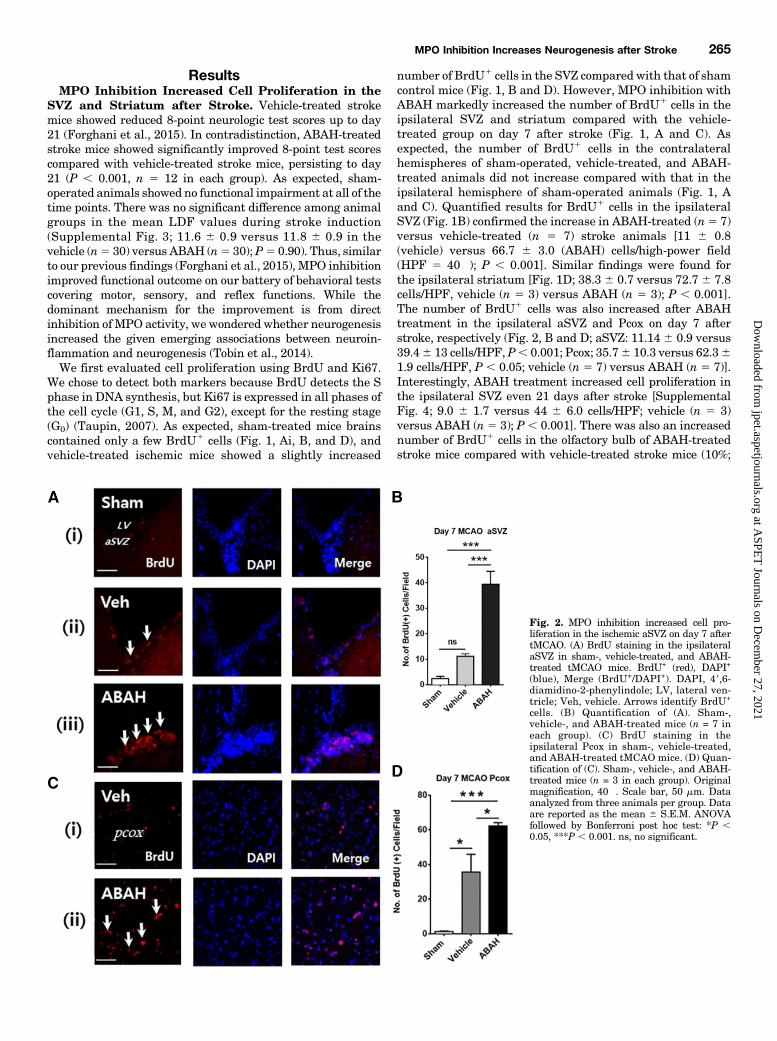
number of BrdU1 cells in the SVZ compared with that of shamcontrol mice (Fig. 1, B and D). However, MPO inhibition withABAH markedly increased the number of BrdU1 cells in theipsilateral SVZ and striatum compared with the vehicle-treated group on day 7 after stroke (Fig. 1, A and C). Asexpected, the number of BrdU1 cells in the contralateralhemispheres of sham-operated, vehicle-treated, and ABAH-treated animals did not increase compared with that in theipsilateral hemisphere of sham-operated animals (Fig. 1, Aand C). Quantified results for BrdU1 cells in the ipsilateralSVZ (Fig. 1B) confirmed the increase in ABAH-treated (n5 7)versus vehicle-treated (n 5 7) stroke animals [11 6 0.8(vehicle) versus 66.7 6 3.0 (ABAH) cells/high-power field(HPF 5 40�); P , 0.001]. Similar findings were found forthe ipsilateral striatum [Fig. 1D; 38.3 6 0.7 versus 72.7 6 7.8cells/HPF, vehicle (n 5 3) versus ABAH (n 5 3); P , 0.001].The number of BrdU1 cells was also increased after ABAHtreatment in the ipsilateral aSVZ and Pcox on day 7 afterstroke, respectively (Fig. 2, B and D; aSVZ: 11.14 6 0.9 versus39.46 13 cells/HPF, P, 0.001; Pcox; 35.76 10.3 versus 62.361.9 cells/HPF, P , 0.05; vehicle (n 5 7) versus ABAH (n 5 7)].Interestingly, ABAH treatment increased cell proliferation inthe ipsilateral SVZ even 21 days after stroke [SupplementalFig. 4; 9.0 6 1.7 versus 44 6 6.0 cells/HPF; vehicle (n 5 3)versus ABAH (n 5 3); P , 0.001]. There was also an increasednumber of BrdU1 cells in the olfactory bulb of ABAH-treatedstroke mice compared with vehicle-treated stroke mice (10%;
Fig. 2. MPO inhibition increased cell pro-liferation in the ischemic aSVZ on day 7 aftertMCAO. (A) BrdU staining in the ipsilateralaSVZ in sham-, vehicle-treated, and ABAH-treated tMCAO mice. BrdU+ (red), DAPI+
(blue), Merge (BrdU+/DAPI+). DAPI, 49,6-diamidino-2-phenylindole; LV, lateral ven-tricle; Veh, vehicle. Arrows identify BrdU+
cells. (B) Quantification of (A). Sham-,vehicle-, and ABAH-treated mice (n = 7 ineach group). (C) BrdU staining in theipsilateral Pcox in sham-, vehicle-treated,and ABAH-treated tMCAO mice. (D) Quan-tification of (C). Sham-, vehicle-, and ABAH-treated mice (n = 3 in each group). Originalmagnification, 40�. Scale bar, 50 mm. Dataanalyzed from three animals per group. Dataare reported as the mean 6 S.E.M. ANOVAfollowed by Bonferroni post hoc test: *P ,0.05, ***P , 0.001. ns, no significant.
MPO Inhibition Increases Neurogenesis after Stroke 265
at ASPE
T Journals on D
ecember 27, 2021
jpet.aspetjournals.orgD
ownloaded from
data not shown). Similarly, there were more Ki671 cells in theipsilateral SVZ on days 7 and 21 in ABAH-treated micecompared with those in vehicle-treated animals, respectively(Fig. 1, Ei and Eii). Quantified results showed that ABAHtreatment significantly increased the number of Ki671 cellscompared with vehicle-treated animals on day 7 after stroke[Fig. 1F; 10.76 0.7 versus 32.06 4.2 cells/HPF; vehicle (n5 3)versus ABAH (n 5 3); P , 0.01]. On day 21 after stroke, thenumber of Ki671 cells remain elevated after ABAH treatment[10.3 6 2.8 versus 30.0 6 1.7 cells/HPF, vehicle (n 5 3) versusABAH (n 5 3); P , 0.01].ABAH Treatment Increased Neurogenesis in the
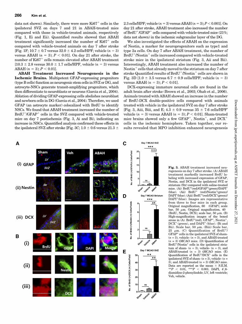
Ischemic Brains. Multipotent GFAP-expressing progenitors(typeB cells) function as neurogenic stem cells in SVZ orDG, andastrocyte-NSCs generate transit-amplifying progenitors, whichthen differentiate to neuroblasts or neurons (Garcia et al., 2004).Ablation of dividing GFAP-expressing cells abolishes neuroblastand newborn cells inDG (Garcia et al., 2004). Therefore, we usedGFAP (an astrocyte marker) colocalized with BrdU to identifyNSCs. We found that ABAH treatment increased the number ofBrdU1/GFAP1 cells in the SVZ compared with vehicle-treatedmice on day 7 postischemia (Fig. 3, Ai and Bi), indicating anincrease in NSCs. Quantified analysis confirmed these effects inthe ipsilateral SVZ after stroke [Fig. 3C; 1.06 0.6 versus 21.36
2.3 cells/HPF; vehicle (n53) versusABAH(n53);P,0.001].Onday 21 after stroke, ABAH treatment also increased the numberof BrdU1/GFAP1 cells compared with vehicle-treated mice (21%;data not shown) in the ischemic subgranular layer of the DG.We also investigated the effects of ABAH on the expression
of Nestin, a marker for neuroprogenitors such as type1 andtype 2a cells. On day 7 after ABAH treatment, the number ofBrdU1/Nestin1 cells increased compared with vehicle-treatedstroke mice in the ipsilateral striatum (Fig. 3, Aii and Bii).Interestingly, ABAH treatment also increased the number ofNestin1 cells that alreadymoved to the striatum on day 7 afterstroke Quantified results of BrdU1/Nestin1 cells are shown inFig. 3D [1.0 6 3.5 versus 6.7 6 0.9 cells/HPF; vehicle (n 5 3)versus ABAH (n 5 3); P , 0.01].DCX-expressing immature neuronal cells are found in the
adult brain after stroke (Brown et al., 2003; Ohab et al., 2006).Animals treated with ABAH showed an increase in the numberof BrdU-DCX double-positive cells compared with animalstreated with vehicle in the ipsilateral SVZ on day 7 after stroke[Fig. 3, Aiii, Biii, and E; 4.3 6 0.9 versus 35 6 7.6 cells/HPF’vehicle (n 5 3) versus ABAH (n 5 3); P , 0.01]. Sham-treatedmice brains showed only a few GFAP1, Nestin1, and DCX1
cells in the ischemic hemisphere. Taken together, our re-sults revealed that MPO inhibition enhanced neurogenesis
Fig. 3. ABAH treatment increased neu-rogenesis on day 7 after stroke. (A) ABAHtreatment markedly increased BrdU la-beling with increased expression of GFAP,Nestin, and DCX in the ipsilateral SVZ orstriatum (Str) compared with saline-treatedmice. (Ai) BrdU+(red)/GFAP+(green)/DAPI+
(blue). (Aii) BrdU+ (red)/Nestin+(green)/DAPI+(blue). (Aiii) BrdU+(red)/DCX+(green)/DAPI+(blue). Images are representativefrom three to four mice in each group.Original magnification, 60� (GFAP); scalebar, 50 mm. Original magnification, 40�(BrdU, Nestin, DCX); scale bar, 50 mm. (B)High-magnification images of the boxedareas in (A). BrdU+(red), GFAP+, Nestin+,DCX+ (green), and DAPI+ (blue). (Bi andBii). Scale bar, 50 mm. (Biii) Scale bar,25 mm. (C) Quantification of BrdU+/GFAP+ cells in the ipsilateral SVZ of sham-(n = 3), vehicle- (n = 3), and ABAH-treated(n = 3) tMCAO mice. (D) Quantification ofBrdU+/Nestin+ cells in the ipsilateral stria-tum of sham- (n = 3), vehicle- (n = 3), andABAH-treated (n = 3) tMCAO mice. (E)Quantification of BrdU+/DCX+ cells in theipsilateral SVZ of sham- (n = 3), vehicle- (n =3), and ABAH-treated (n = 3) tMCAO mice.Data are reported as the mean 6 S.E.M.**P , 0.01, ***P , 0.001. DAPI, 49,6-diamidino-2-phenylindole; LV, left ventricle;Veh, vehicle.
266 Kim et al.
at ASPE
T Journals on D
ecember 27, 2021
jpet.aspetjournals.orgD
ownloaded from

and increased NSC, neuroprogenitor cell, and neuroblastpopulations.MPO Inhibition Increased BDNF, p-CREB, AcH3,
CXCR4, and NeuN in the Ischemic Brain. BDNF is amitogenic factor that promotes the migration of NSCs, andenhances SVZ neuronal progenitor cell proliferation, neuro-genesis, and behavioral recovery (Benraiss et al., 2001;Chiaramello et al., 2007; Kim et al., 2009; Ploughman et al.,2009). We next assessed whether ABAH treatment affects BDNFexpression on day 7 poststroke. We found more BrdU1/BDNF1
cells in the ABAH-treated mice compared with vehicle-treated mice in the ischemic SVZ after stroke, showing thatMPO inhibition increases BDNF expression (Fig. 4, Ai andBi).Quantified data showed that the number of BrdU1/BDNF1
cells significantly increased in the ABAH-treated animals onday7 after tMCAO [Fig. 4C; 3.061.5 versus 3065.8 cells/HPF;vehicle (n 5 3) versus ABAH (n 5 3); P , 0.01].CREB (Ser 133) activation increases the survival of ischemia-
induced neuronal precursors (Zhu et al., 2004; Giachino et al.,2005). ABAH treatment significantly increased the number ofBrdU1/p-CREB1 (Ser 133) cells in the ischemic SVZ comparedwith vehicle-treatedmice on day 7 postischemia (Fig. 4, Aii and
Bii). Quantified results of BrdU1/p-CREB1 (Ser 133) cells inthe SVZ are shown in Fig. 4D [7.3 6 2.7 versus 39.7 6 6.7cells/HPF; vehicle (n 5 3) versus ABAH (n 5 3); P , 0.01].Similarly, on day 21 after stroke, ABAH treatment increasedthe number of BrdU1/p-CREB1 cells compared with vehicle-treated control mice in the SVZ (29%; data not shown). There-fore, MPO inhibition increased key factors in neurogenesis inthe BDNF-p-CREB signaling pathway.An increase in histone acetylation is important for neuronal
lineage progression of neural progenitor cells. We found thatABAH treatment increased the number of BrdU1/AcH31 cellscomparedwith vehicle-treatedmice in the ipsilateral SVZ on day7 poststroke. Most BrdU1 cells colocalized with AcH31 cells inthis region [Fig. 4, Aiii, Biii, and E; 7.7 6 0.7 versus 35 6 4.0cells/HPF; vehicle (n 5 3) versus ABAH (n 5 3); P , 0.001].CXCR4, a receptor of chemokine stromal cell–derived factor
1, is expressed in neuroprogenitors andmigrating neuroblastsin the ischemic hemisphere. ABAH treatment increasedCXCR41 cells in the ipsilateral SVZ compared with vehicle-treated animals on day 7 poststroke, suggesting that MPOinhibitionmay contribute to themigration of NSCs/progenitorcells in to the damaged brain (Fig. 4F).
Fig. 4. ABAH treatment increased BDNF,p-CREB, AcH3, and CXCR4+ immunos-taining in the SVZ on day 7 after tMCAO.(A) Immunostaining for BDNF, p-CREB,and AcH3. BrdU+ (red), DAPI+ (blue). (Ai)BDNF+ (green). (Aii) p-CREB+ (green). (Aiii)AcH3+ (green). Original magnification, 40�.Scale bar, 50 mm. (B) High-magnificationimages of the boxed areas in (A) from theABAH-treated group. Arrowheads identifycells with colocalized expression. (Bi) Scalebar, 50 mm. (Bii) Scale bar, 30 mm. (Biii)Scale bar, 40 mm. (C) Quantification ofBrdU+/BDNF+ cells. Sham-, vehicle-, andABAH-treated mice (n = 3 in each group).(D) Quantification of BrdU+/p-CREB+ cells.Sham-, vehicle-, andABAH-treatedmice (n=3 in each group). (E) Quantification ofBrdU+/AcH3+ cells. Sham-, vehicle-, andABAH-treated mice (n = 3 in each group).Data are reported as the mean 6 S.E.M.**P , 0.01, ***P , 0.001. (Fi) Immunos-taining for CXCR4 in the ipsilateral SVZ onday 7 after stroke. (Fii) High-magnificationimages of the boxed areas in the ABAH-treated group in (Fi). CXCR4 (green), DAPI(blue). Arrows indicate CXCR4+ cells.Scale bar, 25 mm. DAPI, 49,6-diamidino-2-phenylindole; LV, lateral ventricle; Veh,vehicle.
MPO Inhibition Increases Neurogenesis after Stroke 267
at ASPE
T Journals on D
ecember 27, 2021
jpet.aspetjournals.orgD
ownloaded from

Next, we investigated whether ABAH treatment increasesNeuN (a mature neuronal marker)-positive cells after stroke.Treatment with ABAH for 7 days markedly enhanced thenumber of BrdU1/NeuN1 cells in the ipsilateral aSVZ andSVZ compared with sham-operated or vehicle-treated controlanimals (Fig. 5A–E). Quantified results of BrdU1/NeuN1 cellsin the ipsilateral aSVZ confirmed this finding. Interestingly,most of the BrdU1 cells colocalized with NeuN1 cells in theaSVZ of ABAH-treatedmice [Fig. 5C; 2.76 0.3 versus 266 4.4cells/HPF; vehicle (n 5 3) versus ABAH (n 5 3); P , 0.01]. Inthe aSVZ, ABAH treatment also significantly increased thenumber of NeuN1 cells compared with vehicle-treated ani-mals [7.0 6 0.58 versus 26.3 6 3.5 cells/HPF; vehicle (n 5 3)versus ABAH (n 5 3); P , 0.01]. We confirmed similar effectsin the ipsilateral SVZ (Fig. 5D). Quantified results of BrdU1
/NeuN1 cells in the ipsilateral SVZ confirmed these observa-tions [Fig. 5E; 2.0 6 0.58 versus 13.3 6 4.3 cells/HPF; vehicle(n5 3) versus ABAH (n5 3); P, 0.05]. The number of NeuN1
cells in the ipsilateral SVZ also increased with ABAHtreatment [14.3 6 1.9 versus 23 6 0.6 cells/HPF; vehicle(n 5 3) versus ABAH (n 5 3); P , 0.01].MPO Inhibition Decreased the Number of Inflamma-
tory Cells and MMP-9 in the Ischemic Brain. We nextdetermined whether MPO inhibition affects inflammatory cellrecruitment and inflammatory mediators such as MMP-9.Iba-1 is a marker for myeloid cells and has a role in microgliaactivation and phagocytosis (Fumagalli et al., 2015). Amoeboid-shaped and hypertrophic Iba-11 cells were mainly observed in
the ipsilateral striatum. ABAH treatmentmarkedly reduced thenumber of Iba-11 cells compared with vehicle-treated controls[Fig. 6; day7: 23.761.7versus10.360.7 cells/HPF; vehicle (n53)versus ABAH (n5 3);P, 0.001; day 21: 13.36 0.3 versus 3.760.7 cells/HPF; vehicle (n5 3) versus ABAH (n5 3); P , 0.001].Additionally, we investigated the effects of ABAH treatment onthe ED11 (CD68) cells, a marker for monocytes/macrophages.ED11 cells were detected in the ischemic striatum, and thesecells were decreased by ABAH treatment on day 7 after stroke[Fig. 6, Aiii andD; 29.361.2 versus 18.060.6 cells/HPF; vehicle(n 5 3) versus ABAH (n 5 3); P , 0.001].MMPs also play a crucial role in brain inflammation by
increasing blood-brain barrier permeability through the de-struction of extracellular matrix (Park et al., 2009). Neutro-phil infiltration contributes to the increase in MMP-9 in theischemic brain (Justicia et al., 2003) of stroke patients (Loet al., 2003). We found that post-ABAH administration thenumber of MMP-91 cells decreased compared with those ofvehicle-treated animals in the ipsilateral striatum on day7 poststroke (Fig. 6, A, j–l). Quantified results are shown inFig. 6E [23.3 6 3.8 versus 9.0 6 2.5 cells/HPF; vehicle (n 5 3)versus ABAH (n 5 3); P , 0.05]. Our findings revealed thatpostinsult ABAH treatment decreased inflammatory cell re-cruitment and blood-brain barrier disruption.MPO2/2 Stroke Mice Show Increased Cell Prolifera-
tion and Improved Functional Recovery after Stroke.Corroborating our hypothesis,MPO2/2 strokemice also showedan increased number of BrdU1 cells in the vehicle-treated
Fig. 5. ABAH treatment increased the num-ber of BrdU+/NeuN+ cells in the aSVZ and SVZon day 7 after tMCAO mice. (A) Immunostain-ing of BrdU+/NeuN+ cells in the ipsilateralaSVZ in sham, vehicle-treated, and ABAH-treated tMCAO mice. Images are representa-tive from three to four mice in each group.Original magnification, 40�. Scale bar, 50 mm.(B) High-magnification images of the boxedareas in ABAH-treated mice. BrdU+ (red),NeuN+(green), and DAPI+ (blue). Arrowheadsindicate cells with colocalized expression. (C)Quantification of (A). Sham-, vehicle-, andABAH-treated mice (n = 3 in each group). (D)Immunostaining of BrdU+/NeuN+ cells in theipsilateral SVZ (top) and contralateral SVZ(bottom) in sham-, vehicle-, and ABAH-treatedtMCAO mice, respectively. (E) Quantificationof (D) SVZ in BrdU+/NeuN+ colocalizationdata. Sham-, vehicle-, and ABAH-treated mice(n = 3 in each group). Data are reported as themean 6 S.E.M. *P , 0.05, **P , 0.01. DAPI,49,6-diamidino-2-phenylindole; LV, lateralventricle; Veh, vehicle.
268 Kim et al.
at ASPE
T Journals on D
ecember 27, 2021
jpet.aspetjournals.orgD
ownloaded from
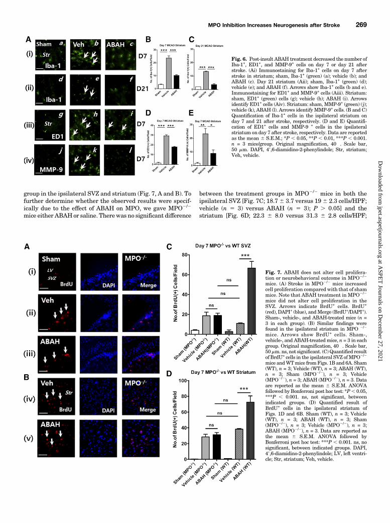
group in the ipsilateral SVZ and striatum (Fig. 7, A and B). Tofurther determine whether the observed results were specif-ically due to the effect of ABAH on MPO, we gave MPO2/2
mice either ABAHor saline. Therewas no significant difference
between the treatment groups in MPO2/2 mice in both theipsilateral SVZ [Fig. 7C; 18.76 3.7 versus 196 2.3 cells/HPF;vehicle (n 5 3) versus ABAH (n 5 3); P . 0.05] and thestriatum [Fig. 6D; 22.3 6 8.0 versus 31.3 6 2.8 cells/HPF;
Fig. 6. Post-insult ABAH treatment decreased the number ofIba-1+, ED1+, and MMP-9+ cells on day 7 or day 21 afterstroke. (Ai) Immunostaining for Iba-1+ cells on day 7 afterstroke in striatum; sham, Iba-1+ (green) (a); vehicle (b); andABAH (c). Day 21 striatum (Aii); sham, Iba-1+ (green) (d);vehicle (e); and ABAH (f). Arrows show Iba-1+ cells (b and e).Immunostaining for ED1+ and MMP-9+ cells (Aiii). Striatum:sham, ED1+ (green) cells (g); vehicle (h); ABAH (i). Arrowsidentify ED1+ cells (Aiv). Striatum: sham, MMP-9+ (green) (j);vehicle (k), ABAH (l). Arrows identify MMP-9+ cells. (B and C)Quantification of Iba-1+ cells in the ispilateral striatum onday 7 and 21 after stroke, respectively. (D and E) Quantifi-cation of ED1+ cells and MMP-9 + cells in the ipsilateralstriatum on day 7 after stroke, respectively. Data are reportedas the mean 6 S.E.M.; *P , 0.05, **P , 0.01, ***P , 0.001.n = 3 mice/group. Original magnification, 40�. Scale bar,50 mm. DAPI, 49,6-diamidino-2-phenylindole; Str, striatum;Veh, vehicle.
Fig. 7. ABAH does not alter cell prolifera-tion or neurobehavioral outcome in MPO2/2
mice. (A) Stroke in MPO2/2 mice increasedcell proliferation compared with that of shammice. Note that ABAH treatment in MPO2/2
mice did not alter cell proliferation in theSVZ. Arrows indicate BrdU+ cells. BrdU+
(red), DAPI+ (blue), andMerge (BrdU+/DAPI+).Sham-, vehicle-, and ABAH-treated mice (n =3 in each group). (B) Similar findings werefound in the ipsilateral striatum in MPO 2/2
mice. Arrows show BrdU+ cells. Sham-,vehicle-, and ABAH-treatedmice, n = 3 in eachgroup. Original magnification, 40�. Scale bar,50mm. ns, not significant. (C)Quantified resultof BrdU+ cells in the ipsilateral SVZ ofMPO2/2
mice andWTmice fromFigs. 1B and 6A. Sham(WT), n = 3; Vehicle (WT), n = 3; ABAH (WT),n = 3; Sham (MPO2/2), n = 3; Vehicle(MPO2/2), n = 3; ABAH (MPO2/2), n = 3. Dataare reported as the mean 6 S.E.M. ANOVAfollowed by Bonferroni post hoc test: *P, 0.05,***P , 0.001. ns, not significant, betweenindicated groups. (D) Quantified result ofBrdU+ cells in the ipsilateral striatum ofFigs. 1D and 6B. Sham (WT), n = 3; Vehicle(WT), n = 3; ABAH (WT), n = 3; Sham(MPO2/2), n = 3; Vehicle (MPO2/2), n = 3;ABAH (MPO2/2), n = 3. Data are reported asthe mean 6 S.E.M. ANOVA followed byBonferroni post hoc test: ***P , 0.001. ns, nosignificant, between indicated groups. DAPI,49,6-diamidino-2-phenylindole; LV, left ventri-cle; Str, striatum; Veh, vehicle.
MPO Inhibition Increases Neurogenesis after Stroke 269
at ASPE
T Journals on D
ecember 27, 2021
jpet.aspetjournals.orgD
ownloaded from

vehicle (n 5 3) versus ABAH (n 5 3); P . 0.05] on day 7 afterstroke. In contradistinction, in wild-type (WT) stroke micetherewas amarked increase in the number of BrdU1 cells afterABAH treatment in both the SVZ and striatum (P , 0.001).Next, we investigated whether MPO2/2 mice demonstrate
improved functional recovery after stroke using the 8-point test.MPO2/2 stroke mice given either ABAH or saline showed noneurobehavioral difference up to day 21 after cerebral ischemia(Fig. 8A). Furthermore, sham-induced WTmice given saline orABAH also demonstrated no neurobehavioral difference be-tween the two groups (Fig. 8B). These results confirmed thatABAH treatment with MPO2/2 mice does not have additionaleffects other thanMPO inhibition, and the changeswe observedwith ABAH treatment inWT stroke mice were due to the effectof ABAH on MPO.We also performed correlational analysis between BrdU1
cells and 8-point scores on day 7 after stroke. We found thata higher number of BrdU1 cells was correlated with betterneurologic outcome (r5 0.86, P , 0.001; Fig. 8C), and ABAH-treated stroke animals had more proliferating cells and betterneurologic outcomes.
DiscussionWe had previously shown that either MPO inhibition or
congenital absence led to a large reduction in infarct size(Forghani et al., 2015). The presentwork reveals that inhibitingthe inflammatory enzyme MPO with the specific irreversibleinhibitor ABAH not only decreased inflammatory cell recruit-ment and inflammatory mediators, but also promoted cellproliferation and NSC/progenitor migration, and enhancedneuronal differentiation, leading to increased neurogenesis inthe ischemic SVZ, aSVZ, striatum, and cortex. We found that
MPO inhibition increased markers for neurogenesis, includingBrdU1, GFAP1, Ki 671, Nestin1, DCX1, BDNF1, p-CREB1
(Ser133), and AcH31 cells in the ipsilateral SVZ, striatum,and DG after ischemia, establishing an inverse relationshipbetween MPO activity and neurogenesis. By treating ischemicMPO2/2 mice with either ABAH or saline and finding nodifference in the number of BrdU1 cells or functional outcome,we confirmed that it was the absence or inhibition of MPO,rather than nonspecific effects of ABAH, that resulted in theobserved effects on neurogenesis. Interestingly, MPO appearsto be involved in all stages of neurogenesis. This is likely theresult of the presence of MPO even up to day 21 after stroke(Breckwoldt et al., 2008), which would continue to adverselyaffect neurogenesis when present.BDNF is involved in neurogenesis, migration, maturation,
and survival of newborn cells in the striatum after ischemicinjury (Benraiss et al., 2001; Pencea et al., 2001). Additionally,the BDNF-tyrosine receptor kinase B signaling cascade is apositive regulator of neurogenesis and oligodendrogenesisin the ipsilateral SVZ and DG, and can result in anti-inflammatory effects and contribute to behavioral improve-ment after permanent MCAO in rats (Kim et al., 2009; Bathand Lee, 2010; Kim and Chuang, 2014). The activation ofCREB (Ser133) by phosphorylation also stimulates neuro-genesis in the ischemic DG after focal ischemia in rodents(Zhu et al., 2004). Our results demonstrated that MPOinhibition increased BDNF-CREB signaling cascade, indicat-ing MPO activity negatively affects this neurogenesis signal-ing pathway. Elevated oxidative stress in stroke can reducehistone acetylation expression, which is important for generegulation. Histone deacetylase inhibitors enhanced histoneacetylation, including AcH3 or acetylatedH4 after stroke (Kimet al., 2007, 2009). Histone deacetylase inhibitors, such asvalproic acid and butyrate, also can decrease MPO activity inlipopolysaccharide-induced lung injury (Ni et al., 2010; Jiet al., 2013). Indeed, we found that MPO inhibition restoredhistone acetylation, which is known to result in a more openchromatin structure and to induce NSC/progenitor production(Kim et al., 2009).In addition, we found that a higher number of BrdU1 cells
correlated with better behavioral outcome, suggesting thatimproving neurogenesis is associated with neurologicimprovement.While we found that MPO activity adversely impacts
neurogenesis after stroke, how MPO activity exerts its effectson neurogenesis is likely complex and beyond the scope of thiscurrent study. Nonetheless, one can hypothesize that MPOactivity increases inflammation that is detrimental to neuro-genesis. In addition to direct the cytotoxic effects of MPOactivity, elevatedMPO activity increases levels of ROS, MMP,inducible nitric oxide synthase, and inflammatory cytokines(e.g., IL-1b, TNF-a) (Ekdahl et al., 2003; Monje et al., 2003;Cacci et al., 2008), creating an environment that is hostile tonewborn cells. MPO inhibition can reduce the number of theseinflammatory mediators. Indeed, we found that MPO in-hibition decreased MMP-9 levels in the infarct (Fig. 6E).Furthermore, we found that ABAH treatment decreased thenumber of inflammatory cells in the ischemic areas (Fig. 6),likely as a result of reduced inflammation. Thus, these directand indirect effects of MPO inhibition combine to provide anenvironment that is more friendly for neurogenesis in theischemic brain.
Fig. 8. MPO2/2 mice showed beneficial effects in functional outcome, andsham-operated mice showed no effect on behavioral function. (A) MPO2/2
mice demonstrated improved 8-point neurologic scores compared withvehicle-treated WT mice after stroke (compare with Supplemental Fig.3A), and ABAH administration did not have any effect in MPO2/2 mice.(B) Sham-treated mice also showed no difference in the neurologic scoreswith ABAH or vehicle treatment. Experimental conditions are the same asin Supplemental Fig. 3A. (C) Correlational analysis between BrdU+ cellnumbers and ABAH-treated 8-point behavioral test scores on day 7 afterstroke (n = 14). Circles, vehicle-treated mice; triangles, ABAH-treatedmice. r = 0.86, P , 0.001.
270 Kim et al.
at ASPE
T Journals on D
ecember 27, 2021
jpet.aspetjournals.orgD
ownloaded from

NADPH oxidase (NOX) is an important pro-oxidant en-zyme, and triggers the generation of superoxide anion andH2O2, which are major sources of ROS (Bedard and Krause2007). The ablation of NOX2 in gp91phox2/2 mice markedlyreduced infarct size and inflammation compared with WTmice (Chen et al., 2009; Wang et al., 2013). The inhibition ofNOX by apocynin treatment reduced ROS and the neuro-protective effect, and improved functional outcome in MCAO(Tang et al., 2007). Interestingly, neural progenitors useNOX-derived H2O2 to maintain neurogenesis (Dickinson et al.,2011; Le Belle et al., 2011), showing that NOX has beneficialroles in ischemic injury. MPO acts downstream of NOX, and,unlike the membrane-bound NOX, MPO is secreted extracel-lularly, where it can cause damage away from the source. NOXdeficiency leads to the often lethal chronic granulomatousdisease in humans and to the chronic granulomatousdisease–like phenotype in mice (Nakano et al., 2008; Sorceand Krause 2009). On the other hand, MPO deficiency has notbeen found to significantly impact mortality (Kutter, 1998;Lanza, 1998; Kutter et al., 2000), making MPO a potentialtarget for human translation. NSCs and neural precursor cellscan undergo asymmetric cell division and maintain NSCpools, allowing for damaged brain repair (Tobin et al., 2014).Thus, part of the beneficial effects of MPO inhibition may befrom increased neurogenesis. While no clinically approveddrug is available to specifically inhibit MPO, several pre-clinical candidates are in development, and one candidate hascompleted a phase IIa trial (Churg et al., 2012; Forbes et al.,2013; Ward et al., 2013).Interestingly, we found that the partial inhibition of MPO
by ABAH led to a greater increase of BrdU1 cells, up to ∼70cells (Fig. 1B). In contrast, the congenital absence of MPO onlyincreased the number of BrdU1 cells to ∼20 (Fig. 7A–D). Thissuggests that congenital deletion of MPO is less favorable tocell proliferation compared with pharmacological partial(∼40% with ABAH) inhibition of MPO (Forghani et al.,2015). Indeed, several studies (Brennan et al., 2001, Kumaret al., 2005) have found that MPO-deficient mice have alteredimmune responses, including increased nitric oxide andT-lymphocyte levels, that may adversely affect neurogenesis.While we found increased neurogenesis associated with MPOinhibition, functional improvement with MPO inhibition wasdetected within 1 day after stroke (Forghani et al., 2015),suggesting that ABAH has neuroprotective effects prior to theonset of neurogenesis. We are currently also investigating thisaspect of MPO inhibition. Taken together, improving ourunderstanding of the actions of MPO and the consequencesof its inhibition in stroke, which resulted in improved func-tional recovery and neurogenesis, could spur further develop-ment of this new class of drugs to treat stroke patients.Conclusion. Our findings indicate that MPO activity is
inversely related to neurogenesis and possibly to neurologicoutcome. MPO inhibition or deficiency in stroke, along withthe associated effects on inflammation, creates an environ-ment that stimulates important endogenous resources topromote many aspects of neurogenesis, including cell pro-liferation, differentiation, and migration, and newborn cellsurvival, providing evidence that links MPO-mediated in-flammation with neurogenesis in stroke.
Authorship Contributions
Participated in research design: Kim, Moskowitz, and Chen.
Conducted experiments: Kim, Wei, Lee, Wu, and Zheng.Contributed new reagents or analytic tools: Kim and Wei.Performed data analysis: Kim, Lee, and Chen.Wrote or contributed to the writing of the manuscript: Kim,
Moskowitz, and Chen.
References
Alvarez-Buylla A and Garcia-Verdugo JM (2002) Neurogenesis in adult sub-ventricular zone. J Neurosci 22:629–634.
Arvidsson A, Collin T, Kirik D, Kokaia Z, and Lindvall O (2002) Neuronal re-placement from endogenous precursors in the adult brain after stroke. Nat Med 8:963–970.
Bath KG and Lee FS (2010) Neurotrophic factor control of adult SVZ neurogenesis.Dev Neurobiol 70:339–349.
Bedard K and Krause KH (2007) The NOX family of ROS-generating NADPH oxi-dases: physiology and pathophysiology. Physiol Rev 87:245–313.
Benraiss A, Chmielnicki E, Lerner K, Roh D, and Goldman SA (2001) Adenoviralbrain-derived neurotrophic factor induces both neostriatal and olfactory neuronalrecruitment from endogenous progenitor cells in the adult forebrain. J Neurosci 21:6718–6731.
Breckwoldt MO, Chen JW, Stangenberg L, Aikawa E, Rodriguez E, Qiu S, MoskowitzMA, and Weissleder R (2008) Tracking the inflammatory response in stroke in vivoby sensing the enzyme myeloperoxidase. Proc Natl Acad Sci USA 105:18584–18589.
Brennan M, Gaur A, Pahuja A, Lusis AJ, and Reynolds WF (2001) Mice lackingmyeloperoxidase are more susceptible to experimental autoimmune encephalo-myelitis. J Neuroimmunol 112:97–105.
Brown JP, Couillard-Després S, Cooper-Kuhn CM, Winkler J, Aigner L, and KuhnHG (2003) Transient expression of doublecortin during adult neurogenesis. J CompNeurol 467:1–10.
Cacci E, Ajmone-Cat MA, Anelli T, Biagioni S, and Minghetti L (2008) In vitroneuronal and glial differentiation from embryonic or adult neural precursor cellsare differently affected by chronic or acute activation of microglia. Glia 56:412–425.
Chamorro A and Hallenbeck J (2006) The harms and benefits of inflammatory andimmune responses in vascular disease. Stroke 37:291–293.
Chen H, Song YS, and Chan PH (2009) Inhibition of NADPH oxidase is neuro-protective after ischemia-reperfusion. J Cereb Blood Flow Metab 29:1262–1272.
Chen JW, Breckwoldt MO, Aikawa E, Chiang G, and Weissleder R (2008)Myeloperoxidase-targeted imaging of active inflammatory lesions in murine ex-perimental autoimmune encephalomyelitis. Brain 131:1123–1133.
Chiaramello S, Dalmasso G, Bezin L, Marcel D, Jourdan F, Peretto P, Fasolo A,and De Marchis S (2007) BDNF/TrkB interaction regulates migration of SVZprecursor cells via PI3-K and MAP-K signalling pathways. Eur J Neurosci 26:1780–1790.
Churg A, Marshall CV, Sin DD, Bolton S, Zhou S, Thain K, Cadogan EB, Maltby J,Soars MG, Mallinder PR, et al. (2012) Late intervention with a myeloperoxidaseinhibitor stops progression of experimental chronic obstructive pulmonary disease.Am J Respir Crit Care Med 185:34–43.
Curtis MA, Kam M, Nannmark U, Anderson MF, Axell MZ, Wikkelso C, Holtås S,van Roon-Mom WM, Björk-Eriksson T, Nordborg C, et al. (2007) Human neuro-blasts migrate to the olfactory bulb via a lateral ventricular extension. Science 315:1243–1249.
Dickinson BC, Peltier J, Stone D, Schaffer DV, and Chang CJ (2011) Nox2 redoxsignaling maintains essential cell populations in the brain. Nat Chem Biol 7:106–112.
Ekdahl CT, Claasen JH, Bonde S, Kokaia Z, and Lindvall O (2003) Inflammation isdetrimental for neurogenesis in adult brain. Proc Natl Acad Sci USA 100:13632–13637.
El Kebir D and Filep JG (2013) Modulation of neutrophil apoptosis and the resolutionof Inflammation through b2 Integrins. Front Immunol 4:60.
Forbes LV, Sjögren T, Auchère F, Jenkins DW, Thong B, Laughton D, Hemsley P,Pairaudeau G, Turner R, Eriksson H, et al. (2013) Potent reversible inhibition ofmyeloperoxidase by aromatic hydroxamates. J Biol Chem 288:36636–36647.
Forghani R, Wojtkiewicz GR, Zhang Y, Seeburg D, Bautz BRM, Pulli B, Milewski AR,Atkinson WL, Iwamoto Y, Zhang ER, et al. (2012) Demyelinating diseases: mye-loperoxidase as an imaging biomarker and therapeutic target. Radiology 263:451–460.
Forghani R, Kim HJ, Wojtkiewicz GR, Bure L, Wu Y, Hayase M, Wei Y, Zheng Y,Moskowitz MA, and Chen JW (2015) Myeloperoxidase propagates damage and is apotential therapeutic target for subacute stroke. J Cereb Blood Flow Metab 35:485–493.
Fumagalli S, Perego C, Pischiutta F, Zanier ER, and De Simoni MG (2015) Theischemic environment drives microglia and macrophage function. Front Neurol 6:81.
Garcia AD, Doan NB, Imura T, Bush TG, and Sofroniew MV (2004) GFAP-expressingprogenitors are the principal source of constitutive neurogenesis in adult mouseforebrain. Nat Neurosci 7:1233–1241.
Giachino C, De Marchis S, Giampietro C, Parlato R, Perroteau I, Schütz G, Fasolo A,and Peretto P (2005) cAMP response element-binding protein regulates differen-tiation and survival of newborn neurons in the olfactory bulb. J Neurosci 25:10105–10118.
Heinecke JW (2002) Tyrosyl radical production by myeloperoxidase: a phagocytepathway for lipid peroxidation and dityrosine cross-linking of proteins. Toxicology177:11–22.
Iosif RE, Ahlenius H, Ekdahl CT, Darsalia V, Thored P, Jovinge S, Kokaia Z,and Lindvall O (2008) Suppression of stroke-induced progenitor proliferation inadult subventricular zone by tumor necrosis factor receptor 1. J Cereb Blood FlowMetab 28:1574–1587.
MPO Inhibition Increases Neurogenesis after Stroke 271
at ASPE
T Journals on D
ecember 27, 2021
jpet.aspetjournals.orgD
ownloaded from

Ji MH, Li GM, Jia M, Zhu SH, Gao DP, Fan YX, Wu J, and Yang JJ (2013) Valproicacid attenuates lipopolysaccharide-induced acute lung injury in mice. In-flammation 36:1453–1459.
Jin K, Sun Y, Xie L, Peel A, Mao XO, Batteur S, and Greenberg DA (2003) Directedmigration of neuronal precursors into the ischemic cerebral cortex and striatum.Mol Cell Neurosci 24:171–189.
Justicia C, Panés J, Solé S, Cervera A, Deulofeu R, Chamorro A, and Planas AM(2003) Neutrophil infiltration increases matrix metalloproteinase-9 in the ischemicbrain after occlusion/reperfusion of the middle cerebral artery in rats. J CerebBlood Flow Metab 23:1430–1440.
Kim HJ and Chuang DM (2014) HDAC inhibitors mitigate ischemia-induced oligo-dendrocyte damage: potential roles of oligodendrogenesis, VEGF, and anti-inflammation. Am J Transl Res 6:206–223.
Kim HJ, Rowe M, Ren M, Hong JS, Chen PS, and Chuang DM (2007) Histonedeacetylase inhibitors exhibit anti-inflammatory and neuroprotective effects in arat permanent ischemic model of stroke: multiple mechanisms of action. J Phar-macol Exp Ther 321:892–901.
Kim HJ, Leeds P, and Chuang DM (2009) The HDAC inhibitor, sodium butyrate,stimulates neurogenesis in the ischemic brain. J Neurochem 110:1226–1240.
Klinke A, Nussbaum C, Kubala L, Friedrichs K, Rudolph TK, Rudolph V, Paust HJ,Schröder C, Benten D, Lau D, et al. (2011) Myeloperoxidase attracts neutrophils byphysical forces. Blood 117:1350–1358.
Kohman RA and Rhodes JS (2013) Neurogenesis, inflammation and behavior. BrainBehav Immun 27:22–32.
Kumar AP, Ryan C, Cordy V, and Reynolds WF (2005) Inducible nitric oxide synthaseexpression is inhibited by myeloperoxidase. Nitric Oxide 13:42–53.
Kutter D (1998) Prevalence of myeloperoxidase deficiency: population studies usingBayer-Technicon automated hematology. J Mol Med (Berl) 76:669–675.
Kutter D, Devaquet P, Vanderstocken G, Paulus JM, Marchal V, and Gothot A (2000)Consequences of total and subtotal myeloperoxidase deficiency: risk or benefit?Acta Haematol 104:10–15.
Lanza F (1998) Clinical manifestation of myeloperoxidase deficiency. J Mol Med(Berl) 76:676–681.
Lau D and Baldus S (2006) Myeloperoxidase and its contributory role in in-flammatory vascular disease. Pharmacol Ther 111:16–26.
Le Belle JE, Orozco NM, Paucar AA, Saxe JP, Mottahedeh J, Pyle AD, Wu H,and Kornblum HI (2011) Proliferative neural stem cells have high endogenous ROSlevels that regulate self-renewal and neurogenesis in a PI3K/Akt-dependantmanner. Cell Stem Cell 8:59–71.
Lo EH, Dalkara T, and Moskowitz MA (2003) Mechanisms, challenges and oppor-tunities in stroke. Nat Rev Neurosci 4:399–415.
Moskowitz MA, Lo EH, and Iadecola C (2010) The science of stroke: mechanisms insearch of treatments. Neuron 67:181–198.
Maki RA, Tyurin VA, Lyon RC, Hamilton RL, DeKosky ST, Kagan VE, and ReynoldsWF (2009) Aberrant expression of myeloperoxidase in astrocytes promotes phos-pholipid oxidation and memory deficits in a mouse model of Alzheimer disease.J Biol Chem 284:3158–3169.
Ming GL and Song H (2011) Adult neurogenesis in the mammalian brain: significantanswers and significant questions. Neuron 70:687–702.
Monje ML, Toda H, and Palmer TD (2003) Inflammatory blockade restores adulthippocampal neurogenesis. Science 302:1760–1765.
Nakano Y, Longo-Guess CM, Bergstrom DE, Nauseef WM, Jones SM, and Bánfi B(2008) Mutation of the Cyba gene encoding p22phox causes vestibular and immunedefects in mice. J Clin Invest 118:1176–1185.
Ni YF, Wang J, Yan XL, Tian F, Zhao JB, Wang YJ, and Jiang T (2010) Histonedeacetylase inhibitor, butyrate, attenuates lipopolysaccharide-induced acute lunginjury in mice. Respir Res 11:33.
Nussbaum C, Klinke A, AdamM, Baldus S, and Sperandio M (2013) Myeloperoxidase: aleukocyte-derived protagonist of inflammation and cardiovascular disease. AntioxidRedox Signal 18:692–713.
Ohab JJ, Fleming S, Blesch A, and Carmichael ST (2006) A neurovascular niche forneurogenesis after stroke. J Neurosci 26:13007–13016.
Park KP, Rosell A, Foerch C, Xing C, Kim WJ, Lee S, Opdenakker G, Furie KL,and Lo EH (2009) Plasma and brain matrix metalloproteinase-9 after acute focalcerebral ischemia in rats. Stroke 40:2836–2842.
Pencea V, Bingaman KD, Wiegand SJ, and Luskin MB (2001) Infusion of brain-derived neurotrophic factor into the lateral ventricle of the adult rat leads to newneurons in the parenchyma of the striatum, septum, thalamus, and hypothalamus.J Neurosci 21:6706–6717.
Ploughman M, Windle V, MacLellan CL, White N, Doré JJ, and Corbett D (2009)Brain-derived neurotrophic factor contributes to recovery of skilled reaching afterfocal ischemia in rats. Stroke 40:1490–1495.
Sorce S and Krause KH (2009) NOX enzymes in the central nervous system: fromsignaling to disease. Antioxid Redox Signal 11:2481–2504.
Tang LL, Ye K, Yang XF, and Zheng JS (2007) Apocynin attenuates cerebral in-farction after transient focal ischaemia in rats. J Int Med Res 35:517–522.
Taupin P (2007) BrdU immunohistochemistry for studying adult neurogenesis: par-adigms, pitfalls, limitations, and validation. Brain Res Brain Res Rev 53:198–214.
Thored P, Arvidsson A, Cacci E, Ahlenius H, Kallur T, Darsalia V, Ekdahl CT,Kokaia Z, and Lindvall O (2006) Persistent production of neurons from adult brainstem cells during recovery after stroke. Stem Cells 24:739–747.
Tobin MK, Bonds JA, Minshall RD, Pelligrino DA, Testai FD, and Lazarov O (2014)Neurogenesis and inflammation after ischemic stroke: what is known and wherewe go from here. J Cereb Blood Flow Metab 34:1573–1584.
Wang Q, Tang XN, and Yenari MA (2007) The inflammatory response in stroke.J Neuroimmunol 184:53–68.
Wang Z, Wei X, Liu K, Zhang X, Yang F, Zhang H, He Y, Zhu T, Li F, Shi W, et al.(2013) NOX2 deficiency ameliorates cerebral injury through reduction of complexinII-mediated glutamate excitotoxicity in experimental stroke. Free Radic Biol Med65:942–951.
Ward J, Spath SN, Pabst B, Carpino PA, Ruggeri RB, Xing G, Speers AE, Cravatt BF,and Ahn K (2013) Mechanistic characterization of a 2-thioxanthine myeloperoxidaseinhibitor and selectivity assessment utilizing click chemistry—activity-based proteinprofiling. Biochemistry 52:9187–9201.
Yamashita T, Ninomiya M, Hernández Acosta P, García-Verdugo JM, Sunabori T,Sakaguchi M, Adachi K, Kojima T, Hirota Y, Kawase T, et al. (2006) Subventricularzone-derived neuroblasts migrate and differentiate into mature neurons in thepost-stroke adult striatum. J Neurosci 26:6627–6636.
Zhang R, Brennan ML, Shen Z, MacPherson JC, Schmitt D, Molenda CE, and HazenSL (2002) Myeloperoxidase functions as a major enzymatic catalyst for initiation oflipid peroxidation at sites of inflammation. J Biol Chem 277:46116–46122.
Zhang R, Zhang Z, Wang L, Wang Y, Gousev A, Zhang L, Ho KL, Morshead C,and Chopp M (2004) Activated neural stem cells contribute to stroke-inducedneurogenesis and neuroblast migration toward the infarct boundary in adult rats.J Cereb Blood Flow Metab 24:441–448.
Zhang RL, LeTourneau Y, Gregg SR, Wang Y, Toh Y, Robin AM, Zhang ZG,and Chopp M (2007) Neuroblast division during migration toward the ischemicstriatum: a study of dynamic migratory and proliferative characteristics of neu-roblasts from the subventricular zone. J Neurosci 27:3157–3162.
Zhao C, Deng W, and Gage FH (2008) Mechanisms and functional implications ofadult neurogenesis. Cell 132:645–660.
Zhu DY, Lau L, Liu SH, Wei JS, and Lu YM (2004) Activation of cAMP-response-element-binding protein (CREB) after focal cerebral ischemia stimulates neuro-genesis in the adult dentate gyrus. Proc Natl Acad Sci USA 101:9453–9457.
Address correspondence to: John W. Chen, Massachusetts GeneralHospital, Harvard Medical School, Richard B. Simches Research Center,185 Cambridge Street, Boston, MA 02114. E-mail: [email protected]
272 Kim et al.
at ASPE
T Journals on D
ecember 27, 2021
jpet.aspetjournals.orgD
ownloaded from