myocardial infarction accelerates atherosclerosis
TRANSCRIPT

Myocardial Infarction Accelerates Atherosclerosis
CitationDutta, Partha, Gabriel Courties, Ying Wei, Florian Leuschner, Rostic Gorbatov, Clinton Robbins, Yoshiko Iwamoto, et al. 2012. Myocardial infarction accelerates atherosclerosis. Nature 487(7407): 325-329.
Published Versiondoi:10.1038/nature11260
Permanent linkhttp://nrs.harvard.edu/urn-3:HUL.InstRepos:10612854
Terms of UseThis article was downloaded from Harvard University’s DASH repository, and is made available under the terms and conditions applicable to Other Posted Material, as set forth at http://nrs.harvard.edu/urn-3:HUL.InstRepos:dash.current.terms-of-use#LAA
Share Your StoryThe Harvard community has made this article openly available.Please share how this access benefits you. Submit a story .
Accessibility

Myocardial infarction accelerates atherosclerosis
Partha Dutta1,*, Gabriel Courties1,*, Ying Wei2, Florian Leuschner1,3, Rostic Gorbatov1,Clinton Robbins1, Yoshiko Iwamoto1, Brian Thompson1, Alicia L. Carlson1, Timo Heidt1,Maulik D. Majmudar1,4, Felix Lasitschka5, Martin Etzrodt1, Peter Waterman1, Michael T.Waring6,7, Adam T. Chicoine6,7, Anja M. van der Laan8, Hans W.M. Niessen9, Jan J. Piek8,Barry B. Rubin10, Jagdish Butany11, James Stone1, Hugo A. Katus3, Sabina A. Murphy12,David A. Morrow12, Marc S. Sabatine12, Claudio Vinegoni1, Michael A. Moskowitz2, MikaelJ. Pittet1, Peter Libby4, Charles P. Lin1, Filip K. Swirski1, Ralph Weissleder1,13, andMatthias Nahrendorf1
1Center for Systems Biology, Massachusetts General Hospital and Harvard Medical School,Simches Research Building, 185 Cambridge St., Boston, MA 02114, USA 2Stroke andNeurovascular Regulation Laboratory, Departments of Radiology and Neurology, MassachusettsGeneral Hospital/Harvard Medical School, 149 13th Street, Charlestown, MA 02129 3Departmentof Cardiology, Medical University Hospital Heidelberg, Im Neuenheimer Feld 410, D-69120Heidelberg, Germany 4Cardiovascular Division, Department of Medicine, Brigham and Women'sHospital, Boston, MA 5Institute of Pathology, University Hospital Heidelberg, Im NeuenheimerFeld 220/221, 69120 Heidelberg, Germany; Cardiovascular Division, Department of Medicine,Brigham and Women's Hospital, Boston, MA 6The Ragon Institute of MGH, MIT and Harvard atMassachusetts General Hospital, Charlestown, MA 02129, USA 7Howard Hughes MedicalInstitute, Chevy Chase, Maryland, USA 8Department of Cardiology, Academic Medical Center,University of Amsterdam, Amsterdam, Netherlands 9Department of Pathology and CardiacSurgery, ICaR-VU, VU University Medical Center, Amsterdam, Netherlands 10Division of VascularSurgery, Peter Munk Cardiac Centre, Toronto General Hospital, University Health Network,Toronto, Ontario M5G-2C4, Canada 11Division of Pathology, Peter Munk Cardiac Centre, TorontoGeneral Hospital, University Health Network, Toronto, Ontario M5G-2C4, Canada 12TIMI StudyGroup, Cardiovascular Division, Brigham and Women’s Hospital, Boston, MA 13Department ofSystems Biology, Harvard Medical School, Boston, MA
SUMMARYDuring progression of atherosclerosis, myeloid cells destabilize lipid-rich plaque in the arterialwall and cause its rupture, thus triggering myocardial infarction and stroke. Survivors of acutecoronary syndromes have a high risk of recurrent events for unknown reasons. Here we show thatthe systemic response to ischemic injury aggravates chronic atherosclerosis. After myocardial
Corresponding authors: Matthias Nahrendorf, Filip K. Swirski, Ralph Weissleder, Center for Systems Biology, 185 Cambridge Street,Boston, MA 02114, Tel: (617) 643-0500, Fax: (617) 643-6133,[email protected]@[email protected].*These authors contributed equally to this work
AUTHOR CONTRIBUTIONSP.D. and G.C. performed experiments, collected and analyzed the data, and contributed to writing the manuscript, R.G. did surgeriesand performed experiments, Y.W., F.L., R.G., C.R., Y.I., B.T., A.L.C., T.H., M.D.M., F.La., M.E., P.W., M.T.W., A.T.C., A.M.L.,H.W.M.N., J.J.P., B.B.R., J.B., J.S., H.A.K., C.V., S.A.M., D.A.M., and M.S.S. performed experiments, collected, analyzed anddiscussed data, M.A.M., M.J.P., P.L., C.P.L., F.K.S. and R.W. conceived experiments and discussed strategy and results; M.N.designed and managed the study and wrote the manuscript which was edited and approved by all co-authors.
COMPETING FINANCIAL INTERESTSMarc S. Sabatine, David A. Morrow and Sabina A. Murphy received grant support from AstraZeneca and GSK. The remainingauthors declare no competing financial interests.
NIH Public AccessAuthor ManuscriptNature. Author manuscript; available in PMC 2013 January 19.
Published in final edited form as:Nature. 2012 July 19; 487(7407): 325–329. doi:10.1038/nature11260.
NIH
-PA Author Manuscript
NIH
-PA Author Manuscript
NIH
-PA Author Manuscript
infarction or stroke, apoE−/− mice developed larger atherosclerotic lesions with a more advancedmorphology. This disease acceleration persisted over many weeks and was associated withmarkedly increased monocyte recruitment. When seeking the source of surplus monocytes inplaque, we found that myocardial infarction liberated hematopoietic stem and progenitor cellsfrom bone marrow niches via sympathetic nervous system signaling. The progenitors then seededthe spleen yielding a sustained boost in monocyte production. These observations provide newmechanistic insight into atherogenesis and provide a novel therapeutic opportunity to mitigatedisease progression.
INTRODUCTIONToday, survival following a first myocardial infarct (MI) approaches 90%. However, re-infarction occurs commonly and carries a high mortality. In a representative trial, newmyocardial ischemia occurred in 54% of patients within the first year after MI1. The largestpopulation study to date showed a 17.4% 1-year risk of re-infarction2. Conventional wisdominfers that these very high rates of secondary events reflect later stages of linear diseaseprogression. This study tested the alternative hypothesis that a first infarct — triggering aburst of acute systemic inflammation aimed at repair of the injured heart —could accelerateatherosclerosis.
Monocytes infiltrate lesions and, together with their lineage descendant macrophages,instigate inflammation and deliver proteolytic enzymes that digest extracellular matrix andrender atherosclerotic plaques unstable3–7. Elevated levels of circulating monocytes providean expanded pool of inflammatory cells available for recruitment to growing arterial lesions,potentially promoting plaque rupture. Leukocytosis post MI predicts an increased risk of re-infarction and death8,9. During acute MI, blood monocyte levels spike, and these cellsaccumulate in the evolving myocardial wound10,11. Thus, the organism experiences an acuteinflammatory event (e.g. MI) superimposed on a pre-existing chronic inflammatory disease(atherosclerosis), both of which involve the same myeloid cell type. Given the frequency ofre-infarction, we investigated whether acute myocardial injury accelerates pre-existingchronic atherosclerosis.
We found that in apoE−/− mice with atherosclerosis, MI increased plaque size and induced a'vulnerable' lesion morphology with higher inflammatory cell content and protease activity,fueled by persistently increased myeloid cell flux to atherosclerotic sites. Earlier clinicalstudies described an increase of hematopoietic stem and progenitor cells (HSPCs) in thecirculation of patients shortly after MI12. We thus hypothesized that release of theseprogenitors may increase the availability of monocytes. We found that in response toheightened sympathetic nervous system (SNS) activity — provoked by pain, anxiety, andheart failure in patients with MI HSPCs departed bone marrow niches and producedprolonged amplified extramedullary monocytopoiesis in mice after coronary ligation.
MI accelerates atherosclerosisProteases, including metalloproteinases and cysteinyl cathepsins, can catabolize theextracellular matrix of the plaque’s fibrous cap and render it prone to rupture13,14.Therefore, protease activity may serve as a marker in mice of processes associated withlesion vulnerability in humans15. To test the hypothesis that MI changes the course ofatherosclerotic disease, we serially imaged protease activity in aortic plaques of apoE−/−
mice, before and 3 weeks after coronary ligation, using fluorescence molecular tomographyfused to X-ray computed tomography (FMT/CT)16. Imaging showed a sharp increase ofplaque protease activity within 3 weeks after MI (Fig. 1a,b). In parallel, expression of theinflammatory cytokine IL-6, MMP-9, myeloperoxidase, and Ly-6C increased in
Dutta et al. Page 2
Nature. Author manuscript; available in PMC 2013 January 19.
NIH
-PA Author Manuscript
NIH
-PA Author Manuscript
NIH
-PA Author Manuscript
atherosclerotic plaques (Supplementary Fig. 1). The number of monocytes/macrophages peraorta expanded, particularly the inflammatory Ly-6Chigh monocyte subset (Fig. 1c). Plaquemonocyte content also increased in apoE−/− mice without MI, reflecting the natural courseof disease in these animals17,18. Yet innate immune cell accumulation accelerateddistinctively after MI, as indicated by the significantly greater slope obtained when fittingthe number of Ly-6Chigh monocytes in the aorta over time (Supplementary Fig. 2).Neutrophil presence in atheromata also increased (Supplementary Fig. 3) while mast cellsdid not (Supplementary Fig. 4). Histologic analysis affirmed increased accumulation ofCD11b+ myeloid cells and larger lesion size after MI (Fig. 1d). The thickness of the fibrouscap decreased, covering larger necrotic cores (Supplementary Fig. 5). Ly-6Chigh monocytesisolated from atherosclerotic lesions exhibited higher levels of mRNAs encodinginflammatory genes. IL-1β and cathepsin B were expressed at higher levels 3 weeks afterMI, whereas arginase-1 and TGFβ, markers associated with alternatively activatedmacrophages, were expressed at lower levels (Supplementary Fig. 6). Monocyte numbers inthe blood and spleen increased consistently for up to 3 months after coronary ligation(Supplementary Fig. 7) but were unaltered in the bone marrow (Supplementary Fig. 8).
Extramedullary monocytopoiesis post MIBecause the spleen has the ability to host extramedullary hematopoiesis19–21, we measuredsplenic monocyte progenitor content following MI. Hematopoietic progenitor cell numbersin the spleen increased post-MI (Fig. 2, Supplementary Fig. 9) but not in the bone marrow(Supplementary Fig. 10). Proliferation of progenitors doubled in the spleen (SupplementaryFig. 11). In patients that died after an acute MI, we found increased numbers of c-kit+ cellsin the spleen, some of which colocalized with the proliferation marker Ki-67(Supplementary Fig. 12).
When we splenectomized mice at the time of MI, atherosclerosis did notaccelerate(Supplementary Fig. 13). The number of progenitor cells in liver tissue after MIwas much lower than in the spleen, however; splenectomy increased progenitor cellpresence in the liver 4 days after MI (Supplementary Fig. 14). We concluded that the infarct-induced monocytosis resulted primarily from augmented production in the spleen, but otherextramedullary sites may contribute22. This observation raised the question whethermonocytes of splenic and bone marrow origin differ qualitatively. Surprisingly, Ly-6Chigh
monocytes isolated from the spleen or bone marrow on day 4 after MI had significantlydifferent mRNA levels in 11 of the 32 genes assessed (Supplementary Fig. 15). For instance,IL-1β and cathepsin B mRNA levels were 60- and 6-fold higher in inflammatory monocytesisolated from the spleen, matching the increased expression of these genes in Ly-6Chigh
monocytes isolated from atherosclerotic plaque after MI (Supplementary Fig. 6). Therefore,post-MI extramedullary myelopoiesis may not only increase the availability of inflammatorycells but also change their functional program. To test whether another form of acute tissueinjury prevalent in atherosclerotic patients would accelerate splenic myelopoiesis, weanalyzed apoE−/−mice 6 weeks after ischemic stroke. The number of myeloid cells andLy-6Chigh monocytes in atherosclerotic plaque increased after stroke, in parallel withexpanded splenic monocytopoiesis (Supplementary Fig. 16).
Bone marrow HSPC release post MISince granulocyte macrophage progenitors (GMP) and macrophage dendritic cellprogenitors (MDP) have a limited self-renewal capacity23,24, we hypothesized that upstreamprogenitors released from their bone marrow niches sustain the splenic proliferative activityafter MI. Indeed, blood levels of HSPCs increased 2, 7 and 24-fold at 6, 48 and 96 hoursafter MI, respectively (Fig. 3a, ). The number of splenic FLK2− HSPCs increaseddramatically after MI (Supplementary Fig. 17). This mobilization of upstream HSPCs with
Dutta et al. Page 3
Nature. Author manuscript; available in PMC 2013 January 19.
NIH
-PA Author Manuscript
NIH
-PA Author Manuscript
NIH
-PA Author Manuscript
high capacity for self-renewal likely explains the long-term boost in splenic monocyteproduction in apoE−/− mice after MI.
Anxiety, pain and impaired left ventricular function during MI can all activate the SNS.Accordingly, levels of tyrosine hydroxylase, the rate-limiting enzyme for production ofnoradrenaline in sympathetic fibers25, increased in the bone marrow of mice after MI andhence indicated a higher sympathetic tone (Fig. 3b). SNS activity may liberatehematopoietic stem cells from their niches by signaling through the β3 adrenergicreceptor26. Nestin+ mesenchymal stem cells express this receptor which regulates theproduction of stem cell retention factors27. Because acute MI raises blood progenitor levelsin patients12, we investigated whether SNS activity causes the release of HSPCs from thebone marrow after MI. Blood HSPCs decreased by 100, 75, and 50% at 6, 48, and 96 hoursafter MI in mice treated with a β3 receptor antagonist (Fig. 3a). The stem cell retentionfactor CXCL12, angiopoietin and stem cell factor (SCF)28 underwent similar regulation(Fig. 3c). Levels of the adhesion molecule VCAM-1, which also retains HSPCs in the bonemarrow, decreased after MI but did not change after β3 receptor blocker administration (Fig.3c). These data indicate that increased sympathetic tone after MI causes withdrawal of stemcell retention factors by β3 adrenoreceptor-expressing niche cells.
Treatment with a β3 adrenergic blocker reduced splenic accumulation of progenitors in wildtype mice shortly after MI (Supplementary Fig. 18) and consequently diminished theiroutput of myeloid cells (Supplementary Fig. 19). In apoE−/− mice 3 weeks after MI, β3blocker treatment reduced the number of GMPs and their progeny in the spleen and blood(Supplementary Fig. 20). Retrospective analysis of a clinical trial29 revealed that prior β-blocker therapy was associated with a reduction in monocytes after an acute coronarysyndrome (Supplementary Table 1). The mechanism that led to this decrease is unclear, alsobecause some clinically used β-blockers have a lower affinity for the β3 receptor subtype30;however, these associative data show an interesting parallel to our findings in mice.
In apoE−/− mice after MI, β3 blocker treatment lowered protease activity, myeloid cellcontent, and mRNA levels of inflammatory cytokines in the plaque (Supplementary Fig. 21).When we adoptively transferred GFP+ GMPs to wild-type mice with MI, β3 blockertreatment did not alter their splenic differentiation (Supplementary Fig. 22). Sympatheticdenervation with 6-hydroxydopamine (6-OHDA)26,31 increased bone marrow mRNA levelsof the stem cell retention factor CXCL12, reduced levels of HSPCs in blood, decreasedcirculating monocyte levels, and attenuated the accumulation of myeloid cells inatherosclerotic lesions (Supplementary Fig. 23). Combination of β3 blockade andsplenectomy showed no additive effects (Supplementary Fig. 24). Neither MI nor β3blockade changed blood cholesterol and HDL levels (Supplementary Fig. 25).
IVM of HSPC departure from bone marrowWe adoptively transferred lineage− c-kit+ Sca-1+ Flk2− HSPCs labeled with a fluorescentmembrane dye (DiD) to examine their release with serial intravital microscopy32. DiD+ cellswere quantified after they had settled into the bone marrow, and then again 4 days after MI.Concomitant with the post-MI increase of progenitors in circulation, 52% of cells that werepresent during the first imaging session departed from the bone marrow, which wasinhibited by the β3 receptor antagonist (Fig. 4). Post-imaging flow cytometry corroboratedthe trafficking of DiD+ cells (Supplementary Fig. 26). We next investigated the relocation ofbone marrow cells to the spleen directly. Lineage− c-kit+ Sca-1+ Flk2 − HSPCs wereharvested from CD45.2+ donors and labeled with a photoconvertible dye before transfer intoCD45.1+ recipients. These cells engrafted into the skull bone marrow, where wephotoconverted them with laser illumination. Only if mice underwent coronary ligation,
Dutta et al. Page 4
Nature. Author manuscript; available in PMC 2013 January 19.
NIH
-PA Author Manuscript
NIH
-PA Author Manuscript
NIH
-PA Author Manuscript
photoconverted CD45.2+ DAPI+ cells were detected in splenic cell suspensions 4 days later(Supplementary Fig. 27).
Splenic HSPC engraftment after MIFinally, we investigated the mechanisms of splenic progenitor seeding. The mRNA levels ofstem cell factor (SCF) increased in splenic tissue after MI in parallel with the number ofSCF+ cells in splenic sections (Fig. 5a,b). Antibody neutralization of SCF decreasedretention and proliferation of adoptively transferred HSPCs in the spleen (Fig. 5c,d).Colocalisation studies identified CD31+ and occasionally nestin+ cells (Supplementary Fig.28a,b) as a source of SCF, in agreement with a recent report on SCF's role in the splenicniche during the steady state33. We found adoptively transferred DiD+ Flk-2− HSPCs cellsin close vicinity to CD31+ cells (Supplementary Fig. 28c). Neutralization of VLA-4, anintegrin involved in stem cell retention34,35, reduced the number of adoptively transferredHSPCs in the spleen after MI, but not in the steady state (Supplementary Fig. 29).
DISCUSSIONThis study reports that acute MI or stroke augments inflammation in atherosclerotic plaquesat a distance. After an ischemic event, atherosclerotic plaque grew faster and displayedhigher protease activity. We identified an increased supply of innate immune cells as adriving force for this phenomenon. On a systems level, pre-existing chronic inflammationflared when the organism experienced an additional acute inflammatory stimulus. Increasedsympathetic nervous system activity after MI released upstream progenitors from bonemarrow niches. On the receiving end, the spleen hosted these cells by increasing SCFproduction, leading to amplified extramedullary myelopoiesis (Paradigm, Fig. 5e). The pro-inflammatory changes in atherosclerotic plaques persisted for several months.
The evolutionary benefit of outsourcing myelopoiesis from the bone marrow may involvethe protection of steady state "housekeeping" in this confined compartment. Unlike the bonemarrow, the spleen is an organ that can rapidly expand in size. In the event of increasedleukocyte need after acute injury, the myelopoietic system may proliferate in extramedullarysites to protect quiescent stem cells and to ensure unimpeded production of red cells,platelets and lymphocytes in the bone marrow.
Despite growing understanding of the chronic inflammatory nature of atherosclerosis3,6,7,specific anti-inflammatory therapy has yet to materialize. Given myeloid cells’ central rolein disease promotion and their rapid turn-over in inflamed tissue, interrupting the monocytesupply chain may attenuate atherosclerosis. In our case, SNS inhibition abrogated stem cellrelease from the bone marrow. Since the regulation of progenitor cell migration ismultifactorial35, there are other targets along this pathway that await exploration, includingchemokine receptors and cytokines involved in stem cell activation. In addition, the innateimmune response unleashed by acute ischemic injury may also change the “fluid phase” ofblood by augmenting circulating acute phase reactants such as fibrinogen and plasminogenactivator inhibitor-1, factors that promote thrombosis and counter endogenous fibrinolysis36.Our study suggests that patients with an ischemic complication of atherosclerosis experiencea particularly vulnerable disease phase, and that interventions aiming at progenitors of innateimmune cells could profoundly impact long-term outcomes.
METHODS SUMMARYWild-type C57BL/6J, C57BL/6.SJL, C57BL/6-Tg(UBC-GFP)30Scha/J and B6.129P2-Apoetm1Unc/J mice were used in these studies, which were approved by the Subcommitteeon Animal Research Care at Massachusetts General Hospital (13th Street, Charlestown,
Dutta et al. Page 5
Nature. Author manuscript; available in PMC 2013 January 19.
NIH
-PA Author Manuscript
NIH
-PA Author Manuscript
NIH
-PA Author Manuscript

MA). The patient studies were conducted in accordance with the Declaration of Helsinki.The studies were approved by the Research Committee of the Department of Pathology ofthe VUmc, Amsterdam and by the Ethikkommission Heidelberg University (Voss Str. 9,Heidelberg, Germany). Detailed procedures are available online at www.nature.com/nature.
Supplementary MaterialRefer to Web version on PubMed Central for supplementary material.
AcknowledgmentsWe thank the CSB Mouse Imaging Program (Jessica Truelove, Derrick Jeon, Jessica Donahoe, Brett Marinelli), andKamila Naxerova for helpful discussions. This work was funded by grants from the National Institute of HealthR01-HL096576, R01-HL095629 (M.N.); R01-EB006432, T32-CA79443 (R.W.). F.L. was funded in part byDeutsche Forschungsgemeinschaft SFB 938/Z2. Figure 5e was produced using Servier Medical Art(www.servier.com).
References1. Goldstein JA, et al. Multiple complex coronary plaques in patients with acute myocardial infarction.
N Engl J Med. 2000; 343:915–922. [PubMed: 11006367]
2. Milonas C, et al. Effect of Angiotensin-converting enzyme inhibition on one-year mortality andfrequency of repeat acute myocardial infarction in patients with acute myocardial infarction. Am JCardiol. 2010; 105:1229–1234. [PubMed: 20403471]
3. Libby P, Ridker PM, Hansson GK. Progress and challenges in translating the biology ofatherosclerosis. Nature. 2011; 473:317–325. [PubMed: 21593864]
4. Weber C, Noels H. Atherosclerosis: current pathogenesis and therapeutic options. Nat Med. 2011;17:1410–1422. [PubMed: 22064431]
5. Randolph GJ. The fate of monocytes in atherosclerosis. J Thromb Haemost. 2009; 7 (Suppl 1):28–30. [PubMed: 19630762]
6. Charo IF, Ransohoff RM. The many roles of chemokines and chemokine receptors in inflammation.N Engl J Med. 2006; 354:610–621. [PubMed: 16467548]
7. Galkina E, Ley K. Immune and inflammatory mechanisms of atherosclerosis (*). Annu RevImmunol. 2009; 27:165–197. [PubMed: 19302038]
8. Ernst E, Hammerschmidt DE, Bagge U, Matrai A, Dormandy JA. Leukocytes and the risk ofischemic diseases. JAMA. 1987; 257:2318–2324. [PubMed: 3553628]
9. Sabatine MS, et al. Relationship between baseline white blood cell count and degree of coronaryartery disease and mortality in patients with acute coronary syndromes: a TACTICS-TIMI 18 (TreatAngina with Aggrastat and determine Cost of Therapy with an Invasive or Conservative Strategy-Thrombolysis in Myocardial Infarction 18 trial)substudy. J Am Coll Cardiol. 2002; 40:1761–1768.[PubMed: 12446059]
10. Nahrendorf M, et al. The healing myocardium sequentially mobilizes two monocyte subsets withdivergent and complementary functions. J Exp Med. 2007; 204:3037–3047. [PubMed: 18025128]
11. Nahrendorf M, Pittet MJ, Swirski FK. Monocytes: protagonists of infarct inflammation and repairafter myocardial infarction. Circulation. 2010; 121:2437–2445. [PubMed: 20530020]
12. Massa M, et al. Increased circulating hematopoietic and endothelial progenitor cells in the earlyphase of acute myocardial infarction. Blood. 2005; 105:199–206. [PubMed: 15345590]
13. Galis ZS, Sukhova GK, Lark MW, Libby P. Increased expression of matrix metalloproteinases andmatrix degrading activity in vulnerable regions of human atherosclerotic plaques. J Clin Invest.1994; 94:2493–2503. [PubMed: 7989608]
14. Libby P. Inflammation in atherosclerosis. Nature. 2002; 420:868–874. [PubMed: 12490960]
15. Chen J, et al. In vivo imaging of proteolytic activity in atherosclerosis. Circulation. 2002;105:2766–2771. [PubMed: 12057992]
Dutta et al. Page 6
Nature. Author manuscript; available in PMC 2013 January 19.
NIH
-PA Author Manuscript
NIH
-PA Author Manuscript
NIH
-PA Author Manuscript
16. Nahrendorf M, et al. Hybrid in vivo FMT-CT imaging of protease activity in atherosclerosis withcustomized nanosensors. Arterioscler Thromb Vasc Biol. 2009; 29:1444–1451. [PubMed:19608968]
17. Tacke F, et al. Monocyte subsets differentially employ CCR2, CCR5, and CX3CR1 to accumulatewithin atherosclerotic plaques. J Clin Invest. 2007; 117:185–194. [PubMed: 17200718]
18. Swirski FK, et al. Ly-6Chi monocytes dominate hypercholesterolemia-associated monocytosis andgive rise to macrophages in atheromata. J Clin Invest. 2007; 117:195–205. [PubMed: 17200719]
19. Robbins CS, et al. Extramedullary hematopoiesis generates Ly–6C(high) monocytes that infiltrateatherosclerotic lesions. Circulation. 2012; 125:364–374. [PubMed: 22144566]
20. Leuschner F, et al. Rapid monocyte kinetics in acute myocardial infarction are sustained byextramedullary monocytopoiesis. J Exp Med. 2012; 209:123–137. [PubMed: 22213805]
21. Swirski FK, et al. Identification of splenic reservoir monocytes and their deployment toinflammatory sites. Science. 2009; 325:612–616. [PubMed: 19644120]
22. Psaltis PJ, et al. Identification of a monocyte-predisposed hierarchy of hematopoietic progenitorcells in the adventitia of postnatal murine aorta. Circulation. 2012; 125:592–603. [PubMed:22203692]
23. Kondo M, et al. Biology of hematopoietic stem cells and progenitors: implications for clinicalapplication. Annu Rev Immunol. 2003; 21:759–806. [PubMed: 12615892]
24. Geissmann F, et al. Development of monocytes, macrophages, and dendritic cells. Science. 2010;327:656–661. [PubMed: 20133564]
25. Zigmond RE, Ben-Ari Y. Electrical stimulation of preganglionic nerve increases tyrosinehydroxylase activity in sympathetic ganglia. Proc Natl Acad Sci U S A. 1977; 74:3078–3080.[PubMed: 19742]
26. Katayama Y, et al. Signals from the sympathetic nervous system regulate hematopoietic stem cellegress from bone marrow. Cell. 2006; 124:407–421. [PubMed: 16439213]
27. Mendez-Ferrer S, et al. Mesenchymal and haematopoietic stem cells form a unique bone marrowniche. Nature. 2010; 466:829–834. [PubMed: 20703299]
28. Mendez-Ferrer S, Lucas D, Battista M, Frenette PS. Haematopoietic stem cell release is regulatedby circadian oscillations. Nature. 2008; 452:442–447. [PubMed: 18256599]
29. Cannon CP, et al. Intensive versus moderate lipid lowering with statins after acute coronarysyndromes. N Engl J Med. 2004; 350:1495–1504. [PubMed: 15007110]
30. Hoffmann C, Leitz MR, Oberdorf-Maass S, Lohse MJ, Klotz KN. Comparative pharmacology ofhuman beta-adrenergic receptor subtypes--characterization of stably transfected receptors in CHOcells. Naunyn Schmiedebergs Arch Pharmacol. 2004; 369:151–159. [PubMed: 14730417]
31. Kruszewska B, Felten SY, Moynihan JAB. Alterations in cytokine and antibody productionfollowing chemical sympathectomy in two strains of mice. J Immunol. 1995; 155:4613–4620.[PubMed: 7594460]
32. Lo Celso C, et al. Live–animal tracking of individual haematopoietic stem/progenitor cells in theirniche. Nature. 2009; 457:92–96. [PubMed: 19052546]
33. Ding L, Saunders TL, Enikolopov G, Morrison SJ. Endothelial and perivascular cells maintainhaematopoietic stem cells. Nature. 2012; 481:457–462. [PubMed: 22281595]
34. Williams DA, Rios M, Stephens C, Patel VP. Fibronectin and VLA-4 in haematopoietic stem cell-microenvironment interactions. Nature. 1991; 352:438–441. [PubMed: 1861722]
35. Lo Celso C, Scadden DT. The haematopoietic stem cell niche at a glance. J Cell Sci. 2011;124:3529–3535. [PubMed: 22083139]
36. Libby P, Theroux P. Pathophysiology of coronary artery disease. Circulation. 2005; 111:3481–3488. [PubMed: 15983262]
Dutta et al. Page 7
Nature. Author manuscript; available in PMC 2013 January 19.
NIH
-PA Author Manuscript
NIH
-PA Author Manuscript
NIH
-PA Author Manuscript
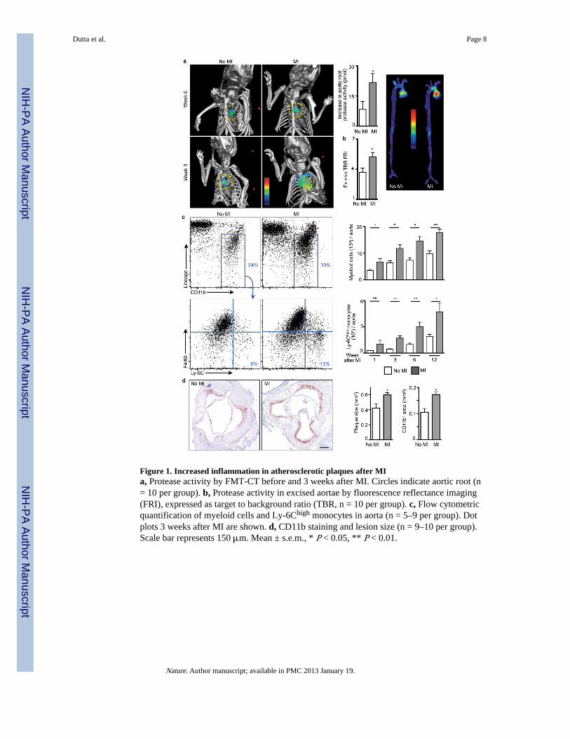
Figure 1. Increased inflammation in atherosclerotic plaques after MIa, Protease activity by FMT-CT before and 3 weeks after MI. Circles indicate aortic root (n= 10 per group). b, Protease activity in excised aortae by fluorescence reflectance imaging(FRI), expressed as target to background ratio (TBR, n = 10 per group). c, Flow cytometricquantification of myeloid cells and Ly-6Chigh monocytes in aorta (n = 5–9 per group). Dotplots 3 weeks after MI are shown. d, CD11b staining and lesion size (n = 9–10 per group).Scale bar represents 150 μm. Mean ± s.e.m., * P < 0.05, ** P < 0.01.
Dutta et al. Page 8
Nature. Author manuscript; available in PMC 2013 January 19.
NIH
-PA Author Manuscript
NIH
-PA Author Manuscript
NIH
-PA Author Manuscript
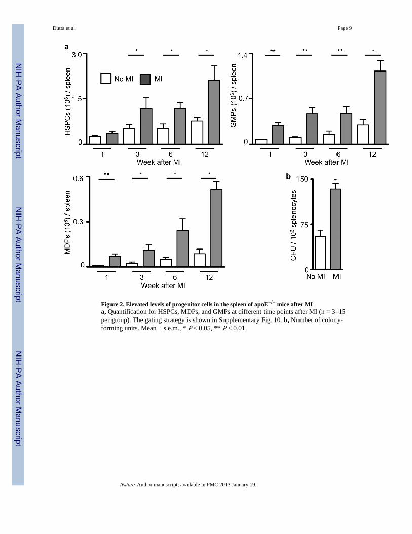
Figure 2. Elevated levels of progenitor cells in the spleen of apoE−/− mice after MIa, Quantification for HSPCs, MDPs, and GMPs at different time points after MI (n = 3–15per group). The gating strategy is shown in Supplementary Fig. 10. b, Number of colony-forming units. Mean ± s.e.m., * P < 0.05, ** P < 0.01.
Dutta et al. Page 9
Nature. Author manuscript; available in PMC 2013 January 19.
NIH
-PA Author Manuscript
NIH
-PA Author Manuscript
NIH
-PA Author Manuscript

Figure 3. β3 adrenergic receptor-mediated progenitor release after MIa, Flow cytometric analyses of HSPC in blood of C57BL/6 mice (n = 6–11 per group). b,Immunostaining for tyrosine hydroxylase (TH). Scale bar represents 10μm. Insets depictlow magnification overview. Bar graph shows quantitation of TH+ area (n = 5 per group). c,Expression of HSPC retention factors (relative to Gapdh) in the bone marrow of C57BL/6mice on day 4 after MI (n = 8 per group). Mean ± s.e.m., * P < 0.05, ** P < 0.01.
Dutta et al. Page 10
Nature. Author manuscript; available in PMC 2013 January 19.
NIH
-PA Author Manuscript
NIH
-PA Author Manuscript
NIH
-PA Author Manuscript

Figure 4. Serial intravital imaging of progenitor release from the bone marrowa, DiD labelled-HSPC Flk2− cells were imaged in the skull bone marrow by intravitalmicroscopy (IVM) before and then again 4 days after MI. DiD labelled-HSPC are white,blood pool red, and bone is blue. The scale bar represents 50 μm. b, Change of HSPCpresence between 1st and 2nd IVM session (n = 3 per group). Mean ± s.e.m., * P < 0.05.
Dutta et al. Page 11
Nature. Author manuscript; available in PMC 2013 January 19.
NIH
-PA Author Manuscript
NIH
-PA Author Manuscript
NIH
-PA Author Manuscript
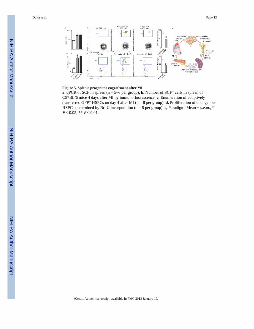
Figure 5. Splenic progenitor engraftment after MIa, qPCR of SCF in spleen (n = 5–6 per group). b, Number of SCF+ cells in spleen ofC57BL/6 mice 4 days after MI by immunofluorescence. c, Enumeration of adoptivelytransferred GFP+ HSPCs on day 4 after MI (n = 8 per group). d, Proliferation of endogenousHSPCs determined by BrdU incorporation (n = 8 per group). e, Paradigm. Mean ± s.e.m., *P < 0.05, ** P < 0.01.
Dutta et al. Page 12
Nature. Author manuscript; available in PMC 2013 January 19.
NIH
-PA Author Manuscript
NIH
-PA Author Manuscript
NIH
-PA Author Manuscript