myofibril growth during cardiac hypertrophy is regulated ... · pdf filemyofibril growth...
TRANSCRIPT
Cellular Signalling 28 (2016) 1015–1024
Contents lists available at ScienceDirect
Cellular Signalling
j ourna l homepage: www.e lsev ie r .com/ locate /ce l l s ig
Myofibril growth during cardiac hypertrophy is regulated through dualphosphorylation and acetylation of the actin capping protein CapZ
Ying-Hsi Lin a,b, Chad M. Warren a,b, Jieli Li a,b, Timothy A. McKinsey c, Brenda Russell a,b,⁎a Department of Physiology and Biophysics, University of Illinois at Chicago, College of Medicine, Chicago, IL 60612-7342, United Statesb Department of Physiology & Biophysics, Center for Cardiovascular Research, University of Illinois at Chicago, Chicago, IL 60612-7342, United Statesc Department of Medicine, Division of Cardiology and Center for Fibrosis Research and Translation, University of Colorado Anschutz Medical Campus, Aurora, CO 80045-0508, United States
⁎ Corresponding author at: Department of PhysiologyIllinois at Chicago, MC 901, 835 S. Wolcott, Chicago, IL 606
E-mail address: [email protected] (B. Russell).
http://dx.doi.org/10.1016/j.cellsig.2016.05.0110898-6568/© 2016 Published by Elsevier Inc.
a b s t r a c t
a r t i c l e i n f oArticle history:Received 7 March 2016Received in revised form 11 May 2016Accepted 12 May 2016Available online 13 May 2016
The mechanotransduction signaling pathways initiated in heart muscle by increased mechanical loading areknown to lead to long-term transcriptional changes and hypertrophy, but the rapid events for adaptation atthe sarcomeric level are not fully understood. The goal of this study was to test the hypothesis that actin filamentassembly during cardiomyocyte growth is regulated by post-translational modifications (PTMs) of CapZβ1. Inrapidly hypertrophying neonatal rat ventricular myocytes (NRVMs) stimulated by phenylephrine (PE), two-di-mensional gel electrophoresis (2DGE) of CapZβ1 revealed a shift toward more negative charge. Consistentwith this, mass spectrometry identified CapZβ1 phosphorylation on serine-204 and acetylation on lysine-199,two residues which are near the actin binding surface of CapZβ1. Ectopic expression of dominant negativePKCɛ (dnPKCɛ) in NRVMs blunted the PE-induced increase in CapZ dynamics, as evidenced by the kinetic con-stant (Kfrap) of fluorescence recovery after photobleaching (FRAP), and concomitantly reduced phosphorylationand acetylation of CapZβ1. Furthermore, inhibition of class I histone deacetylases (HDACs) increased lysine-199acetylation on CapZβ1, which increased Kfrap of CapZ and stimulated actin dynamics. Finally, we show that PEtreatment of NRVMs results in decreased binding of HDAC3 to myofibrils, suggesting a signal-dependent mech-anism for the regulation of sarcomere-associated CapZβ1 acetylation. Taken together, this dual regulationthrough phosphorylation and acetylation of CapZβ1 provides a novel model for the regulation of myofibrilgrowth during cardiac hypertrophy.
© 2016 Published by Elsevier Inc.
Keywords:Actin assemblySarcomereMechanotransductionSignaling pathwaysProteomics
1. Introduction
Transient physiological demands in mechanical loading induce hy-pertrophic growth of cardiomyocytes, increased contractility, and tissueremodeling. The mechanotransduction signaling pathways initiated byloading result in well studied long-term transcriptional changes butalso in rapid transient modifications at the protein level. Here we stud-ied the rapid post-translational modifications (PTMs) of phosphoryla-tion and acetylation of CapZβ1, a capping protein known to regulatethe assembly of myofibrils, which underlie cardiac hypertrophy. Theactin capping protein is a mushroom-like heterodimeric protein (αand β subunits) that binds to the barbed ends of the actin filamentsand slows down their assembly [1]. In muscle cells, this complex wasnamed CapZ because of its localization to the Z-disc [2]. Actin cappingand uncapping by CapZ are highly regulated by several binding proteinsand polyphosphoinositides [3,4,5]. Inmyocytes, both CapZ and actin dy-namics are increased very rapidly by mechanical strain, suggesting thepossible involvement of PTMs in actin filament assembly [4,6].
and Biophysics, University of12, United States.
Phosphorylation is a common PTM, with protein kinase C (PKC) iso-forms being crucial in cardiac contraction and hypertrophy [7,8,9]. ThePKCε isoform is abundantly expressed in human and rodent hearts[10,11]. Activation of PKCε enhances cardiac contractility and remodel-ing [12,13], and has a cardio-protective function in ischemic precondi-tioning [14,15,16]. Transgenic mice that overexpress a constitutivelyactive PKCε enzyme develop cardiac hypertrophy, leading to dilatedcardiomyopathy after many months [17,18]. In cardiomyocytes stimu-lated by mechanical strain, PKCε translocates to the Z-disc and modifiesCapZ, leading to increased CapZ dynamics. This provides a novel mech-anism for PKCε-mediated cardiomyocyte growth [19].
Acetylation of lysine residues is another major post-translationalevent. Acetyl groups are transferred to lysines by histone acetyltransfer-ases (HATs) and removed by histone deacetylases (HDACs) [20]. Themost well characterized function for acetylation is in epigenetic controlof gene expression through acetylation/deacetylation of nucleosomalhistone tails. However, proteomic studies have revealed that thousandsof non-histone proteins also undergo reversible acetylation [21,22,23,24]. The functional consequences of non-histone protein acetylation inthe heart remain poorly characterized.
The goal of the present studywas to test the hypothesis that actinfil-ament assembly is regulated by post-translational modifications of
1016 Y.-H. Lin et al. / Cellular Signalling 28 (2016) 1015–1024
CapZβ1, which are mediated by PKCε and HDACs. In hypertrophyingneonatal rat ventricular myocytes (NRVMs) stimulated by phenyleph-rine (PE), variations of PKCε and HDACs by activators or inhibitorswere applied to test whether phosphorylation or acetylation alterCapZ and actin dynamics. The coordination of phosphorylation andacetylation on CapZβ1 capping was approached by manipulating bothpathways, individually and simultaneously, to study PTMs of CapZβ1in rapidly hypertrophying NRVMs. The data reveal novel functions forPKCε and class I HDACs in the control of CapZβ1 activity and myofibrilformation during cardiac hypertrophy.
2. Materials and methods
2.1. Cell culture
Primary heart cultures were obtained from neonatal rats accordingto Institutional Animal Care and Use Committee and NIH guidelinesfor the care and use of laboratory animals. Hearts were removed andcells isolated from 1 to 2 days old neonatal Sprague-Dawley rats withcollagenase (Worthington) as previously described [25]. The cellswere re-suspended, filtered through a metal sieve to remove large ma-terial and plated at high density (200,000/cm2) in PC-1 medium(Biowhittaker/Cambrex) on fibronectin coated 3.5 cm dishes. Cellswere left undisturbed for 24 h in a 5% CO2 incubator when the unat-tached cells were removed by aspiration and the PC-1 media wasreplenished. Cells were incubated for another 24 h prior to theexperiment.
2.2. Neurohormonal stimulation of NRVM
The neurohormonal treatment times chosen were sufficient to in-duce hypertrophy [3,26] with phenylephrine (10 μM, Sigma-Aldrich)for 24 h prior to experiment analysis.
2.3. Cell size measurement
NRVMs were washed with PBS, fixed with 4% paraformaldehyde(Sigma-Aldrich) for 10 min, placed in cold 70% ethanol, and stored at−20 °C until immunostaining. Primary anti-α-actinin antibody (Cata-log No. ab9465, mouse IgG; Abcam, Cambridge, MA) was diluted(1:200) in 1% BSA in PBS (with 0.1% Triton X-100) and incubated on ashaker table at 4 °C overnight. Cells were then rinsed in PBS at 25 °Cand blocked in 1% BSA in PBS for 1 h at 25 °C. Secondary antibody(Molecular Probes) was diluted at a ratio of 1:500 in 1% BSA in PBSand incubated for 1 h at 25 °C. Cells were washed in PBS. Anti-fade re-agent with DAPI (Molecular Probes) was added and cover slips weremounted on glass slides. For the measurement of hypertrophy, NRVMboundaries were visualized by α-actinin antibody staining and cellarea was measured by Image J.
2.4. HDAC inhibitors
HDAC inhibitors were used at the indicated final concentrations andtreatment time: trichostatin A (TSA) (5 μM, 5 h; Sigma), MGCD0103(500 nM, 24 h; Selleck), theophylline (10 μM, 24 h; Sigma) andtubastatin A (1 μM, 24 h; Selleck).
2.5. Adenoviral and lentiviral constructs and infection
Recombinant adenoviruses for GFP-CapZβ1, constitutively activePKCε (caPKCε) and dominant negative PKCε (dnPKCε) were kindly pro-vided by Dr. Allen Samarel (Loyola University Chicago Stritch School ofMedicine, Maywood, IL) as previously described [3,27]. Two days afterNRVM isolation, NRVMs were infected with CapZβ1 (MOI 20), caPKCε(MOI 100), or dnPKCε (MOI 250) for 60min at 37 °C diluted in PC-1me-dium. The viralmediumwas then replacedwith virus-freemedium, and
cells were left undisturbed for 24 h. Lentiviruses encoding short-hairpin(sh) RNAs targeting HDAC1, HDAC2 and HDAC3 have been previouslydescribed [28].
2.6. Actin-GFP expression
Actin-GFP expressionwas induced by CellLight® Reagents *BacMam2.0* actin-GFP (Invitrogen). Two days after NRVM isolation, theCellLight® Reagent (30 μL per 1,000,000 cells) was used as modifiedfrom the manufacturer's instructions. Infected NRVMs were returnedto the incubator for at least 16 h.
2.7. Fluorescence recovery after photobleaching for CapZ and actindynamics
Fluorescence recovery after photobleaching (FRAP) has yieldedqualitative and quantitative information about the processes that regu-late actin polymerization in living myocytes [29]. The methods andanalysis for FRAP of actin-GFP were described by us [6]. Briefly, bindingof CapZ to the actin filament has two binding states [30], so FRAP curvesof CapZ were fit using non-linear regression in OriginPro (OriginLab,Northampton, MA):
Ifrap tð Þ ¼ 1−C1e−Koff1 t−C2e−Koff2 t: ð1Þ
The average kinetic constant (Kfrap) for dynamics was calculatedusing the following formula:
Kfrap ¼ C1Koff1 þ C2Koff2: ð2Þ
For FRAP of actin-GFP and actin-RFP, since actin binding activity hasone-binding state [31], the equation for curve fitting using non-linearregression in OriginPro was:
Ifrap tð Þ ¼ 1−C1e−Koff1 t: ð3Þ
The average kinetic constant (Kfrap) was calculated using the follow-ing formula:
Kfrap ¼ C1Koff1 ð4Þ
2.8. Co-immunoprecipitation
Cultured NRVM were infected with GFP-CapZβ1 adenovirus. After24 h of infection, cells were washed twice in ice-cold PBS and lysed inRIPA buffer (50 mM Tris-HCl pH 7.4, 150 mM NaCl, 2 mM EDTA, 1%NP40, 0.1% SDS) plus phosphatase inhibitors (Sigma Aldrich, #P5726,P0044) for 1 h at 4 °C under constant agitation. Following protein ex-traction, protein lysates were precleared using 25 μL Protein A/G Plus-Agarose beads (Santa Cruz, #sc-2003) for 1 h at 4 °C. Precleared lysateswere incubated 24 h with 2 μg of HDAC3 antibody (Cell Signaling Tech-nology; 4668) at 4 °C, then immunocomplexes were isolated by addingProtein A/G Plus-Agarose beads overnight at 4 °C. Beads were washedthree times in the binding buffer. SDS-PAGE and Western blottingwere performed using 12% Mini-PROTEAN® TGX™ Gel (Bio-Rad Labo-ratories, #456-1044). Polyvinylidene difluoride (PVDF) membraneswere incubated with GFP primary antibody (Enzo Life Sciences, ADI-SAB-500), followed by horseradish peroxidase (HRP) secondary anti-body (anti-mouse) for 1 h at room temperature. Proteins were finallytreated with an ECL Plus kit and visualized with the aid of ChemiDocXRS+ and analyzed with Image Lab (Bio-Rad Laboratories).
1017Y.-H. Lin et al. / Cellular Signalling 28 (2016) 1015–1024
2.9. Myobrillar protein enrichment
Forty-eight hours after cell culture, NRVMs were washed by PBStwice and added ice-cold 20% sucrose in relaxing solution (0.1 CaCl2,0.1 MgProp, 0.1 NaEGTA, 1 M KProp, 0.1 M Na2SO4, 1 M MOPS, 0.1 MATP) with protease and phosphatase inhibitors (Sigma, St. Louis, MO).After scraped into 1.5 eppendorf, cells slurries were vortexed withhighest speed for 30 s to break cell membrane. Cell slurries were lefton ice for 10min and spinned down at 300g at 4 °C for 5min. To excludethe remaining sucrose, cell slurries werewashed three times by ice-coldrelaxing solution with protease and phosphatase inhibitors. Then, cellslurries were homogenized by tissue homogenizer (Dremel Tissue-Tearor Homogenizer, model 985370) at level 5 for 10 s. Homogenizedslurries were washed by ice-cold relaxing solution three times to ex-clude non-myofibrillar remains, and suspended with 20× amount ofUTC buffer (8 M Urea, 2 M Thiourea, 4% Chaps with protease and phos-phatase inhibitors). After sit on ice for 30min with consistent agitation,insoluble pellets were spinned down at highest speed (13,200g) atroom temperature for 5 min.
2.10. Subcellular fractionation
For subcellular fractionation of myocytes, the CalbiochemProteoExtract Subcellular Proteome Extraction Kit was used (CatalogNo. 539790; EMD Millipore, Billerica, MA), following a previously de-scribed detergent-based protocol [32]. Cellular proteins were sequen-tially extracted into four compartments: cytosolic, membrane/organelles, nuclei, and cytoskeleton. Digitonin-EDTA was used to re-move the cytosol. Triton-EDTA was used to remove the membrane-or-ganelle fraction. Tween/deoxycholate/benzonase was used to removethe nuclei. Finally, SDS was used to remove the cytoskeleton. Cellswere briefly washed three times in PBS between each extraction frac-tion to prevent cross-contamination. After each fraction, cells were ob-served by light microscopy to ensure that they were still attached tothe dish. The accuracy of the fractionationmethodwas verifiedwith an-tibodies to well-documented subcellular distribution markers [heatshock protein (Hsp)70 for cytosol, β1-integrin for membrane, H2B fornucleus, and tropomyosin for myofibrils].
2.11. Immunoblotting
Protein extracts fromwhole cell lysates or different subcellular frac-tions were resolved by SDS/PAGE, transferred to polyvinyl difluoride(PVDF) membrane and probed with antibodies for CapZβ (EMDMillipore, AB6017), HDAC2 (Cell Signaling Technology, 4631), HDAC3(Abcam, ab16047; Cell Signaling Technology, 3949), α-tubulin (SantaCruz Biotechnology Technology, sc-32293), Hsp70 (Santa Cruz Biotech-nology Technology, sc-24), β-integrin (EMD Millipore, MAB1900), H2B(Abcam, ab18977), PCNA (Santa Cruz Biotechnology Technology, sc-25280), cardiac troponin I (Fitzgerald, 10R-T123k) and tropomyosin(provided by Dr. R. John Solaro at the University of Illinois at Chicago).
2.12. Two-dimensional gel electrophoresis (2DGE)
Cells were placed on ice and lysed using ice cold MF buffer (75 mMKCl, 10mM Imidazole, 2 mMMgCl2, 2mMEDTA, 1mMNaN3) contain-ing protease and phosphatase inhibitors. The pellet was thenresolubilized in urea-thiourea-chaps (UTC) buffer (8 M Urea, 2 M Thio-urea, 4% Chaps). The total protein concentration was measured usingthe RCDC™ protein assay (Bio-Rad)with crystalline bovine serumalbu-min as standard. For the first dimension electrophoresis, each proteinsample (~500 μg) was separated in 450 μL of IEF buffer (8 M Urea,2 M Thiourea, 4% Chaps, 1% Destreak and 0.25% (v/v) IPG bufferampholytes GE Healthcare) with 24 cm IPG strips Ph 4–7. The programfor the IEF cell was set up as an active rehydration at 50 V for 10–16 h,250 V rapid 15 min, 10,000 V linear 3 h, 10,000 rapid for 55,000 Vh.
After completion of thefirst dimension electrophoresis, the stripwas in-cubated in EQ buffer (6 M urea, 5%SDS (w/v), 30% glycerol (v/v)) with1% (w/v) DTT), and placed on a shaker at 50 rpm for 15 min. After15 min, the strip gel was taken out and incubated in EQ buffer with2.5% (w/v) iodoacetamide, and then placed on shaker for another15 min (50 rpm). The second dimension electrophoresis was rununder constant 25 mA for 90 min. After the second dimension electro-phoresis was complete, the protein was transferred to PVDF membranewith 10 mM CAPS Ph11.0 transfer buffer and run according to conven-tional protein transfer procedures.
2.13. Mass spectrometry
The protein migrating spots on Coomassie Blue-stained two-dimen-sional gel were cut out and the gel pieces were placed in distilled waterfor processing by the Proteomics Core Facility (UIC). The gel spots werefirst destained to remove Coomassie Blue, followed by reduction withDTT to remove disulfide linkage throughout the protein and then alkyl-ation to confer stability to the proteins prior to protease digestion. Theprotein was subjected to an in-gel digestion with trypsin and the pep-tideswere extracted from the gelmatrix. Electrospray Ionization FourierTransform Ion Cyclotron Resonance Mass Spectrometry (ESI-FTICR,Thermo Scientific) was employed to acquire site-specific informationregarding the PTMs of each of the CapZβ1 sites. MS/MS results were an-alyzed byMascotMS/MS ion search databasewith the following param-eters: fixed modifications: carbamidomethyl; variable modifications:acetyl (K), acetyl (protein N-terminal), oxidation (M), phosphorylation(ST), phosphorylation (Y);mass value:monoisotopic; proteinmass: un-restricted; peptidemass tolerance:±10ppm; fragmentmass tolerance:±0.6 Da; maxmissed cleavages: 1. The results were presented via Scaf-fold v 4.0 program (Proteome Software).
2.14. Statistics
Sample sizes were at least 4 immunoblots, 5 FRAP or 4 MS analysesper group. Spot density of 2D western blot was analyzed by ImageLabsoftware. Values of spot density were analyzed using a paired Student'sT-Test to compare control with a designated treatment. P b 0.05 wasconsidered significant.
3. Results
3.1. Post-translational modifications of CapZβ1 are increased in hypertro-phic NRVMs
We hypothesized that PTMs of CapZβ1 affect actin filament assem-bly dynamics. Myofibrillar proteins extracted from rapidlyhypertrophying NRVMs were separated by two-dimensional gel elec-trophoresis (2DGE, Fig. 1). To enhance CapZβ1 detection, we expressedGFP-CapZβ1 by adenovirus then probedwith aGFP antibody,whichhada higher affinity than the CapZ1β antibody. Western blotting for GFP-CapZβ1 by GFP displayed multiple spots. Three major distinct spotshad consistent presence in all images, with isoelectric points of approx-imately 5.53, 5.44, and 5.33 corresponding to an uncharged form (Spot1), a singly modified form (Spot 2), and a doubly modified form (Spot3), respectively (Fig. 1A). Interestingly, phenylephrine (PE) treatmentof NRVMs resulted in GFP-CapZβ1 2D spots shifting toward a lower iso-electric point (Spots 2 and 3, Fig. 1B). This shift was indicative of morenegative charges on the protein, suggesting that CapZβ1 was post-translationally modified in cardiomyocytes undergoing hypertrophy.
3.2. Identification of PTM sites and locations on CapZ
Mass spectrometry was used to identify the sites and types of PTMson CapZβ1 in NRVMs. Analysis of the three major spots of GFP-CapZβ1on 2D gel stained by Coomassie Blue showed phosphorylation of the
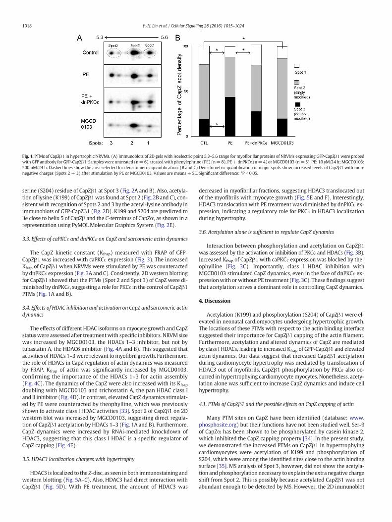
Fig. 1. PTMs of CapZβ1 in hypertrophic NRVMs. (A) Immunoblots of 2D gels with isoelectric point 5.3–5.6 range for myofibrillar proteins of NRVMs expressing GFP-CapZβ1 were probedwith GFP antibody for GFP-CapZβ1. Sampleswere untreated (n=6), treatedwith phenylephrine (PE) (n=8), PE+dnPKCε (n=4) orMGCD0103 (n=5). PE: 10 μM/24 h;MGCD0103:500 nM/24 h. Dashed lines show the area selected for densitometric quantification. (B and C) Densitometric quantification of major spots show increased levels of CapZβ1 with morenegative charges (Spots 2 + 3) after stimulation by PE or MGCD0103. Values are means ± SE. Significant difference: *P b 0.05.
1018 Y.-H. Lin et al. / Cellular Signalling 28 (2016) 1015–1024
serine (S204) residue of CapZβ1 at Spot 3 (Fig. 2A and B). Also, acetyla-tion of lysine (K199) of CapZβ1was found at Spot 2 (Fig. 2B and C), con-sistentwith recognition of Spots 2 and 3 by the acetyl-lysine antibody inimmunoblots of GFP-CapZβ1 (Fig. 2D). K199 and S204 are predicted tolie close to helix 5 of CapZβ and the C-terminus of CapZα, as shown in arepresentation using PyMOL Molecular Graphics System (Fig. 2E).
3.3. Effects of caPKCɛ and dnPKCɛ on CapZ and sarcomeric actin dynamics
The CapZ kinetic constant (Kfrap) measured with FRAP of GFP-CapZβ1 was increased with caPKCɛ expression (Fig. 3). The increasedKfrap of CapZβ1 when NRVMs were stimulated by PE was counteractedby dnPKCɛ expression (Fig. 3A and C). Consistently, 2Dwestern blottingfor CapZβ1 showed that the PTMs (Spot 2 and Spot 3) of CapZ were di-minished by dnPKCɛ, suggesting a role for PKCɛ in the control of CapZβ1PTMs (Fig. 1A and B).
3.4. Effects of HDAC inhibition and activation on CapZ and sarcomeric actindynamics
The effects of different HDAC isoforms onmyocyte growth and CapZstatuswere assessed after treatmentwith specific inhibitors. NRVM sizewas increased by MGCD0103, the HDACs 1–3 inhibitor, but not bytubastatin A, the HDAC6 inhibitor (Fig. 4A and B). This suggested thatactivities of HDACs1–3were relevant tomyofibril growth. Furthermore,the role of HDACs in CapZ regulation of actin dynamics was measuredby FRAP. Kfrap of actin was significantly increased by MGCD0103,confirming the importance of the HDACs 1–3 for actin assembly(Fig. 4C). The dynamics of the CapZ were also increased with its Kfrap
doubling with MGCD0103 and trichostatin A, the pan HDAC class Iand II inhibitor (Fig. 4D). In contrast, elevated CapZ dynamics stimulat-ed by PE were counteracted by theophylline, which was previouslyshown to activate class I HDAC activities [33]. Spot 2 of CapZβ1 on 2Dwestern blot was increased by MGCD0103, suggesting direct regula-tion of CapZβ1 acetylation by HDACs 1–3 (Fig. 1A and B). Furthermore,CapZ dynamics were increased by RNAi-mediated knockdown ofHDAC3, suggesting that this class I HDAC is a specific regulator ofCapZ capping (Fig. 4E).
3.5. HDAC3 localization changes with hypertrophy
HDAC3 is localized to the Z-disc, as seen in both immunostaining andwestern blotting (Fig. 5A–C). Also, HDAC3 had direct interaction withCapZβ1 (Fig. 5D). With PE treatment, the amount of HDAC3 was
decreased in myofibrillar fractions, suggesting HDAC3 translocated outof the myofibrils with myocyte growth (Fig. 5E and F). Interestingly,HDAC3 translocation with PE treatment was diminished by dnPKCɛ ex-pression, indicating a regulatory role for PKCɛ in HDAC3 localizationduring hypertrophy.
3.6. Acetylation alone is sufficient to regulate CapZ dynamics
Interaction between phosphorylation and acetylation on CapZβ1was assessed by the activation or inhibition of PKCε and HDACs (Fig. 3B).Increased Kfrap of CapZβ1 with caPKCɛ expression was blocked by the-ophylline (Fig. 3C). Importantly, class I HDAC inhibition withMGCD0103 stimulated CapZ dynamics, even in the face of dnPKCɛ ex-pressionwith orwithout PE treatment (Fig. 3C). These findings suggestthat acetylation serves a dominant role in controlling CapZ dynamics.
4. Discussion
Acetylation (K199) and phosphorylation (S204) of CapZβ1 were el-evated in neonatal cardiomyocytes undergoing hypertrophic growth.The locations of these PTMs with respect to the actin binding interfacesuggested their importance for CapZβ1 capping of the actin filament.Furthermore, acetylation and altered dynamics of CapZ are mediatedby class I HDACs, leading to increased Kfrap of GFP-CapZβ1 and elevatedactin dynamics. Our data suggest that increased CapZβ1 acetylationduring cardiomyocyte hypertrophy was mediated by translocation ofHDAC3 out of myofibrils. CapZβ1 phosphorylation by PKCɛ also oc-curred in hypertrophying cardiomyocytemyocytes. Nonetheless, acety-lation alone was sufficient to increase CapZ dynamics and induce cellhypertrophy.
4.1. PTMs of CapZβ1 and the possible effects on CapZ capping of actin
Many PTM sites on CapZ have been identified (database: www.phosphosite.org) but their functions have not been studied well. Ser-9of CapZα has been shown to be phosphorylated by casein kinase 2,which inhibited the CapZ capping property [34]. In the present study,we demonstrated the increased PTMs on CapZβ1 in hypertrophyingcardiomyocytes were acetylation of K199 and phosphorylation ofS204, which were among the identified sites close to the actin bindingsurface [35]. MS analysis of Spot 3, however, did not show the acetyla-tion andphosphorylation necessary to explain the extra negative chargeshift from Spot 2. This is possibly because acetylated CapZβ1 was notabundant enough to be detected by MS. However, the 2D immunoblot
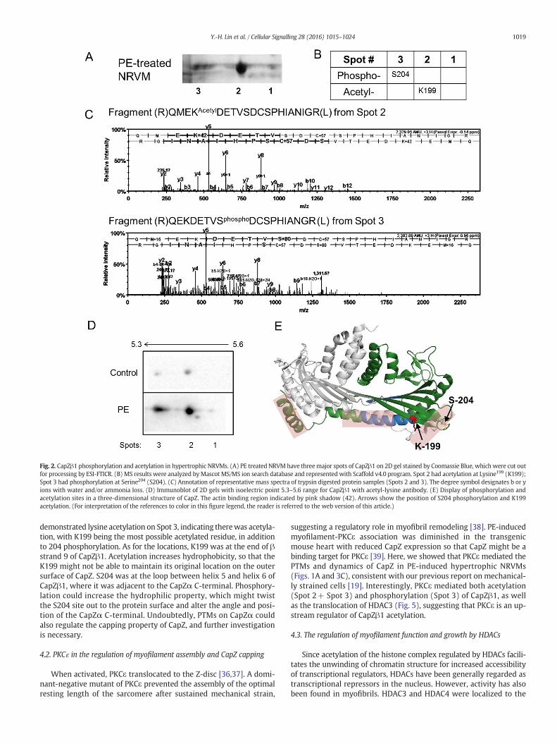
Fig. 2. CapZβ1 phosphorylation and acetylation in hypertrophic NRVMs. (A) PE treated NRVM have threemajor spots of CapZβ1 on 2D gel stained by Coomassie Blue, which were cut outfor processing by ESI-FTICR. (B) MS results were analyzed byMascot MS/MS ion search database and represented with Scaffold v4.0 program. Spot 2 had acetylation at Lysine199 (K199);Spot 3 had phosphorylation at Serine204 (S204). (C) Annotation of representative mass spectra of trypsin digested protein samples (Spots 2 and 3). The degree symbol designates b or yions with water and/or ammonia loss. (D) Immunoblot of 2D gels with isoelectric point 5.3–5.6 range for CapZβ1 with acetyl-lysine antibody. (E) Display of phosphorylation andacetylation sites in a three-dimensional structure of CapZ. The actin binding region indicated by pink shadow (42). Arrows show the position of S204 phosphorylation and K199acetylation. (For interpretation of the references to color in this figure legend, the reader is referred to the web version of this article.)
1019Y.-H. Lin et al. / Cellular Signalling 28 (2016) 1015–1024
demonstrated lysine acetylation on Spot 3, indicating therewas acetyla-tion, with K199 being the most possible acetylated residue, in additionto 204 phosphorylation. As for the locations, K199 was at the end of βstrand 9 of CapZβ1. Acetylation increases hydrophobicity, so that theK199 might not be able to maintain its original location on the outersurface of CapZ. S204 was at the loop between helix 5 and helix 6 ofCapZβ1, where it was adjacent to the CapZα C-terminal. Phosphory-lation could increase the hydrophilic property, which might twistthe S204 site out to the protein surface and alter the angle and posi-tion of the CapZα C-terminal. Undoubtedly, PTMs on CapZα couldalso regulate the capping property of CapZ, and further investigationis necessary.
4.2. PKCɛ in the regulation of myofilament assembly and CapZ capping
When activated, PKCε translocated to the Z-disc [36,37]. A domi-nant-negative mutant of PKCε prevented the assembly of the optimalresting length of the sarcomere after sustained mechanical strain,
suggesting a regulatory role in myofibril remodeling [38]. PE-inducedmyofilament-PKCε association was diminished in the transgenicmouse heart with reduced CapZ expression so that CapZ might be abinding target for PKCε [39]. Here, we showed that PKCε mediated thePTMs and dynamics of CapZ in PE-induced hypertrophic NRVMs(Figs. 1A and 3C), consistent with our previous report on mechanical-ly strained cells [19]. Interestingly, PKCε mediated both acetylation(Spot 2+ Spot 3) and phosphorylation (Spot 3) of CapZβ1, as wellas the translocation of HDAC3 (Fig. 5), suggesting that PKCε is an up-stream regulator of CapZβ1 acetylation.
4.3. The regulation of myofilament function and growth by HDACs
Since acetylation of the histone complex regulated by HDACs facili-tates the unwinding of chromatin structure for increased accessibilityof transcriptional regulators, HDACs have been generally regarded astranscriptional repressors in the nucleus. However, activity has alsobeen found in myofibrils. HDAC3 and HDAC4 were localized to the
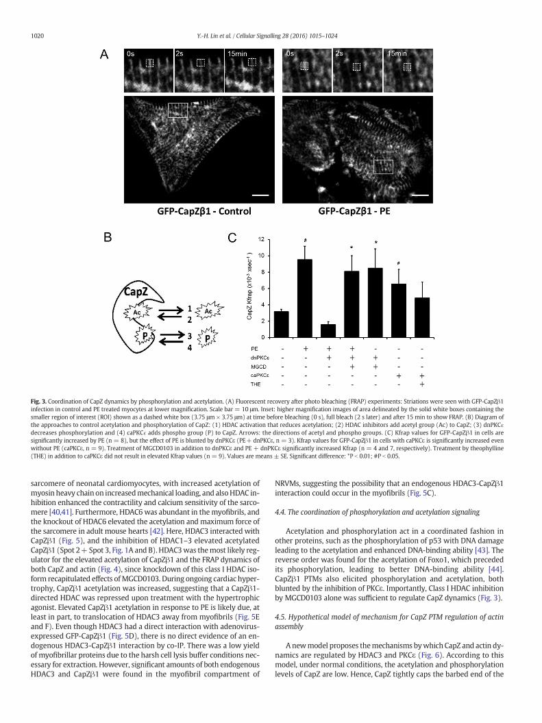
Fig. 3. Coordination of CapZ dynamics by phosphorylation and acetylation. (A) Fluorescent recovery after photo bleaching (FRAP) experiments: Striations were seen with GFP-CapZβ1infection in control and PE treated myocytes at lower magnification. Scale bar = 10 μm. Inset: higher magnification images of area delineated by the solid white boxes containing thesmaller region of interest (ROI) shown as a dashed white box (3.75 μm × 3.75 μm) at time before bleaching (0 s), full bleach (2 s later) and after 15 min to show FRAP. (B) Diagram ofthe approaches to control acetylation and phosphorylation of CapZ: (1) HDAC activation that reduces acetylation; (2) HDAC inhibitors add acetyl group (Ac) to CapZ; (3) dnPKCɛdecreases phosphorylation and (4) caPKCɛ adds phospho group (P) to CapZ. Arrows: the directions of acetyl and phospho groups. (C) Kfrap values for GFP-CapZβ1 in cells aresignificantly increased by PE (n = 8), but the effect of PE is blunted by dnPKCε (PE+ dnPKCε, n = 3). Kfrap values for GFP-CapZβ1 in cells with caPKCε is significantly increased evenwithout PE (caPKCε, n = 9). Treatment of MGCD0103 in addition to dnPKCε and PE + dnPKCε significantly increased Kfrap (n = 4 and 7, respectively). Treatment by theophylline(THE) in addition to caPKCε did not result in elevated Kfrap values (n = 9). Values are means ± SE. Significant difference: *P b 0.01; #P b 0.05.
1020 Y.-H. Lin et al. / Cellular Signalling 28 (2016) 1015–1024
sarcomere of neonatal cardiomyocytes, with increased acetylation ofmyosin heavy chain on increasedmechanical loading, and alsoHDAC in-hibition enhanced the contractility and calcium sensitivity of the sarco-mere [40,41]. Furthermore, HDAC6 was abundant in themyofibrils, andthe knockout of HDAC6 elevated the acetylation and maximum force ofthe sarcomere in adult mouse hearts [42]. Here, HDAC3 interacted withCapZβ1 (Fig. 5), and the inhibition of HDAC1–3 elevated acetylatedCapZβ1 (Spot 2+Spot 3, Fig. 1A and B). HDAC3was themost likely reg-ulator for the elevated acetylation of CapZβ1 and the FRAP dynamics ofboth CapZ and actin (Fig. 4), since knockdown of this class I HDAC iso-form recapitulated effects ofMGCD0103. During ongoing cardiac hyper-trophy, CapZβ1 acetylation was increased, suggesting that a CapZβ1-directed HDAC was repressed upon treatment with the hypertrophicagonist. Elevated CapZβ1 acetylation in response to PE is likely due, atleast in part, to translocation of HDAC3 away from myofibrils (Fig. 5Eand F). Even though HDAC3 had a direct interaction with adenovirus-expressed GFP-CapZβ1 (Fig. 5D), there is no direct evidence of an en-dogenous HDAC3-CapZβ1 interaction by co-IP. There was a low yieldof myofibrillar proteins due to the harsh cell lysis buffer conditions nec-essary for extraction. However, significant amounts of both endogenousHDAC3 and CapZβ1 were found in the myofibril compartment of
NRVMs, suggesting the possibility that an endogenous HDAC3-CapZβ1interaction could occur in the myofibrils (Fig. 5C).
4.4. The coordination of phosphorylation and acetylation signaling
Acetylation and phosphorylation act in a coordinated fashion inother proteins, such as the phosphorylation of p53 with DNA damageleading to the acetylation and enhanced DNA-binding ability [43]. Thereverse order was found for the acetylation of Foxo1, which precededits phosphorylation, leading to better DNA-binding ability [44].CapZβ1 PTMs also elicited phosphorylation and acetylation, bothblunted by the inhibition of PKCε. Importantly, Class I HDAC inhibitionby MGCD0103 alone was sufficient to regulate CapZ dynamics (Fig. 3).
4.5. Hypothetical model of mechanism for CapZ PTM regulation of actinassembly
Anewmodel proposes themechanisms bywhich CapZ and actin dy-namics are regulated by HDAC3 and PKCε (Fig. 6). According to thismodel, under normal conditions, the acetylation and phosphorylationlevels of CapZ are low. Hence, CapZ tightly caps the barbed end of the
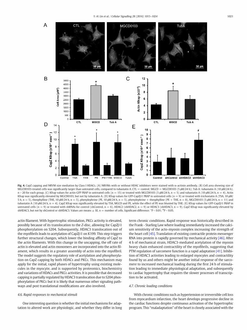
Fig. 4. CapZ capping and NRVM size mediation by Class I HDACs. (A) NRVMs with or without HDAC inhibitors were stained with α-actinin antibody. (B) Cell area showing size ofMGCD0103-treated cells was significantly larger than untreated cells, compared to tubastatin A. CTL = control; MGCD = MGCD0103 (5 μM/24 h); Tub A: tubastatin A (10 μM/24 h).n N 20 for each group. (C) Kfrap values for actin-GFP FRAP in untreated cells (n = 15) or treated with MGCD0103 (5 μM/24 h, n = 5) and tubastatin A (10 μM/24 h, n = 4). ActinKfrap was significantly elevated by MGCD0103, but not by tubastatin A. (D) Kfrap values for GFP-CapZβ1 FRAP in untreated cells (n = 9) or treated with trichostatin A (TSA, 10 μM/5 h, n = 5), theophylline (THE, 10 μM/24 h, n = 5), phenylephrine (PE, 10 μM/24 h, n = 5), phenylephrine + theophylline (PE + THE, n = 8), MGCD0103 (5 μM/24 h, n = 11) andtubastatin A (10 μM/24 h, n = 4). CapZ Kfrap was significantly elevated by TSA, MGCD and PE, while the effect of PE was blunted by THE. (E) Kfrap values for GFP-CapZβ1 FRAP inuntreated cells (n = 9) or treated with shRNAs for control (shControl, n = 6), HDAC2 (shHDAC2, n = 9) or HDAC3 (shHDAC3, n = 9). CapZ Kfrap was significantly elevated byshHDAC3, but not by shControl or shHDAC3. Values are means ± SE. n = number of cells. Significant difference: *P b 0.01; #P b 0.05.
1021Y.-H. Lin et al. / Cellular Signalling 28 (2016) 1015–1024
actin filament. With hypertrophic stimulation, PKCε activity is elevated,possibly because of its translocation to the Z-disc, allowing for CapZβ1phosphorylation on S204. Subsequently, HDAC3 translocation out ofthemyofibrils leads to acetylation of CapZβ1 on K199. This step triggersfurther structural changes, which lower the binding affinity of CapZ tothe actin filaments. With this change in the uncapping, the off rate ofactin is elevated and actinmonomers are incorporated into the actin fil-ament, which results in a greater assembly of actin into the myofibril.The model suggests the regulatory role of acetylation and phosphoryla-tion on CapZ capping by both HDACs and PKCε. This mechanism mayapply for the initial, rapid phases of hypertrophy using existing mole-cules in the myocyte, and is supported by proteomics, biochemistryand variations of HDACs and PKCε activities. It is possible that decreasedcapping is partially regulated byHDAC3 translocation due to S204 phos-phorylation of PKCε but it is likely that numerous other signaling path-ways and post translational modifications are also involved.
4.6. Rapid responses to mechanical stimuli
One interesting question iswhether the initialmechanisms for adap-tation to altered work are physiologic, and whether they differ in long
term chronic conditions. Rapid response was historically described inthe Frank - Starling Lawwhere loading immediately increased the calci-um sensitivity of the acto-myosin complex increasing the strength ofthe heart cell [45]. Translation of existing contractile proteinmessengerRNA into protein is rapidly governed by mechanical activity [46]. After4 h of mechanical strain, HDAC3-mediated acetylation of the myosinheavy chain enhanced contractility of the myofibrils, suggesting thatPTM regulation of sarcomere function is a rapid adaptation [41]. Inhibi-tion of HDAC3 activities leading to enlarged myocytes and contractilityfound by us and others might be another initial response of the sarco-mere to increased mechanical loading during the first 24 h of stimula-tion leading to immediate physiological adaptation, and subsequentlyto cardiac hypertrophy that requires the slower processes of transcrip-tion to be activated.
4.7. Chronic loading conditions
With chronic conditions such as hypertension or irreversible cell lossfrom myocardium infarction, the heart develops progressive decline inthe cardiac functions despite continuous activation of the hypertrophicprogram. This “maladaptation” of theheart is closely associatedwith the
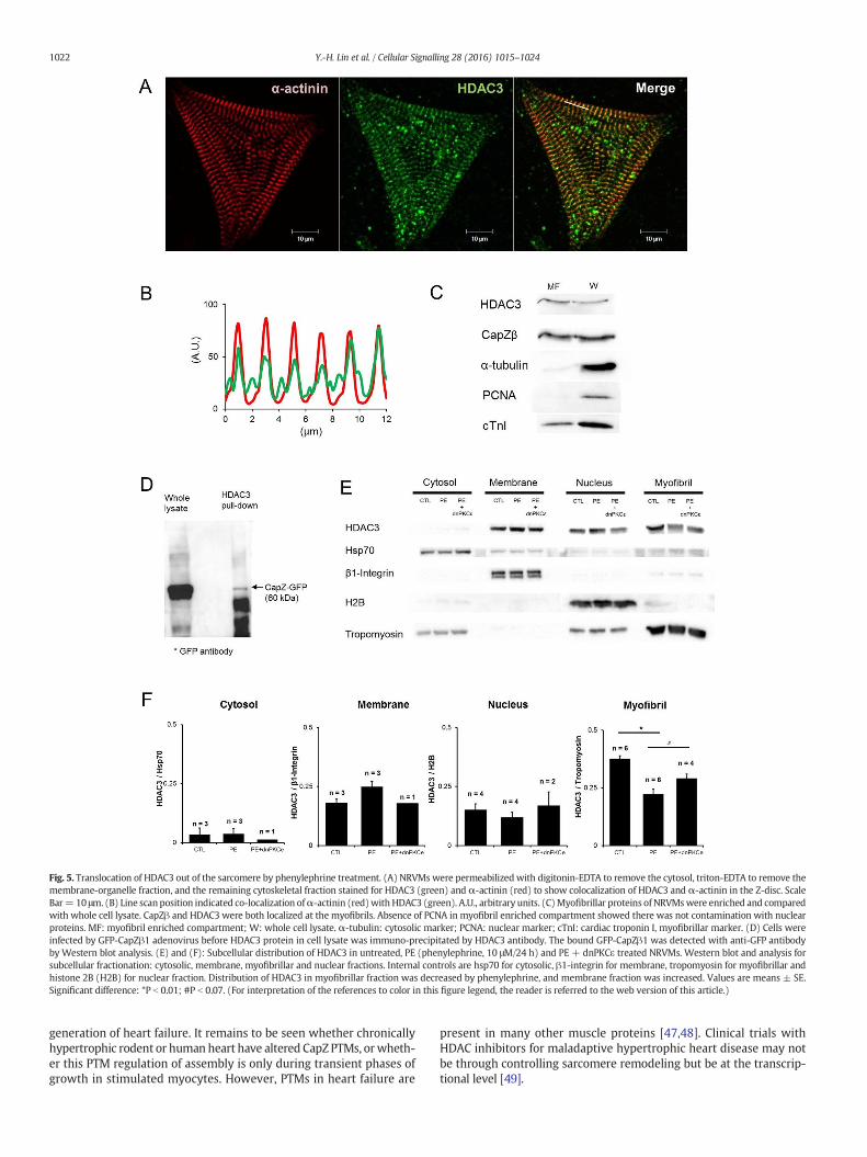
Fig. 5. Translocation of HDAC3 out of the sarcomere by phenylephrine treatment. (A) NRVMs were permeabilized with digitonin-EDTA to remove the cytosol, triton-EDTA to remove themembrane-organelle fraction, and the remaining cytoskeletal fraction stained for HDAC3 (green) and α-actinin (red) to show colocalization of HDAC3 and α-actinin in the Z-disc. ScaleBar= 10 μm. (B) Line scan position indicated co-localization ofα-actinin (red)with HDAC3 (green). A.U., arbitrary units. (C)Myofibrillar proteins of NRVMswere enriched and comparedwith whole cell lysate. CapZβ and HDAC3 were both localized at the myofibrils. Absence of PCNA in myofibril enriched compartment showed there was not contamination with nuclearproteins. MF: myofibril enriched compartment; W: whole cell lysate. α-tubulin: cytosolic marker; PCNA: nuclear marker; cTnI: cardiac troponin I, myofibrillar marker. (D) Cells wereinfected by GFP-CapZβ1 adenovirus before HDAC3 protein in cell lysate was immuno-precipitated by HDAC3 antibody. The bound GFP-CapZβ1 was detected with anti-GFP antibodyby Western blot analysis. (E) and (F): Subcellular distribution of HDAC3 in untreated, PE (phenylephrine, 10 μM/24 h) and PE + dnPKCε treated NRVMs. Western blot and analysis forsubcellular fractionation: cytosolic, membrane, myofibrillar and nuclear fractions. Internal controls are hsp70 for cytosolic, β1-integrin for membrane, tropomyosin for myofibrillar andhistone 2B (H2B) for nuclear fraction. Distribution of HDAC3 in myofibrillar fraction was decreased by phenylephrine, and membrane fraction was increased. Values are means ± SE.Significant difference: *P b 0.01; #P b 0.07. (For interpretation of the references to color in this figure legend, the reader is referred to the web version of this article.)
1022 Y.-H. Lin et al. / Cellular Signalling 28 (2016) 1015–1024
generation of heart failure. It remains to be seen whether chronicallyhypertrophic rodent or human heart have altered CapZ PTMs, orwheth-er this PTM regulation of assembly is only during transient phases ofgrowth in stimulated myocytes. However, PTMs in heart failure are
present in many other muscle proteins [47,48]. Clinical trials withHDAC inhibitors for maladaptive hypertrophic heart disease may notbe through controlling sarcomere remodeling but be at the transcrip-tional level [49].
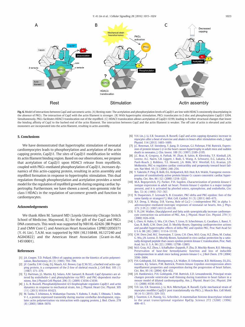
Fig. 6.Model of interactions between CapZ and sarcomeric actin. (A) Resting state: The acetylation and phosphorylation levels of CapZβ1 are lowwith HDAC3 consistently deacetylating inthe absence of PKCε. The interaction of CapZ with the actin filament is stronger. (B) With hypertrophic stimulation, PKCε translocates to Z-disc and phosphorylates CapZβ1 S204.Simultaneously, PKCε facilitates HDAC3 translocation out of the myofibril. (C) HDAC3 translocation allows acetylation of CapZβ1 K199, leading to further structural changes that lowerthe binding affinity of CapZ to the barbed end of the actin filament. The interaction between CapZ and the actin filament is weaker. The off rate of actin is elevated and actinmonomers are incorporated into the actin filament, resulting in actin assembly.
1023Y.-H. Lin et al. / Cellular Signalling 28 (2016) 1015–1024
5. Conclusions
We have demonstrated that hypertrophic stimulation of neonatalcardiomyocytes leads to phosphorylation and acetylation of the actincapping protein, CapZβ1. The sites of CapZβ1 modification lie withinits actin filament binding region. Based on our observations, we proposethat acetylation of CapZβ1 upon HDAC3 release from myofibrils,coupled with PKCε-mediated phosphorylation of CapZβ1, increases dy-namics of this actin-capping protein, resulting in actin assembly andmyofibril formation in response to hypertrophic stimulation. This dualregulation through phosphorylation and acetylation provides a novelmodel for the regulation ofmyofibril growth during ongoing cardiac hy-pertrophy. Furthermore, we have shown a novel, non-genomic role forclass I HDACs in the regulation of sarcomere growth and function incardiomyocytes.
Acknowledgements
We thank Allen M. Samarel MD (Loyola University Chicago StritchSchool of Medicine, Maywood, IL) for the gift of the CapZ and PKCεDNA constructs. This work was supported by NIH HL-62426 (BR Project2 and CMW Core C) and American Heart Association 12PRE12050371(Y.-H. Lin). T.A.M. was supported by NIH (HL116848, HL127240 andAG043822) and the American Heart Association (Grant-in-Aid,14510001).
References
[1] J.A. Cooper, T.D. Pollard, Effect of capping protein on the kinetics of actin polymeri-zation, Biochemistry 24 (3) (1995) 793–799.
[2] J.F. Casella, S.W. Craig, D.J. Maack, A.E. Brown, Cap Z(36/32), a barbed end actin-cap-ping protein, is a component of the Z-line of skeletal muscle, J. Cell Biol. 105 (1)(1987) 371–379.
[3] T.J. Hartman, J.L. Martin, R.J. Solaro, A.M. Samarel, B. Russell, CapZ dynamics are al-tered by endothelin-1 and phenylephrine via PIP2- and PKC-dependent mecha-nisms, Am J Physiol Cell Physiol. 296 (5) (2009) C1034–C1039.
[4] J. Li, B. Russell, Phosphatidylinositol 4,5-bisphosphate regulates CapZβ1 and actindynamics in response to mechanical strain, Am. J. Physiol. Heart Circ. Physiol. 305(11) (2013) H1614–H1623.
[5] M. Taoka, T. Ichimura, A. Wakamiya-Tsuruta, Y. Kubota, T. Araki, T. Obinata, T. Isobe,V-1, a protein expressed transiently during murine cerebellar development, regu-lates actin polymerization via interaction with capping protein, J. Biol. Chem. 278(8) (2003) 5864–5870.
[6] Y.H. Lin, J. Li, E.R. Swanson, B. Russell, CapZ and actin capping dynamics increase inmyocytes after a bout of exercise and abates in hours after stimulation ends, J. Appl.Physiol. 114 (2013) 1603–1609.
[7] J.C. Bowman, S.F. Steinberg, T. Jiang, D. Gennan, G.I. Fishman, P.M. Buttrick, Expres-sion of protein kinase C-β in the heart causes hypertrophy in adult mice and suddendeath in neonates, J. Clin. Invest. 100 (9) (1997) 2189–2195.
[8] J.C. Braz, K. Gregory, A. Pathak, W. Zhao, B. Sahin, R. Klevitsky, T.F. Kimball, J.N.Lorenz, A.C. Nairn, S.B. Liggett, I. Bodi, S. Wang, A. Schwartz, E.G. Lakatta, A.A.Paoli-Roach, J. Robbins, T.E. Hewett, J.A. Bibb, M.V. Westfall, E.G. Kranias, J.D.Molkentin, PKC-α regulates cardiac contractility and propensity toward heart fail-ure, Nat Med. 10 (3) (2004) 248–254.
[9] Y. Takeishi, P. Ping, R. Bolli, D.L. Kirkpatrick, B.D. Hoit, R.A.Walsh, Transgenic overex-pression of constitutively active protein kinase Cε causes concentric cardiac hyper-trophy, Circ. Res. 86 (12) (2000) 1218–1223.
[10] M.A. Bogoyevitch, P.J. Parker, P.H. Sugden, Characterization of protein kinase Cisotype expression in adult rat heart. Protein kinase C-epsilon is a major isotypepresent, and it is activated by phorbol esters, epinephrine, and endothelin, CircRes. 72 (4) (1993) 757–767.
[11] N. Duquesnes, F. Lezoualc'h, B. Crozatier, PKC-delta and PKC-epsilon: foes of thesame family or strangers? J. Mol. Cell. Cardiol. 51 (5) (2011) 665–673.
[12] X.F. Deng, S. Mulay, D.R. Varma, Role of Ca(2+)-independent PKC in alpha 1-adrenoceptor-mediated inotropic responses of neonatal rat hearts, Am. J. Phys.273 (3 Pt 2) (1997) H1113–H1118.
[13] Y. Pi, J.W. Walker, Diacylglycerol and fatty acids synergistically increase cardiomyo-cyte contraction via activation of PKC, Am. J. Physiol. Heart Circ. Physiol. 279 (1)(2000) H26–H34.
[14] L. Chen, H. Hahn, G. Wu, C.H. Chen, T. Liron, D. Schechtman, G. Cavallaro, L. Banci, Y.Guo, R. Bolli, G.W. Dorn 2nd, D. Mochly-Rosen, Opposing cardioprotective actionsand parallel hypertrophic effects of delta PKC and epsilon PKC, Proc Natl Acad SciU S A. 98 (20) (2001) 11114–11119.
[15] G.W. Dorn 2nd, M.C. Souroujon, T. Liron, C.H. Chen, M.O. Gray, H.Z. Zhou, M. Csukai,G. Wu, J.N. Lorenz, D. Mochly-Rosen, Sustained in vivo cardiac protection by a ratio-nally designed peptide that causes epsilon protein kinase C translocation, Proc. Natl.Acad. Sci. U. S. A. 96 (22) (1999) 12798–12803.
[16] M.O. Gray, H.Z. Zhou, I. Schafhalter-Zoppoth, P. Zhu, D. Mochly-Rosen, R.O. Messing,Preservation of base-line hemodynamic function and loss of induciblecardioprotection in adult mice lacking protein kinase C-ε, J Biol Chem. 279 (2004)3596–3604.
[17] P.H. Goldspink, D.E. Montgomery, L.A. Walker, D. Urboniene, R.D. McKinney, D.L.D.L.Geenen, R.J. Solaro, P.M. Buttrick, Protein kinase Cepsilon overexpression altersmyofilament properties and composition during the progression of heart failure,Circ. Res. 95 (4) (2004) 424–432.
[18] J.H. Hankiewicz, P.H. Goldspink, P.M. Buttrick, E.D. Lewandowski, Principal strainchanges precede ventricular wall thinning during transition to heart failure in amouse model of dilated cardiomyopathy, Am. J. Physiol. Heart Circ. Physiol. 294(1) (2008) H330–H336.
[19] Y.H. Lin, E.R. Swanson, J. Li, M.A. Mkrtschjan, B. Russell, Cyclic mechanical strain ofmyocytes modifies CapZβ1 post translationally via PKCε, J. Muscle Res. Cell Motil.36 (4–5) (2015) 329–333.
[20] J. Taunton, C.A. Hassig, S.L. Schreiber, A mammalian histone deacetylase relatedto the yeast transcriptional regulator Rpd3p, Science 272 (5260) (1996)408–411.

1024 Y.-H. Lin et al. / Cellular Signalling 28 (2016) 1015–1024
[21] C. Choudhary, C. Kumar, F. Gnad, M.L. Nielsen, M. Rehman, T.C. Walther, J.V. Olsen,M. Mann, Lysine acetylation targets protein complexes and co-regulates major cel-lular functions, Science 325 (2009) 834–840.
[22] C. Choudhary, B.T. Weinert, Y. Nishida, E. Verdin, M. Mann, The growing landscapeof lysine acetylation links metabolism and cell signalling, Nat Rev Mol Cell Biol. 15(2012) 536–550.
[23] D.B. Foster, T. Liu, J. Rucker, R.N. O'Meally, L.R. Devine, R.N. Cole, B. O'Rourke, The car-diac acetyl-lysine proteome, PLoS ONE 8 (2013), e67513.
[24] A. Lundby, K. Lage, B.T. Weinert, D.B. Bekker-Jensen, A. Secher, T. Skovgaard, C.D.Kelstrup, A. Dmytriyev, C. Choudhary, C. Lundby, J.V. Olsen, Proteomic analysis of ly-sine acetylation sites in rat tissues reveals organ specificity and subcellular patterns,Cell Rep. 2 (2012) 419–431.
[25] S.Y. Boateng, T.J. Hartman, N. Ahluwalia, H. Vidula, T.A. Desai, B. Russell, Inhibition offibroblast proliferation in cardiac myocyte cultures by surfacemicrotopography, AmJ Physiol Cell Physiol. 285 (2003) C171–C182.
[26] P. Simpson, Norepinephrine-stimulated hypertrophy of cultured rat myocardialcells is an alpha 1 adrenergic response, J. Clin. Invest. 72 (1983) 732–738.
[27] J.B. Strait 3rd, J.L. Martin, A. Bayer, R. Mestril, D.M. Eble, A.M. Samarel, Role of proteinkinase C-epsilon in hypertrophy of cultured neonatal rat ventricular myocytes, Am.J. Physiol. Heart Circ. Physiol. 280 (2) (2001) H756–H766.
[28] W.W. Blakeslee, C.L. Wysoczynski, K.S. Fritz, J.K. Nyborg, M.E. Churchill, T.A.McKinsey, Class I HDAC inhibition stimulates cardiac protein SUMOylation througha post-translational mechanism, Cell. Signal. 26 (12) (2014) 2912–2920.
[29] P. Roy, Z. Rajfur, P. Pomorski, K. Jacobson, Microscope-based techniques to study celladhesion and migration, Nat. Cell Biol. 4 (2002) E91–E96.
[30] S. Takeda, S. Minakata, R. Koike, I. Kawahata, A. Narita, M. Kitazawa, M. Ota, T.Yamakuni, Y. Maéda, Y. Nitanai, Two distinct mechanisms for actin capping proteinregulation-steric and allosteric inhibition, PLoS Biol. 8 (2010), e1000416.
[31] T.D. Pollard, M.S. Mooseker, Direct measurement of actin polymerization rate con-stants by electron microscopy of actin filaments nucleated by isolated microvilluscores, J. Cell Biol. 88 (1981) 654–659.
[32] S.Y. Boateng, R.J. Belin, D.L. Geenen, K.B. Margulies, J.L. Martin, M. Hoshijima, P.P. deTombe, B. Russell, Cardiac dysfunction and heart failure are associated with abnor-malities in the subcellular distribution and amounts of oligomeric muscle LIM pro-tein, Am. J. Physiol. Heart Circ. Physiol. 292 (1) (2007) H259–H269.
[33] K. Ito, S. Lim, G. Caramori, B. Cosio, K.F. Chung, I.M. Adcock, P.J. Barnes, A molecularmechanism of action of theophylline: induction of histone deacetylase activity todecrease inflammatory gene expression, Proc Natl Acad Sci U S A. 99 (13) (2002)8921–8926.
[34] D.A. Canton, M.E. Olsten, K. Kim, A. Doherty-Kirby, G. Lajoie, J.A. Cooper, D.W.Litchfield, The pleckstrin homology domain-containing protein CKIP-1 is involvedin regulation of cell morphology and the actin cytoskeleton and interaction withactin capping protein, Mol. Cell. Biol. 25 (9) (2005) 3519–3534.
[35] T. Kim, J.A. Cooper, D. Sept, The interaction of capping protein with the barbed endof the actin filament, J. Mol. Biol. 404 (5) (2010) 794–802.
[36] M.H. Disatnik, G. Buraggi, D. Mochly-Rosen, Localization of protein kinase C iso-zymes in cardiac myocytes, Exp. Cell Res. 210 (2) (1994) 287–297.
[37] S.L. Robia, J. Ghanta, V.G. Robu, J.W. Walker, Localization and kinetics of protein ki-nase C-epsilon anchoring in cardiac myocytes, Biophys. J. 80 (5) (2001) 2140–2151.
[38] H. Mansour, P.P. de Tombe, A.M. Samarel, B. Russell, Restoration of resting sarco-mere length after uniaxial static strain is regulated by protein kinase Cepsilon andfocal adhesion kinase, Circ. Res. 94 (5) (2004) 642–649.
[39] W.G. Pyle, M.C. Hart, J.A. Cooper, M.P. Sumandea, P.P. de Tombe, R.J. Solaro, Actincapping protein: an essential element in protein kinase signaling to the myofila-ments, Circ. Res. 90 (12) (2002) 1299–1306.
[40] M.P. Gupta, S.A. Samant, S.H. Smith, S.G. Shroff, HDAC4 and PCAF bind to cardiac sar-comeres and play a role in regulating myofilament contractile activity, J. Biol. Chem.283 (15) (2008) 10135–10146.
[41] S.A. Samant, V.B. Pillai, N.R. Sundaresan, S.G. Shroff, M.P. Gupta, Histone deacetylase3 (HDAC3)-dependent reversible lysine acetylation of cardiac myosin heavy chainisoforms modulates their enzymatic and motor activity, J. Biol. Chem. 290 (25)(2015) 15559–15569.
[42] J.M. Demos-Davies, B.S. Ferguson, M.A. Cavasin, J.H. Mahaffey, S.M. Williams, J.I.Spiltoir, K.B. Schuetze, T.R. Horn, B. Chen, C. Ferrara, B. Scellini, N. Piroddi, C. Tesi,C. Poggesi, M.Y. Jeong, T.A. McKinsey, HDAC6 contributes to pathological responsesof heart and skeletal muscle to chronic angiotensin-II signaling, Am. J. Physiol. HeartCirc. Physiol. 307 (2) (2014) H252–H258.
[43] K. Sakaguchi, J.E. Herrera, S. Saito, T. Miki, M. Bustin, A. Vassilev, C.W. Anderson, E.Appella, DNA damage activates p53 through a phosphorylation-acetylation cascade,Genes Dev. 12 (18) (1998) 2831–2841.
[44] H. Matsuzaki, H. Daitoku, M. Hatta, H. Aoyama, K. Yoshimochi, A. Fukamizu, Acety-lation of Foxo1 alters its DNA-binding ability and sensitivity to phosphorylation,Proc. Natl. Acad. Sci. U. S. A. 102 (32) (2005) 11278–11283.
[45] A.M. Katz, Ernest Henry Starling, his predecessors, and the “Law of the Heart”, Circu-lation 106 (23) (2002) 2986–2992.
[46] G. Nikcevic, M.C. Heidkamp, M. Perhonen, B. Russell, Mechanical activity in heartregulates translation of alpha-myosin heavy chain mRNA but not its localization,Am J Physiol. 276 (6 Pt 2) (1999) H2013–H2019.
[47] Y. Peng, Z.R. Gregorich, S.G. Valeja, H. Zhang, W. Cai, Y.C. Chen, H. Guner, A.J. Chen,D.J. Schwahn, T.A. Hacker, X. Liu, Y. Ge, Top-down proteomics reveals concerted re-ductions in myofilament and Z-disc protein phosphorylation after acute myocardialinfarction, Mol. Cell. Proteomics 13 (10) (2014) 2752–2764.
[48] D. Wang, C. Fang, N.C. Zong, D.A. Liem, M. Cadeiras, S.B. Scruggs, H. Yu, A.K. Kim, P.Yang, M. Deng, H. Lu, P. Ping, Regulation of acetylation restores proteolytic functionof diseased myocardium inmouse and human, Mol. Cell. Proteomics 12 (12) (2013)3793–3802.
[49] J.Y. Ooi, N.K. Tuano, H. Rafehi, X.M. Gao, M. Ziemann, X.J. Du, A. El-Osta, HDAC inhi-bition attenuates cardiac hypertrophy by acetylation and deacetylation of targetgenes, Epigenetics. 10 (5) (2015) 418–430.