naf-pet hdp-spect fdg-pet fch-pet...naf-pet hdp-spect fdg-pet fch-pet thursday, august 30, 2012...
TRANSCRIPT

NaF-PET
HDP-SPECT
FDG-PET
FCH-PET
Thursday, August 30, 2012
Session 5, 9:30-10:00

Purpose of bone imaging
• 50% of cancer patients
• Prostate, breast, lung cancer
• Identify early bone involvement (staging)
• Determine the full extent of the skeletal disease
– Risk of fracture
– Risk of cord compression
• Treatment monitoring
Patients with bone metastasis only may
survive for years

Pathogenesis of bone metastases
Hematogenous spread via venous system
initiate as intramedullary lesions
90% in the distribution of the red active marrow:
surrounding bone undergoes reactive changes
Osteoclastic / lytic
- rapid growth
Osteoblastic / sclerotic - slower growth
- repair

Lytic or sclerotic
Lytic (osteoclastic)
– All cancer types
– Bladder, kidney, thyroid, multiple myeloma
Sclerotic (osteoblastic)
– Prostate, breast
– Occasionally: lung,
stomach, pancreas, cervix
– Infrequently: CRC
Mixed
Skeletal involvement is seen in
20-70% of all cancer patients

Normal Bone Remodeling
Resorption
Osteoclasts remove bone mineral and
matrix, creating an erosion cavity (3-4
weeks)
Reversal
Mononuclear cells prepare bone
surface for new osteoblasts to begin
building bone
Formation
Osteoblasts synthesize a matrix to
replace resorbed bone with new bone
(3-4 months)
Resting
A prolonged resting period follows until
a new remodeling cycle begins
Organic matrix (35%): ostrocytes, collagen,
glycoprotein
Inorganic matrix (65%) osteoblasts, hydroxyapatit,
Ca++, Mg++, K+, Na+,
strontium, flourid,
fosfor,clorid,
disphosphonates

Methods XR:
bone destruction (30-60% mineral bone loss)
CT:
structural bone changes (non RECIST),
lytic:50-75% destroyed trabecular bone
WB-MRI, DW-MRI:
involvement of bone marrow
99mTcHDP SPECT
NaF PET:
Osteoblast reaction to presence of tumor cells
Binding to calcium phosphate
18F-FDG PET:
deoxyglucose metabolism (upregulated in tumor
and inflammatoy cells)
18F-/11C Choline PET:
incorporated in phosphatidyl cholin (lectin)
in tumor cell membrane due to
upregulation of choline kinase
18F acetate/11C acetate PET:
incorporated cytoplasmatic lipid synthesis
(phosphatidyl cholin and neutral lipids)
due to upregulation of fatty acid synthase

99mTc HDP SPECT: Background pyrophosphate
The pyrophosphate is a byproduct of cellular metabolism:
ATP AMP + pyrophosphate
Pyrophosphate
– a natural circulating inhibtor of mineralization in the blood and urine
– can not enter the bones
(alkaline phosphatase in the lining cells)
o

If a carbon is substituted for the oxygen a
bisphosphonate is formed
The bisphosphonates
• enter the bone
• attach very strongly to the bone mineral (calcium phosphate (hydroxyapatite))
99mTc HDP SPECT: Background bisphosphonates
c o
pyrophosphate disphosphonate

Methylene diphosponate (MDP)
Hydroxymethane Disphosphonate (HDP)
99mTc HDP SPECT: Background bisphosphonates
99mTc HDP SPECT: Background bisphosphonates

Pharmacokinetics of biphosphonates
Intravenous administration
20%-60% cleared to the skeleton (Ca++)
Remainder excreted through the kidneys
Plasma protein binding is often a significant factor
30% immediately after injection
50% by 4 hours
70% by 24 hours
Renal clearance is comparable with GFR and
independent of urine flow rate

Imaging
• Imaging after 3-4 hours
• SPECT improves sensitivity to detect
vertebral lesions 20-50% compared to
planar imaging

Methods XR:
bone destruction (30-60% mineral bone loss)
CT:
structural bone changes (non RECIST)
MRI:
involvement of bone marrow
99mTcHDP SPECT
NaF PET:
Osteoblast reaction to presence of tumor cells
Binding to calcium phosphate
18F-FDG PET:
deoxyglucose metabolism (upregulated in tumor
and inflammatoy cells)
18F-/11C Choline (choline) PET:
incorporated in phosphatidyl cholin (lectin)
in tumor cell membrane due to
upregulation of choline kinase
18F acetate/11C acetate PET:
incorporated cytoplasmatic lipid synthesis
(phosphatidyl cholin and neutral lipids)
due to upregulation of fatty acid synthase

• Stable fluoride is a natural trace element
• > 99% of whole-body fluoride is present in
the skeleton as fluoroapatite
NaF PET: Background

Pharmacokinetics of NaF
• Intravenous administration
• Taken up by red blood cells (erythrocyte concentration is 50% of plasma concentration)
• Transport in red cells accounts for 30% of total flux in blood
• Single passage extraction of whole blood by bone is very rapid and close to 100%
• Renal clearance is dependent on urine flow rate, varying
from 60-90% of GFR at high urine flow to 5% at low flow.
•

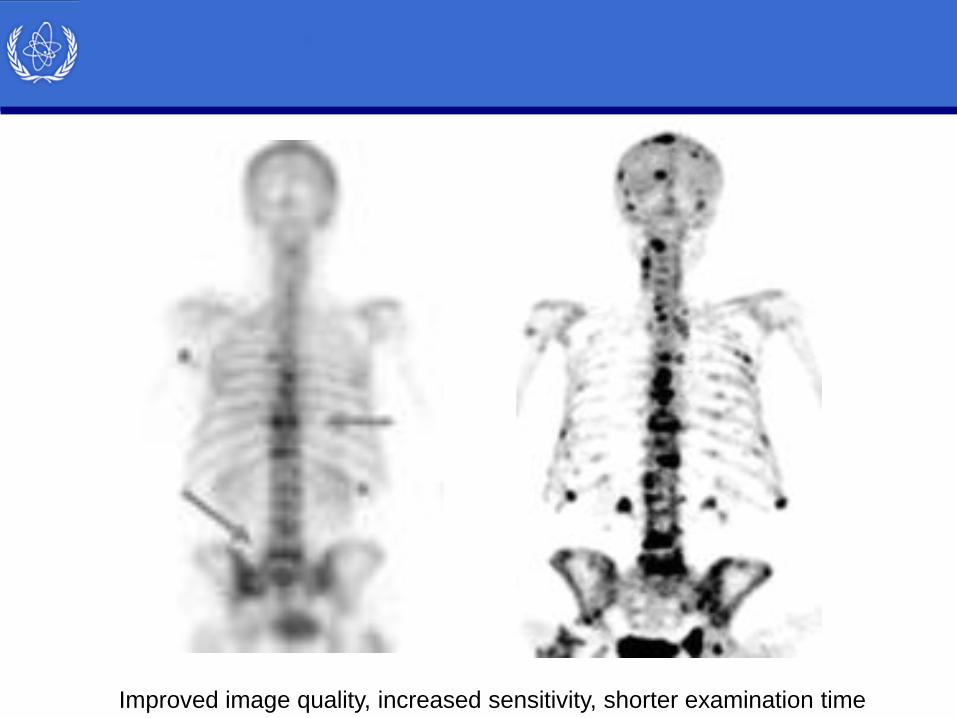
Image quality and sensitivity of PET is 2-3
orders of magnitude compared to
planar/SPECT
The gamma camera (collimator system) aquire ~0.01% of
emitted photons
The PET scanner (coincidence detection) acquire ~1% of
emitted photons
Fluoride PET has higher spatial resolution than bone scan
The favorable kinetic characteristics of sodium fluoride provide
better bone–soft tissue contrast ratio than that of HDP imaging

Improved image quality, increased sensitivity, shorter examination time
Improved image quality, increased sensitivity, shorter examination time

Bone scan 99mTcHDP 24th April 2012
18F NaF PET (Gemini Dual) 31th May 2012
Breast cancer – bone metastases? BS vs NaF
New gamma camera vs old PET-scanner

18FNaF – a forgotten tracer?
The decline in the use of 18F-NaF
for skeletal scintigraphy did not,
reflect limitations of 18F-NaF as
a tracer, but was the result of
the difficulty in imaging 511-keV
photons on a system optimized for
the 140-keV photons of 99mTc
The logistic challenges in the
production and efficient delivery of a
radioisotope with a physical half-life
of 110 min.
Driven by the demand for 18F
FDG technical and logistic
limitations to the routine use of
18F-fluoride for bone imaging
are no longer present
The increasing availability of
PET systems renewed the
interest in using 18F-NaF as a
radiotracer for skeletal imaging
Clinical use of 18F-NaF and BS in oncology
• Primary bone tumors
• Skeletal metastasis in patients with a
range of primary tumors
– Prostate
– Breast
– Lung
• Sclerotic and lytic lesions are visualised

Bone scan: Sclerotic vs lytic
Very sensitive to detect metastases with
sclerotic changes (osteoblastic activity)
Less sensitive for predominately lytic bone
lesions

NaF PET: Sclerotis vs lytic
Because of the increased resolution and
tomographic capability of PET, it has been
suggested that a fluoride bone scan is
more sensitive than a bone scan to detect
the minimal osteoblastic activity
associated with lytic bone metastases

From:Even-Sapir Semin Nucl Med 2007

18F NaF PET

Interpretation of NaF PET
Difficult to differentiate metastases
from benign bone lesions
Cancer patients who are referred
for a BS are usually older,
and have a higher frequency
of degenerative and/or arthritic
bone disease. These benign bone
processes can demonstrate the
same pattern of fluoride uptake
as metastases do, resulting in a
higher chance of false-positive
interpretation if evaluated by PET
alone. CT is often helpfull

Sclerotic, lytic & degenerative lesions
With curtesy of Marius
lytic sclerotic degenerative

Green:
NaF postive sclerotic metastases
Yellow: NaF negative bone cyst
Blue: NaF positive spondylosis
Red: Ureter (physiologic excretion)
Interpretation of NaF PET

January
Interpretation of NaF PET

May
Interpretation of NaF PET

Interpretation of NaF PET

NaF-positive compression fracture

NaF-positive rib fracture

12 April 2012
3 January 2011
NaF-positive rib metastases

Conclusion
Improved sensitivity, specificity, and
diagnostic accuracy of
NaFPET or PET/CT over MDP/HDP bone
scintigraphy have been consistently
reported by numerous investigators in
various cancers including
prostate, breast, lung, and thyroid cancers

Comparison of all studies with data on
18F-Fluoride PET or PET/CT:
11 studies, 425 patients.
350 analyzed on a patient basis,
225 on a lesion basis
Ann Nucl Med 2010

Sensitivity and specificity of NaF PET/CT
• On a patient basis: 96.2% and 98.5%
• On a lesion basis and 96.9% and 98.0%
• The diagnostic accuracy of PET or
PET/CT was significantly higher than that
of the planar and SPECT bone
scintigraphy.
Meta-analysis. Conclusion

Methods XR:
bone destruction (30-60% mineral bone loss)
CT:
structural bone changes (non RECIST)
MRI:
involvement of bone marrow
99mTcHDP SPECT
NaF PET:
Osteoblast reaction to presence of tumor cells
Binding to calcium phosphate
18F-FDG PET:
deoxyglucose metabolism (upregulated in tumor
and inflammatoy cells)
18F-/11C Choline (choline) PET:
incorporated in phosphatidyl cholin (lectin)
in tumor cell membrane due to
upregulation of choline kinase
18F acetate/11C acetate PET:
incorporated cytoplasmatic lipid synthesis
(phosphatidyl cholin and neutral lipids)
due to upregulation of fatty acid synthase

The lesion was suspedted by PET in January
The lesion was visible on CT in April
Bone scintigraphy
FDG-PET and BS

Lytic vs sclerotic and FDG
In osteolytic metastases, FDG uptake is
higher compared to scelorotic lesions
because of the presence of a larger
amount of tumor cells with high
glycolytic rate.
Sclerotic metastases contain smaller
amouts of viable tumor cells and
exhibit therefore less FDG uptake.

Bone metastases
Boadest data
15.000 patients

Bone metastases
FDG-PET BS
Sensitivity %
Patient
Lesion
90
87
86
75
Specificity %
Patient
Lesion
97
97
81
94
Yang H et al. Diagnosis of bone metastases: a meta-analysis comparing 18F FDG PET, CT, MRI
and bone scintigraphy Eur J Radiol 2011; 212604-17
Reliable data on diagnostic accuracy in certain cancer types are of higher
practical relevance

Osseous metastases from breast cancer
Direct comparison between NaF-PET and
FDG-PET has not been published
Compared to BS NaF-PET is superior in lytic
and blastic lesions
FDG-PET is superior in lytic lesions and
similar /inferior in sclerotic lesions
FDG-PET has higher specificiy than NaF-PET
(degenerative lesions) eventhough both tracers are unspecific
NaF vs FDG?
Lytic lesions
BS<NaF
BS<FDG
BS<NaF<FDG
Blastic lesions
BS<NaF
BS=FDG
BS=FDG<NaF

Methods XR:
bone destruction (30-60% mineral bone loss)
CT:
structural bone changes (non RECIST)
MRI:
involvement of bone marrow
99mTcHDP SPECT
NaF PET:
Osteoblast reaction to presence of tumor cells
Binding to calcium phosphate
18F-FDG PET:
deoxyglucose metabolism (upregulated in tumor
and inflammatoy cells)
18Fcholine/11C choline PET:
incorporated in phosphatidyl cholin (lectin)
in tumor cell membrane due to
upregulation of choline kinase
18F acetate/11C acetate PET:
incorporated cytoplasmatic lipid synthesis
(phosphatidyl cholin and neutral lipids)
due to upregulation of fatty acid synthase

Prostate cancer: cholin & acetate
• Bone scan is the most commonly used imaging modality to evaluate osseous metastases.
• Fluoride PET/CT is currently considered the most sensitive method to detect bone metastases in prostate cancer.
• 11C or 18F labeled acetate and choline have been reported to be superior (limited data)
.

[11C]Choline metabolism
cholin kinase,
phosphorylates
the compounds
resulting in the
corresponding
phospoorylcholin
derivatives

Generalized bone metastases detected by FCH PET/CT (MIP-Image)
FCH PET/CT
FCH PET/CT

Prostate cancer: cholin & acetate
• Sensitivity, specificity
NaF: 81%, 93%
FCH 74% (ns), 99% (p=0.01)*
• Absence of tracer uptake in chronic
degenerative lesions is a major advantage
of FCH compared to NaF
.
Beheshti M et al Detection of bone metastases in patients with prostate cancer by 18F fluorocholine and
18F fluoride PET-CT: a comparative stydt. Eur J Nucl med Mol ’imaging 2008;35:1766-74
Austrian study of 38 men compared 18F fluoride PET/CT scanning and 18F-FCH for the detection of bone
metastases from prostate cancer

Prostate cancer: cholin & acetate
18F FCH negative
and 18F fluoride PET–CT
positive degenerative
change in left pubis (white
arrow).
Focal FCH uptake (grey
arrow) in a malignant lesion in
the prostate.
Concordant positive uptake (black arrow)
in a malignant sclerotic lesion (left
acetabulum) in 18F FCH and 18F fluoride
PET–CT.
Degenerative changes malignant lesion
in the prostate
Sclerotic met
18F NaF
Degenerative change
18F FCH

Prostate cancer: cholin & acetate
FCH+:
Bone marrow
metastases without
bone reaction
FCH-
Heavily sclerotic lesions (antiandrogen treatment)
FCH positive malignant focus in the left pubis, turning FCH negative and
sclerotic on subsequent scan (hormone therapy)

Prostate cancer: cholin & acetate
• Metabolic and morphologic changes of
bone metastases are dynamic processes,
and combined imaging is best suited to
capture the natural course of these
changes to allow for management
decisions and accurate assessment of
treatment response.
.

Planar BS
SPECT
FDG-PET
NaF-PET
SPECT?
FCH-PET
Rūpintojėlis
Worring Christ Statue