name and address date - harvest
TRANSCRIPT

UNIVERSITY OF SASKATCHEWAN
This volume is the property of the University of Saskatchewan, and the literaryrights of the author and of the University must be respected. If the reader obtains any assistance from this volume, he must give proper credit in his ownwork.This Thesis by ~'J.:~..ffl~~g .has been used by the following persons, whose signatures attest their acceptance of the above restrictions.
Name and Address Date
•A STUDY OF THE EMBRYONIC DEVELOPMENT
OF THE ROCKY MOC1NTlliN SPOTTED FEVER TICK
Derm~centor andersoni (Stiles)
(Acarina, Ixodidae)
A Thes1s submitted to the Fs.culty of
Graduate Studies in partial fulfilment of .~ .
the requirements for the degree of Master
of Arts in the Department of Biology,
University of Saskatchewan.
by
Martha Pressesky
114469
Saskatoon, Saskatchewan
• January" 1952 .

• "TABLE OF CONTENTS
Page
INTRODUCTION••••••••••••••••••••••••••••••••••••• 1
REVIEW OF LITERATURE ••••••••••••••••••••••••••••• 4
MATERIALS AND n..1ETHODS •••••••••••••••••••••••••"••• 6
ID.mRYOLOGY ••••••••••••••••••••••••••••••••••••••• 11
1. The Egg •••••••••••••••••••••••••••••••••• 11
2. Fertilization and Cleavage ••••••••••••••• 13
3. Formation of the Yolk Cells •••••••••••••• 18
4. Formation of the Endoderm•••••••••••••••• 20
5. Formation of the Mesoderm•••••••••••••••• 21
6. Formation of the Body of the Embryo •••••• 22
(a) The Embryonic Membrane ••••••••••• 22
(b) Formation of the Embryo •••••••••• 24
(c) The Mouthparts ••••••••••••••••••• 27
7. Internal Development ••••••••••••••••••••• 28
(a) The Nervous System•••••••_•••••••• 28
(b) The Digestive Tract and the
Excreto~ System••••••••••••••••• 30
(c) The Stomadaeum and Salivary Glands 34
(d) The Reproductive System••••••••••• 36
8. The Embryonic Integuments ••••••••••••••••• 37
~~Y• • • • • • • • • • • • • • • • • • • • • • • • • • • • • • • • • • • • • • • • • • • ~
RECOmIJENDATIONS ••••••••••••••••••••••••••••••••••• 46
A~O\~~D~NTS•••••••••••••• ~ •••••••••••••••••••• 47
•
\
• INTRODUCTION
The :following work describes some phases of
the embryonic development of the Rocky Mountain spotted
fever tick, Dermacentor andersoni (Stiles), from the
time of oviposition until the embryonic development i~.
completed and the larval tick emerges from the chorion.
Research into the embryonic development of
Dermacentor andersoni was undertaken for several reasons.
The problem was of' interest to the writer, since as
research into the broad field of arthropod embryology,
it was felt to be closely related to the writer's previous
study and research in the field of inseqt embryology. It
was thought that elucidation of the various phases of'
tick embryology might later enable comparison with the
comparatively well established concepts concerning
embryonic development in insects.
An investigation into the literature dealing
with the embryology of arthropods shows that considerable
information has accumulated concerning the embryology of
the Acarina. Most of this, however, deals with the embryonic
development of the mites, and very little describes embryol
ogy as found in the ticks, specifically. In addition to the
scarcity of information on this subject, almost all the
investigations were done in the latter part of the nineteenth
century, and are not readily available. No descriptions
• of the embryonic development of any member of the genus

• - 2
Dermacentor has been found. The limited information as
well as the fact that it is not available ~itbb~rdifficulty
seems to warrant the present effort.
Many members of the order Acarina are of medical
and economic importance. Most ticks are vectors of disease
and this is true of Dermacentor andersoni. This species of
tick is a vector of Rocky Mountain spotted fever, tU1aremia,
Colorado tick fever, and "Qff fever, and is the cause, in
human beings and some domestic animals of the little under
stood condition, ticl: paralysis. It is known that·, in three
of. the diseases named, the causative orgaIiism is transmitted
hereditarily from the adult to the larva through the egg.
The causative organisms thus transmitted are Dermacentroxenus
rickettsi, causing Rocky Mountain spotted fever; Pasteurella
tularemis, causing tulcmemia; and Rickettsia burneti,
causing "Q" fever. In order to determine the re1ationship
of the causative organism to the embryonic tissues, knowledge
of the embryonic development is required.
The Rocky Mountain spotted fever tick is of par
ticu1ar importance in Saskatchewan since the southwestern
part of' the province lies within its area .of distribution.
The Tick and Bubonic Plague SUrvey of the Saskatchewan
Department of' Public Health reports finding Dermacentor
anderson! infected with a highly virulent strain of tularemia
•in a number of localities. This was found at Govenlock,
Maple Creek, Eastend, Shaunavon, and Val Marie in 1950,
and at Eastend in 1951 CDl. ]fiedical reports also indica.te

• - :3
the presence of some Rocky Mountain spotted fever infection
in these areas. Since Dermacentor andersoni plays such an
important role in the transmission of several serious
diseases, and since the causative organisms in several
ins:bances is passed on from the adult to the larva through
the egg stage, it is of importance that the complete
development of the tick Dermacentor andersQni be understood.
Dermacentor andersoni requires two fUll years
to complete its life cycle. It passes thrQugh three morpho
logical sta:ges--the hexapod larva, the eight-legged nymph,
and the sexually mature adult. Each stage requires a
mammalian blood meal before moulting. The rather lengthy
.period required for complete ~elopmentin the post
embryonic life of this tick is in accord with the long
embryonic period of six weeks •
•
• - 4
REVIEVV OF LITERATURE
i'4,~·_-'\.~~~~'_~-~-i
dev~iopment in ticks is a resume of a very detailed investi
gation made by Julius Wagner. His complete work was publish
• ed in 1894 in Russian, and is of a high degree of excellence •
The subject of his investigation was the tick Ixodes calacratus,

• -5
which belongs to the same family (Ixodidae) as Dermacentor
andersoni. The embryonic development of each is similar
in most respects. Wagner, like Korschelt and Heider, has
given a thorough account of the embryonic integuments
"of the Acarina, and his opinion of their significance.
Wagner does not include aIr;{ details of his methods, and
this is very unror-tunat.e , , since the prepat'~1on of the
material for study involves a number of serious difficulties.
The only other reference available was a small
portion of Nordenskiolds brief account "SUr Ovogenese
und Entwicklungsgeschichte von Ixodes r-eduvfua" published
in Helsingfors in 1910. This research is of little value
to the present study since Nordenskiold was concerned
mainly with post-embryonic development.
The foregoing again emphasizes the small amount
of work done in tick embryology •
•

• - 6
MATERIALS AND METHODS
Engorged, fertilized female ticks wre placed in
.small, roUnd, flat, tin containers, kept under glass. Pro
tection from direct daylight was provided by a sheet of heavy
brown paper placed over the glass. It was not necessary to
supply hosts for furthe'r feeding, since only one complete
blood meal is required byt~e adult.
The flat unfed female tick averages 4 mm. in lengtt~
The gravid female onthe other hand measures 15 mm. in le~~,
and its body is very cumbersome. Consequently, the gravid females
scarcely move about unless disturbed. Thus two or three ovi
positing females may be placed in the same container and
their egg masses will remain separate.
Each female may. produce several thousand eggs
during the period of oviposition. The eggs measure on the
average 0.54 mm. in length and 0.35 mm. in width. Ovipositionj
seems to be a continuous process and lasts approximately
thirty days. Several hundred eggs per day are produced
in the first ten days of oviposition, the number becoming
smaller thereafter, and graduaJ..ly falling off to between
tW'enty and thirty eggs per twenty-four hour period. As
oviposition proceeds, the turgid body of the gravid female
slowly shrinks and shrivels, becoming flat, and the color
changes from greyish to yellowish orange. Soon after ovipo~-
• tion has been completed, the female tick dies •
• - 7
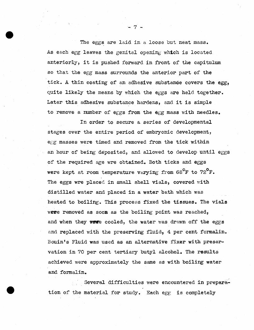
The egg~ are laid in a loose but nea.t mass.
As each egg leaves the genital opening: whtch is located
anteriorly, it is pushed forward in front of the capitulum
so that the egg mass surrounds the anterior part of the
tick. A thin coating of an adhesive substance covers the egg,
quite likely the means by which the eggs are held together.
Later this adhesive substance hardens, and it is simple
to remove a number of eggs from the egg mass with needles.
In order to secure a series of developmental
stages over the entire period of embryonic development,
egg masses were timed and removed from the tick within
an hour of being deposited, and allowed to develop until eggs
of the required age wre obtained. Both ticks and eggs '0 0 were kept at room temperature varying fiom 68 F to 72 F.
The eggs wre placed in small shell viaJ.s, covered with
distilled water and placed in a water bath which was
heated to boiling. This process fixed the tissues. The vials
weee removed as SOOB as the boiling point was reached,
and when they ~ cooled, the water was drawn off the eggs
and replaced with the preserving fluid, 4 per cent formalin.
Bouin'sFluid was used as an alternative fixer with preser
vation in 70 per cent tertiary butyl alcohol. The results
achieved were approximately the same as with boili~ water
and formalin.
• Several difficulties were encountered in prepara
tion of the material :for study. Each egg is completely

• - 8
covered by a tcngh chorion through which fluids penetrate
only with dif-ficulty. The first slides completed showed
that infiltration of the paraffin was inSUfficient. The
sections appeared to be torn and collapsed. In order to
insure a more thorough infiltration some meanS of exposing
the internal tissues is nec essary . This was accomplished
by placing the egg in a minute depression in the surface
of a Luef teblock, and slitting the chorion with a fine
sharp needle under a binocular microscope. This method
was successful in mot-e advanced stages of development ,but
in early stages the puncturing of the chorion was sufficient
to disintegrate the egg oontents. Sometimes, particUlarly
in lad~atnc.ed6 stages, the chorion split and of its
own accord came away from the rest of the egg, the contents
apparently retained by the peripheral development of the
embryo. Such eggs sectioned fairly well. Another method
of removing the chorion to obtain thorough infiltration
was to place the eggs in a solution of sodium hypochlorite,
a method used by Slifer(ll). This solution seems to soften
and dissolve the chorion. It was found that a three percent
solution of sodium hypochlorite affected the chorion of
different eggs in varying degrees--from partial to complete
dissolution. The ease with which infiltration will occur
in an egg thus prep ared can be det ermi ne d by p Lacing the egg
• in a solution of Borax carmine. Eggs from which the chorion
is removed sta in a de ep re d qui ckly. Thoa e whi en nave an

• - 9'
opened chorion gradually acquire a red color. If the
chorion is in tact, the stain does not even adhere to the
external covering of the egg.
All eggs were stained in toto in Borax canaine
before further processing. Then they were dehydrated in
tertiary butyl alcohol, and embedded in the flat surface of
a prepared paraffin block. The paraffin used was Parlax.
Parlax has less tendency toward crystallization than ordinary
paraffin, and has a somewhat ru bbery consistency, which is
an advantage in sectioning. Its melting poL~t is 54°C.
In addition to the difficulty encountered in
infiltrating t~e eggs, a second problem arose in attempting (
to orient the eggs for serial sectioning. Unlike many
arthropod eggs, these possess no characteristic pigmentation
or reticulation which may be used as a guide in orientation.
The ovoid-oblong shape of the egg permits orientation in
relation to either the long or short dimension but the
dorsal, ventral and lateral aspects remain unknown until
the material is studied. In the very early stages no orient
ation is possible.
Serial sections were made in thicknesses varying
fran .seven to ten microns, depending on the degree of
diffieu lty encountered in seeti oning a partieu lar stage.
In staining, Delafield's haema'toxyLfn with eos in counter
• stain was used for the most part and was found to give
satisfactory results. Other stains tested experimentally
• - 10
and found useful were Cresyl Violet, Mallory!s Acid Eosin,
and Bodian's Protargol.
The illustrations were prepared by projecting the
sections on a screen using the Bausch and Lomb triple pur~
pose microprojector, in addition to study by means of a
Spencer oil immersion monocular microscope. The drawings
were made on ordinary bond paper and then transferred to
bristol board. All drawings are somewhat diagrammatic .
•

• - :3..1
EMBRYOLOGY
The eggs of Dermacentor !ndersoni resemble those
described for other Ixodid ticks. They are oblong-ovate
in shape and measure on the average 0.54 mm. in length .and
0.35 mm. in width. The surface of the egg is smooth and
glossy, lacking the sculpture or reticulation characteris
tic of many arthropod eggs. The chorion is a structureless
membrane, transparent and colorl eas , and has a tough, some
what brittle texture. No micropylar opening could be found.
In the egg~ass the eggs adhere lightly one to
another but there are spaces between the eggs so that even
those in the center of the mass are exposed to the air.
At first the eggs are a translucent light yellow
ish brown in color. As embryonic development proceeds the
color changes to a deep rusty brown. The growth of the
Malpighian tubules may be observed externally in the dev
eloping eggs. At first these appear as a pair of elongate
• white marks close together on the ventral surface. Much
later the Malpighian tubules appear as eurviIlgwhi te lines

• - 12
growing cephalad on either side of the embryonic central
nervcu s system.
The chorion appears to be the, only membrane cov
ering the contents of the egg. There is no vitelline
membrane as is found in insect eggs. The yolk is found
directly beneath the chorion. In fresh unpreserved eggs
the yolk granules seem to be spherical. The yolk spheres
are fa. irly large and are of a homogeneous texture. When
fixed and preserved, the yolk granules shrivel and assume
angular and irregular shapes. In early embryonic develop
ment these large angular yolk granules fill the entire egg.
As the development of the embryo advances, the particles
of yolk break down in size and the amount present .beeoaea
progress ively less. Hovvever, some yolk is pre sent in the
mesenteron of the larva as la.te as two weeks after hat ch
Lng .
Eggs which are in a very early stage of' develop
ment do not show the network of p rotop.Lasmi,e strands
genera lly evident in early stage s in insect eggs. The only
eyt oplasm visible at this time is in a small area surround
ing the nuclei.
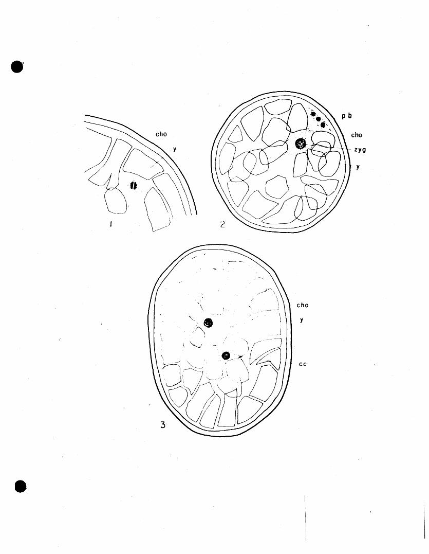
In eggs V'tLieh are less than one hour old the
female pronucleus may be observed in the process of the
first maturation division a.t the periphery of the egg
• (Fig. 1) .
• -13
2. Fertilization and Qleava~e
In De!ffiacento~ ande~son~, it seems most likely
that the union of the egg nucleus and sperm takes place
following oviposition •. This is in agreement with Wagner's
observation in Ixodes caloaratus(!L2) but differs greatly
from the condition found in Dermacentor variabilis by
Zebrowski (14). The latter's investigation of the repro
ductive system of the engorged female of Dermace~tor varia
bilis shows that eggs in the uterus and oviducts pass through
the stages fram zygote to blastoderm formation before
oviposition occurs. In the gravid female the entire rep ro
ductive tract is filled with eggs in the stage of oomplete
blastoderm formation. In the non-gravid female Zebrowski
reported finding spermatozoa all along the course of the
oviducts, which led him to believe that the developed intra
uterine eggs must have been fertilized. The extent to which
embryonic development has proceeded before oviposition, is
in keeping with the comparatively short period required for
the conpLet e life cycle of Dermacentor variabilis. According
to Matheson Dermacentor variabilis requires as little as
fifty-four days for the entire cycle from egg to adult,
providing there are favourable food and climatic conditions
( !7) •
• In the study of the eggs of Dermacentor andersoni
Which were fixed within one hour after oviposition, the
first stage observed is that of the first maturation division

• - 14
of the female pronucleus. The dividing female pronucleus
is located near the periphery (Fig. 1). It was not possible
in this division to count the number of chromosomes, but
it is evident that the number is small.
In eggs four hours old what is believed to be
the synnuc1eus has appeared, and it is possible to see
the three polar bodies which are the products of the re
duction divisiomof the female pronucleus'Fig. 2). The
pol?..r bodies are found at the periphery,just under the
chorion, and the synnucleus, a large nucleus in compari
son to the polar nuclei, is found in the same region but
deeper down among the yolk particles. THe synnucleus is
appar-errtdy in the process of movement to the central part
of the egg, since i,n,~·o""th·er eggs it has been observed in
that location, and from which point subsequent divisions
seem to take ple,ce • Although fertilization has not been
observed by the writer, it is believed to occur within a
short time after oviposition. It is possible that it occurs
during the inward migration of the female nucleus. The
polar bodies have not been observed in subsequent stages
so it is possible that they undergo a rapid disint:eg;r:;ation.
The synnucleus moves toward the centre of the
egg. Here the first division takew place perpendicular to
the long axis of the e.;;g, approximately twelve hours follow
• ing oviposition•
The products of this division migrate to opposite
poles of the egg. Each nucleus is surroundad by a small mass of

• - -15
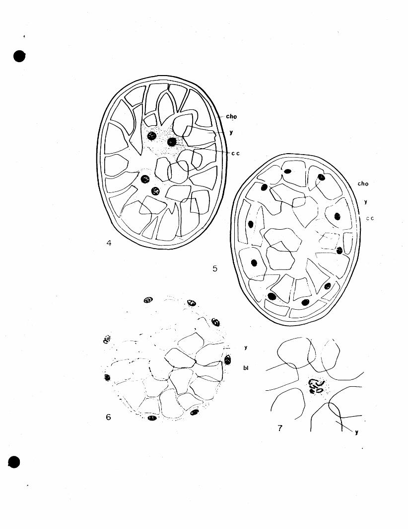
cytoplasm (Fig. 3). 'VVhen these first cleavage nuclei
have moved a.pproximately half the distance to the pole,
each nucleus divides again, this time in a plane perpen
dicular to the plane of the first division (Fig. 4). By
the time the egg is twenty-four hours old another division
has occurred, this division being ina plane perpendicular
to the plane of each of the preceding divisions. The
nuclei continue dividing, at the same time moving toward
the periphery of the egg. They are uniform in appearance,
and by the fourth day of development the eggs have a layer
of cleavage cells slightly below the surface. The cytoplasm
surrounding each nucleus in the layer seems to be attenuated
in a plane parallel to the surfaceo! the egg, (Fig. 5),
and is in contact with the cytoplasm of the adjacent nuclei.
Nuclear division takes place as outwaIrl migration
continues, until, when the cleavage nuclei reach the surface
of the yolk just under the chorion, the yolk is completely
covered with a single layer of cells, uniformly spaced
(Fig. 5). 'Nuclear divisions are only rarely observed, but
in the few'caaeswhere they are seen, they occur in a large
pr~ortion of the cells simUltaneously. This indicates
synchronous cell division. When the serial sections show
the cleavage cells in the process of mitotic division,
those cells dividing Which give a polar view of the metaphase
• plate show the apparent number of chromosomes to be eight
(Fig. 7). Wagner(12) was able to observe some mitotic
divisions in a polar plane in the metaphase stage, and his

• - 1:6
conclusions were that the chromosome number for Ixodes
caIe aretus was either eight or ten.
Considerable interest has centered around the
nature and mode of the eutwa rd movement of the cleavage
cells in arthropod eggs. Gross and Howland (4), in their
study of Proder4~, assume that the outward bound cells move
through the yolk by means of a digestive action. Eastham's
(3) work on Pieris agrees vdth this on the basis that during
the outward migration he has observed a change in the yolk
that has been traversed by the outward migrating cells.
The yolk through which the nuclei have passed appears to
be clearer than that not yet invaded. In Derma. centor
andersoni also, the cleavage nuclei appear to move through
th~ yolk particles by a digestive action (Fig. 5), although
there is no apparent change in the appearane e of the yolk
through which cleavage nuclei have already passed. These
observations are at variance with the condition observed in
Mamestra configurata by Rempel(lOD. The latter found that
there was an attenuation of the yolk in the region of
migration, which he believes is not due to any digestive
action on the part of the cleavage eells, but to mechanieal
displacement of the yolk globules' by the outward migrating
cleavage cells.
The possible cause underlying the peripheral
• migration of the cleavage nuclei is also suggested by the
above workers. Eastham (3) is of the opinion that a

• - 17
centrifugal streaming of the raticu Lar cytoplasm draws
the nuclei through the yolk to the periphery. In
Mamestra, Rempel(~O) describes the cytoplasm surrounding
the migrating cleavage nuclei as "comet shaped" with the
nucleus located in the head of the comet. He feels
that peripheral migration is the result of the independent
outward movement of the cleavage cells, the movement
plausibly under the "guiding influence" of the nuclear
material.
In Dermacentor ang~~oni, neither of the above
assumptions are particularly applicable since, in the ease
of Eastham's theory, there is no reticular cytoplasm visible
in the egg, and in the ease of the second theory, there is
no indication of nuclear control suggested by the position
of the nucleus in a comet -shaped mass of ey t epLasm, However,
nuclear material may be responsible for the peripheral
migration, and the difference in shape of surrounding cyto
plasm may be due to the difference in the length of time
which is required for the completion of outward migration.
In Mamestra the cleavage cells reach the periphery in.
six hours, whereas in Dermacentor andersoni the same stage
requires four days, although the eggs are almost the same
size .
•

• - i8
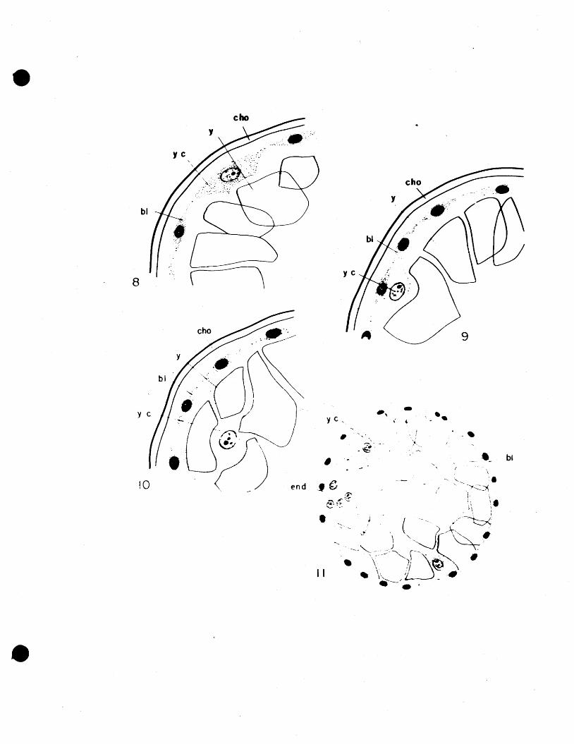
3. Formatien ef the Yolk Cells
As seon as the yolk has been covered by a
layer of uniform evenly-spaced cells, the blastoderm is
complete and the formation of the yolk cells ccmmences.
The first indication of their development is the appear
ance of a new type of cell (Fig. 8), which seems to be
distributed evenly throughout the entire blastoderm. The
nuclei of these cells are much larger than those hereto
fore observed in blastoderm cells, and are of a coarse
granular texture. They do not stain deeply, and one or
two prominent nucleoli may be observed in them. Yolk
cells may be observed in various stages of their develop
ment from the time they first appear in the blastoderm
until they sink down among the yolk granules, and the
blastoderm closes over them. At first the yolk cells sink
just bel~v the blastoderm, still adhering to it, (Fig. 9),
then they separate from it, and gradually sink down into
the yolk (Fig. 10). They seem to be evenly distributed
throughout the yolk. Wagner claims that in Ixodes
calcaratus the yolk undergoes a process of segmentation
into large spherical masses ~EXk, each one including a
yolk cell(12D. Such a condition may oc~~ 'exist in
Del~centor andersoni also, si~ce the evenness of distri
bution of yolk cells might indicate that one yolk cell
• exerts its effect upon a particular quantity of yolk •
However, there is little indication of a secondary seg
mentation of the yolk particles into spherical masses in

• - 19
Dermacentor ander-som , Wagner does not make it clear
whether or not the secondary segmentation of the yolk in
Ixodes calcaratus persists.
By the end of the eighth day the process of yolk
cell fonmation is completed and there are approximately
thirty yolk cells distributed throughout the yolk~. As em-progresses
bryonic development , the yolk cells and the yolk undergo a
number of changes. Gradually, the yolk particles in close
proximity to the yolk cells are broken down and appear as
much smaller particles, and in general, the quantity of yolk
diminishes so that the partially developed egg does not appear
to be so densely filled with yolk. The yolk cells tend to lose
their enveloping cytoplasm as development of the egg proceeds;
the nuclei lose their coarse granular appearance and nucleoli.
Some yolk is present in">lthe mesenteron of the tick larva
even after it emerges from the chorion, and is apparently
utilized iin.'tiie first weeks of larval life prior to the first
blood meal. In tick embryos which are on the point of hatch
ing, a few pale yolk cell nuclei may be observed.
In certain orders of the Insecta the formation of
yolk cells takes place in a different manner. In Collembola,
Orthoptera, Strepsiptra, and Lepidoptera the yolk cells
are formed when some of the cleavage nuclei lag behind the
nuclei moving outward to form the blastoderm. This is
quite different from the method of. formation in
• Dermacentor andersoni • Although the process of having

• - 2'0
the yolk cells difrerentiate from the blastoderm has been
observed in some orders of Arachnida, it has not been
demonstrated to be the general rule (6).
4. Formation of the Endoderm
When the egg is in its eighth day of development
the first cells of the endoderm fonn. Three or four cells,
which in appearance resemble the yolk cells, differentiate
simultaneously on the dorsal side of the egg (Fig. 11), in
the region of the future caudal lobe of the germ band. The
nuclei are large and of a granular texture. They stain
lightly, as do the yolk cell nuclei.
•
The endoderm cells appear in only a small area of
the caudal region, and at first the cells formed lie close
to the cells of the blastoderm. As more such cells appear
those first formed are pushed into the yolk. In sections
through this part of the caudal lobe, either transverse or
longi tudinal, the endoderm. mass gives the impression of
being pyramid-shaped, the base of the mass being at the
periphery of the egg. Those celis which have been pushed
furthest into the yolk show distinct cell walls (Fig. 15)
which are not apparent in the newly formed cells. In this
case, the cell walls may be more in evidence due to pressure
on the membrane from the e ells formed later resulting in
concentration of granular cytoplasm at the cell boundary.
Wagner (Ia considers the stage of immigration
of endod.erm cells to be the gastrula phase.

• - 21
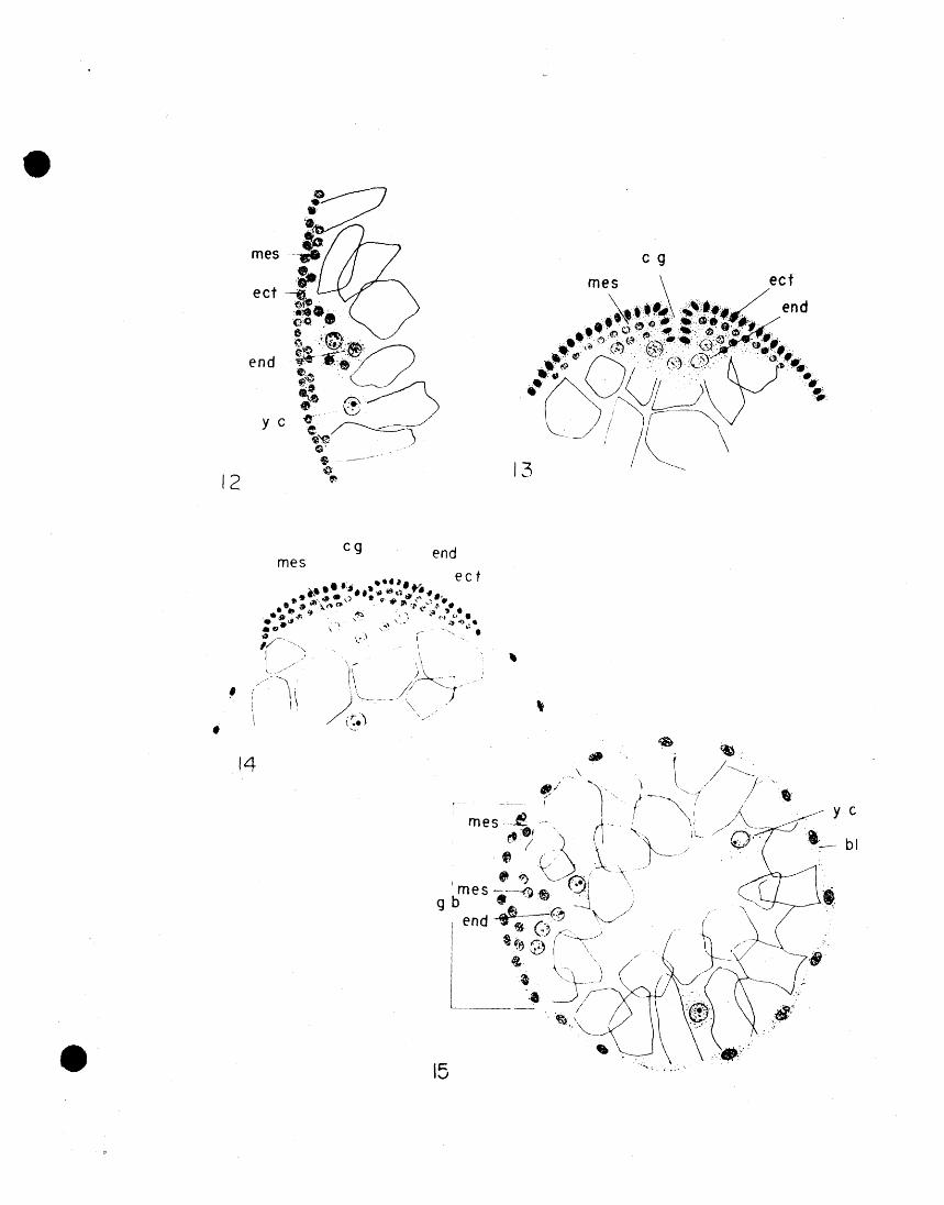
5. Formation of the Mesoderm
Soon afte r the first endoderm cells have been
formed, mesoderm cells begin to appear. They begin as
a disturbance in the evenly distributed olastoderm cells
in the region of the endodenn. The blastoderm cells cov
ering the endoderm. mass prolifera.te and also ccncentrate
in this area, becoming tightly packed (Fig. 12). Then, on
either side of the endodenn mass a small pouch appears into
which mesoderm cells rapidly migrate from the blastoderm.
In this manner, two strips of mesoderm are formed, extend
ing forward from the caudal lobe along the germ band,
toward the caudal pole of the egg. The two pouches in
being filled with mesoderm cells form a pair of caudal
protuberances externally, separated by the caudal groove
(Fig. 13). The caudal groove at the dorsal part of the
egg, just at the point of endoderm formation, is deep,
and constitutes the beginning of the proctodaeal invagin
ation. The caudal groove becomes less distinct toward
the eeudaL pole of the egg (Fig. 14).
The forming mesoderm cells are easily recognized
since they differ greatly in appearance from the endoderm
cells. Mesoderm nuclei are small, of an even texture,
and stain deeply. They are surrounded by a fine layer of
cytoplasm, and cell walls are not discernible.
• When the mesoderm cells migrate into the pouch
like formation on either side of the caudal groove, they

• ... 22
at first tend to line the pouch, so that there is a longi
tudinal cavity within the strip of mesoderm cells. Later
the cavity is obliterated and an inner and outer layer of
mesoderm cells is formed.
6 • Formation of the Body of':Jb,he Embr.yo
(a)T~Embryonic Membrane
When the blastoderm has just been completed, the
cells forming it are uniformly distributed. Siaultaneously
with the development of the endoderm mass, however, the
blastoderm cells begin to concentrate toward the posterior
end of the of the egg's surface to form the germ band.
Due to continuing cell division and cell concentration,
the cells in this area become tightly packed and columnar
in shape, whereas the cells in the remainder of the blasto
derm become squamous and form a membranous layer(Figs. 15, 16).
At this stage the amount of de'lelopment in the tick egg
closely resembles that seen in insect eggs by the time 'the
serosa has been formed. In Dermacentor andersoni that is the
extent of formation of embryonic membrane, whereas in the
insects it is more complex, \I.ri th further development of
an inner membrane known as the amnion. The condition in
Dermacentor andersoni is in agreement with that found in
Ixodes calcaratus by wagner(J2~ and also with other Acarina
• and Araneae .< 6). There are other examples in the Arthropoda
whose development in this respect more complex and like
• - 23
the insects in the development of both a serosa and amnion.
This is true of the Scorpiones (6). Regardless of the
extent of formation of embryonic membranes, in arthropod
eggs which possess a relatively large amount of yolk,
the same purpose is served by the membrane, that is, to
direct the nutritive material toward the developing part
of the embryo. In this vvay the large quantity of yolk
is kept in close proximdty to the developing germ
band. In Insecta the presence of an inner membrane, the
amnion, may be considered to be more specialized,
affording the embryo protection from mechanical
injury.
In the latter part of embryonic develop
ment in Dermacentor anderson! as in other Acarina .and
Araneae ,the squamous portion of the blastoderm forms
the external part of the dorsal and lateral body walls.
In Insecta the embryonic membranes are usually drawn
into the embryo just prior to the dorsal closure of the
body wall, so that the protective function of the membranes
persists until the insect larva is almost ready to aaerge.
The elaborate, persistant double membrane of
the Insecta is a protective device apparently not required
in the development of the Acarina•
•

• - 24
(b ) Format ion of !J1e .Embryo
At first the germ band extends from the point of
endoderm formation caudad to a point slightly beyond the
caudal pole of the egg (Figs. 15, 17). In this position
that part of the germ band which will eventually give rise what is later :found to be
to the head end of the tick lies atl\the caudal pole of the
egg. At this stage the germ band is quite wide, but as
longitudinal gr~~h proceeds it becomes somewhat narrower.
The germ band grows rapidly in length, the head end pushing
cephalad along the ventral surface of the egg, and eventually
passing over the cephalic pole to the dorsal side of the
egg to form the cephalic lobe. The germ band at this stage
is extremely long (Fig. 18). Wagner(12) found a similar
type of germ band development in Ixodes calearatus, and reports thai
Salensky(~2) describes the same for the water mite,
Hyd:rachna.
It is while the germ band is in this elongate
state that the external features appear. Formation of a
pair of protuberances in the caudal lobe is observed, and
the truncated rudiments of the limbs and month parts appear
along the ventral surface of the egg (Fig. 18).
Coincident with the development of the mesoderm
cells in the caudal portion of the embryo, the caudal
groove appears. The groove deepens toward the posterior
• end of the germ band, and at the posterior end forms the
invagination which is the origin of the proctodaeum. The
caudal lobe at this stage appears to be segmented, but
.' - 35
this segmentation is of a t~mporary nature and disappears
with the cephalization of the germ band (Fig. 19).
At the same time, along the whole ventral sur
face of the egg, the appendages appear as six pairs of
small protuberances. While the appendages are still in
this state of development, the germ band contracts
anteriorly •. When the caudal lobe has reached the ventral
part of the egg its cone-like shape (Fig. 19) disappears
and it becomes indistinguishable from the rest of the germ ...... 9~~thband (Fig. 20). As t~1.e ~~1fOC~&8~Nai.aj of the embryo
cephalad continues~,the yolk is pushed into the posterior part of
the egg by the rapid development of the nervous system in
the anterior region.
The concentration of the embryo toward the
anterior end is demonstrated also in the cephalad migrat
ion of the appendages. The anterior two appendage' rudi
menta are destined to form the mouthparts, and becane
located in the cephalic lobe. The remaining four pairs
of limb rudiments elongate, forming the four pairs of
legs. This condition is only temporary, the late embryo
and larva being hexapod. About the twentieth day,
evidence of an embryonic exuviation is present in the form
of a granular layer surrounding the embryo (Fig. 29)
just below the chorion. Since the embryo hatches as a
• hexapod larva (Fig. 21), it is possible that the
regression of the fotirth pair of legs results in this

'. - 2-6
embryonic exuviation. Wagner(12) reports finding four
pairs of legs in the embryg~r~part of its development.
He too, associates the format ion of the larval exuviat ion
(sheath, integument) with the disappearance of the fourth
pair of legs. Wagner(a~) is of the opinion that all
hexapod larvae of ticks pass through an eight-legged
stage prior to the hexapod condition, and that thehex~lY:o~d
larva retains under the ectoderm a "more or less developed
starting point of the fourth pair of legs".
From their survey of the literature available
concerning embryonic development in the Acarina, Korschelt
and Heider (6) state that the hexapod larva occurs in most
families, but is not universal. It is pointed out that
in Fhytopta the larvae have two pairs of legs. The adult
of this form has two pairs of legs also, and Korschelt and
Heider believe that this is a secondary 'oondition in both
the larva and adult, and is in keeping with. other specialized
characteristics of Phyto:pta.
Korschelt and Heider (6) proJ):mte a theory regard
ing larvae with two pairs of legs, in which this form is
co~sidered primitive, and from which has evolved the
hexapod form. This idea was held feasible, sinee the
hexapod larva later changes into the eight-legged form
(the }tLymph). However, the records of several workers
• discredit this theory, by demonstrating that in the
development of the hexapod larva, the embryo passes
• - v.
through a period in which four pairs of legs lllay be I,
observed. Winkler(l3) in his account of the d~velopmentI
of Gamasus grassipes clearly observed the fo~ation of I
four pairs of legs, although the larva is hexapod upon
emergence.Wagner's(l2) report of an eight-legged I
I
condition in the embryo of Ixodes calcaratus and the I
subsequent appearance of the hexapod larva ag~in confirms
the observation that although the larvae of A~arina are
hexapod, they first pass through an eight-legged· stage. I
I
The eight-legged condition in Gamasus grassipds was
observed by Winkler only during early development. Nitzsch
(8) found that the embryo of Pteroptus vespertilionis
commences free life with four pairs of legs, that is,
in the nymphal stage. Nitzsch observed however, that the
embryo passes through a six-legged stage while the egg is
still within the mother. The condition of e~ght legs during
early e'mbryomc development and six legs d~ring late
embryonic development and larval stages demonstrated bt
Dermacentor andersoni', is thus seen to be fairly general
in the Acarina.
( c) The Mouthparts
The anterior two pairs of appendages will form
the capdtuIum, When the stomadaeum begins to invaginate,
• Qnd
both pairs are in a ventralApost-oral position, quite
widely separated along the ventral mid-line of the embryo.
With further development, the first pair migrates antero

-28 dorsally and eventually come to lie in a position just above the
stomadaeum, thus formi~ the paired piercing and cutting organs,
• the chelicerae. The second pair Qf append~es remains in a ventral
post-oral position, widely separated, but eventually each appen
dage grows medially, and they fuse, forming the basis capituli
which surrounds the chelicerae and the stomadaeal invagination.
Figure 23 shows the ante~ior two pairs of appendages in an early
stage. The anterior pair which will form the chelicerae, are
lateral to the oral opening in the process of moving to a
position above it.
The second pair of appendages elongates, lying· in line
with the four pairs of de"eloping legs, and parallel to the long
axis of the egg(Fig. 20). Later, however, the position of the
second pair of appendagea., shifts and they. come to lie in a
position which is at right angles to the long axis of the ~gg.
At this stage a small protuberance forms at the base of each
appendage (Fig. 20). The pair of protuberances grows medially
and elongates to form the hypostome, a median ventral prolo:n-g
ation otnthelcapitulum, seen in Figure 25. The remaining parts
of the second 'appendages form the basis capituli and the tour
segmented appendage, the pedipalpi. The basis capituli is
formed by the erilargement and growth together of the bases of
second appendages, and their growth .around the base of the
chelicerae (Fig. 24). The remaining part of the second append
age becomes the 'four-segmented pedipalp. The pedipalpi, which
were previously at right angles to the long axis of the egg,
again come to lie in a longitudinal position just lateral to
• the mouth opening, and tend to enclose the chelicerae" and
hypostome.
7 • Internal Development(a) The Nervous System
The first evidence of the development of the

• - 2"9 - .
nervous system is a slight invagination of the ectodermal
cells along the mid-line of the ventral part of the germ
band (Fig. 26). This invagination commences soon after
the first appearance of the appendage rud.iments. The strip
of ectodermal cells two or three cells wide which invag
inates, separates the ectodel~ of the germ band into two
longitudinal strips separated by a gradually deepending
neural groove. The neural groove appears externally as
~ continuation of the caudal groove which was observed
in the development of the caudal lobe. The ectodermal
strips on either side of the neural groove proliferate
rapidly so that the nervous system at this stage looks
like a pair of elongate masses, several cells in thickness,
separated by a constantly deepening neural groove (Fig. 27).
At first the nervous system, like the germ band,
is elongate, extending nearly fran the an terior to the
posterior end of the egg. During this stage the "punc t.a.L
substance" app ears. The term "punct.a.L substance" is the
name given by Wagner(12} to a homogeneous granular sub
stance that appears in the nervous system. It tends to
stain lightly. The punctal SUbstance is observed first
in separate areas along the length of each half of the
nervous system. Later, the separate areas of punctal sub
stance fuse transversely (Fig. 28) and then longitUdinally.
• Figure 29 shows how it appears as a lon~itudinal strip in
a sagittal section through one half of the nervous system.

- $0 • As development of the embryo proceeds, the
shortening of the germ band that is noticed in the external
development of the embryo is apparent in the, progressive
concentration cephalad of the nervous system. At the same
time, the neural groove grows deeper and narrower (Fig. 30),
and is finally obliterated. The cells of the early neural
groove may be seen in the interior of the nervous system,
forming a compact m.as s seen just ventral to the st emo da eum
in Fi~~re 30. While the nervous system is concentrating
. anteriorly the s t enodaeum invaginates in the direction of
the nervous system and grows through it (Fig. 31). Thus
the nervous system has the appearance of a large oval mass
divided by the stomodaeum'into a supraoesophageal portion
and a suboesophageal portion (Fig. 32).
Henking (6) speaks of the nervous system of the
mature embryo of Trombidium as a large ventral gangf.i.cnLc
mass and a pair of smaller supraoesophageal ganglia. The
formation of the nervous system in Dermacentor andersoni
is similar to that described by Wagner (12) for Ixodes
cal~aratus. The outstanding feature of the r oma tion of
the nervous system in the Acarina seems to be the marked
cephalization Which takes place following the development
of the el ongate form (Fig. 32).
The th!:
• (b) ADigesti ve Tract and "Excretorl System
The endoderm mass Which forms on the dorsal side

• - 3-1
of the egg at the caudal end of the germ band has already
been described. Although the number of endoderm cells
increases, the endoderrn mass retains its original appearance,
(Fig. 33) that of a pyramid of cells, the apex directed
toward the yolk, until the shifting forward of the caudal
part of the germ. band has commenced.
The first change noticeable in this region, is
the separation of the endoderm mass into two layers (Fig.
54), the layers being in the same plane as the surface of
the egg. The outermost layer divides medially into two
equal parts each' of which eventually gives rise to a
Malpighian tubule. Figure 34 is a. sagittal section cut
lateral to the anal invagination, and shows the origin of
one of the Malpighian tubules.
The growth of the Malpighian tubules may be
studied by observing through the surface of the egg their
progressive increase in length as embryon~c development
proceeds. The tubu les first appear under the ventral sur
face of the egg, toward the posterior end , about the
twelfth day of development. They are milky white in color
and stand out sharply against the rusty brovm of the egg
contents. Each tubule grows forward gradua.lly, one on
each side of the nervo~s system. When the latter concen
trates anteriorly, thus increasing in width as its length
• decreases, the developing Malpighian tubules are corres
pondingly pushed out in a lateral direction. Growth of the

• - 52
tubules takes place in a posterior direction also, toward
the rectal vesicle which, like the tubules, can be observed
tl~ough the ventral surface of the egg as a white~
bilobed structure. The rectal vesicle is well developed
by the twenty-fifth day. By the thirtieth day of develop
ment the Malpighian tubes have reached and joined with
the rectal vesicle.
Although the !~alpighian tubules are 'endederma L
in origin, the rectal vesicle into which they empty
appears to be of ectodermal origin (Fig. 35). It is
formed as an outgrowth of the dorsal part of the procto
daeal invagination which was observed very early in
embryonic development, and which was described in a
previous section.
The formation of loops in the Malpighian tUbes
(Fig. 36) takes place during the last week of embryonic
development. They are located laterally and posteriorly
to the rectal vesicle, and seem to be the result of
growth to accommodate the dorso-ventral mesodermal folds
which at this time grow through the yolk.
Korschelt and Heider (6) report that in
Ranking's study of the mtt e Garn.asus, the conclusion is
drawn that the Malpighian tubules are outgrowths of the
proctodaeum. They report further that in other orders
• within the class Arachnida, namely the Scorpiones and
Araneae, several workers claim that the Malpighian

• - 33
tubules are diverticula of the enteron. From their review
of the literature Kora ch e.Lt and Heider (6) conclude that
the question of the oTigin of the Malpighian tubes in
the Arachnida is not settled beyond doubt.
The inner layer of endoderm (Fig. 34) grows
anteriorly between the great quantity of yolk that is
present and the nervous system, to form at first the
basal part of the large, branched mesenteron. For the
major part of embryonic development, the large volume of
yolk is enclosed only by the ec t oderm , In the last seven
or eight days of development, the yolk mass is broken
do~n into smaller sections by the growth through the
yolk of mesodermal folds. These arise ventrally and grow
dorsad. As the mesodermal folds are formed, the basal
plate of endoderm grows upward around the yolk, enclosing
it. All branches of the mesenteron contain yolk particles
(which may be observed even in hatched larvae two weeks
old). The only exception is the ventral posterior outgrowth
which projects posteriorly as far as the proctodaeum (Fig.
32). This outgrowth is located posterior to the concen
trated nervous system and anterior to the'rectal vesicle.
This posterior outgrowth of the mesenteron does not have
any direct connection with the rectal vesicle but is closely
applied to the proctodaeum just below the origin of the
• rectal vesicle (Fig. 32) •
Wagner (12) is of the opinion that the embryo

• - 34
of Ixodes calcaratus there is no actual opening between
the posterior outgrowth of the mesenteron and the procto
daeum. He has demonstrated to his own satisfaction that
in hat ched larvae the posterior out gr-owth of the mesenteron
is blind. His conclusion is that in the, emb ryo all
excretory function is performed by the Malpighian tubules.
In examining the connection between the posterior out
growth of the mesenteron and the proctodaeum in De~eentor
andersoni no clear passage from one to the other has been
observed, and this writer is therefore inclined to agree
with Wagner thateoceretory function is carried on, in the
embryo at least, by the Malpighian tubes.
(c) The St~daeum and Salivary Glands
The stomodaeal invagination begins later than
the proctodaeal invagination, after the appearance of the
appendage rudiments on the ventral surface of the egg
(Fig. 22). With the development and contraetion and sub
sequent thickening of the nervous system the mesenteron
becanes pushed farther from the oral opening and conse
quent ly the st csicd aeura increases in length. It joins the
mesenteron at the point where it emerges tram the surround
ing nervous tissue. With the translocation of the chel
icerae to a pre-oral position and the growth of the base
of the pedipalpi to form. the ring-like basis capituli·,
• the distal portion of the stomodaeum compresses dorso
ventrally and assumes an arched appearance (Fig. 32). The

• - 35
segmental mesoderm of the oral segments forms the muscles
which operate the blood sucking apparatus. The large
muscles Which activate the chelic erae begin to deve lop
about the twenty-seventh day, and when fully formed
extend posteriorly from the base of the chelicerae and
are inserted in the hypodermis of the anterinr part of
the dorsal body wall. After the formation of the stomo
daeal muscles, the chitinous lining of the anterior part
of the stomodaeum becomes evident.
The paired salivary glands of Dermacentor
andersoni develop as ectodermal invaginations from the
buccal cavity of the basis capitula: into the haemocoele.
TheiDV~inations deepen, growing back into the body cavity
(Fig. 24), and form the main ducts of the salivary glands.
The salivary ducts are located laterally, one on each side
of the central nervous system, and above the anterior
prolongation of the MaLpLghLan tubules (Fig.· 36).
The main duct gives off short lateral branches,
which tenminate in glandular cells. The glandular cells
form first in the anterior part of the body. These may
be first seen about the thirtieth day of embryonic dev
elopment. Toward the end of the embryonic period fully
deve lop ed salivary gland cells may be observed as far
back in the body cavi ty as the posterior outgrowth of
• the mesenteron. The cells may be seen in various stages
of transformation into glandular cells. The terminal

• - 36
cell (Fig. 31) of a lateral duct enlarges, the cytoplasm
becomes eoa r s e in texture and appears to be somewhat
vesicular. The nucleus becomes large and prominent.
The main duct of the salivary gland appears to
be chi tinized in embryos which are thirty-four days old.
The chitinous portion extends only a short distance from
the point of origin, but may be clearly seen in cross
sections through the anterior part of the body (Fig. 24).
Wagner (12) finds that in Ixodes calcaratus, the internal
spiral thickening of the discharge duct does not occur
until the post-embryonic period.
(d) The R~Eroduc~ive System
The development of the reproductive system in
the embryo of Dermacentor andersoni has not been fully
stUdied, but jUdging from the isolated instances in which
parts of the reproductive organs have been observed, their
formation in this tick resembles the development which is
generally found in the Acarina. Korschelt and Heider (6,:)
briefly describe the development of the genital glands as
paired nbean-shaped bodies" in the beginning, and which
"only in the further course of development fuse to foxm
the unpaired genital gland knovm in the adult rt •
• In J?ermacentor and ers on L the geni tal glands are
first noted as paired cell masses Which lie on either side
of the rectal vesicle just anterior to and above it. These
• - .::37
are seen by the twenty-eighth day of development. As in
Ixodes reduvius, reported by Nordenskiold (9), the cells
in the paired masses are of two types, one kind containing
large nuclei, and the other containing small narrow nuclei.
The fonnertype of cell grows around and envelopes the
latter. Wagner identifies the cells with large nuclei as
the primary sex cells. The second type, he claims, are
destined to form the genital ducts.
The writer has not as yet been able to prepare
material from which a complete study of the reproductive
system can be made.
8. The Embrlo~ic Integuments
In the study of the embryology of various
Acarina, many workers have recorded the formation of one
or more delicate structureless membranes just below the
chorion of eggs containing partially developed embryo's.
The origin and significance of this peculiar formation
has been the subject of much speculation by investigators
in the field of arthropod embryology.
The membranes observed have been identified in
various ways by dirferent workers, yet from their des
criptions we may assume that the difrerent terms are
synonymous. ThUS, we have the deutovum and tritovum
mem.branes of Claparede (2); the embryonal sheath of
Renking and Salensky (5); the cuticula. blastodermica

• - 38
mentioned by Wagner (1.2 ); the term. larval integument
employed by Korschelt and Heider {6-}.
A membrane whi ch in som.e respects appears to be
analQgous with these has been observed in Dermacentor
ande~soni. As early as the eighteenth day in some embryos,
the membrane appears surrounding the embryo just under the
chorion (Fig. 29) and seems to be composed of minute chit
inous granules. It follows the external contours of the
body. The membrane is observed in all stages from the time
of its initial appearance until the hatching of the larva.
Wagner (12) describes a membrane in the development
of 1!Qdes ealcaratus as follows: "After the formation of
the mesodermic cells a very thin sheath makes its appearance
under the chorion. This individual embryonal sheath is
visible in sections as a thin granular line, and the indiv
idual parts look from above as if they were covered with
very small grains t1 •
The membrane observ~d by the writer in the study
of Dermacentor andersoni resembles in appearance that, des
cribed by Wagner, but is formed much later in the embryonic
development. The membrane in Ixo<!~ calcaratus must have
appeared as early as the seventh or eighth day.
Korschelt and Heider (6) in their review of the
literature give the following description: Even before the
• truncated limb rudiments have appeared ••.•.. "a delicate
structureless integument separates, in Atax, from the

• - 39
embryo, and surrounds it, like a second egg inte~ment, in
the form of a closed envelope". In other Acarina, this
process takes place lat er, only when the legs are already
present, so that they each project into a sheath formed by
the envelope.
Claparede" (2), in Myobia, describes two successive
cuticular integuments which he terms the deutovum and the
tritovum membranes. In some Acarina where the embryo is
enveloped by two integuments, the embryo may actually leave
the egg shell surrounded by them. In Myobia a modification
of this takes place. The egg shell splits, and this allows
only part of the deutovum to emerge. In many other Acarina,
inclUding A~~ and Trombidium the egg shell is entirely cast
off and the embryo continues to develop surrounded only by
the cuticular deutovum. This situation is not confined to
members of the Acarina but occurs also in some ,Crustacea
(Apus),~o~schelt and Heider report(6).
Wagner(~~) describes the formation ot only one
integument in Ixodes calcaratus. This appears at an early
stage soon after the development of the mesoderm cells.
However, in a later section of his work dealing with the
devel opment of the hexapod 0 ondlti on in Ixod e~ larvae,
Wagner says .• "Ixodes embryos have four pairs of legs. The
last pair has a fUlly developed cavity, and is provided with
• an articulation. It becomes stunted whe~ th~ final larval :x:
sheath is r ormed? . From this, the writer assumes that
x Underlining by writer.

• - 40
W'agner has observed the formation of more than one membrane
in Ixodes • Wagner (12) in a brief review of the literature
presents the views of severaJ. workers with regard to the
origin and significance of the membranes. Henking, Wagner
says, is of the opinion that both the embryonic membrane and
\he'eed1a1s between the different post-embryonic stages are of
the same significance and calls both "apoderma", He believes
that both are formed by free amoeboid cells which arise
from the body of the embryo. He reports having observed
amoeboid cells in both the embryo and during IVIDphal moults.
SUch amoeb.biDi cells have been observed in several Acarina.
Bourgignon, according to if/agner, describes granular corpuscles
surrounding the embryo of Sarcoptes visible on the sixth d~
of development but could not explain their origin or function.
Wagner mentions that Claparede described such cells as 'blood
corpuscles but was unable to trace their development.
Wagner reports. that Salens~, working independently, observed
gra.nular protoplasmic cells forming.:at'~~r:..:~e appearance of
embryonic sheath in Hydra.chna cruent,a, Salensky made a very
thorough examination of tha amoeboid cells, finding the
embryo almost covered with a layer of them, moving by means
of pseudopod formation. Salensky believed that they served
as a nutrient for the developed embryo; but that not all
were consumed and quite a number remained at the co~letion
of embryonic development. Wagner(12) draws the following
• conclusion~ regarding the amoeboid cells described in the
• - 41
"
work of Clarapede, Bourgignon and Salensky: a) t~ese cells
are formed very early in embry~nic development; b) the
number increases by cell division, and they feed and grow
outside the body of the embryo; e) the cells move in an
amoeboid manner; d) 'they do not participate in the form
ation of the embryonic body nor in the elimination of the
deutovum membrane. Henking thought that the cells formed
the embryonic mernbrane, but this cannot be true since the
membrane appears in fqrms such as Wagner's Ixodes and
Clapar~des Myobia which lack the amoeboid cells. In the
present investigation of Dermacentor andersoni, it 1s found
that although the membrane is formed, there is no evidence
of any amoeboid cells, so that this writer must agree with
Wagner's fou~th conclusion. In the three species mentioned
above, at least, the embryonic membrane would be lacking if
its format ion was according to Henk.Lng ' s view.
Wagner believes that the amoeboid cells are
corpuscles which. appear in embryonic development in s orne
eases, as a carry-over from an ancestral form in Which they
had a protective function. It is Wagner's opinion that the
formation of the embryonic membrane, and the moults which
take place in post-embryonic metamorphosis are two different
types of phenomena. He tries to validate this by theorizing
that the ticks' ancestral form. passed tihr-ough a number of
• larval phases which were different from the present post
embryonic metamorphosis from larva to nymph and from nymph
• - 42
to adult. In the passage from one larval stage to the next
the larval skin became detached at certain points from the
body surface, even before the formation of the next larval
integument. In case of injury the larval boqy discharged
leucocytes to the injured part. As different species of
ticks evolved, the embryonic period lengthened so that the
exuviation of the first larval phase took place in the egg.
Also, with some species the formation of leucocytes became
a per-manent rather than an incidental phenomenon, while in
other species leucocytes continued to be produced only after
injury. Thus in species Which form leucocytes at the time
of the embryonal exuviation, and in species which form only
the deutovum membrane, the phenomena are of the same signi
ficance namely, the exuviation at the end of a larval phase.
Wagner holds that there is only one true embryonic
exuviation and that it is the deutovum membrane discussed
above. The tritovum membrane Which occurs in Myobia is
characterized by a histolysis similar to those which are
found When the larva becomes a nymph, and the nymph an
adult. It is therefore, not a second larval sheath, Wagner
claims, but a different type of formation resembling that
found in the metamorphic moults.
• In Dermacentor andersoni the membrane observed by
the writer does not appear before the eighteenth day of the
development. It resembles the deutovum membrane described
by Wagner, but appears much later than the former. Further,
• - 43 ;..
If the membrane (Observed in Dermacentor
andersoni is homologous to the f'fin~l larval sheath" of
Ixodes calcaratus then it appears that the deutovum membrane
of early st~es is omitted. If such is the case, this type
of development could be considered as, a simplification from
that of Ixodes calcaratus, an apparently unnecessary step
having been omitted.
Embryonic development is completed in approximately
thirty-five days. The larval tick emerges through a dorsal
longitu'dinal slit in the chorion. Upon emergence the
globular form of the embryonic tick which was due to the
enclosing chorion, is lost, and the typical dorso-ventrally
flattened appearance of the post-embryonic tick is assumed•
•

• - 44
Sffivll\i!ARY
The egg is oblong-ovate in form, averaging 0.53
lli~. in length and 0.35 rom. in width. The micropyle is not
visible. The eggs are laid in a loose irregular mass.
Fertilization occurs within one hour after oviposition.
lrhe cleavage nuclei seem to digest their way
through the yolk. Upon com~letion of the blastoderm, cells
differentiate from the ordinary blastodel~ cells and
migrate back into the yolk to form the yolk cells.
The geYill band appears first as a short broad area
in the posterior dorsal region. The part of the germ band
which is the caudal pole of the egg grows in a ventral
anterior direction passing over the cephalic pole to the
dorsal side of the egg. The germ band undergoe~ a gradual
shortening process cephalad until the posterior end no
longer extends further along the ventral part of the egg
than the caudal pole.
The endoderm differentiates from the ectoderm at
the caudal end of the elongate germ band, and remains in
the form of a pyramidal mass until the eauda L end of the
germ band has reached the ventral side of the egg.
The mesoderm cells migrate inwards from the ecto
derm. in the region of the endode rra, forming paired lateral
strips that extend forward to the anterior part of the germ
• band .

• - 45
The gena band in the elongate state shows dis
tinct segmentation in the abdominal region.
Proliferation oncliO:-eh' ,.side"8J of the ventral neural
groove ,,~ves rise to the nervous system, which is at first
paired, and lat er rus es and short ens to form a single com
pact mass about the stomodaeum.
The endodern mass divides, one part being the
primordium of the paired Malpighian tubules. In the last
part of embryonic development the Malpighian tubules grow
in a posterior direction and join the rectal vesicle, an
outgrowth of the anal invagination. The remaining endodenn.
tissue grows anteriorly beneath the yolk forming at first
the ventral part of the mesenteron. After the yolk has been
divided by the groW'ththrough it of the mesodermal dorso
ventral folds, the endoderm grows dorsad to enclose the
yolk. In this manner the branched mesenteron is formed.
Six pairs of appendage rudiments appear ventrally
while the germ band is in the elongate state. These move
cephalad with the shortening of the germ band. The anterior
two pairs form the capitulum. The remaining four pairs
become the leg s. In the la tter part of development only
three pairs of legs are present, the fourth pair having
regressed and withdrawn beneath the embryonic ectoderm.
The withdrawal of the fourth pair of legs may be associated
with the embryonic exuviation Which occurs at this time.
• Embryonic development is completed in approximately
thirty-five days.
• - 46 -
RECONiMENDATIONS
This work has been an attempt to describe the
gross morphological changes occurring during the embryonic
development of Dermacentor andersoni. As a:.result, the
general scheme of development is demonstrated but several
important questions still remain unanswered. For instance,
it is still not clear how the development of the reproductive
system occurs, nor has any study been attempted of the
circulatory and muscular ·~ys.:tems. .
The nature of the transmission of infective
organisms from adult through egg to larva is not yet under
stood. The writer feels that only through a detailed study
of the more minute phases of development of infected eggs,
can the true relationship of .the organism to the vector be
established.
It would also be of interest to trace the develop
ment of the post-embryonic period up to the time of the first
blood meal, at least.
It is hoped that the present work might serve as
a basis for the researches suggested above •
•
• ... 47
ACKNOVUEDGMENTS
The writer wishes to extend her sincere thanks
to Dr. J. G. Rempel of the University of Saskatchewan, who
supervised this work. His guidance and encouragement are
deeply appreciated.
Material for this study was supplied by the
Livestock Insect Laboratory at Kamloops. The interest and
assistance of Mr. J. D. Gregson of that laborato~ are
gratefully acknowledged.
This work was made possible through the finan
cial assistance of the National Research Council •
•

• - 48
Literature Cited
1.
2.
3.
4.
5.
6.
7 •
9.
10.
11.
Burgess, G.D.
Claparede, E.
Eastham, L.E.S.
Gross, J.B. and Howland, R.B.
Henking,H.
Korsche1t,E. and Heider, K.
Matheson, R.
Nitzsch, C.J.
Nordenskiold, E.
Rempel, J .G•.
Slifer, Eleanor.
Report of the Tick: .and Bubonic Plague SUrvey. Unpublished data.
1950,1951.
Studien an Acariden.Zeitschr. f. Wiss. Zool. Bd. 18. 1868.(as cited in Korschelt and Heider)
A contribution ~:to ~he embryology ofPieris rapae. Quart. Jour. Micr.Sci. 71: 353-394. 1927.
The early embryology of ProdeniaAnn. Entomo1. Soc. Amer. 33:56-75. 1940.
Beitrage zur Anatomie, Entwick1u~sgeschichte und Biologievon Tromb~dium fuliginosum~ Zeitschr. f. Wiss.Zool. Bd. 37. 1882.(as cited in Korschelt and Heider)
A Textbook of Embryology of theInvertebrates. New York. Vol. 3. 1899
Medical Entomology. Second edition.Comstock. 1950.
Ueber die Fortpflanzung des Pteroptusvespertilionis. Arch1v•f. Naturgesch.Jahrg. 3. 1837.·(as cited iv Korschelt and Heider)
Sur Ovogenese und Entwicklungsgeschichte von Ixodes reduvius.Zool Anzeiger. Band 35: 30- 35. 1910.
A study of the embryology ofMamestra COnfi~lrata (Walker).Canadian Entomologist 83: 1-19. 1951.
Removing the shell from living grasshopper eggs.Science 102:' 282 1945 •
•
• 12. Wagner, J.
-··48a -
Die Embryonalentwicklung von IXQdes calcaratus. Trud. St. Peterb. Obshch. Tom. 24. 1894. (in Russian)
13. ''finkler, Vi. Anatomie der Gamasiden. Arb. Zool. Inst. Wien. Bd. 7 .. 1888.
14. Zebrowski, G. Morphology of the American dog tick, Dermacentor variabi1is. Am. Ent. Soc. 51: 331-369. 1926 •
•

• - 49 -
Abbreviat ions
ab seg bl cap e c oe p o g chel chel m cha c 1 c p deut m dr ect end. eng g b
·hyp L malp mes mesent mes f n c n g :p b pdpproctp sr vs ds gshstomvns yczyg
abdominal segmen ts blastoderm capitulumcleavage cells cephalic polecaudal groove chelicera cheliceral muscle chorion caudal lobe caudal pole deutovum membrane dorsal ectoderm endoderm cells of the early neural groove germ band hypostome legMalpighian tubule mesoderm mesenteron mesodermal fold nerve cells neural groove polar body pedipalp proctodaeumpunctal substance rectal vesicle salivary due t salivary gland cheliceral sheath stanodaeum ventral yolk yolk cell zygote
•
•- 50
Explanation of Figures
1. Egg with female pronucleus in first maturation division.
2. Cross section of egg one hour old. Fertilization has taken piace. Zygote and three polar bodies are present.
3. Egg after first cleavage division. Median vertical section.
4. Egg after second cleavage division. Vertical section.
5. Egg four days old. The cleavage cells have fonned a c'orap.i et e layer below the surface of the egg.
6. Blastoderm complete. Transverse section.
7. Nuc~eus of cleavage cell in mitotic division.
8. Egg six days old. First appearance of yolk cell in blastoderm.
9. Yolk cell has sunk down below blastoderm. Blastoderm cells have closed over it.
10. Yolk cell has sunk down among the yolk particles.
11. Egg eight days old. Beginnir~ of fonnation of mass of endoderm cells. Transverse section showing endoderm mass on dorsal side of egg.
12. Vertical section through mass of endoderm cells showing formation of first mesodel~ cells.
13. Transverse section through the paired caudal protuber~ances at point of endoderm formation. Note the deep caudal groove.
14. Transverse section through paired caudal protUberances at posterior part of endoderm mass, where caudal groove is less marked. At a later stage than Fig. 13.
15. Transverse section through endoderm mass showing width of germ band.
•16. Egg ten days old. LongitUdinal section through the endo
derm mass showing the length of the ge~ band at thisstage of development.

•- 51
17. External view of egg showing extent development in embryo nine days old.
18. Egg eleven days old. External view, maximum length, appendage rudiments
of germ band
germ band at present.
19. Sagittal section through a caudal protuberance. Same stage of development as Fig. 18.
20. Eighteen day embryo. External view of embryo in eightlegged stage.
21. Thirty-three day embryo. External view of embryo in hexapod r ora ,
22. Egg twelve days old. Sagittal section through the stcmodaeal invagination.
23. Transverse section through stomodaeal region. Note lateral position of chelicerae.
24. Transverse section through the capitulum Showing the salivary ducts.
25. Coronal section through the capitulum.
26. Egg nine days old. Ventral part of transverse section showing mid-ventral strip of cells Which invaginates to form the neural groove.
27. Egg eleven days old. Ventral part of transverse section showing proliferation of nerve cells on each side of neural groove.
28. Transverse section through posterior part of embryoshowing relationship between nervous syst em and Malpignian tubules.
29. Egg twenty days old. Sagittal section through the nervous system. Areas of punctal substance visible.
30. Transverse section through nervous system in region through Which stomodaeum passes.
31. Transverse section through nervous system anterior to section shawn in Fig. 30.
• 32. Sagittal section showing relationship of nervous system
and digestive tract. Embryo thirty-three days old •

• - 52
33. Sagittal section through the endoderm mass early im embryonic development.
34. Similar section to that in Fig. 33, but in a further developed st~e, the endoderm mass being divided into an inner and outer layer.
35. Transverse section through posterior part of the egg, the caudal end of the germ band located at the caudal pole of the egg.
36. ftree dimensional view of embryonic nervous system and digestive tract. Embryo thirty-three days old•
•
•zyg
y
cho
y
cc
•
•
•
•
bl
8
\ ~-,y c , _ __ bl
•
••
•
12
• 14

•ce p
dr
16
17
•

• ce p
vn
/9
•

•••
• ·~·,'...
'
,,-...... • a e e. te
.
, .~
~ :":.: mes3~; ~- stom .........
_ ·.~n~:
. ~.... c
;..'.._.c,-.. .-.y
-.. 23 22 •
cop
••_ ••~... .(S •.." ~..~~....~ ..... .
"'". F=....• 0_... (••< ••••• '·0
• •• ." ~I• ~ • - cho • $ \ .••• ) ••~ ",0 ·.. .~ ~• ;6. ." ... ,. ''-- ~ ':.... · I'l'" •• ~~.., ~ • _r.e,
• :~ ••-'/ ",... .,:", ;,.-...9 • •. v 'II!! ~'!# •• -, ~ s d
• •• •• stom
24
• ,
ng nc ng ect26 mes27
•

•
//:-:<~
//··· · ·)·,· ·~Q·&; :~·~· ~ :>'"
t, .~ \ )() , . sh.' .r.:>. ..:•• ' " ., .••••.• 0 '•.• ~., " .' ·'t ...~ • ~,;,,~IIIfo •••:.·...·~··~..!'~': chel
, • r>. Y ...... •~.;~: . , • ~t'<)~ l~ 10,:.\•••• •':r"'" pdp ,. I"'<, • • ••••••.• .I .• ..-. ", •••••••• '.I • '. ,,' r:>, ..".' '. '. • ••••,: ill '.I <. jJ • '0 •••• ..' • (J'"~ -', .\ <I, ,.;..•:'. '. • • • ' .. ".,. \ ~/ ,.. ' ..' '" '.t.-~ ~ v' .,.~ •••••• . ': " · 8'.~~ ~ r ')' : ' ';. • • ••••ect I' •..:..... ..I.. . 0) \: •• ~" • • • • ~ :"-r'"-- n C. \J' ··:.;J-. • • •• • ' \, ,,/' \..: <:» '. ••.••••••• . \deut m I\.. -~ u/,,( / \J('
,
,l,~~::...:::::· '. \
\. U' \ ·.~- ",. - P 5cho • . (2,\ ~ •.':' •••: ~I
~I 8··::;~·~·:~:..·~~t:r~• ,""--J' .~... • •• • . It"I, .f··:····· .......'. • /,,'_' .i,'. " e·' .~\ ". ,,~ 0 ~:~::;:~·:*i;-% ;~ :~. \2)"0' $:.~~~.\'t.:' ..".:'. •"-:!'.....O: ,.....
•. "'~~• '" .t~'" ..'. .. ,t'.,'" ;,' ,
, .....•,~'.!..::~:~..~....~...,./~29
L

•
nc
rv
"- proct
-- mes f
• 32
•
31

•
ect
mes
malp
end
34
s c gme \
~. . ." ~r v ~'."'.' ••"' ., .. . • o o ,,.,,', <l)o-Gl.
•. ~~~.~.~ 0- '1;•. o· .: ........•~o ..o ...·(i)o····· .. ·.:oo
:~;~Q~:~l:':;..•~·~,~:~,OJ'. "i' ('> £, o .. '" 0:_\ ';0 'tl-.:
• .... I..,'e...~ "J rt~.... ; '. C' "'~, .~ ~ "S'. It • ,_~ • ,"" . ; '",.'. ~.: "!".G '.'~:::'i, .'' , malp'... : .. ...., "", "",,,0 ~ ••• 0:., -., , <_ ,';.; ~,". '.. 0 e7::~... ' ~::: d - .- :...~:" .~ en.:,.• t! '\)' ......
-••..•..•.'\), , ',. "- y
• . .
...•......', .......•. . ~ : . : .
. ......• ..;.'
...'~.: -~..... .,~ •., 'r.~•.;.:::.•.•. .. .... "'. '. \ -.....t .. n c
35 ng

. ·~ " Il ;-
'
~
.'
• 36