nano- and biosensors for the detection of sars-cov-2
TRANSCRIPT

3092 | Mater. Adv., 2020, 1, 3092--3103 This journal is©The Royal Society of Chemistry 2020
Cite this:Mater. Adv., 2020,
1, 3092
Nano- and biosensors for the detection ofSARS-CoV-2: challenges and opportunities
Siavash Iravani
Nanotechnology and biotechnology are currently being focused on pathogenic viruses, and researchers
are ready to use these approaches to detect viral infections. Indeed, during pandemics, innovative nano-
based structures and nanobiotechnology can be employed for the rapid, sensitive, and reliable detection
of pathogenic viruses to control and prevent/reduce their spread, which is important in the case of
the COVID-19 pandemic. Generally, the currently employed detection technique for COVID-19 is
quantitative real-time polymerase chain reaction (qRT-PCR) technology, but it is labor-intensive, time-
consuming, and cannot be promptly used in remote or resource-limited settings. This may lead to
obstacles in obtaining actual data on the infectivity and transmission of SARS-CoV-2. Accordingly, nano-
and biosensors should have sufficient sensitivity, selectivity, user-friendliness, scalability, authenticity,
portability, specificity, and rapid/robust properties, with the potential for highly qualified and reliable
screening, and great sensitivity, with minimal false positive/negative responses. This paper summarizes
important alternative nano- and biosensor-based diagnostics approaches in comparison with the
conventional methods used for detecting severe acute respiratory syndrome coronavirus 2 (SARS-CoV-2).
Additionally, current important challenges and future perspectives related to the development of these
innovative sensors for the detection of SARS-CoV-2 are discussed.
1. Introduction
Severe acute respiratory syndrome coronavirus 2 (SARS-CoV-2)is an enveloped, single-stranded, and positive (+)-sense RNAvirus, belonging to the beta-coronavirus (CoV) genera in thefamily Coronaviridae. The genome of this and other emergingpathogenic human coronaviruses (CoVs) encodes four majorstructural proteins, including spike (S), envelope (E), membrane (M),and nucleocapsid (N), about 16 nonstructural proteins (nsp1–16),and five to eight accessory proteins; importantly, the S protein playsa critical role in viral attachment, fusion, entry, and transmission.1–3
It includes an N-terminal S1 subunit responsible for virus-receptorbinding and a C-terminal S2 subunit responsible for virus-cellmembrane fusion. S1 is further divided into an N-terminal domainand a receptor-binding domain. During the infection, CoV firstbinds to the host cell through an interaction between itsS1-receptor-binding domain and the cell membrane receptor,causing conformational alterations in the S2 subunit that resultin virus fusion and entry into the target cell.1,4–6
Considering the urgency of global healthcare, varioustechnologies, including whole-genome sequencing and com-puted tomography (CT) imaging have been employed for thediagnosis of infected humans. Importantly, rapid and sensitive
diagnosis and detection are urgently needed for epidemiologicalmeasurement, infection control, antiviral treatment and vaccineresearch.2,7 Considerable efforts have also been made to detectand prevent various patterns of community transmission(Fig. 1). Generally, diverse diagnostic and testing kits/assays,such as real-time reverse transcriptase polymerase chain reaction(PCR), enzyme-linked immunosorbent assay (ELISA)-based immuno-assays, thermal screening guns, and point-of-care (POC) tests, havebeen employed or are under further investigation for the detection ofSARS-CoV-2 and characterizing the cellular and antibody responsesto viral infections. However, these methods have some draw-backs and restrictions/limitations, including high costs, non-specificity, false positive/negative responses, long duration oftesting, and they are labor intensive.2,8
Typically, methods for the detection of viral infections arebased on molecular diagnostics, with the potential to detect thepresence of the pathogen, either by recognizing its geneticmaterial or the unique markers of the pathogen itself. In thecase of COVID-19, molecular diagnosis mainly relies on thedetection of RNA of the SARS-CoV-2 virus. However, detectionbased on viral proteins can be considered, but unlike nucleicacids, proteins cannot be directly amplified; with no amplification,direct detection of trace amounts of viral proteins is verychallenging, and may have some limitations for detection.9
The molecular-based methods generally require samples (con-taining viruses) from patients, including nasopharyngeal swabs
Faculty of Pharmacy and Pharmaceutical Sciences, Isfahan University of Medical
Sciences, Iran. E-mail: [email protected]
Received 15th September 2020,Accepted 31st October 2020
DOI: 10.1039/d0ma00702a
rsc.li/materials-advances
MaterialsAdvances
HIGHLIGHT
Ope
n A
cces
s A
rtic
le. P
ublis
hed
on 0
2 N
ovem
ber
2020
. Dow
nloa
ded
on 3
/23/
2022
11:
51:4
4 A
M.
Thi
s ar
ticle
is li
cens
ed u
nder
a C
reat
ive
Com
mon
s A
ttrib
utio
n-N
onC
omm
erci
al 3
.0 U
npor
ted
Lic
ence
.
View Article OnlineView Journal | View Issue

This journal is©The Royal Society of Chemistry 2020 Mater. Adv., 2020, 1, 3092--3103 | 3093
(most reliable) or sputum samples. Moreover, these molecular-based techniques are comparatively more sensitive and arequicker than immunoassays, and could be utilized not onlyin a facile form towards the manual detection of a virus, butalso as an embedded component of more complex systems.Despite these promising applications, most of them still havediverse potential limitations in accuracy, repeatability, specifi-city, and sensitivity, often caused by the high genetic variabilityof several viruses. On the other hand, real-time polymerasechain reaction technology (RT-PCR) has been generally deployedas a routine diagnosis method for the detection of CoVs.10
However, some false positive/negative responses are observed,
especially in the case of COVID-19. Although, RT-PCR analysesare broadly applied, the testing capacity and availability cannotmeet the unprecedented global demands for rapid, low-cost,reliable, and broadly accessible molecular diagnosis. Somechallenging issues regarding the collection/treatment of specimensand the amplification and identification of viral RNA, as well as thevalidation procedure of clinical sensitivity/specificity, still remain.Some important diagnostic methods for the detection of SARS-CoV-2 are summarized in Fig. 2.11–13 Currently, COVID-19detection is mainly based on the combination of some techniqueswhich include RT-PCR, chest X-ray, CT scans, and identificationof some main biomarkers in the blood; the detection of the level
Fig. 1 Some important biosensing methods and surface analysis techniques with the potential for detecting SARS-CoV-2.
Fig. 2 Important detection methods and their key features.
Highlight Materials Advances
Ope
n A
cces
s A
rtic
le. P
ublis
hed
on 0
2 N
ovem
ber
2020
. Dow
nloa
ded
on 3
/23/
2022
11:
51:4
4 A
M.
Thi
s ar
ticle
is li
cens
ed u
nder
a C
reat
ive
Com
mon
s A
ttrib
utio
n-N
onC
omm
erci
al 3
.0 U
npor
ted
Lic
ence
.View Article Online

3094 | Mater. Adv., 2020, 1, 3092--3103 This journal is©The Royal Society of Chemistry 2020
of biomarkers, including procalcitonin (low level), C-reactiveprotein (elevated level), lymphocyte counts (low level), andinterleukins 6 and 10 (high concentrations) are important.11,14
Additionally, ELISA and nucleic acid mediated assays provideconsiderable amplification of detection signals, permittingindirect detection of specific proteins; they require affinityligands (e.g., antibodies, receptors, aptamers, and peptides) forbinding with the precise viral proteins (the binding affinity/specificity are important issues).9
Detection based on non-contact optical approaches is importantfor controlling the spread of viruses and surface disinfection.In this regard, strategies based on nanomaterials and nano-technology are efficiently applied for the rapid detection ofviruses.15 Indeed, there are various nano-based structures,including metallic nanoparticles (NPs), graphene oxide (GO),graphene, quantum dots (QDs), polymeric nanomaterials, nano-composites and carbon nanotubes for the diagnosis of viralinfectious diseases and the detection of pathogenic viruses.3 Forinstance, gold NPs combined with silver staining have beenapplied for the identification of HPV (human papillomavirus) in
cervical carcinoma cells.16 These metallic nanoparticle (NP)-baseddetection methods are reported for various types of clinicallyrelevant viruses with a particular focus on NP bio-hybrid systems,virus detection targets, and assay modalities.17 DNA, RNA, anti-bodies and antigens conjugated to the surface of various NPs canbe utilized for rapid, sensitive, specific, direct and facile detectionwith extraordinary multiplexing potentials.18–20 Furthermore, thefunctionalization of nanomaterials with antibodies or nucleicacids can be employed for nano-based detection techniques,through antigen-binding/colorimetric assays and light/photo-thermal systems and platforms (Fig. 3). It should be noted thatbecause of the superior surface-to-volume ratios, nanostructurematerials can take part in remarkably suitable surface interactionsbetween sensors and analytes, or any chemical constituents thatshould be measured or evaluated, permitting highly-qualified andsensitive viral detection with a good reliability and selectivity.3,21
On the other hand, biosensors are suitable for viral detection witha good sensitivity/selectivity, and can be employed for immediatemeasurements with robust and easy to perform processes. Theyhave remarkable potential for use in on-site field detection and
Fig. 3 Nano-based sensors for the detection of SARS-CoV-2 based on viral tagging and diagnostic assays: Ab, antibody; FRET, Forster resonance energytransfer; LSPR, localized surface plasmon resonance; NPs, nanoparticles; PNA, peptide nucleic acid; and PPT, photothermal therapy. Reproduced withpermission from ref. 21.
Materials Advances Highlight
Ope
n A
cces
s A
rtic
le. P
ublis
hed
on 0
2 N
ovem
ber
2020
. Dow
nloa
ded
on 3
/23/
2022
11:
51:4
4 A
M.
Thi
s ar
ticle
is li
cens
ed u
nder
a C
reat
ive
Com
mon
s A
ttrib
utio
n-N
onC
omm
erci
al 3
.0 U
npor
ted
Lic
ence
.View Article Online

This journal is©The Royal Society of Chemistry 2020 Mater. Adv., 2020, 1, 3092--3103 | 3095
POC analyses (Tables 1 and 2).22–25 This paper provides recentadvances in the design of nano- and biosensors, as well as theirimportant challenges and opportunities for SARS-CoV-2 detec-tion and COVID-19 diagnosis.
2. Nano- and biosensors forSARS-CoV-2 detection
Owing to the possibility of such pandemics in the future and inorder to control and prevent the further spread of viral diseases,there is an urgent and vital demand to develop rapid, sensitiveand low cost methods for the detection of viruses and diagnosisof related viral infections. Nano- and biosensors can helpprovide rapid and sensitive detection of SARS-CoV-2 virus particles,helping to better control, diagnose and treat COVID-19.22,24,35
Generally, biosensor platforms for the detection of SARS-CoV-2are based on three important aspects, including the target foridentification (e.g., viral RNA and proteins, or human immunoglo-bulins), identification methods (based on aptamers, antibodies,nucleic acid probes, receptors), and the amplification of signalsand transduction systems (based on electrical, surface plasmonresonance, electrochemical, optical, mechanical systems, andfluorescent signals).36 The interaction between the antibody andantigen or the receptor and related ligand can be identifiedthrough the conformational alterations of sensor proteins.Additionally, the detection based on enzymatic reactions isone of the most important recognition methods, as an example,the detection of proteolytic cleavage by precise protease.36
Overall, an ideal biosensor should have some important features,including being mass producible, autonomous, possessing aremarkable sensitivity/selectivity, quick response time, multiplexingcapabilities, multiple sensing modes, disposable, long shelf-life, costeffective, and easy to use.11,23
2.1. For the detection of surface antigens and/or the wholevirus
The detection of SARS-CoV-2 in medical samples was performedusing a field-effect transistor (FET)-based biosensing gadget,and its performance/efficacy was evaluated by applying a culturedvirus, antigen protein and nasopharyngeal swab samples fromCOVID-19 patients.32 For fabrication of the biosensor, thegraphene sheets of the FET were coated by precise Ab againstthe SARS-CoV-2 spike protein, and these sheets were decoratedwith the SARS-CoV-2 spike Ab via 1-pyrenebutyric acidN-hydroxysuccinimide ester as a probe linker. Thus, the pro-duced FET tool could detect the SARS-CoV-2 spike protein atconcentrations of 1 fg mL�1 in phosphate-buffered saline and a100 fg mL�1 medical transfer vehicle. In addition, this sensorsuccessfully detected SARS-CoV-2 in a culture with a limit ofdetection (LOD) of about 1.6 � 101 pfu mL�1 and medical testswith a LOD of about 2.42 � 102 copies mL�1. The FET biosensorexhibited a suitable sensitivity for the identification of COVID-19with no sample pretreatment or labeling, but different materialsmay be considered for improvement of the signal-to-noiseratio.32 Importantly, the device showed no measurable cross-reactivity with the Middle East respiratory syndrome coronavirus
Table 1 Some important biosensors for the detection of CoVs
Recognition element/analyte Detection methodLimit of detection(LOD) Range of detection Ref.
Anti-SARS-CoV N protein/SARS-CoV N protein Optical-LSPR 1.00 pg mL�1 0.100 pg mL�1–1.00 ng mL�1 26SARS-CoV N protein/quantum dot-conjugatedRNA aptamer
Optical-confocal laser scanningmicroscopy
0.100 pg mL�1 0.1–50 pg mL�1 27
SARS-CoV 2 cDNA/SARS-CoV 2 nucleic acid Optical-LSPR 0.220 pM 0.100 pM–1.00 mM 10SARS-CoV oligonucleotide probe Optical-LSPR 2.00 nM 1.00 nM–1 mM 28SARS-CoV NG-8 aptamer/SARS-CoV helicaseprotein
Piezoelectric immunosensor 3.50 ng mL�1 0.050–1.00 mg mL�1 29 and 30
MERS-CoV and human CoV proteins/antibodyfor each virus
Electrochemical square wavevoltammetry (SWV)
0.400 pg mL�1 0.010–1.00 � 104 ng mL�1 31
Table 2 Some important biosensing techniques used for SARS-CoV-2 detection and their properties
Biosensor type Properties Ref.
Plasmonic biosensors These biosensors are label-free and remarkably sensitive, and they can be employed for varioustypes of clinically interesting target analytes; human serum samples can be utilized withoutdilution for detecting nucleocapsid antibodies (specific against SARS-CoV-2) via applying asurface plasmonic resonance (SPR) biosensor.
10
Field-effect transistor (FET)-basedbiosensing
FET-based biosensing platforms have various promising advantages, including the capability ofbeing highly sensitive and to detect small volumes of target analyte instantaneously. These types ofbiosensors have the potential to be employed in clinical analysis, POC tests, and on-site diagnostics.
32
Electrochemical biosensors Electrochemical biosensors are used by researchers owing to their simplicity, high sensitivity/specificity, ease of operation, cost-effectiveness, and ease of miniaturization and bulkfabrication. These biosensors also have POC usability in homes/clinics.
33
Surface-enhanced Raman scattering(SERS)-based biosensors
These biosensors are used by researchers owing to their remarkably sensitive and quantitativedetermination of analytes using SERS-encoded NPs (SERS tags) as an alternative to colloidal goldto report a signal. There are three basic parts of SERS tags: gold/silver NPs as Raman enhancedsubstrates, adsorbed Raman reporter dyes to generate characteristic SERS signals, and preciseantibodies for binding the targets.
34
Highlight Materials Advances
Ope
n A
cces
s A
rtic
le. P
ublis
hed
on 0
2 N
ovem
ber
2020
. Dow
nloa
ded
on 3
/23/
2022
11:
51:4
4 A
M.
Thi
s ar
ticle
is li
cens
ed u
nder
a C
reat
ive
Com
mon
s A
ttrib
utio
n-N
onC
omm
erci
al 3
.0 U
npor
ted
Lic
ence
.View Article Online
3096 | Mater. Adv., 2020, 1, 3092--3103 This journal is©The Royal Society of Chemistry 2020
(MERS-CoV) antigen, demonstrating the remarkable potential ofthis sensor to identify the SARS-CoV-2 antigen protein from thatof MERS-CoV.32
A biosensor for detection of the SARS-CoV-2 S1 spike proteinexpressed on the surface of the virus has been introduced.37
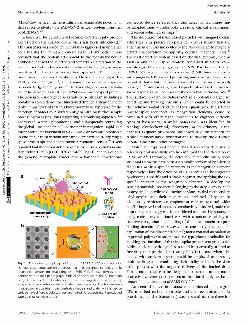
This biosensor was based on membrane-engineered mammaliancells bearing the human chimeric spike S1 antibody. It wasrevealed that the protein attachment to the membrane-boundantibodies caused the selective and remarkable alteration in thecellular bioelectric characteristics evaluated by applying an assaybased on the bioelectric recognition approach. The preparedbiosensor demonstrated an ultra-rapid behavior (B3 min) with aLOD of about 1 fg mL�1, and a semi-linear range of responsebetween 10 fg and 1 mg mL�1. Additionally, no cross-reactivitycould be detected against the SARS-CoV-2 nucleocapsid protein.The biosensor was designed as a ready-to-use platform, including aportable read-out device that functioned through a smartphone ortablet. It was revealed that this biosensor may be applicable for thedetection of SARS-CoV-2 surface antigens with no further sampleprocessing/managing, thus suggesting a promising approach forwidespread screening/monitoring, and subsequently controllingthe global CoV pandemic.37 In another investigation, rapid anddirect optical measurement of SARS-CoV-2 viruses was introducedin one step, almost without any sample preparation by applying aspike protein specific nanoplasmonic resonance sensor.38 It wasreported that the sensor detected as few as 30 virus particles in onestep within 15 min (LOD = 370 vp mL�1) (Fig. 4). Analysis of boththe generic microplate reader and a handheld smartphone
connected device revealed that this detection technique maybe adopted rapidly under both a regular clinical environmentand resource-limited settings.38
The decoration of nano-based particles with magnetic char-acteristics with precise receptors for viruses means that theattachment of virus molecules to the NPs can lead to magneticextraction/separation by applying external magnetic fields.21
The viral detection system based on the viral genome, such as+ssRNA and the S (spike)-protein contained in SARS-CoV-2,was designed by applying magnetic NPs. For the detection ofSARS-CoV-2, a giant magneto-resistive (GMR) biosensor alongwith magnetic NPs showed promising and sensitive biosensingpotential, but additional evaluations should be systematicallymanaged.39 Additionally, the G-quadruplex-based biosensorshowed remarkable potential for the detection of SARS-CoV-2.40
The surface protein of SARS-CoV-2 is a potential target fordetecting and treating this virus, which could be detected bythe exclusive spatial structure of the G-quadruplex. The selectedG-quadruplex sequences, as recognition elements, could becombined with other signal molecules to engineer differenttypes of biosensors, in which SARS-CoV-2 was identified byreading electrochemical, florescent or colorimetry signalchanges. G-quadruplex based biosensors have the potential toreplace antibody-based detection and to develop the detectionof SARS-CoV-2 and other pathogens.40
Molecular imprinted polymer based sensors with a uniqueselectivity and sensitivity can be employed for the detection ofSARS-CoV-2.41 Previously, the detection of the Zika virus, Ebolavirus and Norovirus have been successfully performed by selectingtheir DNA or virus specific aptamers as the recognition element,respectively. Thus, the detection of SARS-CoV-2 can be suggestedby choosing a specific and suitable polymer and applying the CoVspecific aptamer as the recognition element. To develop thesensing materials, polymers belonging to the acrylic group, suchas acrylamide, acrylic acid, methyl acrylate, methyl methacrylate,ethyl acrylate and their mixtures are preferred. They can beadditionally reinforced on graphene or conducting metal oxidesto offer improved and enhanced conductivity.41 Indeed, molecularimprinting technology can be considered as a suitable strategy toapply molecularly imprinted NPs with a unique capability forselective recognition and binding of the spike protein receptor-binding domain of SARS-CoV-2.42 In one study, the potentialapplication of the biocompatible polymeric material as molecularimprinted polymer-based monoclonal-type plastic antibodies forblocking the function of the virus spike protein was proposed.42
Additionally, these designed NPs could be potentially utilized asfree-drug therapeutics for treating COVID-19, and when alsoloaded with antiviral agents, could be employed as a strongmultimodal system combining their ability to block the virusspike protein with the targeted delivery of the loaded drug.Furthermore, they can be designed to become an immuno-protective vaccine or a molecular imprinted polymer-basedsensor for the detection of SARS-CoV-2.42
An electrochemical immunosensor fabricated using a goldNPs modified carbon electrode and the recombinant spikeprotein S1 (as the biomarker) was reported for the detection
Fig. 4 The one-step rapid quantification of SARS-CoV-2 virus particlesvia low-cost nanoplasmonic sensors: (a) the designed nanoplasmonicresonance sensor for evaluating the SARS-CoV-2 pseudovirus con-centration; and (b) a photograph (middle) of one piece of the Au nanocuparray chip with a drop of water on top. The scanning electron microscopyimage (left) demonstrates the replicated nanocup array. The transmissionmicroscopy image (right) demonstrates that air and water on the devicesurface have different colors, green and red/pink, respectively. Reproducedwith permission from ref. 38.
Materials Advances Highlight
Ope
n A
cces
s A
rtic
le. P
ublis
hed
on 0
2 N
ovem
ber
2020
. Dow
nloa
ded
on 3
/23/
2022
11:
51:4
4 A
M.
Thi
s ar
ticle
is li
cens
ed u
nder
a C
reat
ive
Com
mon
s A
ttrib
utio
n-N
onC
omm
erci
al 3
.0 U
npor
ted
Lic
ence
.View Article Online

This journal is©The Royal Society of Chemistry 2020 Mater. Adv., 2020, 1, 3092--3103 | 3097
of MERS-CoVs.31 This immunosensor holds promise for thesimultaneous detection of various CoVs (20 min), and demon-strated a linear response of 0.001 to 100 ng mL�1 and 0.01 to10 000 ng mL�1 for detecting MERS-CoVs and human CoVs,respectively. Additionally, the resulting LOD values were 1.0 and0.4 pg mL�1 for MERS-CoVs and human CoVs, respectively.31 Inanother study, a gold-NPs based electrochemical biosensor wasconstructed for detection of the SARS-CoV-2 spikeS1 protein anti-gen. The biosensor was fabricated by applying a fluorine-doped tinoxide based substrate, and gold NPs were utilized as a signalamplifier owing to the significant electrical conductivity.33 Toproduce the biosensing platform, gold NPs (B29 nm) were dropcast, and then the monoclonal antibodies against SARS-CoV-2 wereimmobilized to prepare the immunosensor. The LOD of theimmunosensor was about 10 fM for detection of the COVID-19antigen (spike protein), and it could be employed for detection ofthe COVID-19 antigen (spike protein) at a concentration up to120 fM, and more than three times.33 Additionally, a cheap andsensitive cobalt-functionalized TiO2 nanotubes-based electro-chemical sensor was designed for the detection of SARS-CoV-2via sensing of the spike present on the surface of the virus
within about 30 s. The sensor specifically detected the S-receptorbinding domain protein of SARS-CoV-2, even at a very lowconcentration (14–1400 nM); the sensor exhibited a linearresponse for the detection of the viral protein over the concen-tration range.43
For rapid and sensitive detection of the SARS-CoV-2 spike 1(S1) protein, an innovative method was introduced by applyingthe SARS-CoV-2 receptor ACE2, which can produce matchedpairs with commercially available antibodies (Fig. 5).44 ACE2and S1-mAb were paired with each other for capture anddetection in a lateral flow immunoassay (LFIA) that did notcross-react with the SARS-CoV Spike 1 or MERS-CoV Spike 1protein. As a result, the SARS-CoV-2 S1 (o5 ng of recombinantproteins/reaction) was identified by the ACE2-based LFIA. TheLOD was about 1.86 � 105 copies mL�1 in clinical specimensfrom COVID-19 patients, without any cross-reactivity for nasalswabs from healthy subjects.44
2.2. For the detection of antibodies
A rapid and sensitive LFIA was introduced by applyinglanthanide-doped polystyrene NPs to detect anti-SARV-CoV-2
Fig. 5 (a) The detection of SARS-CoV-2 spike 1 antigens using a cellular receptor (ACE2)-based LFIA. (b) The designed ACE2-based LFIA included asample pad, conjugate pad, nitrocellulose membrane, and absorbent pad. The test line placed on the nitrocellulose membrane contains ACE2 for thedetection of the SARS-CoV-2 spike antigen. The anti-IgG antibody was applied in the control line; and the detection result was achieved within 20 min.Reproduced with permission from ref. 44.
Highlight Materials Advances
Ope
n A
cces
s A
rtic
le. P
ublis
hed
on 0
2 N
ovem
ber
2020
. Dow
nloa
ded
on 3
/23/
2022
11:
51:4
4 A
M.
Thi
s ar
ticle
is li
cens
ed u
nder
a C
reat
ive
Com
mon
s A
ttrib
utio
n-N
onC
omm
erci
al 3
.0 U
npor
ted
Lic
ence
.View Article Online

3098 | Mater. Adv., 2020, 1, 3092--3103 This journal is©The Royal Society of Chemistry 2020
IgG in human serum.45 A recombinant nucleocapsid phospho-protein of SARS-CoV-2 was dispensed onto a nitrocellulosemembrane to capture the specific IgG. Mouse anti-humanIgG antibody was labeled with self-assembled lanthanide-doped polystyrene NPs, which served as a fluorescent reporter.This assay can be employed as a rapid and sensitive method forthe detection of anti-SARS-CoV-2 IgG in human serum and canpermit positive detection in suspicious cases; additionally, itcan be promising for evaluating COVID-19 progression andanalyzing a patients’ response to treatment.45 Additionally, alateral flow combined IgG–IgM immunochromatographic assaywas reported for rapid and simultaneous identification of IgMand IgG antibodies against SARS-CoV-2 in clinical blood sampleswithin 15 min.46 After clinical evaluations, it was revealed that thesensitivity and specificity of this assay were 85.29% and 100.00%,respectively. It should be noted that the detection of SARS-CoV-2related IgM tends to indicate a recent exposure to the virus, whiledetection based on IgG demonstrates exposure to the virus sometime ago. Thus, compared with a single IgG and IgM evaluation,the combined IgG–IgM immunochromatographic strip test hada better sensitivity, and it showed potential for monitoringCOVID-19 patients.46 For detection of IgM and IgG antibodies con-currently against SARS-CoV-2 (B15 min), researchers developed arapid and simple POC lateral flow immunoassay that is applicableat different stages of viral infection. Accordingly, the sensitivity andspecificity were 88.66% and 90.63%, respectively. This colloidalgold-based test kit was based on the conjugation of gold NPs toIgM/IgG antibodies in human serum, plasma and whole bloodsamples, but it appears that the specificity is not sufficient forCOVID-19. Thus, it can cause some false responses for patientswith irrelevant infections.47 Furthermore, a multiplexed grating-coupled fluorescent plasmonics biosensor platform was intro-duced for rapid and accurate measurement of antibodiesagainst SARS-CoV-2 in human blood serum and dried bloodspot samples.48 This technique could be employed for thesuccessful detection of IgM, IgG and IgA antibody–antigeninteractions. Accordingly, the biosensor could evaluate anti-body–antigen binding interactions for multiple targets in asingle sample, and showed a high selectivity and sensitivitywhen measuring serum IgG levels against three SARS-CoV-2
antigens (spike S1, spike S1S2, and the nucleocapsid protein);the platform could result in a quantitative, linear response for serumsamples diluted to as low as 1 : 1600 dilution. For evaluation of thetest efficacy with other sample matrices, dried blood spot sampleswere utilized and analyzed using the biosensor, yielding 100%selectivity and 86.7% sensitivity for diagnosing prior COVID-19.48
The properties of nanomaterials, such as a high surface-to-volume ratio, quantum size effects, remarkable adsorption andreactive capacity, as compared to their bulk form, are imperative tothe design of biosensing techniques.3,49,50 Additionally, the size andshape of nanomaterials and nanoarchitectures can be optimizedand designed, and thus surface modification/immobilization withvarious biological species through covalent or non-covalent bondingis possible to improve the biosensing features in terms of the LOD(increased up to several order of magnitudes), significant sensitivity/selectivity and rapid response towards the sample analytes.49,50 Inone study, an opto-microfluidic sensing platform with gold nanos-pikes was developed for detection of the presence and amount ofantibodies specific to the SARS-CoV-2 spike protein in 1 mL ofhuman plasma diluted in 1 mL of buffer solution, within 30 min(Fig. 6).51 The target antibody concentration can be correlated withthe LSPR wavelength peak shift of the gold nanospikes caused bythe local refractive index alteration because of the antigen–anti-body binding. The label-free microfluidic platform demonstrateda LOD of 0.08 ng mL�1, falling under the clinically relevantconcentration range.51 This platform provided a promising POCtesting tool to complement standard serological assays and canmake SARS-CoV-2 quantitative diagnostics easier, cheaper, andfaster. However, it is critical to validate it for antibody tests forthe COVID-19 pandemic, and also the electrodeposition processshould be optimized to produce gold nanostructures with asmaller spacing and a higher aspect ratio, thus the antibody–antigen binding can generate a larger shift in the LSPR peak andimprove the signal-to-noise ratio of the sensor.51
2.3. For the detection of cytokines
Application of biosensors for the constant evaluation of cyto-kine levels have demonstrated remarkable potentials for thediagnosis of COVID-19 progression/severity stages and analysisof the efficacy of anti-inflammatory treatments.13 For instance,
Fig. 6 An opto-microfluidic sensing platform prepared for the rapid detection of antibodies against the SARS-CoV-2 spike protein in diluted humanplasma with remarkable sensitivity. The sensing principle is based on LSPR, involving gold nanospikes in a microfluidic device, coupled with an opticalprobe. Reproduced with permission from ref. 51.
Materials Advances Highlight
Ope
n A
cces
s A
rtic
le. P
ublis
hed
on 0
2 N
ovem
ber
2020
. Dow
nloa
ded
on 3
/23/
2022
11:
51:4
4 A
M.
Thi
s ar
ticle
is li
cens
ed u
nder
a C
reat
ive
Com
mon
s A
ttrib
utio
n-N
onC
omm
erci
al 3
.0 U
npor
ted
Lic
ence
.View Article Online

This journal is©The Royal Society of Chemistry 2020 Mater. Adv., 2020, 1, 3092--3103 | 3099
needle-shaped microelectrodes were suggested for the identifi-cation of changes in Interleukin-6 (IL-6) levels in real time.12
Additionally, by applying aptamer-modified graphene as theconducting channel, a wearable and deformable graphene-based field-effect transistor biosensor was designed for sensi-tive, consistent and time-resolved detection of cytokines inhuman biofluids (Fig. 7).52 The designed biosensor showedadvantages, including a remarkable mechanical durability andconsistent sensing responses, while conforming to non-planarsurfaces such as the human body and withstanding largedeformations. Additionally, a nonionic surfactant was deployedto reduce the nonspecific adsorption of the biosensor, hence,enabling cytokine detection (inflammatory cytokines such asTNF-a and IFN-g) in artificial tears (as a biofluid). As a result,this biosensor demonstrated a high potential for use in theconsistent and sensitive detection of TNF-a and IFN-g (with theLOD down to 2.75 and 2.89 pM, respectively).52 It appears thatbiosensors should be further evaluated and developed for clinicalapplications; wearable biosensors are promising candidates forcontinuous monitoring and follow up of patients with COVID-19.13
2.4. For the detection of nucleic acids
A colorimetric assay based on gold NPs was developed, andcapped by appropriately constructed thiol-modified antisenseoligonucleotides with sufficient specificity for the N-gene(nucleocapsid phosphoprotein) of SARS-CoV-2; accordingly it
showed potential for the selective and visual naked-eye diagnosisof COVID-19 (B10 min), but its performance can be influenced bythe quantity of loaded virus (Fig. 8).53 The thiol-modified antisenseoligonucleotide-capped gold NPs agglomerated selectively in thepresence of the target RNA sequence of SARS-CoV-2 and showedan alteration in its surface plasmon resonance (SPR).53 Addition-ally, the utilization of RNaseH could cleave the RNA strand fromthe RNA-DNA hybrid, causing a visually detectable precipitate fromthe solution mediated by the additional agglomeration among thegold NPs. The selectivity of the assay has been checked in thepresence of MERS-CoV viral RNA with a LOD of about 0.18 ng mL�1
of RNA having a SARS-CoV-2 viral load. Thus, this study reportedselective and visual naked-eye detection of SARS-CoV-2, with norequirement for any sophisticated instrumental techniques.53
Interestingly, a double-operational plasmonic biosensorcombining the LSPR sensing transduction and the PPT influence,offered a promising substitute for the diagnosis of COVID-19,clinically.10 The delicate recognition of certain arrangements fromSARS-CoV-2 via nucleic acid hybridization was performed by usingthe 2-D gold nano-islands decorated with complementary DNAreceptors.10 The thermos-plasmonic heat was produced on the chipfor an improved sensing performance, which upon illumination attheir plasmonic resonance frequency and under local PPT heat,could improve the in situ hybridization temperature and enable theprecise discernment of two similar gene sequences. This biosensorshowed remarkable sensitivity for the examined SARS-CoV-2sequences with a much lower detection limit at a concentration of0.22 pM, and permitted specific target recognition in a multigenemixture.10 In this study, Qui et al.,10 reported that by using LSPRexcited at two different wavelengths and the plasmonic resonancesof PPT, remarkable stability, reliability and sensitivity properties indiagnosis are achievable.
A paper-based colorimetric assay for DNA detection basedon pyrrolidinyl peptide nucleic acid (acpcPNA)-induced NPaggregation has been proposed as an alternative to conven-tional techniques for the detection of SARS-CoV-2.54 These PNAprobes are an attractive alternative to DNA and RNA probesowing to the chemical and biological stability and simplerelated synthetic methods; they can also hybridize successfullywith the complementary DNA strands. Indeed, the acpcPNAprobe comprises a single positive charge from the lysine at theC-terminus and results in aggregation of the citrate anion-stabilized silver NPs in the absence of complementary DNA.54
In the presence of the target DNA, the anionic DNA-acpcPNAduplex was generated, which can cause the dispersion of silverNPs as a result of the electrostatic repulsion, providing anenhancement in the detectable color change. This techniquewas introduced for detecting and analyzing synthetic MERS-CoV,Mycobacterium tuberculosis, and human papillomavirus DNA;however, it may also be suitable for the detection ofSARS-CoV-2. The acpcPNA probe showed significant selectivityfor the complementary oligonucleotides over single-base-mismatch,two-base-mismatch, and noncomplementary DNA targets.Importantly, some parameters for controlling the sensitivity/selectivity issues of this assay should be noted, such as the silverNP concentration, DNA strand mismatches, ionic strength, and
Fig. 7 An ultra-flexible graphene field-effect transistor biosensor: (a) aschematic diagram of the biosensor produced on an ultrathin film. A photo-graph of the flexible device conformably attached onto (b) a human wrist and(c) an artificial eyeball. (d) The stretchable biosensor can be stretched with theactivity of the human body. Reproduced from ref. 52, (CC BY 4.0).
Highlight Materials Advances
Ope
n A
cces
s A
rtic
le. P
ublis
hed
on 0
2 N
ovem
ber
2020
. Dow
nloa
ded
on 3
/23/
2022
11:
51:4
4 A
M.
Thi
s ar
ticle
is li
cens
ed u
nder
a C
reat
ive
Com
mon
s A
ttrib
utio
n-N
onC
omm
erci
al 3
.0 U
npor
ted
Lic
ence
.View Article Online

3100 | Mater. Adv., 2020, 1, 3092--3103 This journal is©The Royal Society of Chemistry 2020
PNA concentration.54 Additionally, aptamers and molecularbeacons have been suggested instead of PNA.21
3. Challenges and opportunities
One of the important challenges for constructing suitablebiosensors is to capture a signal of very low magnitude thattakes place between the biological species (bio-receptor andanalyte).24 To solve the problem, nanomaterials can beemployed as labels to obtain a remarkable improvement inthe signal, high enough to be easily detectable.50,55,56 As anexample, metallic nanomaterials (e.g., gold or silver NPs) andquantum dots can be analyzed and employed for labeling byattaching them onto the targeted DNA/bio-recognizing probe.55,56
This can cause a synergetic effect owing to the nano-labelingconsequences to significantly amplify the electrochemical signal,and make it possible to design ultrasensitive and selectivelabeled-biosensing strategies.56 In this regard, future studiesshould be performed based on taking advantage of the variousattractive physicochemical characteristics of nanoscale materials(especially optical, electrical, magnetic, and opto-magneticproperties) for the development of nano-enabled biosensingapproaches for the specific detection of viruses, especiallyMERS-CoV, SARS-CoV and SARS-CoV-2 (Table 3).3,57 However,limited analyses have been performed to develop thesenanomaterials-based biosensors for SARS-CoV-2 detection.These techniques can be applied instead of PCR-based testing forCOVID-19, with the advantages of simplicity, cost-effectiveness, fastresponse and real-time diagnostic procedures. These nanomaterial-enabled biosensors primarily rely on the nucleic acid and protein(antigen/antibody) mediated detection of SARS-CoV-2, although theyhave not yet yielded 100% accuracy owing to the contamination ofthese highly sensitive bio-receptors, and ultrasensitive, rapid,
and portable SARS-CoV-2 sequence detection methods are highlydemanded. For instance, the technique based on CRISPR-Cas12,58
or in the case of nanomaterial-based biosensors based on theaerosol mediated diagnosis method, have the advantages of a fastresponse, sensitivity, and a lack of sample perturbation.59 Impor-tantly, the scale up of these detection methods is very important;in the lab scale, these biosensors have exhibited an approvedstability, fast response time, and high sensitivity/selectivity.50
However, this may change in the case of real sample evaluations,and several factors, such as the characteristics of the targetantigen/protein/antibody, nanomaterial features, and other impor-tant biomolecules may influence the obtained results, as well asthe performance of the nanomaterial based biosensing devices.50
Nano and biosensors should be technically developed forhighly efficient detection of SARS-CoV-2 in various clinicalsamples, including urine, blood, saliva, and nasopharyngealswabs.50,60 One of the important limitations for developingbiosensors based on biological reagents (such as antibodies,DNA, and antigens) is the time consuming and costly procedureof extraction and/or preparation of the target biological metrics/reagents.61 Thus, designing nanomaterials-enabled biosensorsthat do not need extra extraction procedures is very necessary.For instance, a rapid electrochemical diagnostic kit composed offixed/screen printed electrodes was constructed for the detectionof SARS-CoV-2 via the differentiable fingerprint of the viralglycoproteins at various voltage positions.61 The sensor wasactivated upon coating a layer of coupled graphene oxide withsensitive chemical compounds along with gold nanostars; thistechnique, without the need for extraction procedures and bio-markers, could identify trace amounts of the virus in about 1 minwith a high sensitivity. The electrochemical detection mechanisticevaluations showed the surface-confined mechanism via theadsorption procedure, providing differentiable detection of patho-genic viruses within selected aquatic biological media.61
Fig. 8 The process of detecting SARS-CoV-2 RNA mediated using appropriately constructed thiol-modified antisense oligonucleotide (ASO)-cappedgold (Au) NPs with remarkable selectivity. Reproduced with permission from ref. 53, copyrightr 2020 American Chemical Society.
Materials Advances Highlight
Ope
n A
cces
s A
rtic
le. P
ublis
hed
on 0
2 N
ovem
ber
2020
. Dow
nloa
ded
on 3
/23/
2022
11:
51:4
4 A
M.
Thi
s ar
ticle
is li
cens
ed u
nder
a C
reat
ive
Com
mon
s A
ttrib
utio
n-N
onC
omm
erci
al 3
.0 U
npor
ted
Lic
ence
.View Article Online

This journal is©The Royal Society of Chemistry 2020 Mater. Adv., 2020, 1, 3092--3103 | 3101
The detection limit is very important because of the relativelylow viral concentration in patient samples; it appears thatmagnetic, plasmonic, and electrochemical devices have exhibitedthe lowest LOD, indicating they are promising techniques.66
However, an important drawback regarding these magnetic andplasmonic methods is that they typically require specializedinstrumentation both for production and operation, creatingchallenging portability issues. Therefore, improved proceduresare highly necessary to make them more portable, and alsoimprove their sensitivity to be applicable to POC use.66 Further-more, some techniques based on colorimetric, electrochemical,and lateral flow assays have demonstrated greater portability,permitting better operation in the field as they do not need alaboratory infrastructure and instrumentation to obtain theresults.67,68 On the other hand, the plasmonic and magnetictechniques require a laboratory infrastructure, and also have theadvantage of a higher throughput, allowing more samples tobe evaluated in one step; the electrochemical and plasmonictechniques allow a better and fastest readout compared with the
colorimetric and lateral flow methods.24,58,66 Additionally, todesign well-organized biosensors, one of the most importantparts is transporting the targeted molecules to the functionalizedsurface of them.69 In this regard, robust simulation (reliable, fast,and stable numerical modelling) of these systems and theirrelated chemical reactions can help improve the designed bio-sensors. For instance, computational fluid dynamics (CFD) havebeen employed for various related biomedical studies involvingdesign, validation and proof-of-concept.69
4. Conclusions and future outlooks
Rapid, convenient, and large-scale diagnosis is vital for thetreatment and management of COVID-19 (especially asympto-matic and/or early-stage patients) resulting from SARS-CoV-2,especially for reducing and controlling its spread. Traditionaltechniques for the detection of respiratory viruses are generallycostly, time consuming, and labor intensive, and they require
Table 3 Some important examples of nano-based sensors for the detection of SARS-CoV-2
Nanomaterial Biomarker Findings Target Ref.
Magnetic NPs Poly(amino ester) withcarboxyl group-coatedmagnetic NPs
Linear correlation: 10 to 105 copies of SARS-CoV-2pseudovirus particles; 10-copy sensitive; NPs enabledimproved RNA extraction
SARS-CoV-2 virus RNA 62
Biotinylated probe LOD was about 0.4 fM; total assay time was about100 min; dynamic detection range covers three ordersof magnitude; the process was based on circle to circleamplification based optomagnetic biosensing
RdRp sequence(synthetic complementaryDNA of SARS-CoV-2)
63
Gold NPs Thiol-c-DNA receptors/nucleic acid
LOD of about 0.22 � 0.08 pM; detection range wasabout 0.1 pM to 1 mM; the process was based on thecombined effects of PPT and LSPR mediatedbiosensing
ORF1ab-COVID, and Egenes from SARS-CoV-2;viral sequences includingRdRp-COVID
10
Thiol-modified antisenseoligonucleotides specificfor the N-gene (nucleo-capsid phosphoprotein) ofSARS-CoV-2
Detection limit of about 0.18 ng mL�1; detection range:0.2–3 ng mL�1; assay time was about 10 min; theprocess was based on plasmonic effect basedcolorimetric biosensing
RNA [N-gene ofSARS-CoV-2]
53
Oligo probe Detection limit of about 0.5 ng; total assay time wasabout 30 min; the process was based on plasmoniceffect based colorimetric biosensing
RNA [RdRp gene ofSARS-CoV-2]
64
Recombinant antigen ofSARS-CoV-2 and rabbit-IgG
Assay time was about 15 min; the process was based oncolorimetric dependent lateral flow immunoassaybased biosensing
IgM and IgG antibodies 47
Monoclonal antibodiesagainst SARS-CoV-2
LOD of about 10 fM; sensitive for detecting SARS-CoV-2antigens; amperometric biosensing technique
SARS-CoV-2 spike antigenin saliva samples
33
Screen printed carbonelectrode (SPCE)-basedsensing device
Monoclonal antibodiesagainst SARS-CoV-2
Detection of viral antigen at 10 fM concentration instandard buffer sample; LOD was about 90 fM in thecase of spiked saliva samples using an SPCE mediatedhomemade biosensor device; detection of antigentraces in patient saliva within one min; stability forup to 28 d; amperometric biosensing technique
SARS-CoV-2 spike antigenin saliva samples
33
Polymer NPs coatedwith dye streptavidin(Crimson red)
Rabbit anti-fluoresceinantibody, sheep anti-digoxigenin antibody, andbiotinylated bovine serumalbumin
LOD was about 12 copies (for each detection target) perreaction; total diagnostic test was completed within 1 hfrom sample collection to result interpretation;analytical sensitivity was 100%; specificity was100%; the assay required simple heating equipmentto maintain a constant temperature of 63 1C for40 min; the process was based on multiplex reversetranscription loop mediated isothermal amplificationcoupled with a NP-based lateral flow biosensor
ORF1ab and N genes ofSARS-CoV-2
65
Graphene Spike protein antibody FET-based biosensing method for the detectionof the SARS-CoV-2 spike protein; LOD was about1.6 � 101 pfu mL�1 in culture medium, whereasit was 2.42 � 102 copies mL�1 in clinical samples.
SARS-CoV-2 antigenprotein
32
Highlight Materials Advances
Ope
n A
cces
s A
rtic
le. P
ublis
hed
on 0
2 N
ovem
ber
2020
. Dow
nloa
ded
on 3
/23/
2022
11:
51:4
4 A
M.
Thi
s ar
ticle
is li
cens
ed u
nder
a C
reat
ive
Com
mon
s A
ttrib
utio
n-N
onC
omm
erci
al 3
.0 U
npor
ted
Lic
ence
.View Article Online

3102 | Mater. Adv., 2020, 1, 3092--3103 This journal is©The Royal Society of Chemistry 2020
specialized laboratory equipment and expertise. Nano- andbiosensors can be considered as sensitive, selective, and afford-able analytical diagnostic systems for the detection of SARS-CoV-2and related viral infections. Indeed, the low-level detection of atargeted disease biomarker can be highly suitable for the betterevaluation, management, and treatment of viral disease progres-sion. The surface functionalization and modification of NPs canfacilitate the faster and more specific detection of SARS-CoV-2,even in the early stages of infection. Moreover, to improve thediagnostic performance and accuracy, combinational diagnosis/detection procedures for various kinds of biomarkers, viaapplying multiplex nano- and biosensors, could be a usefulstrategy. On the other hand, to improve the reliability andreproducibility of these sensors, machine learning-based signalprocedures and the related obtained details are an essentialarea of focus for researchers.
It appears that more elaborate academic analyses and studiesshould be systematically carried out to solve these challengingissues, especially with regards to false negative/positive results,validation processes, detection speed, simplicity, selectivity,sensitivity, cost-effectiveness, and public usability. Nanotech-nology can enable extreme progress regarding the fabrication ofinnovative diagnostic sensors, the integration of novel devices,better optimization/validation, and improvements in sensingperformances at the POC. Future studies should be planned tofind and design innovative and next-generation non-invasive,specific, affordable, and fast biosensing methodologies andtechnologies for diagnostic applications, to manage pandemicsand life-threatening infectious diseases in particular. Remarkably,sensing devices with suitable cost-effectiveness, miniaturization,and user-friendly features have received considerable academicattention, and companies and industry in particular are focusedon these smart nano- and biosensors (with high sensitivity/selectivity features) for use as highly qualified and intelligentdiagnostic systems/platforms. Importantly, owing to the occurrenceof asymptomatic SARS-CoV-2 patients, public and home-use nano-and biosensors may enable immediate detection of this virus andaccordingly provide better control and management of COVID-19.In this regard, novel smartphone-based biosensors and colorimetricstrips targeting antibodies/antigens have shown potential for use inthe home and for simple POC testing.
Conflicts of interest
There are no conflicts to declare.
References
1 S. Jiang, C. Hillyer and L. Du, Trends Immunol., 2020, 41,355–359.
2 G. Jamalipour Soufi, A. Hekmatnia, M. Nasrollahzadeh,N. Shafiei, M. Sajjadi, P. Iravani, S. Fallah, S. Iravani andR. S. Varma, Appl. Sci., 2020, 10, 3641.
3 M. Nasrollahzadeh, M. Sajjadi, G. Jamalipour Soufi, S. Iravaniand R. S. Varma, Nanomaterials, 2020, 10, 1072.
4 H. F. Florindo, R. Kleiner, D. Vaskovich-Koubi, R. C. Acurcio,B. Carreira, E. Yeini, G. Tiram, Y. Liubomirski and R. Satchi-Fainaro, Nat. Nanotechnol., 2020, 15, 630–645.
5 S. Talebian and J. Conde, Matter, 2020, 3, 598–601.6 M. D. Shin, S. Shukla, Y. H. Chung, V. Beiss, S. K. Chan,
O. A. Ortega-Rivera, D. M. Wirth, A. Chen, M. Sack, J. K.Pokorski and N. F. Steinmetz, Nat. Nanotechnol., 2020, 15,646–655.
7 F. Cui and H. S. Zhou, Biosens. Bioelectron., 2020, 165,112349.
8 B. Udugama, P. Kadhiresan, H. N. Kozlowski, A. Malekjahani,M. Osborne, V. Y. C. Li, H. Chen, S. Mubareka, J. B. Gubbayand W. C. W. Chan, ACS Nano, 2020, 14, 3822–3835.
9 W. Feng, A. M. Newbigging, C. Le, B. Pang, H. Peng, Y. Cao,J. Wu, G. Abbas, J. Song, D.-B. Wang, M. Cui, J. Tao,D. L. Tyrrell, X.-E. Zhang, H. Zhang and X. C. Le, Anal.Chem., 2020, 92, 10196–10209.
10 G. Qiu, Z. Gai, Y. Tao, J. Schmitt, G. A. Kullak-Ublick andJ. Wang, ACS Nano, 2020, 14, 5268–5277.
11 N. Bhalla, Y. Pan, Z. Yang and A. F. Payam, ACS Nano, 2020,14, 7783–7807.
12 C. Russell, A. C. Ward, V. Vezza, P. Hoskisson, D. Alcorn,D. P. Steenson and D. K. Corrigan, Biosens. Bioelectron.,2019, 126, 806–814.
13 S. M. Russell, A. Alba-Patino, E. Baron, M. Borges, M. Gonzalez-Freire and R. de la Rica, ACS Sens., 2020, 5, 1506–1513.
14 T. Ai, Z. Yang, H. Hou, C. Zhan, C. Chen, W. Lv, Q. Tao,Z. Sun and L. Xia, Radiology, 2020, 200642.
15 N. Rabiee, M. Bagherzadeh, A. Ghasemi, H. Zare, S. Ahmadi,Y. Fatahi, R. Dinarvand, M. Rabiee, S. Ramakrishna,M. Shokouhimehr and R. S. Varma, Int. J. Mol. Sci., 2020,21, 5126.
16 I. Zehbe, G. W. Hacker, H. Su, C. Hauser-Kronberger,J. F. Hainfeld and R. Tubbs, Am. J. Pathol., 1997, 150, 1553.
17 M. S. Draz and H. Shafiee, Theranostics, 2018, 8, 1985.18 X. Wang, L.-H. Liu, O. Ramstroem and M. Yan, Exp. Biol.
Med., 2009, 234, 1128–1139.19 P. Alivisatos, Nat. Biotechnol., 2004, 22, 47–52.20 N. L. Rosi and C. A. Mirkin, Chem. Rev., 2005, 105, 1547–1562.21 S. Talebian, G. G. Wallace, A. Schroeder, F. Stellacci and
J. Conde, Nat. Nanotechnol., 2020, 15, 618–621.22 H. A. Hussein, R. Y. A. Hassan, M. Chino and F. Febbraio,
Sensors, 2020, 20, 4289.23 R. Samson, G. R. Navale and M. S. Dharne, 3 Biotech, 2020,
10, 385.24 M. Asif, M. Ajmal, G. Ashraf, N. Muhammad, A. Aziz, T. Iftikhar,
J. Wang and H. Liu, Curr. Opin. Electrochem., 2020, 23, 174–184.25 I. Santiago, ChemBioChem, 2020, 21, 2880–2889.26 J. C. Huang, Y.-F. Chang, K.-H. Chen, L.-C. Su, C.-W. Lee,
C.-C. Chen, Y.-M. A. Chen and C. Chou, Biosens. Bioelectron.,2009, 25, 320–325.
27 C. Roh and S. K. Jo, J. Chem. Technol. Biotechnol., 2011, 86,1475–1479.
28 L. Shi, Q. Sun, J. He, H. Xu, C. Liu, C. Zhao, Y. Xu, C. Wu,J. Xiang, D. Gu, J. Long and H. Lan, Biomed. Mater. Eng.,2015, 26, S2207–S2216.
Materials Advances Highlight
Ope
n A
cces
s A
rtic
le. P
ublis
hed
on 0
2 N
ovem
ber
2020
. Dow
nloa
ded
on 3
/23/
2022
11:
51:4
4 A
M.
Thi
s ar
ticle
is li
cens
ed u
nder
a C
reat
ive
Com
mon
s A
ttrib
utio
n-N
onC
omm
erci
al 3
.0 U
npor
ted
Lic
ence
.View Article Online

This journal is©The Royal Society of Chemistry 2020 Mater. Adv., 2020, 1, 3092--3103 | 3103
29 T. Lee, J.-H. Ahn, S. Y. Park, G.-H. Kim, J. Kim, T.-H. Kim,I. Nam, C. Park and M.-H. Lee, Micromachines, 2018, 9, 651.
30 D. R. B. Albano, K. Shum, J. A. Tanner and Y. S. Fung,17th Int. Meet. Chem. Sensors - IMCS, AMA, Viena, Austria,2018, 211–213.
31 L. A. Layqah and S. Eissa, Mikrochim. Acta, 2019, 186, 224.32 G. Seo, G. Lee, M. J. Kim, S. H. Baek, M. Choi, K. B. Ku, C. S. Lee,
S. Jun, D. Park, H. G. Kim, S. J. Kim, J. O. Lee, B. T. Kim,E. C. Park and S. I. Kim, ACS Nano, 2020, 14, 5135–5142.
33 S. Mahari, A. Roberts, D. Shahdeo and S. Gandhi, bioRxiv,2020, DOI: 10.1101/2020.04.24.059204.
34 C. Wang, C. Wang, X. Wang, K. Wang, Y. Zhu, Z. Rong,W. Wang, R. Xiao and S. Wang, ACS Appl. Mater. Interfaces,2019, 11, 19495–19505.
35 L. Krejcova, P. Michalek, M. Merlos Rodrigo, Z. Heger,S. Krizkova, M. Vaculovicova, D. Hynek, V. Adam andR. Kizek, Nanobiosens. Dis. Diagn., 2015, 4, 47–66.
36 K.-H. Liang, T.-J. Chang, M.-L. Wang, P.-H. Tsai, T.-H. Lin,C.-T. Wang and D.-M. Yang, J. Chin. Med. Assoc., 2020, 83,701–703.
37 S. Mavrikou, G. Moschopoulou, V. Tsekouras and S. Kintzios,Sensors, 2020, 20, 3121.
38 L. Huang, L. Ding, J. Zhou, S. Chen, F. Chen, C. Zhao, J. Xu,W. Hu, J. Ji, H. Xu and G. L. Liu, Biosens. Bioelectron., 2020,112685.
39 A. Islam and Z. Ahsan, Am. J. Nanosci., 2020, 6, 6–13.40 H. Xi, M. Juhas and Y. Zhang, Biosens. Bioelectron., 2020,
167, 112494.41 T. N. Chatterjee and R. Bandyopadhyay, Trans. Indian Natl.
Acad. Eng., 2020, 1–4, DOI: 10.1007/s41403-41020-00125-41407.42 O. I. Parisi, M. Dattilo, F. Patitucci, R. Malivindi, V. Pezzi,
I. Perrotta, M. Ruffo, F. Amone and F. Puoci, BioRxiv, 2020,DOI: 10.1101/2020.05.28.120709.
43 B. S. Vadlamani, T. Uppal, S. C. Verma and M. Misra,Sensors, 2020, 20, 5871.
44 J.-H. Lee, M. Choi, Y. Jung, S. K. Lee, C.-S. Lee, J. Kim,J. Kim, N. H. Kim, B.-T. Kim and H. G. Kim, Biosens.Bioelectron., 2021, 171, 112715.
45 Z. Chen, Z. Zhang, X. Zhai, Y. Li, L. Lin, H. Zhao, L. Bian, P. Li,L. Yu, Y. Wu and G. Lin, Anal. Chem., 2020, 92, 7226–7231.
46 L. Zeng, Y. Li, J. Liu, L. Guo, Z. Wang, X. Xu, S. Song, C. Hao,L. Liu, M. Xin and C. Xu, Mater. Chem. Front., 2020, 4, 2000–2005.
47 Z. Li, Y. Yi, X. Luo, N. Xiong, Y. Liu, S. Li, R. Sun, Y. Wang,B. Hu, W. Chen, Y. Zhang, J. Wang, B. Huang, Y. Lin,J. Yang, W. Cai, X. Wang, J. Cheng, Z. Chen, K. Sun,W. Pan, Z. Zhan, L. Chen and F. Ye, J. Med. Virol., 2020,92, 1518–1524.
48 N. C. Cady, N. Tokranova, A. Minor, N. Nikvand, K. Strle,W. T. Lee, W. Page, E. Guignon, A. Pilar and G. N. Gibson,Biosens. Bioelectron., 2021, 171, 112679.
49 G. Maduraiveeran, M. Sasidharan and V. Ganesan, Biosens.Bioelectron., 2018, 103, 113–129.
50 M. Srivastava, N. Srivastava, P. K. Mishra and B. D. Malhotra,Sci. Total Environ., 2021, 754, 142363.
51 R. Funari, K.-Y. Chu and A. Q. Shen, Biosens. Bioelectron.,2020, 169, 112578.
52 Z. Wang, Z. Hao, S. Yu, C. Huang, Y. Pan and X. Zhao,Nanomaterials, 2020, 10, 1503.
53 P. Moitra, M. Alafeef, K. Dighe, M. B. Frieman and D. Pan,ACS Nano, 2020, 14, 7617–7627.
54 P. Teengam, W. Siangproh, A. Tuantranont, T. Vilaivan,O. Chailapakul and C. S. Henry, Anal. Chem., 2017, 89,5428–5435.
55 C. Kokkinos, Nanomaterials, 2019, 9, 1361.56 Y. Saylan, O. Erdem, S. Unal and A. Denizli, Biosensors, 2019,
9, 65.57 T. Ozer, B. J. Geiss and C. S. Henry, J. Electrochem. Soc., 2020,
167, 037523.58 C. Lucia, P.-B. Federico and G. C. Alejandra, bioRxiv, 2020,
DOI: 10.1101/2020.02.29.971127.59 J. A. Huffman, A. E. Perring, N. J. Savage, B. Clot, B. Crouzy,
F. Tummon, O. Shoshanim, B. Damit, J. Schneider,V. Sivaprakasam, M. A. Zawadowicz, I. Crawford, M. Gallagher,D. Topping, D. Doughty, S. C. Hill and Y. Pan, Aerosol Sci.Technol., 2020, 54, 465–495.
60 M. Fani, M. Zandi, S. Soltani and S. Abbasi, Biotechnol. Appl.Biochem., 2020, DOI: 10.1002/bab.2033.
61 S. A. Hashemi, N. G. G. Behbahan, S. Bahrani, S. M.Mousavi, A. Gholami, S. Ramakrishna, M. Firoozsani,M. Moghadami, K. B. Lankarani and N. Omidifar, Biosens.Bioelectron., 2021, 171, 112731.
62 Z. Zhao, H. Cui, W. Song, X. Ru, W. Zhou and X. Yu, bioRxiv,2020, DOI: 10.1101/2020.02.22.961268.
63 B. Tian, F. Gao, J. Fock, M. Dufva and M. F. Hansen, Biosens.Bioelectron., 2020, 165, 112356, DOI: 10.1016/j.bios.2020.112349.
64 V. Kumar, S. Mishra, R. Sharma, J. Agarwal, U. Ghoshal,T. Khanna, L. K. Sharma, S. K. Verma and S. Tiwari, bioRxiv,2020, DOI: 10.1101/2020.06.30.172833.
65 X. Zhu, X. Wang, L. Han, T. Chen, L. Wang, H. Li, S. Li,L. He, X. Fu, S. Chen, M. Xing, H. Chen and Y. Wang,Biosens. Bioelectron., 2020, 166, 112437.
66 C. Tymm, J. Zhou, A. Tadimety, A. Burklund and J. X. J.Zhang, Cell. Mol. Bioeng., 2020, 13, 313–329.
67 V. N. Goral, N. V. Zaytseva and A. J. Baeumner, Lab Chip,2006, 6, 414–421.
68 A. Parihar, P. Ranjan, S. K. Sanghi, A. K. Srivastava andR. Khan, ACS Appl. Bio Mater., 2020, DOI: 10.1021/acsabm.0c01083.
69 F. Shahbazi, M. Jabbari, M. Nasr Esfahani and A. Keshmiri,Biosens. Bioelectron., 2021, 171, 112716.
Highlight Materials Advances
Ope
n A
cces
s A
rtic
le. P
ublis
hed
on 0
2 N
ovem
ber
2020
. Dow
nloa
ded
on 3
/23/
2022
11:
51:4
4 A
M.
Thi
s ar
ticle
is li
cens
ed u
nder
a C
reat
ive
Com
mon
s A
ttrib
utio
n-N
onC
omm
erci
al 3
.0 U
npor
ted
Lic
ence
.View Article Online