nanocapsulated curcumin: oral chemopreventive...
TRANSCRIPT

Chemico-Biological Interactions 195 (2012) 206–214
Contents lists available at SciVerse ScienceDirect
Chemico-Biological Interactions
journal homepage: www.elsevier .com/locate /chembioint
Nanocapsulated curcumin: Oral chemopreventive formulationagainst diethylnitrosamine induced hepatocellular carcinoma in rat
Debasree Ghosh a, Somsubhra Thakur Choudhury a, Swarupa Ghosh a, Ardhendu K. Mandal a,Sibani Sarkar a, Aparajita Ghosh a, Krishna Das Saha b, Nirmalendu Das a,⇑a Drug Development/Diagnostics & Biotechnology Department, Indian Institute of Chemical Biology, 4, Raja S.C. Mullick Road, Kolkata 700032, Indiab Cancer & Cell Biology Department, Indian Institute of Chemical Biology, 4, Raja S.C. Mullick Road, Kolkata 700032, India
a r t i c l e i n f o
Article history:Received 19 August 2011Received in revised form 16 November 2011Accepted 8 December 2011Available online 16 December 2011
Keywords:Oxidative stressReactive oxygen speciesDiethylnitrosamineHepatocellular carcinomaDrug deliveryNanocapsules
0009-2797/$ - see front matter � 2011 Elsevier Irelandoi:10.1016/j.cbi.2011.12.004
Abbreviations: Cur, curcumin; PLGA, polylactide ccapsulated curcumin; HCC, hepatocellular carcinomFTIR, Fourier Transform Infra Red; ROS, reactive oxpermeability and retention effect; BSA, bovine serumdimethylammonium bromide; CM-H2DCFDA, 5-(and-rodihydro-fluorescein diacetate acetyl ester; AFM, atserum aspartate transaminase; AP, alkaline phosptransaminase; RLW, relative liver weight; DPH, diphenide dismutase; H2O2, hydrogen peroxide; GSH, reducenylidene fluoride; BCIP/NBT, 5-bromo-4-chloro-30-nitro-blue tetrazolium chloride; NOS 2, nitric oxide sy⇑ Corresponding author. Address: Drug Developmen
Department, Indian Institute of Chemical Biology, 4, RaKolkata 700032, India. Tel.: +91 33 2499 5715; fax: +9
E-mail addresses: [email protected],(N. Das).
a b s t r a c t
Toxic outcome of chemical therapeutics as well as multidrug resistance are two serious phenomena fortheir inacceptance in cancer chemotherapy. Antioxidants like curcumin (Cur) have gained immenseimportance for their excellent anticarcinogenic activities and minimum toxic manifestations in biologicalsystem. However, Cur is lipophilic and thus following oral administration hardly appears in blood indi-cating its potential therapeutic challenge in cancer therapy. Nanocapsulated Cur has been used as a drugdelivery vector to focus the effectiveness of these vesicles against hepatocellular carcinoma. The theme ofwork was to evaluate effectiveness in oral route of polylactide co-glycolide (PLGA) Nanocapsulated cur-cumin (Nano Cur) against diethylnitrosamine (DEN) induced hepatocellular carcinoma (HCC) in rat.
Nano Cur of average diameter 14 nm and encapsulation efficiency of 78% were prepared. Fourier Trans-form Infra Red (FTIR) analysis revealed that there is no chemical interaction between drug and the poly-mer. Three i.p. injections of the chemical hepatocarcinogen DEN at 15 days interval causes hepatotoxicity,the generation of reactive oxygen species (ROS), lipid peroxidation, decrease in plasma membrane micro-viscosity and depletion of antioxidant enzyme levels in liver. Nano Cur (weekly oral treatment for16 weeks at 20 mg/kg b.wt) in DEN induced HCC rats exerted significant protection against HCC andrestored redox homeostasis in liver cells. Nanocapsulated Cur caused cancer cell apoptosis as visualizedby ApoBrdU analysis. Histopathological analysis confirmed the pathological improvement in the liver.Nano Cur was found to be a potential formulation in oral route in combating the oxidative damage ofhepatic cells and eliminating DEN induced hepatocellular cancer cells in rat whereas identical amountof free Cur treatment was found almost ineffective.
� 2011 Elsevier Ireland Ltd. All rights reserved.
1. Introduction countries. Angiogenesis plays a significant role in the aggressive-
Hepatocellular carcinoma (HCC) is the fifth most commoncancer worldwide, with a particularly high prevelance in Asian
d Ltd. All rights reserved.
o-glycolide; Nano Cur, Nano-a; DEN, diethylnitrosamine;
ygen species; EPR, enhancedalbumin; DMAB, Didodecyl-
6)-chloromethyl-20 ,70-dichlo-omic force microscopy; AST,hatase; ALT, serum alanineyl hexatriene; SOD, superox-d glutathione; PVDF, polyvi-
indolylphoshate p-toluidine/nthase; Cyt C, cytochrome C.t/Diagnostics & Biotechnologyja S.C. Mullick Road, Jadavpur,1 33 2473 5197/2472 3967.
ness of HCC. The only potential curative modality for HCC is sur-gery, including transplantation, yet the recurrence rate for thisparticular cancer is high and long-term survival rate is rather poor[1]. Both conventional chemotherapy and radiotherapy have beenfound to be ineffective or only minimally effective in patients withunresectable HCC. Chemoprevention has been considered to be thebest strategy in lowering the present prevalence of the disease inview of the limited treatment and negative prognosis of liver can-cer [2]. The major risk factors of liver cancer are hepatitis viralinfection, alcohol, fungal toxins (aflatoxins), toxic industrial chem-icals, air and water pollutants [3].
ROS play a major role in the induction of carcinogenesis [4].They are derived from the metabolism of molecular oxygen. ROSinclude superoxide anion radical (O2
�.), singlet oxygen (O2), hydro-gen peroxide (H2O2), and the highly reactive hydroxyl radical(�OH). ROS normally exists in all aerobic cells in balance with bio-chemical antioxidants. Oxidative stress occurs when this criticalbalance is disrupted because of excess ROS, antioxidant depletion,
D. Ghosh et al. / Chemico-Biological Interactions 195 (2012) 206–214 207
or both. Cellular targets attacked and damaged by ROS are lipids,proteins, sugars and nucleic acids. Various carcinogens exert theireffects by generating excessive ROS during their metabolism whichcause oxidative damage to cellular DNA which modulates cell sig-nals and activate gene expression [5] that result in mutations lead-ing to sustained proliferation of cancer cells and inhibitingproapoptotic signals responsible for apoptosis and therefore, playan important role in the initiation, promotion and progression ofmultistage carcinogenesis. Free radicals accelerate the rate of pro-gression of benign tumors to malignant neoplasm.
Chemical carcinogen diethylnitrosamine (DEN) is a potenthepatocarcinogenic nitrosamine present in tobacco smoke, groundwater having high level of nitrates, cheddar cheese, cured and friedmeals, alcoholic beverages, occupational settings, cosmetics, agri-cultural chemicals and pharmaceutical agents [3,6]. The rat modelof DEN-induced HCC is considered as one of the most accepted andwidely used experimental models to study hepatocarcinogenesis[7]. Human livers metabolize nitrosamines similar to that of rat li-ver and also exhibit considerable similarities with regard to mor-phology, genomic alterations and gene expression, despite theirdifferent disease etiologies [8]. DEN metabolism in the liver bycytochrome isoform 2E1 (CYP 2E1) generates reactive oxygen spe-cies (ROS) causing oxidative stress [9]. DEN, being a genotoxic car-cinogen, forms alkyl DNA adducts, induces chromosomalaberrations, micronuclei and sister chromatid exchanges in therat liver [10]. These mutations induced by DEN are responsiblefor the development of hepatocarcinogenesis. Administration ofherbal antioxidants has been shown to be preventive agentsagainst DEN induced hepatocarcinoma [9].
Growing evidence suggests that populations with greater reli-ance on fruits, vegetables and spices in the diet experience a re-duced risk for the major cancers. The nutraceuticals derived fromnutritional sources are naturally multitargeting, and are lessexpensive, safer and immediately available [11]. The antitumoreffects of plant polyphenols and quinone derivatives have beencharacterized in several cell culture and animal cancer models[12]. Curcumin (Cur) [diferuloylmethane] is a polyphenol derivedfrom the plant Curcuma longa, commonly called turmeric. It hasexcellent antioxidant, anti-inflammatory and anti-tumorigenicproperties [13]. Last 50 years extensive research has indicatedthat this polyphenol can both prevent and treat cancer. Cur cansuppress tumor initiation, promotion and metastasis [14]. It actson a variety of signal transduction pathways and molecular tar-gets involved in the development of cancer [11,15]. The anti-initiation effect may result from its ability to inhibit the forma-tion of DNA damage, whereas the antipromotion effect may bemediated through antiproliferation or apoptosis promotion ofthe initiated cells [16]. It is also known to reverse multidrugresistance of cancer cells. Oral Cur administration has shown apromising outcome in preventing the development of cancers ofthe skin, soft palate, stomach, duodenum, colon, liver, lung andbreasts of rodents [17].
However, simple application of antioxidants is not effective tocounter the hepatic damage, for poor oral availability of antioxi-dants to interact with hepatocellular membranes [18]. Despitethe multiple medicinal benefits of the antioxidant Cur, it is lipo-philic and has a poor systemic bioavailability following oral dosingdue to its poor gastrointestinal absorption due to which very highdoses (>1.5 g/day in humans) [19] are required to produce anymedicinal effect and thus comprises its potential therapeutic useswhich is a major challenge in cancer related therapy till date [20].However its bioavailability increases when mixed in oils and hencevarious formulations are being made to capture the benefit of thiscompound. To maximize the therapeutic efficacy, the sufficientdose of drug should be specifically delivered to a target with min-imal exposure to non-target cells.
Hence, there is a need to develop a delivery system for vectoringcurcumin to hepatic cells. In this context, nanocapsules have beenused here as potent drug carriers because of their several impor-tant technological advantages, e.g. long shelf life, high carriercapacity, feasibility of incorporation of both hydrophobic andhydrophilic substances and feasibility of variable routes of admin-istration including oral route [18]. Also, nanocapsules are known toaccumulate mostly at tumour sites because of poor lymphaticdrainage of macromolecules in solid tumors and the enhanced per-meability and retention effect (EPR) of tumor cells due to whichthe nanocapsules can extravasate through enlarged pores in thecapillary endothelium of tumor cells [21].
Since poor systemic bioavailability following oral dosing of Curlimits its potential therapeutic uses, many groups have focussed onways to improve its bioavailability. Co-administration of oral Curwith piperine appeared to increase serum concentrations of Cur[22]. Till date effectiveness of liposomal and nanoparticulatedCur has been seen against pancreatic cell lines [23,24] but not inan in vivo model of HCC. Recently nanoparticulated Cur has beentested against a number of cancer cell lines [20]. Oral anticancerformulations are preferred by the patients. However poor gastroin-testinal absorption of Cur following oral dosing made it indispens-able to formulate Cur nanoparticles for the treatment ofhepatocellular carcinoma in vivo. In this context, present in vivostudy has been designed to optimise the dose of the antioxidantCur in polylactide-co-glycolide (PLGA) nanocapsules to judge theefficacy of oral nanocapsulated Cur over free Cur feeding againstDEN induced hepatocellular carcinoma in rat model. According tothe best of our knowledge and belief, this is the first in vivo studydemonstrating the chemopreventive activity of curcumin encapsu-lated in biodegradable nanoparticles against chemically inducedhepatocarcinoma.
2. Materials and methods
2.1. Materials
N-Nitrosodiethylamine (DEN), bovine serum albumin (BSA),Polylactide-co-glycolide (PLGA) (Resomer RG 85:15H), Didodecyl-dimethylammonium bromide (DMAB), and curcumin were pur-chased from Sigma–Aldrich (St. Louis, MO, USA). CM-H2DCFDAwas purchased from Invitrogen. Ethyl acetate (AR Grade) was pur-chased from Rankem Fine Chemicals (New Delhi, India). Chloro-form and Methanol were purchased from E. Merck. All otherreagents were of analytical grade.
2.2. Preparation of Nano Cur
A modified emulsion–diffusion–evaporation method [25] wasused to make the Cur nanocapsule. In brief, 100 mg of PLGA wasdissolved in 5 ml of ethyl acetate at room temperature. Cur(10 mg) was dissolved in 1 ml of ethyl acetate. The organic solutionof PLGA and drug in ethyl acetate was then emulsified with 10 mlof an aqueous phase containing Didodecyldimethylammoniumbromide (DMAB). The resulting o/w emulsion was stirred at roomtemperature for 3 h before homogenizing at 15,000 rpm for 5 minwith a high-speed homogenizer (Polytron PT4000; PolytronKinematica, Lucerne, Switzerland). The organic solvent wasremoved by constant stirring on a water bath set at 40 �C. The sus-pension was ultracentrifuged at 105,000g in Sorval RC 5B Plususing the rotor Sorval T-865 for 1 h. The pellet of nanocapsuleswas washed with PBS twice and re-suspended in 2 ml PBS [26].
To estimate the intercalated drugs in nanocapsules, the pelletswere then dissolved in 2 ml of ethyl acetate and kept for 3 daysat 4 �C. The O.D. was measured at kmax (Cur) 422 nm and percent

208 D. Ghosh et al. / Chemico-Biological Interactions 195 (2012) 206–214
of incorporation was calculated. The percent encapsulation ofdrugs in nanocapsule was found to 78% of the added amount.
2.3. Characterization of Nano Cur
2.3.1. Atomic force microscopy (AFM)The AFM observations were performed with an Agilent Technol-
ogies, 5500 Pico Plus AFM system. All the images were obtainedwith the Aquastic mode using cantilevers having resonance fre-quency150–300 kHz, Tip height 10–15 lm and Tip length225 lm. Mica was chosen as a solid substrate and used immedi-ately after cleavage in a clean atmosphere. During this character-ization experiment, the probe and cantilever were immersedcompletely in the water solution. The nanocapsule suspension onmica was dried in air (65% humidity) for 30 min. Images were cap-tured and analysed using Picoscan 5.33 software of MolecularImaging Corporation.
2.3.2. Fourier transform infrared spectroscopy (FTIR)Potassium bromide (KBr) technique was used for FTIR analysis.
First KBr was dried at 105 �C and grounded finely. Drug sample wasadded to it (Sample: KBr = 1:3) and was finely grounded again. Itwas compressed under high pressure to prepare pellets of10.0 mm and 1–2 mm thick. The pellets were scanned over a rangeof 4000 cm-1 to 400 cm-1 and spectra was recorded using FTIR (Jas-co FTIR 460 Plus, Japan).
2.4. Animals and treatment
Adult male Swiss Albino rats, each weighing approximately100–120 g, were acclimatized to conditions in the laboratory(26–28 �C, 60–80% relative humidity, 12-hr. light/dark cycle) for7 days prior to the commencement of the treatment during whichthey received food and drinking water. All the rats used in thisstudy received proper care and handling in compliance with Ani-mal Ethics Committee. India, Registration No. 147/99/CPC SEA, In-dia. All animal experiments were conducted following theguideline of the ‘‘Principles of laboratory animal care’’ (NIH publi-cation No. 85-23, 1985) and only after receiving the approval of theinstitutional animal ethics committee.
Animals were randomly selected for groups and carcinogen anddrug were administered as per individual body weight of rat. Ratswere divided into five groups with five animals in each group. Ratsin the normal group (Group A) were injected (i.p.) with three dosesof olive oil (0.5 ml) at an interval of 15 days. All rats in experimen-tal groups were injected with three doses of DEN (i.p.) (200 mg/kgb. wt. in 0.5 ml olive oil) at 15 days interval. Group B animals werekept as DEN administered control. Animals in Group C were treatedwith empty nanocapsule (0.5 ml) orally once in a week from theday of 1st DEN administration upto 16 weeks, Group D animalswere orally treated with free Cur (0.5 ml suspension of 0.2% Tween80 aqueous solution containing 20 mg Cur/kg b.wt) once in a weekfrom the day of 1st DEN administration upto 16 weeks. Group Eanimals were treated orally with nanocapsulated Cur (0.5 ml con-taining 20 mg Cur/kg b.wt) as a weekly dose from the day of 1stDEN administration upto 16 weeks. All animals were kept withnormal diet and drinking water without any treatment for the next2 weeks and sacrificed thereafter [9].
2.5. General procedures
At the end of 18 week starting from the 1st day of DEN admin-istration, the final body weight was measured and blood was col-lected from heart in the rats of each group. Serum aspartatetransaminase (AST), alkaline phosphatase (AP), serum alaninetransaminases (ALT) were determined using a standard kit manu-
factured by Coral Clinical Systems; India [9]. After collection ofblood, all rats were dissected and their livers were isolatedpromptly and washed with cold physiological saline. Final liverweights of all animals were recorded and relative liver weights(RLW) were calculated. A part of the organ was immediately usedfor mitochondria preparation and another part was fixed in 10%formaldehyde and processed for histological examination. The restpart was kept at �80 �C for further experiments.
2.6. Liver mitochondria preparation
Liver mitochondria were isolated from the rat tissue by conven-tional differential centrifugation [27,28]. The tissue was homoge-nized in 250 mM sucrose, 1 mM EDTA and 5 mM HCl–Tris buffer(pH 7.4). The 600 g pellet was discarded and mitochondria werepelleted at 6800 g, washed twice with the same solution, and thefinal pellet suspended in 240 mM sucrose, 34 mM KCl,5 mM mgCl2, 9 mM HCl–Tris, 1 mM EDTA, 6 mM Na2HPO4–KH2PO4
(pH 7.4) (1 ml buffer/g liver). All experiments were performedwithin 3 h following mitochondrial isolation.
2.7. Liver cytosolic fraction preparation
A portion of the liver was homogenized in 0.25 M sucrose solu-tion. The homogenate was centrifuged at 8,200g for 10 min. using aSorvall SS34 rotor. The supernatant obtained was again spun at105,000g for 1 h in an OTD-50B Sorvall ultracentrifuge (4 �C)[18]. The supernatant from the second centrifugation was collectedas the cytosolic fraction of liver cells.
2.8. Biochemical analysis and enzyme assays
2.8.1. Mitochondrial ROS level measurementIntracellular ROS level was measured in liver mitochondria [29].
Briefly, isolated mitochondria (0.4 mg protein/ml) were loadedwith the cell permeant probe CM-H2DCFDA (5-(and-6)-chloro-methyl-20,70-dichlorodihydro-fluorescein diacetate acetyl ester)(2 lM) for 15 min at 30 �C in dark, and fluorescence was measuredthrough a spectrofluorometer (LS 3B, Perkin Elmer, USA) by using499 nm as excitation and 520 nm as emission wavelengths. Thedata were normalized to normal values, and the normal was ex-pressed as a value of 100%.
2.8.2. Mitochondrial membrane fluidity measurementThe fluorescence depolarisation, associated with the hydropho-
bic fluorescence probe diphenyl hexatriene (DPH), was used tomonitor the changes in the fluidity of the lipid matrix accompany-ing the gel to liquid crystalline phase transition. Mitochondrialmembrane fractions of hepatic cells were incubated at 37 �C bythe addition of DPH dissolved in tetrahydrofuran (DPH/lipid molarratio 1:500). The excitation and the emission maxima were kept at365 and 430 nm, respectively. The fluorescence anisotropy wascalculated by using the equation,
r ¼ ðIII � I?Þ=ðIII þ 2I?Þ
where III and I\ are the fluorescence intensities in parallel and per-pendicular to the direction of polarization of the excited light. Themicro viscosity parameters [(ro/r) � 1]�1 were calculated in eachcase, knowing the maximal limiting fluorescence anisotropy ro
which for DPH is 0.362 [30].
2.8.3. Lipid peroxidation assayLipid peroxidation in the whole liver membrane was deter-
mined by measuring the amount of conjugated diene by the meth-od of Mandal et al. using a spectrophotometer [31]. Lipids incyclohexane solvent were assayed at 234 nm and the results were

D. Ghosh et al. / Chemico-Biological Interactions 195 (2012) 206–214 209
expressed as lmol of Lipohydroperoxide/mg protein by using 2mof 2.52 � 104 l/mol/cm. Total protein was measured by the methodof Lowry et al. [32].
2.8.4. Estimation of SOD and catalaseThe assay of Superoxide Dismutase (SOD) (EC 1.15.1.1) in cyto-
solic fractions of liver homogenate was estimated by following themethod of Markland and Markland [33]. The activity of SOD wasexpressed as percentage inhibition of pyrogallol autooxidation de-tected in Reyleigh UV 2601 double beam spectrophotometer. Oneunit of SOD is described as the amount of enzyme required to bring50% inhibition of pyrogallol autooxidation per 2 ml of assay mix.
A part of the cytosolic fraction was used for the assay of catalaseactivity spectrophotometrically by the method of Moragon et al.[34] with slight modifications. The activity was assayed by moni-toring the decrease in absorbance at 240 nm as a consequence ofhydrogen peroxide (H2O2) consumption and enzyme activity is ex-pressed as lmol of H2O2 decomposed per minute per milligram ofprotein.
2.8.5. Reduced glutathione (GSH) assayGlutathione level of a part of tissue homogenate was deter-
mined by the method of Davila et al. [35] with the help of a spec-trophotometer by using tetrachloroacetic acid with EDTA asprotein precipitating reagent. After completion of the total reac-tion, solutions were read at 412 nm. Absorbance values were com-pared with a standard curve generated from known GSHconcentration to evaluate liver homogenate GSH levels.
2.9. Immunoblotting
Liver tissues were homogenized (1:10, W/V) in 10 mM Tris–HCl,pH 7.4, 150 mM NaCl, containing freshly added protease and phos-phatise inhibitors and cytosols were prepared by centrifugation at15,000g for 10 min at 4 �C. Protein concentrations were deter-mined in cytosolic fractions [32]. SDS/PAGE was performed by sub-jecting 30 lg total protein under reducing conditions on 10%polyacrylamide gels, followed by electrophoretic transfer to poly-vinylidene fluoride (PVDF) membrane (Sigma) at 15 V for 20 minby using Semi dry (Bio-Rad) transblot apparatus. Membrane wasblocked in 4% BSA in PBS (overnight) at 4 �C, followed by incuba-tion with the primary rabbit anti-cytochrome C (Cyt C) antibody(1:100) in blocking solution (0.2% Tween 20 in PBS and 2% BSA)and NOS 2 (1:1000) for 3 h at room temperature. After five washeswith 0.2% Tween20 in PBS, the membrane was incubated in sec-ondary alkaline phosphatase conjugated anti rabbit goat IgG anti-body (1:1000) for 1 h 30 min. After five washes with 0.2% T-20 inPBS and 2% BSA, proteins were visualized by the development ofcolour using Sigma premixed 5-bromo-4-chloro-30-indolylphos-phate p-toluidine/nitro-blue tetrazolium chloride (BCIP/NBT) sub-strate solution. Colour intensity of bands were analysed withImage J software system.
2.10. Histological studies
Liver sections were fixed in 10% formalin overnight, and paraffinwax embedded. Sections were stained with hematoxylin and eosin(H&E) [36] for histopathological examination.
2.11. In situ DNA fragmentation assay
Apoptosis was determined by Apo-BrdU in situ DNA Fragmenta-tion Assay Kit [Biovision K401-60].
2.12. Statistical analysis
The mean and standard error were calculated for all data. Statis-tical analysis was performed with Student’s t-test. In all instances,P < 0.05 was considered as the minimum level of significance.
3. Results
3.1. Characterization of Nano Cur and their encapsulation efficiency
AFM analysis of the Cur nanocapsules revealed spherical shapednanocapsules with mean average diameter of 15 nm (Fig. 1a). Thesize of 30 nm diameter is the result of aggregation of two nanocap-sules of 15 nm diameter. The height of the nanocapsules above thesubstratum was found to be 3 Å.
The FTIR analysis of PLGA, Nano Cur and free Cur (Fig. 1b–d)indicates no drug-polymer interactions in the nanocapsulated for-mulation Nano Cur.
The encapsulation efficiency of the Cur nanoparticles was foundto be 78%.
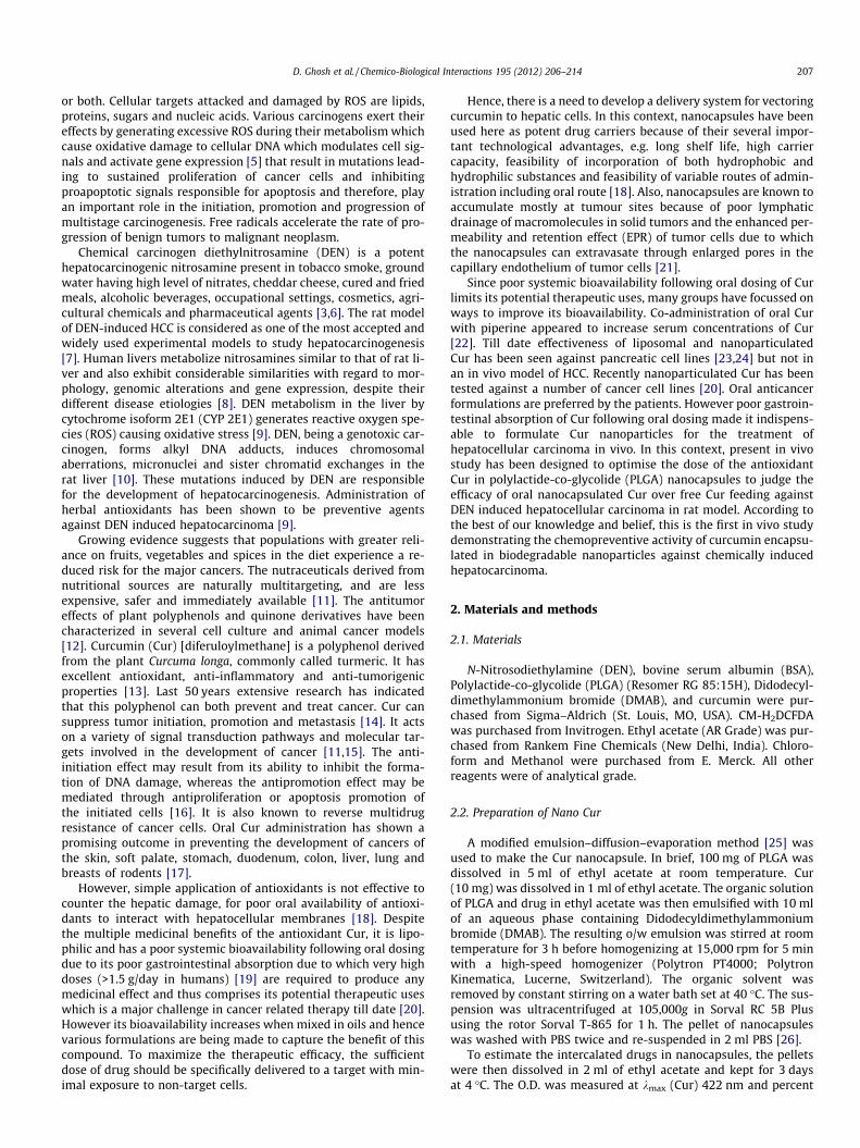
3.2. Effect of Nano Cur on RLW and marker enzymes of liver
DEN (3 doses of i.p. 200 mg/kg b.wt at 15 days interval) treat-ment causes an increase in relative liver weight (RLW) and markerenzymes of liver in rats. Nano Cur prevented reduction in RLW andmarker enzymes of liver in rats significantly in comparison to DENadministered control group. Free Cur treatment or empty nanocap-sules were not effective in giving any significant prevention fromDEN administered control group of rats (Table 1).
3.3. Effect of Nano Cur on the generation of mitochondrial ROS andmicroviscosity of liver
DEN administration induced a significant increase of ROS with a sig-nificant decrease in the membrane microviscosity of liver tissue in thecontrol as compared with rats of the normal group. Free Cur or emptynanocapsules treatment were unable to prevent mitochondrial ROSgeneration and microviscosity parameters as compared to DEN admin-istered control group. However, Nano Cur showed a significant preven-tion in mitochondrial ROS generation with an elevation ofmicroviscosity as compared to the control group (Table 2).
3.4. Effect of Nano Cur on lipid peroxidation and antioxidant status inliver
Conjugated diene is an important index of lipid peroxidation.DEN administered control rats showed a significant elevation inconjugated diene formation. Free Cur or empty nanocapsules didnot show any significant prevention. However Nano Cur showeda significant prevention from increase in the levels of conjugateddiene in the liver cell membrane (Table 3).
Antioxidant enzyme assay showed a significant reduction in thelevels of the enzymatic antioxidants, SOD and CAT as well as re-duced GSH in the DEN administered control group of rats. FreeCur and empty nanocapsules did not show any major deviationin the expression levels of these enzymes from those of the controlgroup. However Nano Cur treatment prevented reduction of theseantioxidant enzyme levels (Table 3).
3.5. Effect of Nano Cur on the expression of inducible nitric oxidesynthase (NOS 2) expression in cytosol and release of cytochrome C(Cyt C) from the liver mitochondrial membrane
Western Blot analysis showed a significant increase in NOS 2expression in the liver cell in DEN administered control group of

Fig. 1. AFM images of nanoparticles (a) under water obtained 30 min afterdeposition on mica sheet (Topography and amplitude flattened). Graph indicatesheight of nanoparticles from substratum i.e. mica sheet. Arrow indicates the heightmeasured. FTIR images of PLGA (b), NanoCur (c) and Free Cur (d).
210 D. Ghosh et al. / Chemico-Biological Interactions 195 (2012) 206–214
rats. Free Cur did not show any significant decrease in the expres-sion levels of NOS 2 in the liver. However nanocapsulated Curshowed a significant decrease in the NOS 2 levels in the liver ascompared to DEN administered control group of rats (Fig. 2a).
Release of Cyt C, from the mitochondrial membrane to the cyto-sol is a key event in cell apoptotic signalling following cell death. Itwas observed that normal cells released Cyt C in the cytosol as aconsequence of the normal process of Programmed Cell Death(PCD). DEN administered rat showed an increase in the Cyt C levelsas compared to normal rats. However Nano Cur treatment led to asignificant higher release in Cyt C as compared to DEN inducedcontrol group of rats (Fig. 2b).
3.6. Effect of Nano Cur on histopathological and histochemical analysisof liver sections
Haematoxylin–Eosin-stained liver sections of normal rat(Fig. 3a1) showed hepatocytes are arranged in cords around hepa-tic vein forming hepatic lobules. Portal tracts are normal. DEN in-jected animals shows dialated hepatic veins (Fig. 3a2) andhepatic micronodules separated by thin fibrous septum(Fig. 3a3). In higher magnification fatty changes in hepatocyteswith irregular nuclear membrane, with some eosinophilic cyto-plasm are evident (Fig. 3a4). Free drug treatment to DEN injectedanimals was unable to produce any significant change to DEN in-duced hepatocyte pathomorphological structures. Hepatic micro-nodules separated by thin fibrous septa, with dialated hepaticveins and some amount of periportal fibrosis are also evident(Fig. 3a5). In higher magnification fatty changes are also evident.However nuclear membrane has not lost its structural integrity(Fig. 3a6). However Nano Cur have prevented the liver from devel-oping hepatocarcinoma. Normal looking hepatic vein, portal tractsand hepatocytes are present here (Fig. 3a7).
ApoBrdU staining has shown some amount of apoptosis in nor-mal cells as a consequence of normal Programmed Cell Death(Fig. 3b1). We observed increased BrdU positive cells indicatingapoptosis in liver of Nano Cur treated group in comparison toDEN administered control group (Fig. 3b2 and b4). Free Cur treat-ment showed visibly less apoptotic cells in liver as compared toNano Cur (Fig. 3b3).
4. Discussion
The limited progress achieved by cancer therapy in the lastthree decades has increased the interest of researchers in cancerchemoprevention. The process of carcinogenesis can take severaldecades to complete; hence it makes more sense to prevent cancerat its earliest stages by using low-toxic chemicals than to wait untilthe disease has reached its final stages, where it becomes necessaryto use more toxic chemicals.
A dose escalation study of Cur has been done at three differentdoses of curcumin encapsulated in nanocapsules (5, 10 and 20 mg/kg b.wt). The effective dose of Cur has been found to be 20 mg/kgb.wt in combating DEN induced hepatocellular carcinoma in ratmodel. The data’s for the effective dose have only been shown inthe article. Pharmacologically, Cur has been found to be safe evenat very high doses according to the USA-FDA in both animals aswell as humans [17].
The small particle size (average diameter 15 nm) of the Curnanocapsules formulated by us might be responsible for its longercirculation time and also for targeting the liver tumor by seepagethrough the leaky vasculature characteristic of the tumor tissues[37]. Also the uniformity in the dimensions of the different nano-capsules indicates the uniformity of the nanocapsules formed. FTIRanalysis revealed no interaction between the polymer and the drug
Table 1Effect of Cur (Free and Nanoencapsulated forms) on% of increase in RLW and marker enzymes of liver in DEN induced hepatocellular carcinoma.
Groups Liverweight (g)
RLW % of increasein RLW
Alkalinephosphatase(KA Units)
Serum aspartatetransaminase (IU/L)
Serum alaninetransaminase (IU/L)
Normal 8.2 ± 1.1 4.2 ± 0.2 – 30.2 ± 1.6 115.4 ± 5.3 36.4 ± 2.5DEN (A) 10.5 ± 1.9 6.9 ± 0.7** 64.2 82.4 ± 2.3* 256.6 ± 6.2* 130.1 ± 3.2*
A + empty nanocapsule 10.4 ± 2.0 6.8 ± 0.3 61.9 76.5 ± 1.5 248.3 ± 7.4 125.2 ± 3.4A + Free Cur 10.1 ± 2.4 6.5 ± 0.4 54.7 71.3 ± 2.1 225.4 ± 3.5 113.3 ± 2.2A + Nano Cur 8.8 ± 2.2 4.7 ± 0.1## 11.9 37.1 ± 3.2# 122.2 ± 5.5# 32.4 ± 1.5#
Relative liver weight (RLW) = liver weight/final body weight � 100.Results are expressed as mean ± S.E, of five animals.
* P < 0.0001 is significantly different from normal.** P < 0.01 is significantly different from normal.# P < 0.0001 significantly different from the DEN treated control group (A).
## P < 0.05 significantly different from the DEN treated control group (A).
Table 2Effect of Cur in free and nanoencapsulated forms on changes in the generation of ROS and mitochondrial membrane microviscosity ([r0/r � 1]�1) in rat hepatocarcinoma.
Groups DCF fluorescence (% of normal) Membrane microviscosity ([r0/r � 1]�1)
Normal 100 ± 7.43 0.656 ± 0.032DEN treated(A) 250 ± 12.30* 0.244 ± 0.014*
(A) + empty nanocapsule treated 248 ± 11.72 0.256 ± 0.023(A) + free Cur treated 230 ± 7.56 0.306 ± 0.011(A) + Nano Cur treated. 120 ± 5.34# 0.590 ± 0.010#
Results are expressed as mean ± S.E, of five animals.* P < 0.0001 is significantly different from normal.
# P < 0.0001 significantly different from the DEN treated control group (A).
Table 3Effect of Cur (free and nanoencapsulated forms) on lipid peroxidation and antioxidant enzymes in liver of DEN induced rats.
Groups Conjugated diene(lg/mg protein)
SOD level (% inhibitionof pyrogallol autooxidation)
Catalase activity(lmol of H2O2
reduced/min/mg protein)
GSH level(mg/mg protein) [10�4]
Normal 1.6 ± 0.11 74.3 ± 1.47 8.5 ± 0.18 0.0016 ± 0.0002DEN treated (A) 3.6 ± 0.22* 33.3 ± 2.40* 2.6 ± 0.35* 0.0003 ± 0.0001**
A + Empty Nanocapsule treated 3.4 ± 0.23 33.6 ± 1.85 2.7 ± 0.31‘ 0.0003 ± 0.0001A + Free Cur 3.2 ± 0.12 39.6 ± 2.72 3.3 ± 0.17 0.0006 ± 0.0001A + Nano Cur 2.1 ± 0.17# 71.1 ± 1.73## 8.2 ± 0.35## 0.0014 ± 0.0002###
Results are expressed as mean ± S.E, of five animals.* P < 0.0001.
** P < 0.001 is significantly different from normal.# P < 0.001 significantly different.
## P < 0.0001.### P < 0.01significantly different from the DEN treated control group (A).
D. Ghosh et al. / Chemico-Biological Interactions 195 (2012) 206–214 211
in Nano Cur, which indicates that Cur can be incorporated in thenanocapsules without altering its individual structural identity(Fig. 1).
Relative liver weight is an important parameter in judging thepathological condition of the liver. Lowering in the relative liverweight of rats by Nano Cur treatment is an important indicationof the pathological improvement of the liver. Reduction of SGPT,SGOT and AP levels, important markers of liver function tests, byNano Cur indicate its preventive effect in hepatocarcinoma(Table 1).
Oxidative stress is an obvious outcome of human cancer. It isproduced either through an increased ROS generation with orwithout an alteration of antioxidant defence of the target cells ortissues [38,39]. Mitochondria are the major sites of cellular ROSproduction. ROS binds with a number of cellular components ofcell including lipid, protein, DNA, carbohydrate, thiols, andother low-molecular-weight antioxidants causing oxidation of
macromolecules and ultimately leading to pathogenesis. DENadministration generates a high amount of ROS in the hepatocarci-noma cells and the high amount of ROS generation is also respon-sible for a decrease in membrane fluidity due to rigidity of themitochondrial membrane. Nano Cur treatment prevented the oxi-dative stress to a major extent by inhibiting ROS production, whichin turn prevented decrease in membrane fluidity as compared toDEN treated control group of animals (Table 2).
Conjugated diene is one of the major end products of peroxida-tive degradation of the polyunsaturated fatty acid constituents ofbiological membranes and has mutagenic and carcinogenic proper-ties as evident from in vitro systems and in experimental animals[40]. The present study shows a significant prevention of conju-gated diene formation on Nano Cur treatment which is believedto be due to ROS generation in the liver (Tables 2 and 3). The detox-ification ability of the liver resides in its rich metabolizing enzymeprofile and intracellular antioxidant reserve. The antioxidant

Fig. 2. Western blot analysis showing expression of Nos 2 (a) and Cyt C (b) proteinin cytosolic fraction of liver tissue. Lane 1: Olive oil treated normal (A), Lane 2: DENtreated (B), Lane 3: DEN + Nano Cur treated (C). Histogram showing representativepixel intensities (arbitrary units of densitometric analysis using Image J software) ofthe immunoblot performed with different individual rats. Values are mean ± S.E. of5 rats. ⁄P < 0.0001 significantly different from DEN treated control and #P < 0.0001significantly different from Nano Cur.
212 D. Ghosh et al. / Chemico-Biological Interactions 195 (2012) 206–214
reserve of liver includes two important enzymatic antioxidants,SOD, Catalase and an important non-enzymatic antioxidant re-duced glutathione (GSH). SOD and Cat are the major free radicalscavenging enzymes present in the biological systems. GSH playsa crucial role in the detoxification process of majority of alkylatingagents including DEN. It neutralizes the electrophilic site by pro-viding a –SH group and renders the metabolite more water soluble[6]. Our results agree with the hypothesis that DEN treatment inrat causes depletion of enzymatic and non-enzymatic antioxidantdefence in hepatic tissue and induces hepatocarcinoma in rat (Ta-ble 3) with a substantial increase of mitochondrial ROS as indicatedby (Table 2). Numerous reports have demonstrated that Cur mayact as an antioxidant or as a pro-oxidant. In our experiments NanoCur is acting as an antioxidant by preventing generation of highcellular levels of ROS and hence prevents the process of carcino-genesis, therefore acting as a cancer chemopreventive agent(Tables 2 and 3).
NOS 2 is an inflammation responsive enzyme, involved inwound healing, angiogenesis and carcinogenesis [41]. The induc-ible isoform (NOS 2) is expressed de novo by cell types, includingmacrophages and hepatocytes, in pathological stressed conditions[41] and is responsible for enhanced tumor growth in hepatocellu-lar carcinoma [42]. NOS 2 upregulation and increased nitric oxide(NO) production affect the redox balance of cells, NOS being adownstream target of ROS, can induce protein, lipid, and DNAmodifications [43]. Estimating the levels of NOS 2 expression istherefore an important parameter for anticancer formulations.DEN administration increase the NOS 2 levels indicating the gener-ation of ROS promoting carcinogenesis and possibly leading to-wards angiogenesis. A low level of NOS 2 in the Nano Cur treatedgroup is indicative of the low levels of ROS generation which mighthave helped prevent tumor growth and inhibit angiogenesis(Fig. 2a).
Cancer therapy is modulated either by a suppression of prolifer-ation of cancer cells or by apoptosis of the cancer cells. The anti-tu-mor effect of curcumin has also been attributed in part to thesuppression of cell proliferation, reduction of tumor load andinduction of apoptosis in various cancer models both in vitro andin vivo [44]. The mechanism of cancer cell apoptosis is mitochon-dria-dependent or mitochondria independent [14]. Release of cyto-chrome C, from the mitochondrial membrane to the cytosol is a keyevent in cell death, which can lead the cell towards apoptosis ornecrosis. In our experiments, increased cytochrome C levels abovethe normal cells in DEN administered control group of rats indi-cates the death of some cells on exposure to the genotoxic carcin-ogen. Increased cytosolic cytochrome C levels above the DENadministered control group of rats on Nano Cur treatment indi-cates an increased amount of death of the liver cells as comparedto DEN administered rats. ApoBrdU positive cells indicates apopto-sis. We observed increased BrdU positive cells in Nano Cur treatedgroup as compared to DEN administered control group indicatingDNA fragmentation leading to apoptosis. Since Cur preferably in-duces apoptosis in highly proliferating cells, [44,45] death is muchmore pronounced in tumor cells than normal ones. (Fig. 3b, b1 andb2) Hence it can be attributed that this higher amount of Brdu po-sitive cells indicates apoptosis of the initiated cancer cells on NanoCur treatment. Nano Cur follows the identical mechanism that curfollows for cancer cell killing. Since cytochrome C release in thecytosol in earlier studies has been proved to be associated withthe intrinsic pathway [14], possibly in our experiment the intrinsicpathway is involved in the induction of apoptosis by Nano Cur.Cytochrome C release in the cytosol from the mitochondrial innermembrane on curcumin treatment converts procaspase 9 to cas-pase-9, with activation of caspase-3, ultimately leading the livercells towards apoptosis [14]. In our studies release of cytochromeC in the cytosol from the mitochondrial inner membrane indicatesthat Nano Cur might have followed the same mechanism of apop-tosis as reported in earlier studies with curcumin alone.
Curcumin at low doses has been shown to possess antioxidantproperty with chemopreventive effect in the development of can-cers of the skin, soft palate, stomach, duodenum, colon, liver, lungand breast of rodents [17]. Apoptosis inducing property was shownby curcumin in HL-60 (human acute myelogenous leukemia) celllines at doses of 25 lM of curcumin in a time dependent mannerby Anto et al. [46]. In our study we found the antioxidant effectof curcumin in Nano Cur treated animals (Table 2 and 3; Fig. 2a).Besides acting as an antioxidant it might have exerted its antiapo-ptotic effect on the transformed malignant cells that proliferate athigh rates [44,45]. The apoptosis inducing property of NanoCur (Figs. 2b, 3b) might have been due to the inhibition of NFkappa b, a feature commonly seen in the antiproliferative andapoptosis-inducing effects of curcumin. This NF kappa b inhibition

Fig. 3. Histological and histochemical examination of liver sections of experimental rats. (a) Eosin–haematoxylin stained liver sections [a1] Olive oil treated Control (10�)showing normal liver architecture, [a2] DEN treated control (10�) showing (;) dialated hepatic veins [a3] DEN treated control (10�), Inset showing hyperplastic nodules, [a4]DEN treated control (40�), showing ( ) fatty changes, [a5] DEN + Free Cur treated (10�) showing hyperplastic nodules (inset) with marked periportal fibrosis (;), [a6]DEN + Free Cur treated (40�) showing ( ) atypical nuclei and fatty changes ( ), [a7] DEN + Nano Cur treated (10�), showing architecture similar to normal liver. (b)Representative photomicrographs (20�) of BrdU positive cells observed by double staining with BrdU (under FITC filter) and PI staining of liver of normal (b1), DEN treated(b2), DEN + Free Cur treated (b3) and DEN + Nano Cur treated (b4) rats.
D. Ghosh et al. / Chemico-Biological Interactions 195 (2012) 206–214 213
might have led the cancerous cells towards apoptosis and cyto-chrome C release into the cytosol.
Hepatocarcinoma in rat was also examined by histopathologicalexamination. A single dose of DEN administration (200 mg/kg b.wt,i.p. injection) initiates hepatocarcinogenesis, promoted by pheno-barbital in drinking water (orally in drinking water for 14 weeksand animals were sacrificed at the end of 15/16 weeks startingfrom the day of DEN administration) has been reported earlier bySreepriya et al., Bishayee et al. [47,2]. DEN administration (3 dosesi.p) at 15 days interval has also been reported earlier by Mandalet al. [9]. Development of hyperplastic nodules is a characteristicfeature of DEN induced hepatocellular carcinoma shown earlierby Bishayee et al., Das et al., Mandal et al. [2,40,7]. Also the pres-ence of atypical nuclei is another marker of hepatocellular carci-noma shown by Sreepriya et al. [47]. Our studies have showndistorted histopathological changes in the liver with formation ofhyperplastic nodules (Fig. 3a3) and atypical nuclei (Fig. 3a6) onDEN administration which are indications of DEN induced hepato-carcinogenesis. Remarkable pathological improvement was no-ticed in rats treated with Nano Cur whereas the same dose offree Cur did not show any encouraging results (Fig. 3a).
It appears that when any essential component of a signal trans-duction pathway of a cell is rendered hyperactive or autonomous,it may acquire the ability to drive the cell into unchecked prolifer-ation leading to tumor promotion. Cur attenuates or suppresses thehyperactivity of these components of signal transduction andmaintains simultaneously the normal cell function [15,11]. Our re-sults show that in animals these biodegradable Cur nanocapsulesare more bioavailable than free curcumin without any toxic effectsat the dose employed herein. Due to their greater bioavailabilitythese Cur nanocapsules have proved to be much more effectivethan free Cur at much lower dose than the effective dose of freeCur in combating DEN induced hepatocarcinoma in rats.
5. Conclusions
Curcumin in nanocapsulated drug delivery system has provedto be an effective free radical quencher, could protect the rat liverfrom DEN induced altered hepatic functioning, prevented DEN in-duced hyperplastic nodule formation, checked any upregulationof iNOS expression which in a way prevented angiogenesis in tu-mor sites, as well as promoted apoptosis of the initiated cancercells as evidenced by DNA fragmentation, Cyt C release. Nano Curhas shown its effectivity against DEN induced hepatocellular carci-noma in rat. Though further studies on quantitating bioavailabilityremains to be done Nano Cur in oral route might be a promisinganticancer alternative to prevent HCC.
Conflict of interest statement
The authors declare that they have no conflict of interest.
Acknowledgements
This work has been supported financially by the Council of Sci-entific and Industrial Research (CSIR), Government of India, CSIR-SRF Project and Network project (IAP 0001), IICB Kolkata. Theauthors acknowledge Dr. Anjan Kumar Das, Associate Professor,Department of Pathology, Calcutta National Medical College andHospital, Kolkata for histopathological interpretation, Mr. T.Mur-uganandan, Technical Officer for AFM operation and Mr. SatyabrataSamaddar for FTIR operation.
References
[1] P. Chintana, Role of Curcumin on tumor angiogenesis in hepatocellularcarcinoma, Naresuan. Univ J. 16 (3) (2008) 239–254.

214 D. Ghosh et al. / Chemico-Biological Interactions 195 (2012) 206–214
[2] A. Bishayee, N. Dhir, Resveratrol-mediated chemoprevention ofdiethyhlnitrosamine-initiated hepatocarcinogenesis: inhibition of cellproliferation and induction of apoptosis, Chem. Biol. Interact. 179 (2009)131–144.
[3] V. Sivaramkrishnan, P.N.M. Shilpa, V.R.P. Kumar, S.N. Devaraj, Attenuation ofN-nitrosodiethylamine-induced hepatocellular carcinogenesis by a novelflavonol-Morin, Chem. Biol. Interact. 171 (2008) 79–88.
[4] J.E. Klaunig, L.M. Kmendulis, B.A. Hocevar, Oxidative stress and oxidativedamage in carcinogenesis, Toxicol. Path. 38 (2010) 96–109.
[5] G. Waris, H. Ahsan, Reactive oxygen species: role in the development of cancerand various chronic conditions, J. Carcinogenesis 5 (14) (2006).
[6] C. Gupta, A. Vikram, D.N. Tripathi, P. Ramarao, G.B. Jena, Antioxidant andantimutagenic effect of quercetin against DEN induced hepatotoxicity in rat,Phytother. Res. 24 (1) (2010) 119–128.
[7] W. Hai, C. Kim, S. Song, C. Kang, Study on mechanism of multistephepatotumorigenesis in rat; development of hepatotumorigenesis, J. Vet. Sci.2 (2001) 53–58.
[8] F. Feo, P.M. Pascale, M.M. Simile, M.R. De Miglio, M.R. Muroni, D. Calvisi,Genetic alterations in liver carcinogenesis; implications for new preventiveand therapeutic strategies, Crit. Rev. Oncog. 11 (2000) 19–62.
[9] A.K. Mandal, S. Das, M. Mitra, R.N. Chakrabarti, M. Chatterjee, N. Das, Vesicularflavonoid in combating diethylnitrosamine induced hepatocarcinoma in ratmodel, J. Exp. Ther. Oncol. 7 (2008) 123–133.
[10] M.C. Jagadeesh, M. Sreepriya, G. Bali, D. Manjulakumari, Biochemical studieson the effect of curcumin and embelin during N-nitrosodiethylamine/Phenobarbital induced-hepatocarcinogenesis in wistar rats, African J.Biotechnol. 8 (18) (2009) 4618–4622.
[11] S.C. Gupta, J.H. Kim, R. Kanappan, S. Reuter, P.M. Dougherty, B.B. Aggarwal,Role of nuclear factor- kb-mediated inflammatory pathways in cancer-relatedsymptoms and their regulation by nutritional agents, Exp. Biol. Med. 236(2011) 658–671.
[12] M.A. Indap, M.S. Barkume, Efficacies of plant polyphenolics on sodiumbutyrate induced anti-tumoractivity, Indian J. Exp. Biol. 41 (2003) 861–864.
[13] A. Altunbas, S.J. Lee, S.A. Rajasekaran, J.P. Schneider, D.J. Pochan, Encapsulationof curcumin in self-assembling peptide hydrogels as injectable drug deliveryvehicles, Biomaterials 32 (2011) 5906–5914.
[14] B.B. Aggarwal, A. Kumar, A.C. Bharti, Anticancer potential of curcumin:Preclinical and clinical studies, Anticancer Res. 23 (2003) 363–398.
[15] S.C. Gupta, J.H. Kim, S. Prasad, B.B. Aggarwal, Regulation of survival,proliferation, invasion, angiogenesis, and metastasis of tumor cells throughmodulation of inflammatory pathways by nutraceuticals, Cancer MetastasisRev. 29 (2010) 405–434.
[16] M.H. Pan, W.L. Chang, S.Y. Lin Shiau, C.T. Ho, J.K. Lin, Induction of apoptosis bygarcinol and curcumin through cytochrome C release and activation ofcaspases in human leukemia HL-60 cells, J. Agric. Food. Chem. 49 (2001)1464–1474.
[17] G. Sa, T. Das, S. Banerjee, J. Chakraborty, Curcumin: from exotic spice tomodern anticancer drug, Al. Ameen. J. Med. Sci. 3 (1) (2010) 21–37.
[18] D. Ghosh, S. Ghosh, S. Sarkar, A. Ghosh, N. Das, K.D. Saha, A.K. Mandal,Quercetin in vesicular delivery systems: Evaluation in combating arsenic-induced acute liver toxicity associated gene expression in rat model, Chem.Biol. Interact. 186 (2010) 61–71.
[19] R.A. Sharma, S.A. Euden, S.L. Platton, D.N. Cooke, A. Shafayat, H.R. Hewitt, T.H.Marczylo, B. Morgan, D. Hemingway, S.M. Plummer, M. Pirmohamed, A.J.Gescher, W.P. Steward, Phase I clinical trial of oral curcum: biomarkers ofsystematic activity and compliance, Clin. Cancer Res. 10 (2004) 6847–6854.
[20] P. Anand, H.B. Nair, B. Sung, A.B. Kunnumakkara, V.R. Yadav, R.R. Tekamal, B.B.Aggarwal, Design of curcumin-loaded PLGA nanopaticles formulation withenhanced cellular uptake and increased bioactivity in vitro and superiorbioavailability in vivo, Biochem. Pharmacol. 79 (2010) 330–338.
[21] T. Lammers, W.E. Hennink, G. Storm, Tumor targeted nanomedicines:principles and practice, Br. J. Cancer 99 (2008) 392–397.
[22] J. Shaikh, D.D. Ankhola, V. Beniwal, D. Singh, M.N.V. Ravi Kumar, Nanoparticleencapsulation improves oral bioavailability of curcumin by at least 9-foldwhen compared to curcumin administered with piperine as absorptionenhancer, Eur. J. Pharm. Sci. 37 (2009) 223–230.
[23] L. Li, S.F. Braiteh, R. Kurzrock, Liposome encapsulated curcumin. In vitro andin vivo effects on proliferation, apoptosis, signaling and angiogenesis, Cancer104 (6) (2005) 1322–1331.
[24] S. Bisht, G. Feldmann, S. Soni, R. Ravi, C. Karikkar, A. Maitra, Polymericnanoparticles-encapsulated curcumin (nanocurcumin): a novel strategy forhuman cancer therapy, J. Nanobiotechnol. 5 (3) (2007) 1–18.
[25] S. Hariharan, V. Bhardwaj, I. Bala, J. Sitterberg, U. Bakowsky, M.N.V. Ravi Kumar,Design of Estradiol loaded PLGA Nanoparticulate formulations: a potent oraldelivery system for Hormone therapy, Pharm. Res. 23 (1) (2006) 184–195.
[26] S. Lala, S. Gupta, N.P. Sahu, D. Mandal, N.B. Mondal, S.P. Moulik, M.K. Basu,Critical evaluation of the therapeutic potential of basic acid incorporated inoil-in-water microemulsions Poly-D, L-lactide nanoparticles againstexperimental leishmaniasis, J. Drug Target. 14 (2006) 171–179.
[27] M. Dubin, P.H. Carrizo, A.M. Biscardi, S.H. Fernandez Villamil, A.O.M. Stopanni,Effect of 5-nitroindole on adenylate energy charge, oxidative phosphorylationand lipid peroxidaion in rat hepatocytes, Biochem. Pharmacol. 48 (1994)1483–1492.
[28] N.V. deWitte, A.O.M. Stopanni, M. Dubin, 2-Phenyl-b-lapachone can affectmitochondrial function by redox cycling mediated oxidation, Arch. Biochem.Biophys. 432 (2004) 129–135.
[29] C. Betainder, E. Fontaine, C. Keriel, X.M. Leuerve, Determination ofmitochondrial oxygen species: methodological aspects, J. Cell Mol. Med. 6(2002) 175–187.
[30] A.K. Mandal, N. Das, Sugar coated liposomal flavonoid: a unique formulation incombating carbontetrachloride induced hepatic oxidative damage, J. DrugTarget. 13 (2005) 1305–1315.
[31] A.K. Mandal, J. Sinha, S. Mandal, S. Mukhopadhyay, N. Das, Targeting ofliposomal flavonoid to liver in combating hepatocellular oxidative damage,Drug Delivery 9 (2002) 181–185.
[32] O.H. Lowry, N.J. Rosebrough, A.L. Farr, R.G. Randel, Protein measurement withFolin Phenol reagent, J. Biol. Chem. 193 (1951) 265–277.
[33] S. Markland, G. Markland, Involvement of superoxide anion radical in theautooxidation of pyrogallol and a convenient assay of Superoxide Dismutase,Eur. J. Biochem. 47 (1974) 469–474.
[34] A.C. Moragon, G.N. De Lucas, F.M. Encarnicon Lopez, A.S. Rodriguez-Manzaneque, F.J.A. Jimenez, Antioxidant enzymes occupational stress andburnout in works of a prehospitalary emergency service, Eur. J. Emerg. Med. 12(2005) 11–115.
[35] J.C. Davila, P.J. Davis, D. Acosta, Changes in glutathione and cellular energy aspotential mechanisms of Papaverine-induced hepatotoxicity in vitro, Toxicol.Appl. Pharmacol. 108 (1) (1991) 28–36.
[36] S.N. Kim, K.C. Park, H.J. Kim, K.H. Cho, J.H. Chung, K.H. Kim, H.C. Eun, J.S. Lee,K.D. Park, Effects of collagen IV and laminin on reconstruction of human oralmucosa, J. Biomed. Mater. Res. 58 (2001) 108–112.
[37] N.P. Praetorius, T.K. Mandal, Engineered nanoparticles in cancer therapy,Recent Patents on Drug Delivery Formulation. 1 (2007) 37–51.
[38] M.A. Trush, T.W. Kensler, An overview of the relationship between oxidativestress and chemical carcinogenesis, Free Radic. Biol. Med. 10 (1991) 201–209.
[39] C. Rice-Evans, R. Burdon, Free radical–lipid interactions and their pathologicalconsequences, Prog. Lipid Res. 32 (1993) 71–110.
[40] T. Das, F. Patra, B. Mukherjee, Effect of antisense oligomer in controlling c-raf.1overexpression during diethylnitrosamine-induced hepatocarcinogenesis inrat, Cancer Chemother. Pharmacol. 65 (2010) 309–318.
[41] M.A. Rahman, D.K. Dhar, E. Yamaguchi, et al., Coexpression of Inducible NitricOxide Synthase and COX-2 in Hepatocellular carcinoma and surrounding liver:Possible involvement of COX-2 in the angiogenesis of Hepatitis C Virus-positive cases, Clin. Cancer Res. 7 (5) (2001) 1325–1332.
[42] M.L. Majano, C. Garcia-Monzon, M. Lopez-Cabrera, E. Lara-Pezzi, E. Fernandez-Ruiz, Inducible nitric oxide synthase expression in chronic viral hepatitis,evidence for a virus induced gene upregulation, J. Clin. Invest. 101 (1998)1343–1352.
[43] S.A. Glynn, B.J. Boersma, T.H. Dorsey, M. Yi, H.G. Yfantis, L.A. Ridnour, D.N.Martin, C.H. Switzer, R.S. Hudson, D.A. Wink, D.H. Lee, R.M. Stephens, S. Ambs,Increased NOS 2 predicts poor survival in estrogen receptor-negative breastcancer patients, J. Clin. Invest. 120 (11) (2010) 3843–3854.
[44] G. Sa, T. Das, Anti cancer effects of curcumin: cycle of life and death, CellDivision 3 (14) (2008) 1–14.
[45] T. Choudhuri, S. Pal, T. Das, G. Sa, Curcumin selectively induces apoptosis inderegulated cyclin D1-expressed cells at G2 phase of cell cycle in a p-53dependent manner, J. Biol. Chem. 280 (2005) 20059–20068.
[46] R.J. Anto, A. Mukhopadhyay, K. Denning, B.B. Aggarwal, Curcumin(diferuloylmethane)induces apoptosis through activation of caspase-8, BID cleavage and cytochromeC release: its suppression by ectopic expression of Bcl-2 and Bcl-xl,Carcinogenesis 23 (1) (2002) 143–150.
[47] M. Sreepriya, G. Bali, Effects of administration of embelin and curcumin on lipidperoxidation, hepatic glutathione antioxidant defence and hematopoietic systemduring N-nitrosodiethylamine/phenobarbital-induced hepatocarcinogenesis inwistar rats, Mol. Cell. Biochem. 284 (2006) 49–55.