naosite: nagasaki university's academic output...
TRANSCRIPT

This document is downloaded at: 2018-05-14T15:39:59Z
Title Phospholipid Metabolism in the Lung of Fat-Fed Rats
Author(s) Katoh, Yasutaka
Citation Acta Medica Nagasakiensia. 1967, 11(3-4), p.222-236
Issue Date 1967-03-25
URL http://hdl.handle.net/10069/17438
Right
NAOSITE: Nagasaki University's Academic Output SITE
http://naosite.lb.nagasaki-u.ac.jp

Acta med. nagasaki. : 222 --236
Phospholipid Metabolism in the Lung of Fat-Fed Rats
Yasutaka KATOH *
Second Department of Internal Medicine, Nagasaki University School of Medicine.
Nagasaki, Japan
Received for publication February 20, 1967
Phospholipid metabolism in the lung was studied because the lung was the first organ at which the ingested lipids arrived.
It was revealed in this study that the lung contained phosphatidyl cho- line, ethanolamine, phosphoinositol, sphingomyelin and lysolecithin, that the amount of phosphatidyl serine were very small in the lung, and that a
phosphatidic acid was discovered in the lung after the administration of fats. Furthermore, an unknown phospholipid subfraction was revealed in the lung.
Fatty acid composition of phospolipid subfractions was investigated one and six hours after the administration of trilinolein.
The elongation of fatty acids occured also in the lung. Phosphatidyl ethanolamine and an unknown subfraction were active in the elongation but
phosphatidyl choline was stable. It was resulted by ligation of the thoracic duct that appearance of a
phosphatidic acid was delayed, and that saturated fatty acid increased, unsaturated decreased.
These findngs suggest that the lung has an ability of lipid synthesis, and that phosphatidyl ethanolamine and an unknown subfraction were active
in lipid metabolism although phosphatidyl choline was stable.
INTRODUCTION
It is well established that the liver plays an important role in lipid metabolism. On the other hand, little attention has been given to a
possible role of the lung in the storage or excretion of lipids. In considering the mechanism involved in the absorption of lipids,
most of the ingested lipids pass through the lung before the liver by way of the thoracic duct. Therefore, it is naturaly considered that the lung may have some roles in lipid metabolism because the lung is the first organ at which the ingested lipids (chylomicrons) arrive.
In the literature, there were scattered reports indicating the lung
had also an important role in lipid metabolism.
*加 藤 泰 孝
222
PHOSPHOLIPID METABOLISM IN THE LUNG OF FAT-FED RATS
SEEMANl) administered cholesterol into a peripheral artery or into the portal vein, and found that even if cholesterol had to pass through the liver bofore the l.ung its primary site of deposit was still the lung.
CroNI2) found that cholesterol in the lung was located in the reticu-loendothelial cells of the alveolar wall.
SCHRADE3) administered olive oil to rats and found that the amount of total lipids increased makedly in the lung.
OSAJIMA et al4) observed that the amount of total lipids, cholesterol
and phospholipids increased in the lung when triolein or trilinolein was administered but did not increase when tripalmitin or tristearin was adm in istered .
INDERBITZEN5) observed that the disapperance of chylomicrons was delayed when one lung was removed, and arrived at the conclusion that the lung removed chylomicrons from the circulation.
In view of the fact obtained by the investigation Lipoprotein-Lipase
Activity, HElNEMANN6) and YAMADA7) concluded that the lung also play-ed an important role in lipid metabolisrn.
On the other hand, fatty liver was observed at a great rate in patients with pulmJnary tuberculosis8) . This clinical observation may suggest the role of the lung in lipid metabolism.
The present study was un,dertaken in an attempt to investigate a possible role of the lung in lipid metabolism, especially from the stand-
point of phospholipid metabolism.
MATERIALS AND METHODS
Male albino rats of Wistar strain, weighing 160g to 260g, were selected for the present investigation and were fed ad libitum a com-mercial laboratory chow (Oriental chow).
At this investigation, rats were divided into three groups. Group I was left overnight with water but without food, and then killed by exsanguination. This group was used for the control. Group 11 and group 111 also were left overnight with water but without food, and then either 0.5g of tripalmitin in 4 ml of dehycol solution or 0.5g of trilinolein in 4 ml of dehycol solution was administered through a rub-ber inserted into the stomach.
The animals were lightly anesthetized with ether so that they were fully recovered by the time a cathether was removed.
Rats of group 11 and 111 were killed by exsanguination six hours after tripalmitin or trilinolein was administered. It was on the basis of the preliminary observation that rats of group 11 and 111 were killed six hours after the administration of fats.
In order to investigate the effect of the thoracic duct on lipid meta-
bolism in the lung, Iigation of the thoracic duct was accomplished by

Y. KATOH
the procedure of BOLLMAN. After the operation, the rats were main-tained on the same chow and water for a week, and then trilinolein was administered after fasting for twelve hours. At time intervals of one, three and six hours after the administration of trilinolein the rats were killed.
At the time of killing, Iungs were removed and total lipids were extracted from the lungs by a modification of the method of FoLcH-LEEslo) . The amounts of total lipids were calculated by a balance and phosphrus was measured by the method of ALLEN. The extracted lipi-ds were dissolved in 10ml of chloroform and this solution were used for the samples.
Chromatographic technique :
Glass plates (・20cm x 20cm and 5cm x 20cm) were throughly clea-ned with a detergent (H2S04 -potassium dichromate solution). The <'neutral" plates were prepared by making a slurry 30g of Kiesel Gel G with 65ml distilled water and applying it according to the method of STAHLll). After a layer of Kiesel Gel G had been applied the p]ates were left on the tray at room. temperature for 30 minutes and then inserted on the light-alloy drying rack. The rack with the plates was inserted into the drying cabinet, which had been heated to 110C'. The door of the cabinet should be opened for the first ten minutes of dry-ing to let the stream escape, and furthermore the plates were activated
for 30 minutes. The hot rack was next removed, cooled for 10 minu-tes and stored in a cabinet with silica gel and phosphorus pentoxide. When it took longer than three days before the samples were applied on the plates, the plates had to be again activated at 80C' for 40 minu-
tes just prior to the application of the samples. Depending on the amount of minor constituent to be detected, various volumes (10pl-200pD of the samples were applied 2 cm from the bottom of the plates with a calibrated sv. ringer for gas-1.iquid chromatography.
If it was necessary to detect a minor constituent on thin-layer chromatogram, the samples were applied in greater quantities although a major constituent could not be clearly separated.
Chromatographic chambers were prepared 60 minutes prior to the insertion of the plates and futhermore were lined on three sides with a filter paper in order to prevent drying of the solvent at the front. The edge of a thin-layer should be stripped off using the thumb as a guide in order to prevent the edge effect occured in the developing.
The plates were developed at room temperature until the solvent front reached 12cm from the origin of spots.
At this investigation, a solvent mixture of chloroform-methanol-water(65 : 25 : 4) was used for the separation of phospholipid.12)
Detection of spots : In this investigation six different detection

PHOSPHOLIPID METABOLISM IN THE LUNG OF FAT-FED RATS
methods were employed.
(1) Ninhy~drin to detect amino-phospholipids. Phospholipids with free amino-group were revealed by ninhydrin spray (0.2% ninhydrin in buthanol). (After spraying, the plates were heated for 5 minutes at 100C' ) .
Red-violet spots appeared on a white background.
(2) Choline-containing phospholipids were detected by dragendorff reagent (Bi). The plates were sprayed with a mixture of 4 ml of solution I, I ml of solution 11 and 20 ml distilled water. Solution I contained 1.7g of Bi (N03 )3 ' 5 H2 O diluted to 100 ml with 20 ~~ v/v acetic acid. Solution 11 contained 40 g of KJ in 100 ml water. As the plates were dried at room temperature, choline-containing phospholipi-ds produced orange spots.
(3) Phosphoinositol was detected by ammonium silver nitrate (Ag). The plates were sprayed with a mixture equal volumes of 0.1 N AgN03 and 7 N ammonium hydroxide. The plates were than heate_d at 110C' until dark brown spot appeared on a white background.
(4) Lipids containing phosphorus(phospholipids) were detected by molybdic acid reagent (Mo). The plates were sprayed with a solution of 5 ml of 60~~ w/v perchloric acid, 10 ml of I N HCl, and 25 ml of 4% w/v ammonium molyb date. Blue spots appeared on a white back-ground as the plates were dried at room temperature.
(5) Lipids were detected nonspecifically by iodine solution (12)-The plates were sprayed with a solution of 1.0g diluted 100 ml metha-nol. Brown spots appeared on a white background as the plates were dried at room temperature and the excess of iodine was evaporated.
(6) Lipids were detected semiquantitatively by sulfonic acid. The plates were sprayed with 50% sulfonic acid and then heated for 20 minute. s at 100C'. During the first few minutes of the charring opera-tion, free sterols and sterol esters gave a typical Lieberman-Burchard c,olor reaction, and at a later stage glycolipids turned deep purple and phospholipids, pale brown.
Two-dimentional chromatography : As all of phospholipids could not be separated by one dimentional chromatography. Since phospha-tidyl ethanolamine, serine and inositol could not be separated, two-dimentional thin-1ayer chromatography had to be used at the same time. The plates were developed at first with chloroform-methanol-7 N ammonium hydroxide (60 :35 : 5) and then dried at room tempera-ture. For two・・dimentional chrorDatography, the plates were rotated 90 clockwise and placed in another developing chamber containing a solvent mixture of chloroform-methanol-7 N ammonium hydroxide (35 : 60 : 5).
Another solvent mixtures were used for two-dimentional thin-1ayer

226 Y. KATOH Vol. 11
chromatography. The solvent mixture contained chloroform-methanol-glacial acetic acid-wather (250 : 74 : 19 : 3) or chloroform-methanol
-7 N ammonium hydroxide (230 : 90 : 15). In this type of two-dimentional chromatography, the "basrc" plates were used The "ba sic" plates were prepared by making a slurry of 30g of Kiesel Gel G with 65 ml of 0.01 M Na2 C03
Fatty acid composition : Fatty acid composition of phospholipid subfractions separated by thin-layer chromatography were investigated by gas-liquid chromatography.
The samples were applied as many small spots to 20cm x 20cm plates and then developed with chloroform-methanol-water (65 : 25 : 4). The plates were dried and partially masked by covering aluminium foil over the surface of silicic acid so that only one spot remained uncove-re,d. Exposure to iodine solution showed the position of the lipids, and on this basis the cover area of silicic acid containing the unchan-
ged lipids were scrapped off by a spatel. It could be checked by spraying the remainder of the plate with 50~ sulfonic acid that the scrapped silicic acid contained only a phospholipid subfraction. This silicic acid containing a separated phospholipid subfraction was trans-
ferred to a small glass column I cm in diameter and was eluted with chloroform-methanol-wat.er (65 : 25 : 4) and then with methanol.
The eluted solvents were evaporated in vacuum and used for the inYestigation of fatty acids.
RESULTS
By thin-1ayer chromatogarphy lung lipids was separated and it~* re-sults were shown in Fig (1) and Fig (2).
Five spots appeared on the chrornatogram in group I (Fig 1). A spot which did not appeared in group I was revealed in group 11 and group 111 (Fig 2). That is to say, a new spot appeared only when fat was administered to rats. The applied samples on the chromatogram contained the same volumes in three groups . Spots on the chrornato-gram were investigated and its results were listed in Table (1). The spot at the solvent front was neutral lipids and cholesterol because the
spot gave a typical Liebermann-Burchard color reaction by 50% sulfonic acid, and the other spots gave pale brown. Furthermore, each spot was scrapped off and it was investigated by the method of WAGNER16) whether it contained phosphorus or not. All spots except the spot at the solvent front contained phosphorus. On the other hand, only the solvent elute,d from the spot at the solvent front contained cholesterol
and did not contain phosphorus. By the above exarnination, it was revealed that five spots and a

1967
Solvent f ront
No. 5
No. 4
No. 3
No. 2
No. l
PHOSPHOLIPID
Fig .
METABOLISM
(1)
(1) (ll)
IN THE LUNG OF FAT・FED
Fig. (2)
RATS
(ll) (m)
2 27
Table. 1
Neutral lipid
Cholesterol
Phosphatic acid
Unknown Cephalin
Choline
S phingomyelin
Lysophosphatidyl Choline
12
+ + + + + + + +
H2S04
brow n
deep purple
brol~rn
ll
ll
ll
ll
ll
Mo
+ '+
+ + +
Nln
+
Bi
+ + +
Ag
new spot were phospholipids. Phospholipids were identified by observing the color reaction and
its results were listed in Table (1).
N0.1, 2 and 3 spot gave an orange color by spraying Dragendorff reagent. N0.4 spot became red-violet by spraying ninhydrin reagent.
As compared with a relative pure phosphatidyl choline and ethano-lamine obtained from soya-bearn lecithin, Rf value of N0.3 spot was identical with that of soya-be.=arn phosphatidyl choline, and Rf value of
N0・4 spot was identical with that of soya-bearn phosphatidyl ethanola-mine .
Two-dimentional thin-layer chromatogram of the lipids extracted
Y. KATOH
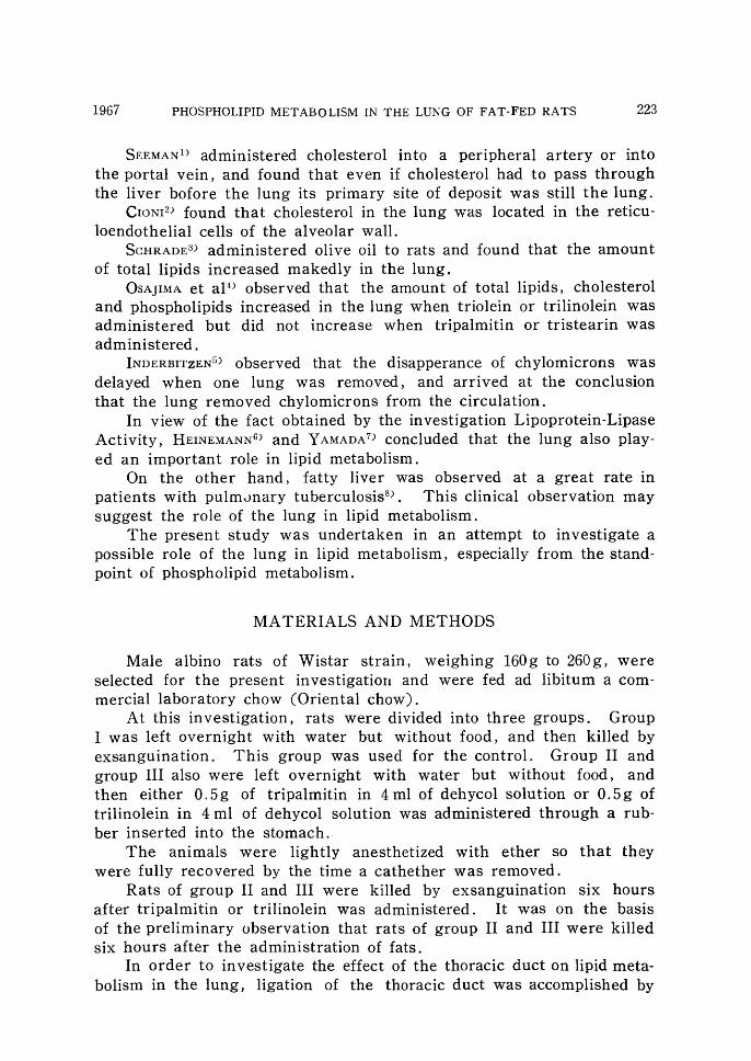
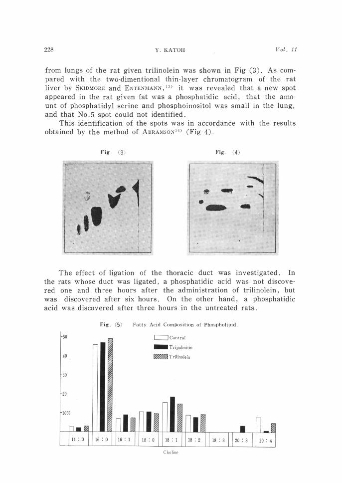
from lungs of the rat given trilinolein was shown in Fig (3). As com-
pared with the two-dimentional thin-layer chromatogram of the rat liver by SKIDMORE and ENTENMANN,13) it was revealed that a new spot appeared in the rat given fat was a phosphatidic acid, that the amo-unt of phosphatidyl serine and phosphoinositol was small in the lung, and that N0.5 spot could not identified.
This identification of the spots was in accordance with the results
obtained by the method of ABRAMSONl4) (Fig 4).
Fig. (3) Fig. (4)
the
red
was acid
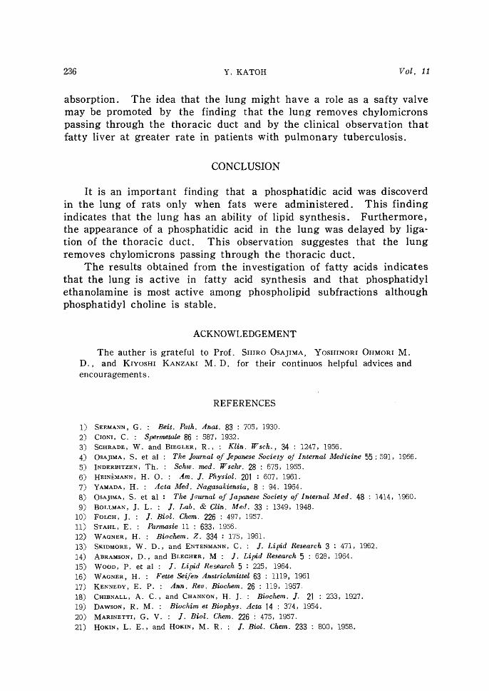
The effect of ligation of rats whose duct was ligated one and three hours after discovered after six hours.
was discovered after three
Fig. (5) Fatty Acld
**..~ *~"*~;~:'i~
* ~* * * +~~ ' '<" *i~;'*~'#: *~ =~'~*='=~'~~~"*"=~~'"'*'{~='*'~>'#~:'>~ :**~"*;"'
' ' _ *"*~~*** ==*='**"'**=+*=' *='*<~" ~ ~~i*'*~::'*~:*"*~~~~=~~i~~= ~~?~:'=~ =~*+'~ '~' ~>~~*'*=i*=**~'*'#*'~+~i** * '~'#'+*~ = *
*:~~* ;~:*~~ ****" '*' ' *'* <* * ~~~:**'*~:~;+*~:x~::"~** *'
{ * *s;~~i*~';.'*""~!'~*'+*':'*~i.'*1 *
S ~""'~;:~j""'i~:~:";~;~~~"~;~~i'~';~"""""'~~s" ~ ~;:';i~:~"~~;:";';~i~~;""~""~~~'"~"~;~~~~;;::;:;" ~~"'~:""~'f'
~'+'*~~;'*=' *"" ;~~~""~~':';"~i~<"'#~~~~;~'}~~~"~~ ~:":~'~~'*"~;s"~'~ *~ ' ='~
'.**.~*~'*~***~:** *'=**~:**~~+'** '~<~*~~** ' ' ~~:' * =* '*"~~'>=~~~~~'~~;~;~~t"":'~~~:"~i:~~~;;i~;;~ "*:";":~i>'f'~~"*;"i~/'~i"'<' ' ~~' ~
* ~~~~' ~ ' ** **. ** * ' ' i='~~~~~"'=~~'~""' :~;'i;'="";~'~;";j";r~~~'~'j'~"""=~:f~;~;;~~;;~~t;"~~~;% ~'~' ~ ' '
';':*~s' 2'*'^*='~=~~<+*:'~~i~~*~ *~*'~'~"~'~i;'~;~' ' *~#~ ~{;~'~~** * ~ '~'
'>~~~i~~*='*~===;'" * ' *"*~^> ~ {~~
the thoraciC duct was mvestlgated In ' a phOSPhatidic acid Was not diScove-the adminiStratiOn of trilinolein' but
On the other hand' a phOsphatidic hOurs in the untreated rats'
compos*ition ot PhosphohPid'
Choline

1967 PHOSPHOLIPID METABOLISM IN THE LUNG OF FAT・FED RATS 229
Fatty acid composition of phospholipid subfractions was investiga・
ted. The results oヒtained from the rat given tripalmitin were show
in Fig(5).
In phosphatidyl choline, the concentration of20 :4decreased and
20:3increased. In phosphatidンl ethanolamine,the decrease of20:4and the increase of20:3became more obvious than in phosphatidyl
choline. In an unknown spot,the concentration of16:0and20:3increased and the concentration of18:0,18=1and18:2decreased. The result obtained from the rats given trilinolein were shown in
Fig(5).
Fig.(5) Fatty Acid Composit1on of Phospholipid
50
40
E二ニコControl
01■■■Tripalmitin
璽Trihnolein
30
20
10%
14 0 16 0 16 1 18 0 18 1 18 2 18 3 20 3 20.4
Ethanolamine
50
Fig。(5) Fatty Acid Composition of Phospholipid
[二ニコC・ntr・1
-Tripalmitin
翻Trilinolein40
30
20
10%
14 0 16 0 16 1 18 0 18 1 18 2 18 3 2013 20’4
{7nkno~vn

230 Y.KATOH 70」.11
In phosphatidyl choline,the concentration of 16:0slightly increa-
sed but the concentration of 18:2 did not increase. In phosphatidyl
ethanolamine,the concentration of16:0and18:2decreased and theconcentration of20:4increased.
In an unknown spot,the concentration of16:0and18:1increa-se(1and the concentration of18:2 and20:4did not change.
Fatty acid composition was investigated one and six hours after
the administration of trilinolein,and its results were shown in Fig(6).
Fatty acid composition of the lung lipid one hour after the admi一
50
Fig.(5) Fatty Acid Composition of Phospholipid
口C・ntr・1
■■■Tripざ1mitin
r読※纒Trilindein
ビ「
40
30
20
10%
14:0 16:0 1611 18 0 18 1 18 2 18=3 20:3 20:4
Phosphatidic Acid
Fig.(6) Fatty Acid Composition of Phospholipid
[二ニコC・ntr・1
50
-at 1 hour
翻at 6 hours
40
30
20
10%
14:・0 16 :0 16 :1 18 :0 18 二1 18 :2 18:3 20 :4
Choline

1967 PHOSPHOLIPID METABOLISM IN THE LUNG OF FAT・FED RATS 231
nistration of trilinolein was compared with that of the lung lipid six
hours.
In phosphatidyl choline, concentration of l6:0 decreased and the
concentration of20:4increased.In phosphatidyl ethanolamine,theconcentration of20:4increased markedly,the concentration of16:0decreased at the same degree as the increase of20=4,but the con-
centration of18:2did not change. In an unknown spot,the concen-
tration of 18:1and 18:2 increased and the concentration of 16:0decreased.
Ligation of the thoracic duct was accomplished in order to investi
50
40
30
20
10%
Fig.(6) Fatty Acid Composition of Phospholipid
[ニコC・ntr・1
■■■a宣1h・11r
翻at6h・urs
14 0 16 0 16 1 18 :0 18 1
ノz
18 2 18 3
F4
勿
彪.タ形
膨
20 4
Ethanolamine
Fig.(6) Fatty Acid Composition of Phospholipid
50 口C・ntr・1
■■■at 1hour
40
0
團at 6 hours
20
0%
14 0 16 0 16.1 18:0 18 :1 18 2 18二3 20 :4
UnknOW皿

Y. KATOH
gate the effect of the thoracic duct on lipid metabolism in the lung, anc fatty acid composition of the lung lipid in the rats given the surgical operation was compared with that of the lung lipid in the rats whose thoracic duct remained intact. Its results were shown in Fig(7 ).
' In phosphatidyl choline, the concentration of 16 : O and 16 : 1 increased slightly, the concentration of 18 : I increased, and the con-centration of 18 : 2 and 20 : 4 were unchanged. In phosphatidyl ethano-lamine, the concentration of 16 : O and 20 : 4 decreased and the decrea-
se of 20 : 4 was marked. In an unknown spot, the concentration of 16 : O and 18 : O increased, the concentration of 18 : 1, 18 : 2 and 20 : 4
decreased, and the decrease of 16 : O was marked. In phosphatidic acid, the concentration of 16 : O, 16 : 1, 18 : I and
18 : 2 increased and the concentration of 18 : O and 20 : 4 decreased.
Fig. (7) Fatty Ac*d C*mpos*t*o~ 'f Phosph~hp,d
Choline
Fig. (7) Fatty Acid.Composition of Phospholipid
50 C]Ligation of th~. thoracio duct was accomp]ished
~~The thoracid du~t iva~ intact 40
30
20
lO"/"
14 : O 16 : O 16 : 1 i8 : o 18 : 1 18 : 2 18 : 3 20 : 3 20 : 4
Ethanolamine

1967 PHOSPHOLIPID METABOLISM IN THE LU N G OF FAT-FED RATS 233
Fig. (7) Fatty Acid Composition of Pho-.pholipid
Inositol Unknown
Fig. (7) Fatty Acid Composition of phospholipid
[~ll 50
~~
Ligation of
The thoracic
t he thoracic duct
duct was intact
was accomplished
40
30
20
10010
14 : O 16 : O 16 : 1 18 : O 18 : 1
,
18 : 2 18 : 3 o 20 : 4
Phosphatidic Acid
DISCCUSI ")N
It was revealed in this investigation that the lung contained phos-phatidyl choline, phosphatidyl ethanolamine, phosphoinositol, sphingo-myelin and lysolecithin, that the amounts of phosphatidyl serine were very small in the lung, and that a phosphatidic acid was discovered in the lung after the administration of fats.
It is of interest that a new phospholipid subfraction was discove-red in the rat lung after the administration of fats, and that this

234 Y. KATOH Vol. 11
subfraction was considered to be a phosphatidic acid judging from its Rf value, fatty acid composition and two-dimentional thin-layer chro-matographic behavior.
There are reports that a phosphatidic acid is revealed in the lung of normal rats but the arnount of phosphatidic acid is so small in the lur*g that it is not detected by the analytical method used in the present investigation.
KENNEDY et all7) presented data which they regarded as an eviden-ce that a phosphatidic acid intermediated in phospholipid metabolism.
Since CI{IBNALL and CHANNONl8) isolated a phosphatidic acid from cabbage leaves in 1927, DAwSONl9) and MARINETTI20) investigated whe-ther a phosphatidic acid was present in animal tissue, and were of the opinion that there was not a phosphatidic acid but was a closely related substance.
On the other hand, ~IOKIN and HOKlN21) investigated whether a phosphatidic acid was present in animal tissues in vivo, and found that acetylcholine stimulated the incoporation of P into a highly labelled
lipid of brain and pancreas slices which was presumed to be a phos-phatidic acid since it yieled glycerophosphate by the hydrolytic method
of DAwsoN. And HoKIN and HOKIN arrived at the conclusion that a phosphatidic
acid was present in vivo and that it was not only an intermediator in phospholipid biosynthesis but also played a role in the mechanism of active transport across cell membranes.
On the basis that a phosphatidic acid plays such an important role, it is very interest that the amounts of phosphatidic acid increased
in the lung when fats were administered. This_ finding indicates that the lung also has an ability of lipid synthesis.
It was obsefved in the preliminary investigation that the amount of neutral lipids, cholesterol and phospholipids did not increase in the
lung when tripalmitin or tristearin was administered to the rats. However, the amounts of phosphatidic acid in the present investigation even if tripalmitin was administered.
The increase of phosphatidic acid must indicate that lipid synthesis increases in the lung: even if tripalmitin was administered.
In order to resolve this gap, the rate of increase of a phosphatidic
acid must be measured when tripalmitin or trilinolein was administe-red. As the rate was not measured at the present investigation, this problem remains unresolved.
The appearance of a phosphatidic acid was delayed in the rats in which ligation of the thoracic duct was accomplished. In the rats i,n which the thoracic duct rernained intact, a phosphatidic acid was re-vealed already 3 hours after the administration of trilinolein. In the rats in which ligation of the thoracic duct was accomplished, however,

1967 PHOSPHOLIPID METABOLISM IN THE LUNG OF FAT・FED RATS 235
a phosphatidic acid was revealed 6 hours after the administration of trilinolein .
This result indicates that the lung removes chylomicrons passing through the thoracic duct and may suggest particularly, the significan-ce of the thoracic duct.
The results obtained from the investigation of fatty acid composi-tion indicate that the lung also plays a role in fatty acids synthesis.
The increase of the adrninistered fatty acid (18 : 2) was not obser-ved in each phospholipid subfraction, but the results obtained from the investigation of fatty acid composition of phosphatidyl ethanolamine or the unknown spot suggest that the conversion or elongation of fatty acid occurs in the lung.
The results obtained from the investigation of fatty acid composi-tion one hour after the administration of trilinolein showed that the concentration of 20 : 4 increased markedly at one hour rather at 6 hours, and suggest that the elongation or utilization of chylomicrons occurs already in the early stage.
It was resulted by ligation of the thoracic duct that the concentra-tion of 16 : O increased and the concentration of 18 : I , 18 : 2 and 20 : 4
decreased. This finding might indicate that the lung can not utilize well lipid in the rats in which ligation of the thoracic duct was accom-
plished, and might be related with the fact that lung removes and utilizes chylomicrons passing through the thoracic duct.
The effect of ligation of the thoracic duct on fatty acids occ_ured in
phosphatidyl ethanolamine and the unknown spot but did not occur in phosphatidyl choline.
Relation between phosphatidic acid and inositol metabolism is well established and an increased phosphatidic acid should be reflected in phosphoinositol if the eatablished pathway for phosphatidic acid and phosphoinositol is applied to the lung. But phosphoinositol could not be separated by the analytical method used in the present investigation
and was mixed with phosphatidyl ethanolamine. However, the unk-nown spot showed an interest finding. The concentration of 18 : 2 (the administered fatty acid) increased only in the unknown spot one hour after the administration of trilinolein. This finding might indicat~ the close relation between phosphatidic acid and the unknown spot.
In generaly it was concluded that phosphatidyl ethanolamine and the unknown spot were active in lipid metabolism although phosphati-dyl choline was stable.
It was revealed in the present investigation that the lung also could have an ability of lipid synthesis. Of course, the lung can not be given the first situation in lipid metabolism but can have a role as
a safty valve to prevent a temporary overwork of the liver in lipid
Y. KATOH
absOrptiOn. The idea that the lung might have a role as a safty valve may be promoted by the finding that the lung remOVes chylomicrOnS paSSing through the thOracic duct and by the clinical observation that fatty liver at greater rate in patientS With pulmonary tuberculosis.
CONCLUSION
It is an important finding that a phOSphatidic acid waS diSCOVerd in the lung of ratS only when fatS Were adminiStered. This finding indiCates that the lung has an ability Of lipid SyntheSiS. Furthermore, the appearance of a phosphatidic acid in the lung waS delayed by liga-tion of the thoracic duct. This observation SuggesteS that the lung remOVeS ChylomicrOnS paSSing through the thOracic duct.
The reSultS obtained from the investigation of fatty acids indicateS
that the lung iS aCtive in fatty acid syntheSis and that phosphatidyl ethanolamine iS moSt actiVe among phospholipid subfractiOnS although phoSphatidyl choline iS Stable.
ACKNOWLEDGEMENT The auther is grateful to Prof. SHIRO OSAJIMA. YOSHINORI OHMORI M.
D., and KIYOSHI KANZAKI M. D. for their continuos helpful advices and encouragements .
REFERENCES
1) SEEMANN, G. : Beit. Path. A,~at. 83 : 705, 1930.
2) CroNI. C. : Spermetale 86 : 587, 1932. : Klin. Wsch., 34 : 1247, Ig56. 3) SCHRADE, W. and BIEGLER, R.,
4) OSAJIMA, S. et al : The Journal of Jepal~ese Society oj Internal Medicil~e 55 : 591, 1966.
5) INDERBITZ"-N, Th. : Schw. med. Wschr. 28 : 675, 1955.
6) HEIN~MANN, H. O. : Am. J. PhysioL 201 : 60y, 1961. 1) YAMADA, H. : Acta Med. Nagasakie,ssia, 8 : 94. 1964. 8) OSAJIMA, S. et al : The Jaurnal of Japanese Society of 1lrternal Med. 48 : 1414, 1960.
9) BOLLMAN, J. L. : J. Lab. & Cli,~. Med. 33 : 1349, 1948. 10) FoLCH, J. : J. Biol. Chem. 226 : 4g7, 1957.
ll) STAHL, E. : Parmasie ll : 633, 1956. 12) WAGNER, H. : Biochem. Z. 334 : 175, 1961. 13) SKIDMORE, W. D., and ENTENMANN, C. : J. Lipid Research 3 : 4~1, 1962. 14) ABRAMSON, D., and BLECHER. M : J. Lipid Research 5 : 628, 190'4. 15) WooD, P. et al : J. Lipid Research 5 : 225, 1964. 16) WAGNER. H. : Fette Seifel~ Anstrichmittel 63 : Ill9, 1961
17) KENNEDY, E. P. : Alel~. Rev. Biochem. 26 : I19, 1957-
18) CHIBNALL, A. C., and CHANNON, H. J. : Biochem. J. 21 : 233, 1927. 19) DAwsoN, R. M. : Bioch,im et Biophys. Acta 14 : 374, 1954.
20) MARINETTI, G. V. : J. Biol. Chem. 226 : 475, 1957. 21) HoKIN, L. E., and Ho~clN. M. R. : J. Biol. Chem. 233 : 800, Ig58.