naosite: nagasaki university's academic output...
TRANSCRIPT

This document is downloaded at: 2019-03-17T21:32:25Z
Title A Case Report of the Pleural Aspergillosis
Author(s) Itoh, Shigehiko; Ohe, Hisakuni; Nakamura, Akihiro; Yoshimoto,Masahiko; Masuyama, Yasuharu; Ohe, Nobuharu
Citation Acta Medica Nagasakiensia. 1992, 37(1-4), p.150-152
Issue Date 1992-12-25
URL http://hdl.handle.net/10069/17579
Right
NAOSITE: Nagasaki University's Academic Output SITE
http://naosite.lb.nagasaki-u.ac.jp

Acta Med. Nagasaki 37:150-152
A Case Report of the Pleural Aspergillosis
Shigehiko Itoh,* Hisakuni Ohe,* Akihiro Nakamura,* Masahiko Yoshimoto,** Yasuharu Masuyama,**
Nobuharu Ohe* *
Department of Surgery* and Internal Medicine** Kikakyushu City Yahata Hospital, Kitakyushu.
Summary: A case of pleural sapergillosis, which is a relatively less
common pulmonary aspergillosis, is reported. A 31-year-old male
patient was admitted for right pneumothorax with pleural fluid.
Having complication of multiple bulla at the apex pulmonis, he under-
went resection of partial lung and thickened pleura. Examination
result of cultured pleural fluid was negative. However, intrapleural
aspergillosis was histologically recognized, hence a diagnosis of pleural
aspergillosis was made.
Introduction
Compared with other types of pulmonary aspergillosis,
pleural aspergillosis is less common and mainly causes
pleurisy and accounts for pleura fluid. Most pleural asper-
gillosis cases develop in pulmonary tuberculosis or lung carcinoma after surgery. Our recent experience of a case of
pleural aspergillosis is presented along with literature refer-ences.
Case Report
A 31-year-old male was admitted to our hospital for the exertional dyspnea. On past history, he had undergone conservative therapy for left pneumothorax in 1984, and surgery for recurrence of left pneumothorax in a 1986.
On physical examination, he was 170 cm tall and weighed 52 kg, leptosome. Blood pressure: 110/70, pulse: regular, body temperature: 36.8 °C , weak respiratory sound at the right superior lung field noted by auscultation. No superficial lymph node was found by palpation.
There were no abnormalities detected by hematological and biochemical examinations.
Simple rentogenologic examination of chest (Fig. 1.): Pleural effusion and pneumothorax with air fluid level localized in the right superior lung field was noted. At the same time multiple bulla was observed in the superior lung field. Computed tomography of the chest (Fig. 2.): In the
multiple bulla occupying one third of the right thoratic
UtiC
92
;58
Fig.l.
27:7,6.0
RN 0,0
+ D
Posterior-anterior roentogenogram of chest
E
1.1r1D11,211'11
IS'T:1;17) .11501111-c: 120k
Fig. 2. Computed tomography of the chest
H
3 1 r'
L
F r
S. Itoh et al.: A Case Report of the Pleural Aspergillosis
cavity and in the posterior mediastinum, retention of pleu-
ral effusion was observed. No calcification of pleura was
indicated.
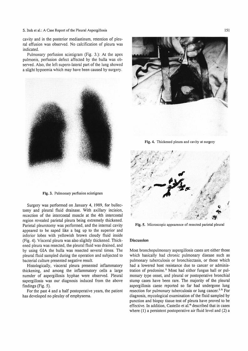
Pulmonary perfusion scintigram (Fig. 3.): At the apex
pulmonis, perfusion defect affected by the bulla was ob-
served. Also, the left super0-1ateral part of the lung showed
a slight hypoemia which may have been caused by surgery.
Fig. 3. Pulmonary perfusion scintigram
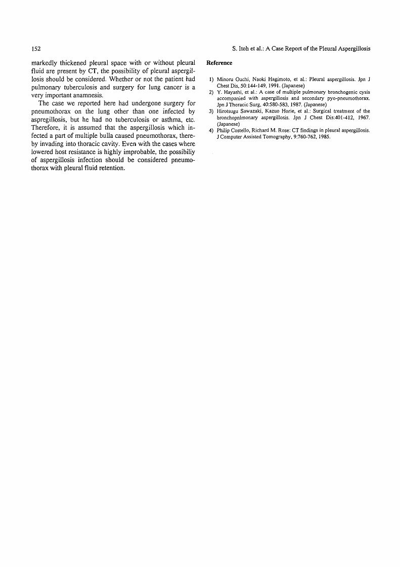
Surgery was performed on January 4, 1989, for bullec-
tomy and pleural fluid drainase. With axillary incision,
recection of the intercostal muscle at the 4th intercostal
region revealed parietal pleura being extremely thickened.
Parietal pleurotomy was perforrned, and the internal cavity
appeared to be saped like a bag up to the superior and
inferior lobes with yellowish brown cloudy fluid inside
(Fig. 4). Visceral pleura was also slightly thickened. Thick-
ened pleura was resected, the pleural fluid was drained, and
by using GIA the bulla was resected several times. The
pleural fluid sampled during the operation and subjected to
bacterial culture presented negative result.
Histologically, visceral pleura presented inflammatory
thickening, and among the inflammatory cells a large
numder of aspergillosis hyphae were observed. Pleural
sapergillosis was our diagnosis induced frpm the above
findings (Fig. 5).
For the past 4 and a half postoperative years, the patient
has developed no pleulsy of emphysema.
151
*::~=:::.
-~'1:ii
¥;
Fig. 4. Thickened pleura and cavity at surgery
~=
~i=="'=-~: ' =' ~' = ':. ' ='~;~'F"- :
*=~ '
ri r ~~'f ' / " "*
~ '= = =~:;~:* ' =!
1~ P ~"
~i~i ..' ~
(b * + :=:=1i~;'~~
; '~ '~;1.' ~ :+=+=*+~ ~ ii
Fig' 5. MiCroscopic appearence of resected Parietal pleural
Discussion
Most bronchopulmonary aspergillosis cases are either those
which basically had chronic pulmonary disease such as
pulmonary tuberculosis or bronchiectasis, or those which
had a lowered host resistance due to cancer or adminis-
tration of predonine.*) Most had either fungus ball or pul-
monary type onset, and pleural or postoperative bronchial
stump cases have been rare. The majority of the pleural
aspergillosis caese reported so far had undergone lung resection for pulmonary tuberculosis or lung cancer.2.3) For
diagnosis, mycological examination of the fluid sampled by
punction and biopsy tissue test of pleura have proved to be
effective. In addition, Castello et al.') described that in cases
where (1) a persistent postoperative air fluid level and (2) a
1 52 S. Itoh et al. A Case Report of the Pleural Aspergillosis
markedly thickened pleural space with or without pleural
fluid are present by CT, the possibility of pleural aspergil-
losis should be considered. Whether or not the patient had
pulmonary tuberculosis and surgery for lung cancer is a
very important anamnesis.
The case we reported here had undergone surgery for
pneumothorax on the lung other than one infected by aspregillosis, but he had no tuberculosis or asthma, etc.
Therefore, it is assumed that the aspergillosis which in-
fected a part of multiple bulla caused pneumothorax, there-
by invading into thoracic cavity. Even with the cases where
lowered host resistance is highly improbable, the possibiliy
of aspergillosis infection should be considered pneumo-
thorax with pleural fluid retention.
Reference
l) Minoru Ouchi, Naoki Hagimoto, et al.: Pleural aspergillosis. Jpn J
Chest Dis, 50:144-149, 1991. (Japanese)
2) Y. Hayashi, et al.: A case of multiple pulmonary bronchogenic cysis
accompanied with aspergillosis and secondary pyo-pneumothorax. Jpn J Thoracic Surg, 40:580-583, 1987. (Japanese)
3) Hirotsugu Sawazaki, Kazuo Horie, et al.: Surgical treatment of the
bronchopnlmonary aspergillosis. Jpn J Chest Dis:401-412, 1967. ( Japanese)
4) Philip Costello. Richard M. Rose: CT findings in pleural aspergillosis.
J Computer Assisted Tomography, 9:760-762, 1985.