naosite: nagasaki university's academic output...
TRANSCRIPT
This document is downloaded at: 2019-03-15T18:48:18Z
Title Pathological Study of the Intestinal Infarction -especially Non -occlusiveIntestinal Infarction
Author(s) Takahira, Ryoji
Citation Acta medica Nagasakiensia. 1983, 28(1-4), p.65-88
Issue Date 1983-10-25
URL http://hdl.handle.net/10069/15641
Right
NAOSITE: Nagasaki University's Academic Output SITE
http://naosite.lb.nagasaki-u.ac.jp
Acta Med. Nagasaki 28: 64-88
Pathological Study of the Intestinal Infarction
-especially Non -occlusive Intestinal Infarction
Ryoji TAKAHIRA
Department of Pathology, Atomic Disease Institute,
NAGASAKI University School of Medicine, NAGASAKI, Japan
Received for Publication, January 5, 1983
A part of this study was presented at the 7lst Annual Meeting of
the Japanese Pathological Society, April 7 , 1982
SUMMARY
Among intestinal infarctions, there is a relatively high incidence of non-occlusive
intestinal infarctions, which have no evidence of apparent mesenteric vascular occlusion . In these cases, etiology and/or pathogenesis are not known, though some authors described
congestive heart failure, digitalis intoxication, minute vessel occlusion , vasospasmus and so on as the trigger of the disorders. The purpose of this study is to examine the vas-
cular factors which have not been reported systematically.
Fifteen autopsy cases were used for this study including five cases of vascular
occlusive intestinal infarction (two of arterial occlusions, two of venous occlusions and
one of strangulation) and ten non-occlusive ones. Specimens for pathological study were
obtained from both mesenteries and intestinal walls.
As to the lesions of the intestinal walls, there were some differences between arterial
and venous occlusions. Arterial occlusion showed mucosal necrosis and submucosal con-
gestion while venous occlusion showed marked edema and hemorrhage of intestinal walls
with less common necrosis of the mucosa.
Ten cases of non-occlusive intestinal infarctions consisted of eight diffuse and two
segmental cases. In the latter, in addition to marked stenotic atherosclerosis of the prox-
imal superior mesenteric artery(SMA), the branch of the SMA toward the impaired parts
of intestine was also narrowed by arteriosclerosis. Three of eight diffuse infarction cases
also had stenotic proximal SMA and one of them had diffuse arteriosclerotic narrowing
from proximal SMA to small mesenteric arteries (vasa recta). One of the other five
cases presented microthrombi in the intestinal wall, suggesting Disseminated intravascular
coagulation (DIC). The remaining four cases had no obvious vascular changes.
In clinical aspects, six of ten non-occlusive intestinal infarction cases had cardiac
diseases such as congestive heart failure, myocardial infarction and pericardial effusion . Hypertension was also found in five of ten cases. Hypotension (shock) before intestinal
symptoms became manifest was not seen in any cases.
高 平 良 二
INTRODUCTION
Circulatory disturbances of the digestive tracts, especially of intestines, are one of
the fatal disorders even nowadays when medical science has progressed rapidly. Strangula-
tion ileus and mesenteric vascular obstruction have reportedly been the main clinico-pa-
thological features of intestinal infarction. In recent years, circulatory disturbances of
intestine with no evidence of vascular occlusion have attracted considerable attention, and
clinical and experimental reports have increased in number. Some authors estimated that
systemic circulatory disturbances, i. e. , cardic disease, shock, etc. , are the important
etiologic factors, and others attached importance to microthrombi of the intestinal
wall. However, unanimous concordance has not been attained.
Many of the studies in the literature dealt with large intestinal ischemia, i. e. ,
ischemic colitis mostly using surgical materials without systematic pathological studies
of mesenteric vessels. As to the small intestines, non-occlusive intestinal infarctions
were reported mainly in 1960's but systematic pathological studies of mesenteric vessels
were not performed. These authors suggested that vasospasmus occurring at systemic cir-
culatory disturbance was the pathogenetic factor.
In the cases of non-occlusive intestinal infarctions, pathophysiological conditions
such as advanced age, heart failure and hypotension were pointed out by many authors
as important etiologic factors. These conditions are readily acceptable. Using autopsy
cases whereby the mesenteric vessels as well as intestines could be examined, I examin-
ed non-occlusive intestinal infarctions and also the intestinal changes in occlusive and
non-occlusive intestinal infarctions, to investigate the vascular changes for the factors of
non-occlusive intestinal infarctions.
MATERIALS AND METHODS
From the autopsy cases of the Nagasaki University School of Medicine and its re-
lated hospitals, intestines which had mesenteries together were reexamined to possible ex-
tent, paying much attention macroscopically to erosion, ulcer, hemorrhage and infarct.
Excepting etiologically evident cases such as intestinal amebiasis and fungus infection,
intestines seemingly with circulatory disturbances were collected. Moreover intestinal in-
farction cases were picked up from autopsy protocols and Annual of the Pathological
Autopsy Cases in Japan and some of them were added to the material after microscopical
reexamination. The cases wherein intestines had no mesenteries or whole bowels were not
preserved were excluded from this study because the relation between infarcted bowels and mesenteric vessels could not be examined.
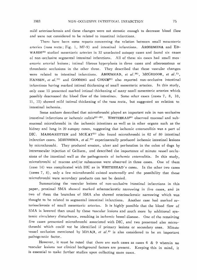
Macroscopical observation of intestinal walls and cross cut surfaces of mesenteric
vessels were made, and then as shown in Fig. 1, nine specimens of mesentery, three of
mesocolon, three to six of small intestine and three of large intestine were taken as a
rule. When further examination was needed, more specimens were prepared accordingly.
Specimens for histopathological study were embedded in paraffin and sliced into 3 to 5
u sections. Hematoxylin-EOSIN stain, Elastica Van-GIESON stain and Azan-MALLORY
stain were performed as a rule, and other special stains were performed as needed. In-
testines and mesenteric vessels were examined by light microscopy.
Clinical findings were taken from medical charts and partly from autopsy protocols.
RESULTS
In 289 autopsy cases randomly selected and examined macroscopically, 63 had in-
testinal lesions such as congestion, hemorrhage and ulcers. From these, 15 cases in all
wherein both intestines and mesenteries could be examined were collected as the materials
of this study.
Etiologically these 15 cases consisted of five vascular occlusive intestinal infarctions
(two of arterial occlusion, two of venous occlusion and one of strangulation) and 10
vascular non-occlusive intestial infarctions (Table 2 & 3).
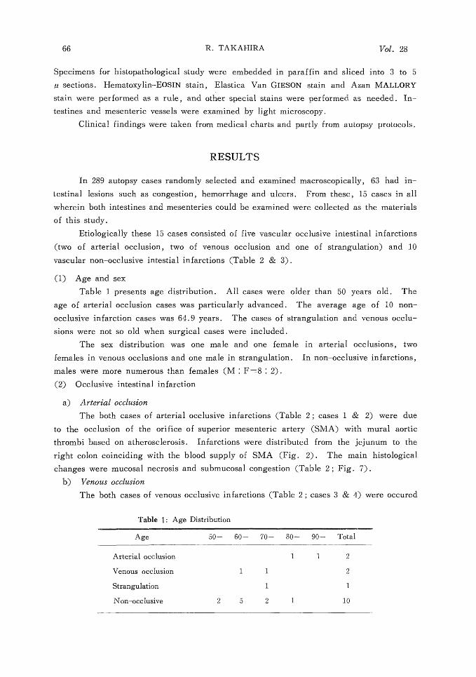
(1) Age and sex
Table 1 presents age distribution. All cases were older than 50 years old. The
age of arterial occlusion cases was particularly advanced. The average age of 10 non-
occlusive infarction cases was 64.9 years. The cases of strangulation and venous occlu-
sions were not so old when surgical cases were included.
The sex distribution was one male and one female in arterial occlusions, two
females in venous occlusions and one male in strangulation. In non-occlusive infarctions,
males were more numerous than females (M : F=8: 2).
(2) Occlusive intestinal infarction
a) Arterial occlusion
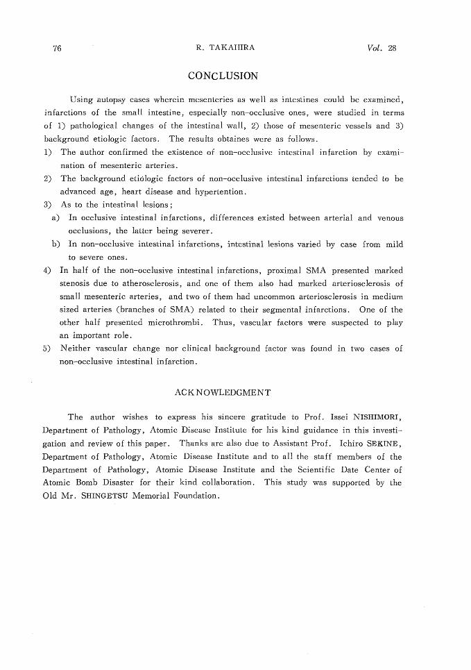
The both cases of arterial occlusive infarctions (Table 2 ; cases 1 & 2) were due
to the occlusion of the orifice of superior mesenteric artery (SMA) with mural aortic
thrombi based on atherosclerosis. Infarctions were distributed from the jejunum to the
right colon coinciding with the blood supply of SMA (Fig. 2). The main histological
changes were mucosal necrosis and submucosal congestion (Table 2 ; Fig. 7) .
b) Venous occlusion
The both cases of venous occlusive infarctions (Table 2 ; cases 3 & 4) were occured
Table 1: Age Distribution
Age 50- 60- 70- 80- 90- Total
Arterial occlusion 1 1 2
Venous occlusion 1 1 2
Strangulation 1 1
Non-occlusive 2 5 2 1 10
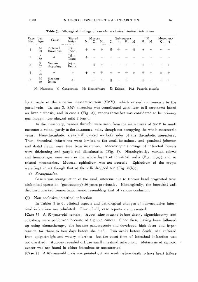
Table 2: Pathological findings of vascular occlusive intestinal infarctions
Case Sex Cause Site of Mucosa Submucosa PM Mesentery No. Age lesion N. C. H. C. E. H. N. H. N. C. H.
I M Arterial Jej.- + + + It -1-I- - ~I + - - - 93 thr ombus Asc.
2 F ~i Jej ' + - - + - - - - - - - 89 Trans .
3 F Venous Jej . - +f + + - - ~} - + + 67 thrombus Ileum .
4 73 + + * + - * f * + ± +
5 M Strangu- ,/ + + I F - I1 - H - + ± 79 lation
N: Necrosis C: Congestion H: Hemorrhage E: Edema PM: Propria muscle
by thrombi of the superior mesenteric vein (SMV), which existed continuously to the
portal vein. In case 3, SMV thrombus was complicated with liver cell carcinoma based on liver cirrhosis, and in case 4 (Fig. 3), venous thrombus was considered to be primary
one though liver showed mild fibrosis.
In the mesentery, venous thrombi were seen from the main trunk of SMV to small
mesenteric veins, partly to the intramural vein, though not occupying the whole mesenteric
veins. Non-thrombotic areas still existed on both sides of the thrombotic mesentery.
Thus, intestinal infarctions were limited to the small intestines, and proximal jeiunum
and distal ileum were free from infarction. Macroscopic findings of infarcted bowels
were thickening and purple-red discoloration (Fig. 3). Histologically, marked edema
and hemorrhage were seen in the whole layers of intestinal walls (Fig. 8(a)) and in
related mesenteries. Mucosal epithelium was not necrotic. Epithelium of the crypts
were kept intact though that of the villi dropped out (Fig. 8(b)).
c) Strangulation
Case 5 was strangulation of the small intestine due to fibrous band originated from
abdominal operation (gastrectomy) 20 years previously. Histologically, the intestinal wall
disclosed marked hemorrhagic lesion resembling that of venous occlusion.
(3) Non-occlusive intestinal infarction
In Tables 3 to 6, clinical aspects and pathological changes of non-occlusive intes-
tinal infarctions are tabulated. First of all, case reports are presented.
[Case 6] A 62-year-old female. About nine months before death, sigmoidectomy and
colostomy were performed because of sigmoid cancer. Since then, having been followed
up using chemotherapy, she became pancytopenic and developed high fever and hypo-
tension for three to four days before she died. Two weeks before death, she suffered
from epigastralgia and watery diarrhea, but the onset time of intestinal infarction was
not clarified. Autopsy revealed diffuse small intestinal infarction. Metastasis of sigmoid
cancer was not found in either intestines or mesenteries.
[Case 7] A 67-year-old male was pointed out one week before death to have heart failure
because of cough and stridor. Three days after he was ambulanced because of epigas-
tralgia, vomiting and diarrhea. In his hospital days, he had high fever and leucocytosis,
and then gradually became hypotensive. He died four days after developing abdominal
symptoms. Autopsy revealed infarction of the small intestine and right colon. Though
a clot of blood was obtained from the SMA, it was not thought to be thrombus as it
was pulled out easily.
[Case 8] A 63-year-old male, being admitted because of mitral regurgitation and steno-
sis, had meteorismus complicated with hypotension, high fever and positive occult blood
for a week before he died. At autopsy, diffuse intestinal infarction was found extending
from the small intestine to the caecum. In spite of left atrial thrombus, SMA did not
present any vascular occlusive lesions such as thrombi and emboli.
[Case 9] A 61-year-old male was admitted because of orthostatic disturbance, and fur-ther examination revealed thrombotic obstruction of the lower abdominal aorta, and a
diagnosis of LERICHE's syndrome was made. Angiography was performed then but
obstruction of SMA was not observed. He died next day showing ileus, hypotension,
high fever and tarry stool. At autopsy, intestinal infarction extending from the ileum
to the ascending colon was observed. Thrombo-emboli were not found in SMA or SMV.
[Case 10] A 53-year-old male was admitted because of heart failure and atrial fibril-
lation. He had hypertension in the past history, but after admission, blood pressure
gradually dropped and he died. Abdominal signs and symptoms were not noted clinically. At autopsy, diffuse acute and old myocardial infarction was found. Also in intestines,
diffuse infarction extending from the small intestine to the large intestine was observed.
There were mural thrombi in the abdominal aorta under the orifice of renal arteries.
Hematologically, there was hemoconcentration, but thromboembolism was not found in
mesenteric vessels.
[Case 11] An 84-year-old male died three days after admission because of heart failure and bronchopneumonia. Abdominal symptoms were not recognized. Autopsy revealed
old myocardial infarction and myocardial hypertrophy. Diffuse infarction of the small
intestine and focal infarction of the large intestine were also found.
[Case 12] A 72-year-old male, being admitted because of liver cirrhosis, developed ileus and died after 14 hours. At autopsy, hemorrhagic infarction measuring about 30
cm in length was observed in the jejunum.
[Case 13] A 52-year-old male, being admitted because of lung cancer, died of respira-
tory failure. Abdominal signs and symptoms were not noted. At autopsy, two segmental
infarctions, about 20 cm and 25 cm in length, were found in the center of small intestine
(Fig. 4).
[Case 14] A 61-year-old female was operated for pancreatic abscess five years before.
A half year ago, an abnormal shadow of chest X-P was pointed out. Further examination
revealed cholangiocarcinoma, and then chemotherapy was commenced. It was complicated
with DIC, pleural effusion and pneumothorax. Three days before death, severe abdom-
inal pain and melena occurred. She also suffered from shock, renal failure and DIC
again, and then died. At autopsy, small and large intestines showed infarction.
[Case 15] A 74-year-old male had received drug therapy for arteriosclerotic heart disease
and hypertension for four years. He was admitted because of heart failure four months
ago and discharged after hospital therapy. Three days before death, he was admitted
again because of abdominal pain and asthma attack. Abdomen became distended and
blood pressure gradually dropped before he died. At autopsy, ischemic change was
found in small and large intestines in addition to adenocarcinoma of the lung and general
congestion.
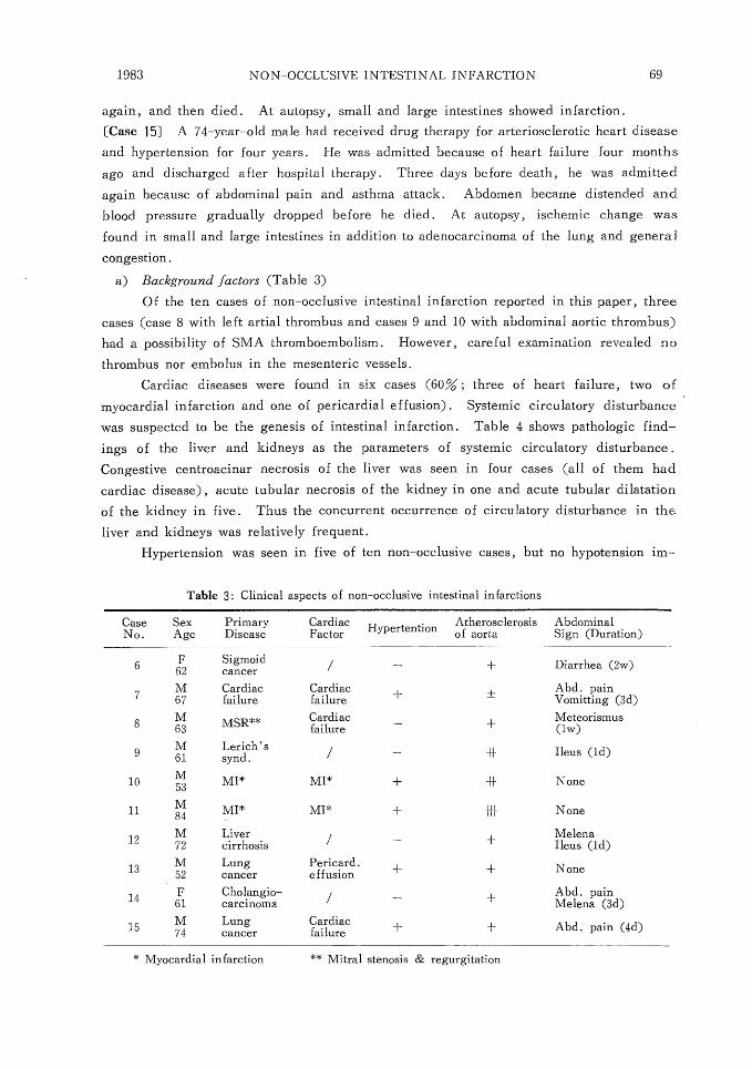
a) Background factors (Table 3)
Of the ten cases of non-occlusive intestinal infarction reported in this paper, three
cases (case 8 with left artial thrombus and cases 9 and 10 with abdominal aortic thrombus)
had a possibility of SMA thromboembolism. However, careful examination revealed no
thrombus nor embolus in the mesenteric vessels.
Cardiac diseases were found in six cases (60% ; three of heart failure, two of
myocardial infarction and one of pericardial effusion). Systemic circulatory disturbance
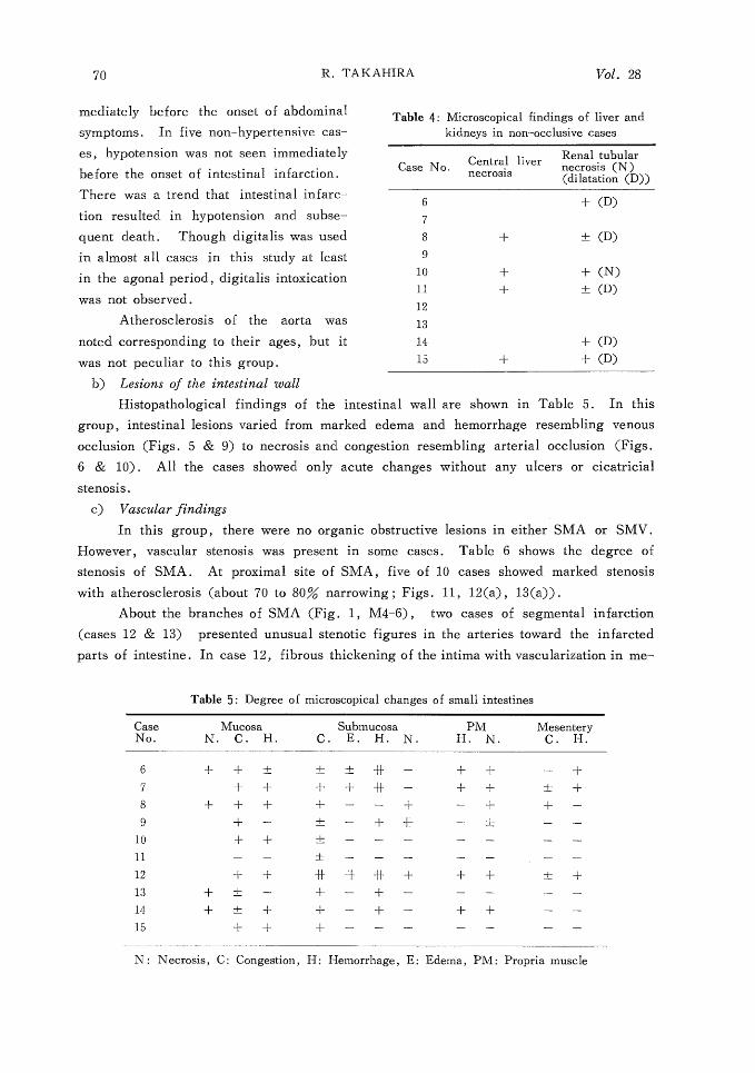
was suspected to be the genesis of intestinal infarction. Table 4 shows pathologic find-
ings of the liver and kidneys as the parameters of systemic circulatory disturbance.
Congestive centroacinar necrosis of the liver was seen in four cases (all of them had
cardiac disease), acute tubular necrosis of the kidney in one and acute tubular dilatation
of the kidney in five. Thus the concurrent occurrence of circulatory disturbance in the
liver and kidneys was relatively frequent.
Hypertension was seen in five of ten non-occlusive cases, but no hypotension im-
Table 3: Clinical aspects of non-occlusive intestinal infarctions
Case Sex Primary Cardiac Hypertention Atherosclerosis Abdominal No. Age Disease Factor of aorta Sign (Duration)
6 F Sigmoid / - + Diarrhea (2w) 62 ca M Cardiac Cardiac + Abd. pain 7
67 failure failure + Vomitting (3d)
M ** Cardiac Meteorismus 8 63 MSR failure + (1w)
9 M Lerich's _ H Ileus (ld) 61 synd .
10 M MI* MI* + -H- None 11 M MI* MI* + {H- None 12 M Liver _ + Melena 72 cirrhosis Ileus (id)
13 M Lung Pericard. + + None 52 cancer effusion
14 F Cholangio- / _ + Abd. pain 61 carcinoma Melena (3d)
15 M Lung Cardiac + + Abd. pain (4d) 74 cancer failure
* Myocardial infarction ** Mitral stenosis & regurgitation
mediately before the onset of abdominal
symptoms. In five non-hypertensive cas-
es, hypotension was not seen immediately
before the onset of intestinal infarction.
There was a trend that intestinal infarc-
tion resulted in hypotension and subse-
quent death. Though digitalis was used in almost all cases in this study at least
in the agonal period, digitalis intoxication
was not observed.
Atherosclerosis of the aorta was
noted corresponding to their ages, but it
was not peculiar to this group.
b) Lesions of the intestinal wall
Table 4: Microscopical findings of liver and kidneys in non--occlusive cases
Central liver Renal tubular Case No. necrosis (N) necrosis (dilatation (D))
6 + (D)
7
8 + ± (D)
9 10 + + (N)
11 + ± (D)
12
13
14 + (D) 15 + + (D)
Histopathological findings of the intestinal wall are shown in Table 5. In this
group, intestinal lesions varied from marked edema and hemorrhage resembling venous occlusion (Figs. 5 & 9) to necrosis and congestion resembling arterial occlusion (Figs.
6 & 10). All the cases showed only acute changes without any ulcers or cicatricial
stenosis.
c) Vascular findings
In this group, there were no organic obstructive lesions in either SMA or SMV.
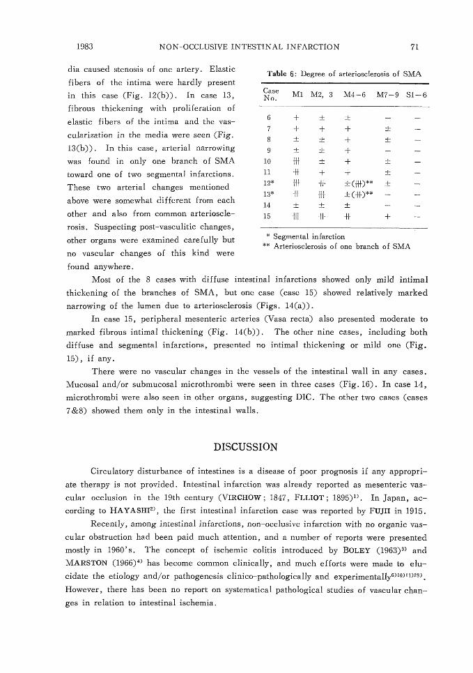
However, vascular stenosis was present in some cases. Table 6 shows the degree of
stenosis of SMA. At proximal site of SMA, five of 10 cases showed marked stenosis
with atherosclerosis (about 70 to 80% narrowing ; Figs. 11, 12(a), 13(a)).
About the branches of SMA (Fig. 1, M4-6), two cases of segmental infarction
(cases 12 & 13) presented unusual stenotic figures in the arteries toward the infarcted
parts of intestine. In case 12, fibrous thickening of the intima with vascularization in me-
Table 5: Degree of microscopical changes of small intestines
Case Mucosa Submucosa PM Mesentery No. N. C. H. C. E. H. N. H. N. C. H.
6 + + ± ± ± -~-}- - + + - +
7 + + + + -H - + + ± +
8 + + + + - - + - + + -
9 + - ± - + * - ± - -
10 + + ± - - - - - - -
11 - - ± - - - - - - -
12 + + * * -f I- + + + ± +
13 + 4- - + - + - - - - -
14 + -!- + + - + - + + - -
15 + + + - - - - - - -
N: Necrosis, C: Congestion, H: Hemorrhage, E: Edema, PM: Propria muscle
dia caused stenosis of one artery. Elastic
fibers of the intima were hardly present
in this case (Fig. 12(b)). In case 13,
fibrous thickening with proliferation of
elastic fibers of the intima and the vas-
cularization in the media were seen (Fig.
13(b)). In this case, arterial narrowing
was found in only one branch of SMA
toward one of two segmental infarctions.
These two arterial changes mentioned
above were somewhat different from each
other and also from common arterioscle-
rosis. Suspecting post-vasculitic changes,
other organs were examined carefully but
no vascular changes of this kind were
found anywhere.
Table 6: Degree of arteriosclerosis of SMA
Case M1 M2, 3 M4-6 M7-9 S1-6 No.
6 + ± ± - -
7 + + + ± -
8 ± ± + ± -
9 ± ± + - -10 ± + ± -
11 H + + ± -
12* }~ _~ ()** ± -
13* -f l- i H ± (*)' ' - -
14 ± ± ± - -
15 }H {I }{ + -
* Segmental infarction ** Arteriosclerosis of one branch of SMA
Most of the 8 cases with diffuse intestinal infarctions showed only mild intimal
thickening of the branches of SMA, but one case (case 15) showed relatively marked
narrowing of the lumen due to arteriosclerosis (Figs. 14(a)).
In case 15, peripheral mesenteric arteries (Vasa recta) also presented moderate to
marked fibrous intimal thickening (Fig. 14(b)). The other nine cases, including both
diffuse and segmental infarctions, presented no intimal thickening or mild one (Fig.
15), if any.
There were no vascular changes in the vessels of the intestinal wall in any cases.
Mucosal and/or submucosal microthrombi were seen in three cases (Fig. 16). In case 14,
microthrombi were also seen in other organs, suggesting DIC. The other two cases (cases
7&8) showed them only in the intestinal walls.
DISCUSSION
Circulatory disturbance of intestines is a disease of poor prognosis if any appropri-
ate therapy is not provided. Intestinal infarction was already reported as mesenteric vas-
cular occlusion in the 19th century (VIRCHOW ; 1847, FLLIOT ; 1895)1). In Japan, ac-
cording to HAYASHI2), the first intestinal infarction case was reported by FUJII in 1915.
Recently, among intestinal infarctions, non-occlusive infarction with no organic vas-
cular obstruction had been paid much attention, and a number of reports were presented
mostly in 1960's. The concept of ischemic colitis introduced by BOLEY (1963)3) and
MARSTON (1966)4) has become common clinically, and much efforts were made to elu-
cidate the etiology and/or pathogenesis clinico-pathologically and experimentallys)10)11)23 .
However, there has been no report on systematical pathological studies of vascular chan-
ges in relation to intestinal ischemia.
In this report, relations between non-occlusive. intestinal infarction and its vascu-
lature were studied, using autopsy materials in whih both intestines and mesenteries could
be examined systematically. In addition to this, intestinal lesions were compared between
occlusive and non-occlusive infarctions. Fifteen cases were collected for this study.
1) INCIDENCE
MAEZAWA, et al.') selected 307 cases of intestinal infarction from the Annual of
the Pathological Autopsy Cases in Japan in a decade from 1962 to 1971. This figure re-
portedly represented 0.15% of total autopsies in each year on an average. MIYAJI, et
al.') reported that intestinal disorders due to circulatory disturbance were found in 5.5%
of the serial autopsies at a medical center for aged persons. Except for the aforemention-
ed aged group, intestinal infarctions are not thought to be so numerous among autopsy
cases but there must be more cases if non-autopsied and surgically treated cases are in-
cluded. More attention should be paid as MAEZAWA said. Furthermore, it is anticipa-
ted that incidence of intestinal infarction would gradually increase with the advancement
of age in the society.
The incidence of non-occlusive intestinal infarctions among whole intestinal infar-
ctions varies by author ; 10 to 52% in the literature by MCGREGOR, et al.'), 21% by
BERGER and BYRNE9), 50% by PIERCE, et al.") and OTTINGERI°), 67% by MIYAJI,
et al .6 and 26% by HORIE, et al. Thus the incidence of non-occlusive cases among
intestinal infarctions seems to be relatively high. Although this study is not appro-
priate to estimate the incidence because materials were not collected from serial autopsi-
es, non-occlusive cases were more than occlusive ones as far as the number is concern-
ed.
2) AGE AND SEX
Concerning the age at onset of intestinal infarctions, almost all authors agreed that this
disease occurs in old-aged persons. OTTINGER and AUSTEN10) classified intestinal in-
farctions by pathogenesis and estimated the average age to be 77 in arterial thrombus, 68
in arterial embolus, 72 in venous thrombus and 75 in non-occlusive intestinal infarction.
There seem to be no significant difference among the groups. In non-occlusive intestin-
al infarctions, the average age of onset reportedly was 69 by BERGER and BYRNE9), 64
by MUSA12) and 67 by HEER, et al. 13>
In this report, all 15 cases, especially arterial occlusion cases, were old. As to the
10 non-occlusive cases, the average age was 64, which was not different from that in
the previous reports.
The ratio of male to female in the non-occlusive series showed no definite tendency
in previous reports. MUSA12) presented 31:0 in favor of males, while OTTINGER and
AUSTEN10) presented more females than males (39:28). In this study, eight were ma-
les and two were females.
3) LESIONS OF INTESTINAL WALL
a) vascular occlusive infarction
In the two cases of venous occulsive infarctions in this report, the intestinal wall
showed marked thickening and purple-red discoloration macroscopically, and marked he-
morrhage and edema microscopically. In spite of severe intestinal lesions mentioned a-
bove, L' he mucosal epithelium remained intact at crypts and dropped out at villi due to
hemorrhage. This means that the mucosal epithelium itself was not necrotic.
On the other hand, in the two cases of arterial occlusive infarctions, mucosal ne-
crosis and submucosal congestion were the main microscopical findings while thickening
of the intestinal wall, edema and hemorrhage were not notable. Mesenteries were involved
in circulatory disturbance in venous occlusion but not in arterial occlusion. Thus differ-
ences of intestinal and mesenteric lesions between arterial and venous occlusions were
observed but no such difference could be found in previous reports. Since hemodynamic
mechanism of ischemia is different between them involving complicated factors such as
degree and duration of ischemia, period of examination, etc. , it is readilly recognized
that intestinal lesions mentioned above might also be different.
MARCUSON, et al. 141 examined changes of the colon due to venous occlusion in
dogs and estimated that mucosal necrosis was rare in venous occlusion being different
from arterial occlusion, and that venous occlusion produced stagnant hypoxia while arterial
occlusion produced ischemic hypoxia.
b) Non-occlusive infarction
In 10 cases of non-occlusive intestinal infarctions in this paper, the degree of in-
testinal lesions varied from marked hemorrhage and edema resembling venous occlusion
to submucosal congestion like in arterial occlusion. In general, intestinal lesions in non-
occlusive infarctions were reported to be limited to the mucosa. PIERCE and BROCKEN-
BROUGH8) described that the detph of the diseased bowels had no difference between
occlusive and non-occlusive infarctions, though MING's description showed more super-
ficial lesions in nonocclusive infarctions. On the other hand, HEER, et al.") observed
two cases of mesenteric involvement in 36 non-occlusive infarction cases.
4) BACKGROUND FACTORS
FOGARTY, et al.") attributed the genesis of non-occlusive intestinal infarction to
1) congestive heart failure, 2) digitalis intoxication and 3) hemoconcentration, after ex-
amination of their 18 cases.
Cardiac diseases, especially heart failure, arrythmia and myocardial infarction,
were the important background factors in many previous reports. They were presented
in 30 of 31 cases by MUSA, et al. 11), 9 of 10 cases by GROSH, et al. 15) and 21 of 23
cases by BERGER, et al.9). BERGER, et al.') stated that heart failure produced low
cardiac output and then compensatory mesenteric arterial vasospasmus might have occurred
to maintain the blood flow of the vital organs, such as the brain and the heart, resulting
in intestinal ischemia. They also stated that intestinal ischemia led to spasmus of the
intestinal wall and further decreased blood flow of the intestine, and that intestinal in-
farction produced shock state and subsequent systemic circulatory disturbance, which in
turn decreased the blood flow of the intestine.
GROSH, et al.") raised a question in their report whether mucosal necrosis of the
intestines induced by heart diseases was due to increased portal venous pressure or to
mesenteric vascular angiospasmus, but they are of the view that angiospasmus of the
arterioles in the intestinal wall was an important factor, quoting the examination by
CORDAY and others.
Digitalis is considered to produce intestinal ischemia by its pharmacological effect
of constricting the mesenteric arteries. HOBBOUSHE, et al. 17) reported a patient ad-
ministered with digitalis because of mitral stenosis and heart failure, whose angiography
before laparotomy for intestinal infarction disclosed segmental spasmus of the medium
and small sized mesenteric arteries.
As mentioned above, angiospasmus is considered in many reports as an important
etiologic factor of non-occlusive intestinal infarction. In this paper, cardiac diseases
might be considered as background factors being found in 6 of the 10 cases (60% ; two
of myocardial infarction, three of congestive heart failure and one of pericardial effu-
sion), though angiospasmus could not be demonstrated in histopathological examination
Digitalis was used in almost all cases but digitalis intoxication was not found even in
heart failure patients. Other background factors mentioned in previous reports were
diuretic agents, vasopressors, hypertension and arteriosclerosis.
However, OTTINGER, et al.10) described that about one-fourth of patients showed
no background diseases in non-occlusive intestinal infarctions like in two patients of this
paper. It is felt that more efforts are required to elucidate other etiologic factors of this disease.
5) VASCULAR FACTORS
Reports on non-occlusive intestinal infarctions associated with vasculature of SMA
are scarce. FOGARTY and FLETCHERI6> observed chronic occlusive changes of SMA in
14 cases among the 18 non-occlusive intestinal infarctions, but no case of over 50% nar-
rowing of the lumen. OTTINGER and AUSTEN10) observed sclerosis and/or stenosis of
SMA in 10 of their 67 cases. HEER, et al.") observed atherosclerosis in 33% and em-
phasized the importance of vascular factors in non-occlusive intestinal infarctions. In these reports, the vascular changes were seen only in proximal region of SMA. We
also observed marked stenosis in the same region in three cases of diffuse infarction and
two of segmental infarctions.
Arterial lesions in the branches of SMA (Fig. 1 ; M4-6) were not reported previ-
ously. In the two cases of segmental intestinal infarction in this study (cases 12 & 13),
unusual stenotic changes were seen in the branches of SMA toward the diseased bowels.
Although these arterial lesions themselves might be exceptional changes being uncommon
arteriosclerosis, they were suspected to be valuable cases in terms of the relation between
intestinal infarctions and vascular changes. The other branches of SMA in these cases
and all branches of SMA in the cases having diffuse infarction, except case 15, presented
mild arteriosclerosis and these changes were not stenotic enough to decrease blood flow
and were not considered to be related to intestinal infarctions.
There have been some reports concerning the relation between small mesenteric
arteries (vasa recta; Fig. 1, M7-9) and intestinal infarctions. AROSEMENA and ED-
WARDS19) studied mesenteric arteries in 32 unselected autopsy cases and found six cases
of non-occlusive segmental intestinal infarctions. All of these six cases had small mes-
enteric arterial lesions ; intimal fibrous hyperplasia in three cases and atheromatous or
thrombotic occlusions in the other three. They described that these vascular changes
were related to intestinal infarctions. ABOUMRAD, et al.20), MCGREGOR, et al.'1),
HANSEN, et al.") and GOODING and COUCH") also reported non-occlusive intestinal
infarctions having marked intimal thickening of small mesenteric arteries. In this study,
only case 15 presented marked intimal thickening of many small mesenteric arteries which
possibly descreased the blood flow of the intestines. Some other cases (cases 7, 8, 10,
11, 12) showed mild intimal thickening of the vasa recta, but suggested no relation to
intestinal ischemia.
Some authors described that microthrombi played an important role in non-occlusive
intestinal infarctions or ischemic colitis23)-25) WHITEHEAD24) observed mucosal and sub-
mucosal microthrombi in the ischemic intestines as well as in other organs such as the
kidney and lung in 20 autopsy cases, suggesting that ischemic enterocolitis was a part of
DIC. MARGARETTEN and MCKAY25) also found microthrombi in 62 of 80 intestinal
infarction cases. MIZUSHIMA, et al.") experimentally produced ischemic intestinal lesions
by microthrombi. They produced erosion, ulcer and perforation in the colon of dogs by
intravascular injection of Gelform, and described the importance of minute vessel occlu-
sions of the intestinal wall as the pathogenesis of ischemic entercolitis. In this study,
microthrombi of mucosa and/or submucosa were observed in three cases. One of them
(case 14) was complicated with DIC as in WHITEHEAD's cases. In the other two cases
(cases 7, 8), only a few microthrombi existed scatteredly and the possibility that these
microthrombi were secondary products can not be denied.
Summarizing the vascular lesions of non-occlusive intestinal infarctions in this
paper, proximal SMA showed marked atherosclerotic narrowing in five cases, and in two of them the branches of SMA also showed arteriosclerotic narrowing which was
thought to he related to segmental intestinal infarctions. Another case had marked ar-
teriosclerosis of small mesenteric arteries. It is highly possible that the blood flow of
SMA is lowered than usual by these vascular lesions and much more by additional sys-
temic circulatory disturbance, resulting in ischemic bowel disease. One of the remaining
five cases presented microthrombi associated with DIC, and two presented also micro-
thrombi which could not be identified if primary lesions or secondary ones . Minute vessel occlusion mentioned by MIYAJI, et al.s) is also considered to be an important
pathogenetic factor.
However, it must be noted that there are such cases as cases 6 & 9 wherein no vascular lesions nor clinical background factors are present . Keeping this in mind, it is essential to make further studies upon collecting more cases .
CONCLUSION
Using autopsy cases wherein mesenteries as well as intestines could be examined,
infarctions of the small intestine, especially non-occlusive ones, were studied in terms
of 1) pathological changes of the intestinal wall, 2) those of mesenteric vessels and 3)
background etiologic factors. The results obtaines were as follows.
1) The author confirmed the existence of non-occlusive intestinal infarction by exami-
nation of mesenteric arteries.
2) The background etiologic factors of non-occlusive intestinal infarctions tended to be
advanced age, heart disease and hypertention.
3) As to the intestinal lesions ;
a) In occlusive intestinal infarctions, differences existed between arterial and venous
occlusions, the latter being severer.
b) In non-occlusive intestinal infarctions, intestinal lesions varied by case from mild
to severe ones.
4) In half of the non-occlusive intestinal infarctions, proximal SMA presented marked
stenosis due to atherosclerosis, and one of them also had marked arteriosclerosis of
small mesenteric arteries, and two of them had uncommon arteriosclerosis in medium
sized arteries (branches of SMA) related to their segmental infarctions. One of the
other half presented microthrombi. Thus, vascular factors were suspected to play
an important role.
5) Neither vascular change nor clinical background factor was found in two cases of
non-occlusive intestinal infarction.
ACKNOWLEDGMENT
The author wishes to express his sincere gratitude to Prof. Issei NISHIMORI,
Department of Pathology, Atomic Disease Institute for his kind guidance in this investi-
gation and review of this paper. Thanks are also due to Assistant Prof. Ichiro SEKINE, Department of Pathology, Atomic Disease Institute and to all the staff members of the
Department of Pathology, Atomic Disease Institute and the Scientific Date Center of
Atomic Bomb Disaster for their kind collaboration. This study was supported by the
Old Mr. SHINGETSU Memorial Foundation.
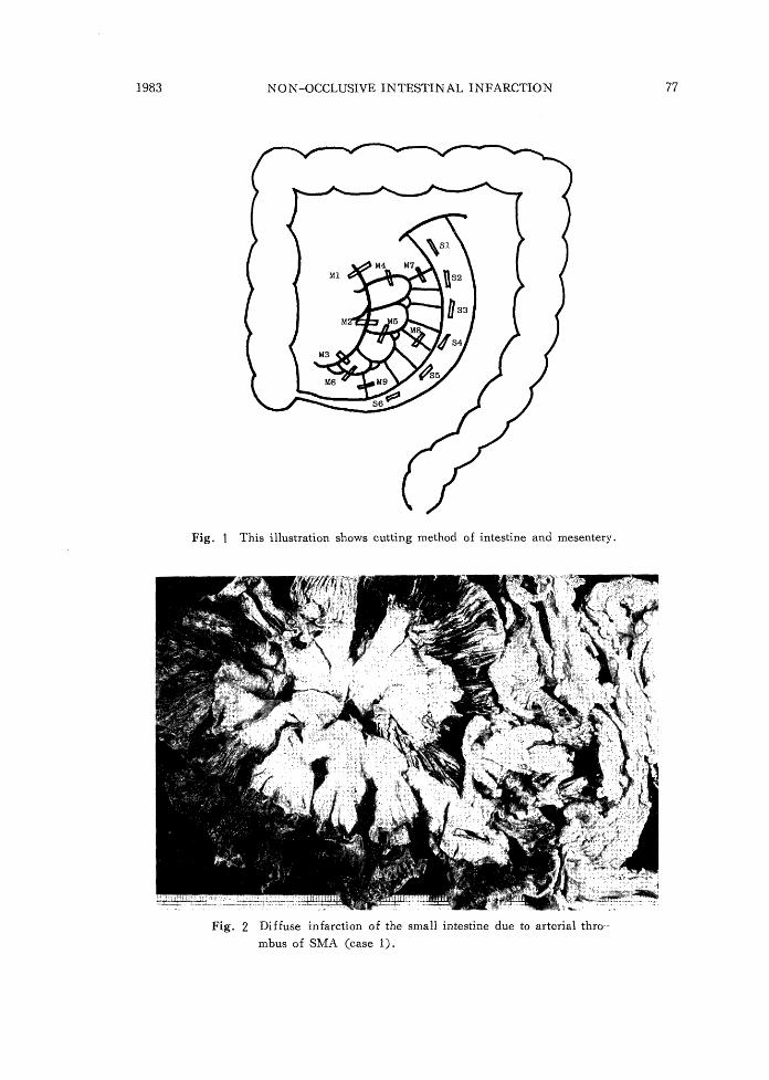
Fig. 1 This illustration shows cutting method of intestine and mesentery.
Fig. 2 Diffuse infarction of the small intestine due to arterial thro-
mbus of SMA (case 1).
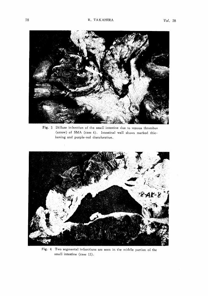
Fig. 3 Diffuse infarction of the small intestine due to venous thrombus
(arrow) of SMA (case 4). Intestinal wall shows marked thic- kening and purple-red discoloration.
Fig. 4 Two segmental infarctions are seen in the middle portion of the
small intestine (case 13).
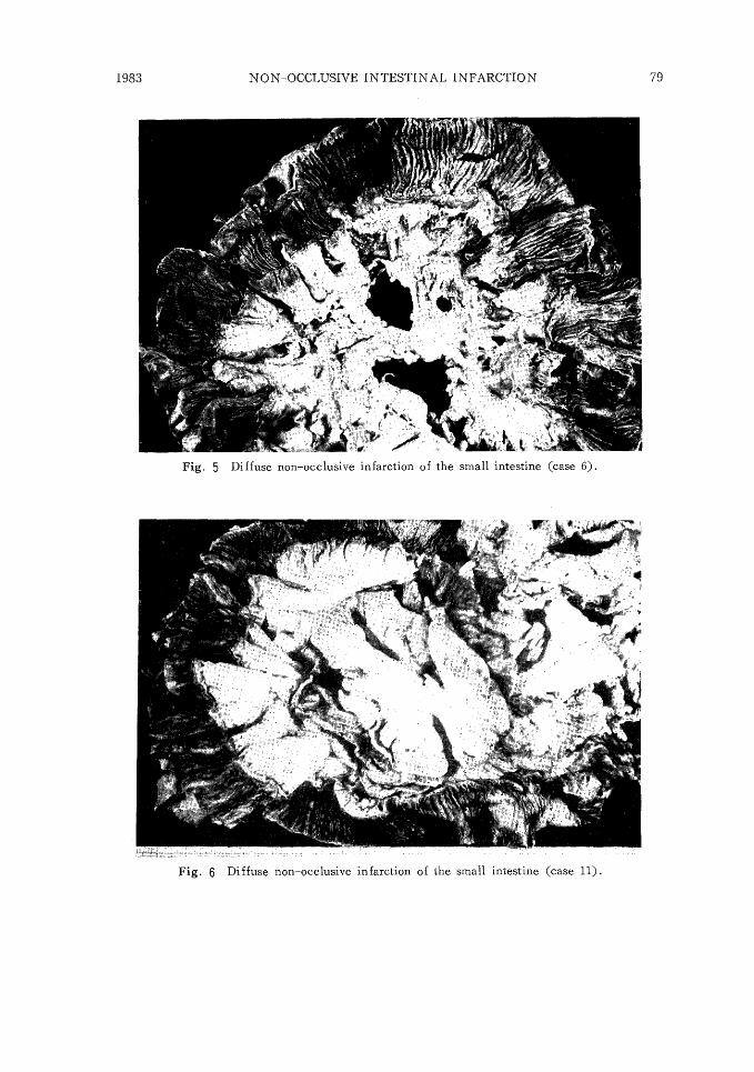
Fig. 5 Diffuse non-occlusive infarction of the small intestine (case 6).
Fig. 6 Diffuse non-occlusive infarction of the small intestine (case 11).
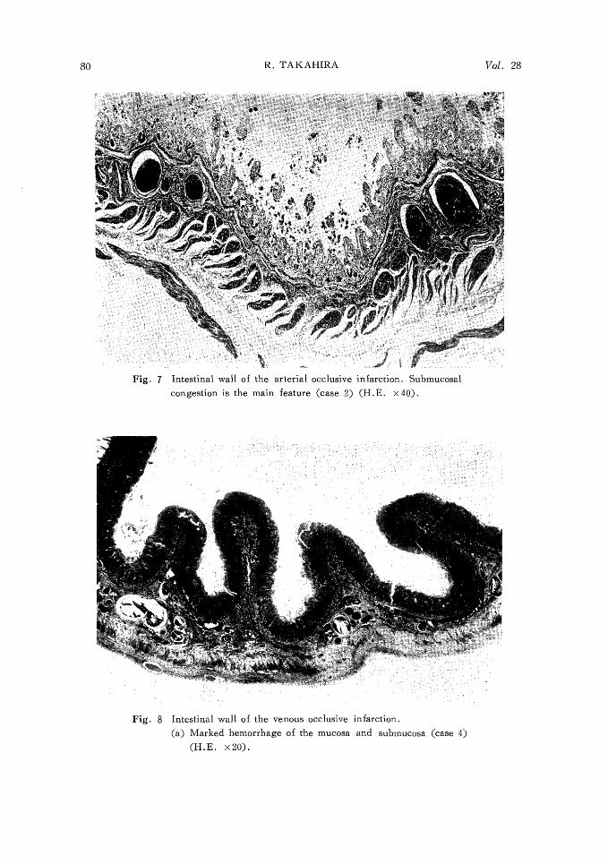
Fig. 7 Intestinal wall of the arterial occlusive infarction. Submucosal
congestion is the main feature (case 2) (H.E. x40).
Fig. 8 Intestinal wall of the venous occlusive infarction.
(a) Marked hemorrhage of the mucosa and submucosa (case 4)
(H.E. x20).
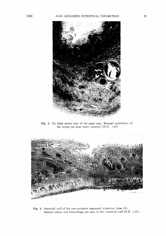
Fig. 8 (b) High power view of the same case. Mucosal epithelium of the crypts are kept intact (arrows) (H . E . X40).
Fig. 9 Intestinal wall of the non-occlusive segmental infarction (case 12). Marked edema and hemorrhage are seen in the intestinal wall (H.E. X20).
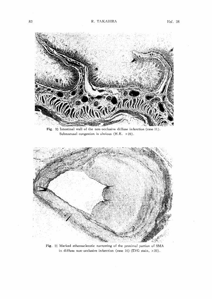
Fig. 10 Intestinal wall of the non-occlusive diffuse infarction (case 11).
Submucosal congestion is obvious (H. E. x20).
Fig. 11 Marked atherosclerotic narrowing of the proximal portion of SMA
in diffuse non-occlusive infarction (case 10) (EVG stain, x20).
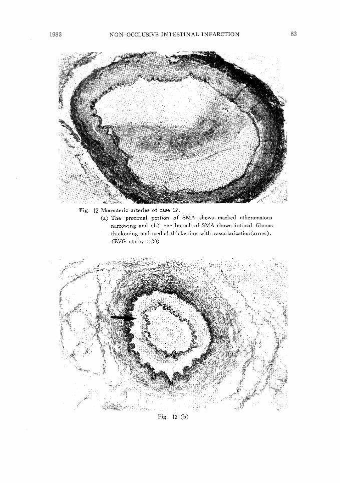
Fig. 12 Mesenteric arteries of case 12.
(a) The proximal portion of SMA shows marked atheromatous narrowing and (b) one branch of SMA shows intimal fibrous
thickening and medial thickening with vascularization(arrow).
(EVG stain, X20)
Fig. 12 (b)
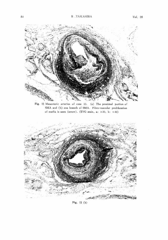
Fig. 13 Mesenteric arteries of case 13. (a) The proximal portion of
SMA and (b) one branch of SMA. Fibro-vascular proliferation
of media is seen (arrow). (EVG stain, a: X20, b: X40)
Fig. 13 (b)
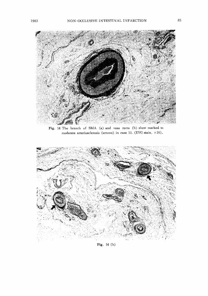
Fig. 14 The branch of SMA (a) and vasa recta (b) show marked to
moderate arteriosclerosis (arrows) in case 15. (EVG stain, x 20)
Fig. 14 (b)

Fig. 15 None to mild intimal thic kening of the small mesenteric arteries (vasa recta) (case 10) (EVG stain, x50).
Fig. 16 Fibrin thrombi (arrows) in intestinal wall of case 14 (H.E. x100).
REFERENCES
1) GRENDELL, J. H. and OCKNER, R. K.: Mesenteric venous thrombosis. Gastroen-
terology 82: 358-372, 1982
2) HAYASHI, S.: Ischemic lesions in the intestinal tract. -clinical and experimental
studies-. The Journal of Japanese College and Angiology 18 (2): 105-110, 1978
3) BOLEY, S. T., SCHWARTZ, S., LASH, J. and STERNHILL, V.: Reversible vascular
occlusion of the colon. Surg. Gynec. Obstet. 116: 53-60, 1963
4) MARSTON, A., PHEILS, M.T. and THOMAS, M.L.: Ischemic colitis. Gut 7: 1-10,
1966
5) MAEZAWA, H., SHIMAMOTO, t., MASHITA, N . , ODAKURA, T. and NAKASHIMA,
M.: Ischemic lesions of the mesenteric vascular region (author translation). Japan
Medical Journal 2614: 11-16, 1974
6) MIYAJI, M., ITO, M., SUZUKI, K., YAGI, H. and TAKEUCHI, T.: Vascular
disorder of the intestine in the aged. Stomach and Intestine 14(15): 615-626, 1979
7) MCGREGOR, D. H., PIERCE, G. E., THOMAS, J. H. and TILZER, L. L.: Obstructive
lesion of distal mesenteric arteries. Arch. Pathol. Lab. Med. 104: 79-83, 1980
8) PIERCE, G. E. and BROCKENBROUGH, E. C.: The spectrum of mesenteric infarction.
Amer. J. Surg. 119: 233-239, 1970
9) BERGER, R. L. and BYRNE, J. J.: Intestinal gangrene associated with heart disease.
Surg. Gynec. Obstet. 112: 529-533, 1961
10) OTTINGER, L. W. and AUSTEN, W. G.: A study of 136 patients with mesenteric
infarction. Surg. Gynec. Obstet. 124: 251-261, 1967
11) HORIE, Y., MISHIMA, Y., MUTO, T., SHIGEMATSU, H. and YAMASHIRO, M.:
Clinically study of the ischemic intestinal disease. Jap. J. of Gastroenterology 76(9):
1768 -1781, 1979
12) MUSA, B. U.: Intestinal infarction without mesenteric vascular occlusion. A report
of 31 cases. Ann. Intern. Med. 63(5): 783-792, 1965
13) HEER, F. W., SILEN, W. and FRENCH, S. W.: Intestinal gangrene without appar-
ent vascular occlusion. Amer. J. Surg. 110: 231-238, 1965
14) MARCUSON, R. W., STEWART, K. O. and MARSTON, A.: Experimental venous
lesions of the colon. Gut 13: 1-7, 1972
15) GROSH, K. L., MANN, R. H. and O'DONNFLL, W. M.: Nonthrombotic intestinal
infarction in the heart disease. Amer. J. Med. Sci. 250: 613-620, 1965
16) FOGARTY, T. J. and FIETCHER, W. S.: Genesis of nonocclusive mesenteric ische-
mia. Amer. J. Surg. 111: 130-137, 1966
17) HABBOUCHE, F., WALLACE, H. W., NUSBAUM, M., BAUM, S., DRATCH, P. and
BLAKEMORE, W. S.: Nonocclusive mesenteric vascular insufficiency. Ann. Surg.
180: 819-822, 1974
18) SHAREFKIN, J. B. and SILEN, W.: Diuretic agents. Inciating factors in nonocclusive
mesenteric infarction? J. A. M. A. 229 (11): 1451-1453, 1974
19) AROSEMENA, E. and EDWARDS, J. F.: Lesion of the small mesenteric arteries
underlying intestinal infarction. Geriatrics 22: 122-138, 1967
20) ABOUMRAD, M. H., FINE, G. and HORN, R. C.: Intimal hyperplasia of small
mesenteric arteries. Arch. Path. 75: 196-200, 1963
21) HANSEN, H. J. B., JORGENSEN, S. J. and ENGELL, H. C.: Acute mesenteric infa-
rction caused by small vessel disease. Acta. Chir. Scand. 472: 109-111, 1976
22) GOODING, R. A. and COUCH, R. D.: Mesenteric ischemia without vascular occlu-
sion. Arch. Surg. 85: 186-191, 1962
23) MIZUSHIMA, K . , OKAMURA, K . , HARADA, K . , HAYASHI, H . , SHIBATA, Y.,
MATSUDA, T. and NAMIKI, M.: An experimental study of the etiology of ischemic
lesion in the digestive tract. SP.mach and Intestine 17 (5): 627-636, 1979
24) WHITEHEAD, R.: Ischemic enterocolitis; an expression of the intravascular coagula-
tion syndrome. Gut 12: 912-917, 1971
25) MARGARETTEN, W. and MCKAY, D.G.: Thrombotic ulceration of the gastrointestinal
tract. Arch. Intern. T1Ied. 127: 250-253, 1971
26) MARSTON, A.: Acute intestinal failure. in intestimal ischemia. 70-104, Ed. by A.
Marston, Edward Arnord, 1st Ed., London, 1977