nasal augmentation with temporalis fascia wrapped...
TRANSCRIPT

Z.U.M.J.Vol. 21; No.4 July; 2015 Nasal Augmentation With Temporalis Fascia……
http://www.zumed.zu.edu.eg/ 373
NASAL AUGMENTATION WITH TEMPORALIS FASCIA WRAPPED DICED
CARTILAGE GRAFT VERSUS CLASSIC CARTILAGE GRAFT
Mosaad EL-Sesy MD, Ahmed Ashraf EL-Hamshary MD, Ahmed Shehata MD. Mohamed EL-
Sayed MDand Taha Mohamed MD Otorhinolaryngology Department, faculty of medicine, Benha University, Egypt
ABSTRACT Objective: The aim of this work is to compare between the use of deep temporal fascia wrapped diced
cartilage graft and classic cartilage graft in nasal augmentation as regards: Graft criteria , Post–operative
residual deformity and the need for revision rhinoplasty.
Patients and methods: The study was conducted on 30 patients with dorsal nasal deformity
requiring augmentation rhinoplasty. Patients were classified into 2 groups: group I (15 patients) the
augmentation was done by deep temporal fascia-wrapped diced cartilage (DC-F), group II (15 patients) the
augmentation was done by one classic cartilage technique. Post-operative follow up of all patients was
performed based on clinical, postoperative photography after one & six months and Magnetic Resonance
Imaging (MRI) one &six months postoperatively to assess the rate of graft resorption .
Results: We found that dorsal augmentation with both grafts had a statistically significant effect on
nasofrontal angle, nasofacial angle, dorsal projection at rhinion, mid dorsal projection and tip projection by
changing their measurements toward the normal range. But the advantages of Temporalis fascia- wrapped
diced cartilage graft technique ‘Turkish Delight’ are: more easy, adjustable graft volume, good handling ,
effective, better results and less morbidity of open rhinoplasty.
Conclusion: We conclude that the technique of using temporalis fascia –wrapped diced cartilage
“Turkish Delight” offers good aesthetic results in augmentation of dorsal nasal deformities when
compared with classic cartilage technique. Also decrease the need for revision rhinoplasty.
Keywords: Turkish Delight, Rhinoplasty, Dorsal nasal deformity.
INTRODUCTION
asal structural integrity is maintained by
a network of bony and cartilagenous
structures connected to each other by dense
fibrous tissue and lined internally by a
flexible mucoperichondrium. The septal
cartilage firmly interlocks with the nasal
bones and bone septum to form a support wall
for the middle vault and the nasal tip (1)
.
Rhinoplasty may require the addition
of material to augment the nose for both
aesthetic and functional indications.
Functional reasons for augmentation include
providing structural support for areas
deficient of tissues, such as the upper or lower
lateral cartilages. Augmentation also may be
required for aesthetic reasons to increase the
projection of the nasal dorsum on the profile
view or to augment the nasal tip. In addition,
grafts may be placed to camouflage
irregularities of the bony dorsum and /or of
the upper or lower lateral cartilages (2)
.
A lot of materials can be used to
augment the nose. Implant materials may be
categorized as autologous tissue (cartilage,
bone, fascia, and dermis), homograft
materials (preserved, irradiated cartilage or
bone, preserved acellular dermis or alloderm,
and others), and alloplastic materials (3)
.
Many surgeons prefer to use
autologous tissue whenever possible due to
complete tissue immunogenicity, lowest rates
of resorption and extrution, also autologous
materials are believed to have unique ability
to adapt to the host bed (4)
.
With the increased popularity of diced
cartilage for dorsal nasal augmentation, there
has been greater focus on wrapping methods
that enclose the cartilaginous fragments and
homogenously stabilize them inside the area
of defect (5)
.
Many techniques have been described
to avoid post-rhinoplasty dorsal irregularities.
Among them the use of diced cartilage grafts
wrapped with surgicel or fascia(6)
.
Diced cartilage graft can be prepared
from any type of cartilage (septal, conchal or
costal). This type of graft is very easy to
apply and can be molded externally with
fingers after placement giving smooth surface
with desirable form. It can be also a good
alternative to the classic cartilage grafts such
N

Z.U.M.J.Vol. 21; No.4 July; 2015 Nasal Augmentation With Temporalis Fascia……
http://www.zumed.zu.edu.eg/ 374
as block or crushed cartilage and to prosthesis (7)
.
In 2000, EROL'S had described
(Turkish Delight) which is a modified
technique for using diced cartilage after its
wrapping in surgicel to create a moldable
cartilage graft for dorsal nasal augmentation (7)
.
A series of clinical failures were noted
after the use of surgicel wrapped diced
cartilage, hypothesis for these failures was
that surgicel incited a foreign body reaction
leading to graft inflammation and subsequent
cartilage absorption (8)
.
In 2006, Clavert et al., (9)
had proved
that the use of fascia wrapped diced cartilage
is more superior to surgicel wrapped diced
cartilage being an autologous tissue and more
histologically stable (10)
.
The aim of this work is to compare between
the use of deep temporal fascia wrapped diced
cartilage graft and classic cartilage graft in
nasal augmentation.
PATIENTS AND METHODS
It is prospective study in which thirty
patients were attended the outpatient clinic of
Benha University hospital in the period from
August 2011 to August 2013. The study was
approved by medical ethical committee.
Thirty adult patients (10 males and 20
females) suffered from dorsal nasal deformity
(saddling or irregular nasal dorsum) required
augmentation rhinoplasty, were included in
this study.
Inclusion criteria:
1. Patients suffered from dorsal nasal
deformity (saddling or irregular nasal
dorsum) requiring augmentation
rhinoplasty.
2. Healthy patients with good general
condition.
Exclusion criteria:
1. Pregnant females.
2. Nasal trauma less than six month duration.
3. Uncontrollable systemic diseases.
4. Patients who are psychotic or have
personality disorders.
They were classified into two groups;
each group consisted of 15 patients selected
on the base of sealed envelope method.
The first group (I) included 15 adult
patients of both sexes. In this group the
augmentation was done by deep temporal
fascia-wrapped diced cartilage (DC-F).
The second group (II) included 15
adult patients of both sexes. In this group the
augmentation was done by one Classic
cartilage technique.
► Pre-operative assessment:
This was done for assessment of the
patient’s general condition and to assess the
nose for any preexisting nasal complaint.
All patients were subjected to:
I. History taking: A full ENT history was taken.
II. Full ENT Examination: Include endoscopic nasal examination
and nasal aesthetics evaluation.
II I. Patient counseling:
Helping patients to understand the
deformity and the aim of the surgery as well
as discussing the benefits and risks of the
surgery.
IV. Consent:
Written informed consent was
provided by each patient. Also a photographic
record consent was taken.
V. The Photographic Record:
Standard and uniform color
photographs were taken pre-operative, for all
patients in frontal, lateral, oblique and basal
views.
VI. Laboratory investigation: Routine
laboratory investigations were done.
VII. Pre- operative antibiotics:
All the patients were given
preoperative antibiotics starting an hour
before the operation.
Surgical Technique
► Anaethesia:
All surgeries were performed under
general anesthesia with endotracheal tube.
► Trimming of the vibrissae:
This was carried out using a long,
slightly curved, blunt scissors. The cut-off
vibrissae were removed using a cotton-wool
applicator with some Vaseline.
►Local infiltration:
A total amount of 3 ml of Lidocaine
HCl 2% with epinephrine 1:100,000 were
used for septorhinoplasty, as much as 10 ml
local anesthesias were used with a 22 G
needle.
Sites of injection:

Z.U.M.J.Vol. 21; No.4 July; 2015 Nasal Augmentation With Temporalis Fascia……
http://www.zumed.zu.edu.eg/ 375
a. The caudal septal end is infiltrated b.
Infiltration of the marginal incision site
c. Infiltration of the nasal Base d.
Paranasal infiltration
► Harvesting of: (According)
Costal cartilage graft.
Nasal septal cartilage graft.
Auricular cartilage: by a pre or post-
auricular approach.
Deep temporal fascia.
I- Group-I (DC-F group): Fig.(1)
Operated by closed rhinoplasty
(endonasal approach).
Incision:
Unilateral intercartilaginous incision with
scalpel no 15.
Then the dorsum was rasped to
facilitate the take and the future
vascularization of the graft.
Preparation of graft:
Cartilage was harvested from the
septum in eleven patients and from the
conchal bowl in four patients (in septal
cartilage-depleted patients).
The cartilage was placed on a firm
cutting board and diced with no. 11 scalpel, to
produce pieces with an average size of 0.5-1
mm. Add saline while dicing the cartilage,
this helps in holding the pieces together, so
that it firmly adheres to the underlying
surgical sheet and moistened with an
antibiotic.
Preparation of the Turkish delight .
Rectangle of deep temporal fascia
(approximately 5 x 5 cm) was harvested by
means of single V-shaped incision overlying
the temporal fossa.
.
After harvesting, the fascia was
wrapped around 1-ml tuberculin syringe and
secured in place using 5-0 PDS suture. In this
manner, we form a sausage-like sheath that
then filled with the desired amount of diced
cartilage and sutured closed at both ends to
prevent extrusion of the inserted contents.
Two 3-0 PDS sutures were left in the cephalic
edge of the composite graft to assist in its
precise placement later.
The graft was then molded from
outside by hand; this is an important step in
the procedure, because it ensures that no
ridges or contour irregularities occur.
Pl acement of Turkish-Delight graft:
The graft was pulled percutaneously
into the pocket through the intercartilagenous
incision with the help of the 2 PDS sutures
placed previously. Adjustments to the final
position and form of the graft were carried out
by external manual manipulation after the
nose had been closed.
► Suturing: The rim
(intercartilaginous) incision was closed by
interrupted sutures, using 5/0 vicryl sutures.

Z.U.M.J.Vol. 21; No.4 July; 2015 Nasal Augmentation With Temporalis Fascia……
http://www.zumed.zu.edu.eg/ 376
Fig.(1)
b. Pieces of cartilage a. Wrapping of temporalis fascia around tuberculin
syringe
c. Injection of cartilage pieces in fascia pocket d. Turkish Delight
e. Infiltration at the site of incision f. Intercartilaginous incision
g. Placing of the graft h. Closure of the incision

Z.U.M.J.Vol. 21; No.4 July; 2015 Nasal Augmentation With Temporalis Fascia……
http://www.zumed.zu.edu.eg/ 377
II- GROUP-II: (Classic cartilage graft Group).Operated by open rhinoplasty: Fig.(2)
Fig.(2)
b- Infiltration at the site of marginal incision a- Infiltration at the columella
c- Infiltration at the dorsum
d-Transcolumellar inverted v shaped icision
e- Exposure of the nasal dorsum f- Placing of the cartilage graft
g- Fixation of the graft by a needle
h- Closure of the incision
I. Placing of steristrips j. Placing of external nasal splint

Z.U.M.J.Vol. 21; No.4 July; 2015 Nasal Augmentation With Temporalis Fascia……
http://www.zumed.zu.edu.eg/ 378
Postoperative care:
• The patients were hospitalized for 24-48
hours for observation of their vital signs,
pulse, blood pressure, respiratory rate and
temperature.
• Post operative medications:
o The pre-operative antibiotic was
continued IV for 2 days then they
were shifted to the oral forms for one
week
o Systemic analgesic anti-inflammatory
for 5 days.
o Normal saline or normal water nasal
wash after removal of the nasal pack
for 2 weeks.
• The anterior nasal packs were removed after
24 to48 hours.
Follow up program:
� Postoperative Photography
Photos were taken after six months
to evaluate aesthetic results and to
measure post-operative: nasofrontal angle,
nasofacial angle, radix projection, dorsal
projection at rhinion, mid dorsal
projection and tip projection in lateral
view.
� Magnetic Resonance Imaging (MRI)
MRI of the nasal bone skeleton
(axial and sagittal), were performed
after one month and six months post-
operative for some random cases to
measure the rate of resorption of the
cartilage.
STATISTICAL ANALYSIS
The data were recorded on an
“Investigation report form”. These data were
tabulated, coded then analyzed using the
computer program SPSS (Statistical package
for social science) version 16 .
RESULTS
Thirty adult patients (10 males and 20
females) suffered from dorsal nasal deformity
(saddling or irregular nasal dorsum) required
augmentation rhinoplasty, were included in
this study.
Twenty patients were females and ten
patients were males. Their ages ranged from
18 years to 48 years old.
They were classified into two groups;
each group consisted of 15 patients selected
on the base of sealed envelope method.
The first group (I) included 15 adult
patients of both sexes (nine females and six
males). Their ages ranged from 20 to 48 years
old with mean of 24.3 years old with SD ±4.5.
In this group the augmentation was done by
deep temporal fascia-wrapped diced
cartilage (DC-F graft). The second group (II) included 15
adult patients of both sexes (eleven females
and four males). Their ages ranged from18 to
35 years old with mean of 27.3 years old with
SD ±8.6. In this group the augmentation was
done by Classic cartilage technique.
Table (1)Comparison between two study groups as regards age and sex
Group I
(n=15)
Group II
(n=15) Test of sig. p-value
Age (mean ±SD) 24.3±4.5 27.38.6 1.2 >0.05
Sex
No. (%)
Female 9(60%) 11(73.3%) 0.5 >0.05
Male 6(40%) 4(26.7%)
Table (2) Showed descriptive statistics for the donor Site of graft.
Group I I Group I Donor site
10 11 Septum
3 4 Concha
2 0 Costal cartilage
The results include aesthetic photographic analysis in lateral view, in which we measure:
Nasofrontal angle (NFA) Nasofacial angle (NF)
Radix projection Dorsal projection at rhinion
Mid dorsal projection Tip projection
MRI done for random selected cases (3 in each group) to measure the rate of resorption of the cartilage.

Z.U.M.J.Vol. 21; No.4 July; 2015 Nasal Augmentation With Temporalis Fascia……
http://www.zumed.zu.edu.eg/ 379
Photographic measurement angles. Table (3,4)
I- Nasofrontal angel measurement.
Table (3) Comparison of study groups as regards nasofrontal angle measurement
Group I
(n=15)
Group II
(n=15)
Student t test p-value
Mean S. D Mean S. D
Pre-operative 137.73 6.273 138.40 7.199 0.3 >0.05
Post-operative 142.33 6.079 141.00 7.101 0.6 >0.05
Paired t test 8.1 2.5
p-value <0.001 <0.05
II- Nasofacial angle measurement.
Table (4)Comparison of study groups as regards nasofacial angle measurement
Group I
(n=15)
Group II
(n=15)
T p-value
Mean S. D Mean S. D
Pre-operative 30.27 3.369 31.00 3.207 0.6 >0.05
Post-operative 24.87 2.875 27.93 4.464 2.2 <0.05
Paired t test 8.003 3.7
p-value <0.001 <0.01
Photographic measurement lengths. Table (5,6)
1- Dorsal projection at rhinion measurement
Table (5)Comparison of study groups as regards Dorsal projection at rhinion
Group I (n=15)
Group II (n=15)
T p-value
Mean S. D Mean S. D
Pre-operative 46.73 10.250 52.47 8.585 1.7 >0.05
Post-operative 40.80 11.365 43.60 8.935 0.8 >0.05
Paired t test 6.5 6.3
p-value <0.001 <0.001
2 - Radix projection measurement
Table (6)Comparison of study groups as regards Radix projection measurement
Group I (n=15)
Group II (n=15)
T p-value
Mean S. D Mean S. D
Pre-operative 37.00 8.718 37.00 5.529
Post-operative 35.40 8.959 35.27 6.808 0.1 >0.05
Paired t test 1.8 2.2
p-value >0.05 <0.05
3- Mid dorsal projection measurement
Table (7)Comparison of study groups as regards Mid dorsal projection measurement
Group I (n=15)
Group II (n=15)
T p-value
Mean S. D Mean S. D
Pre-operative 58.27 10.957 64.13 7.809 1.7 >0.05
Post-operative 51.80 11.681 52.27 8.681 0.1 >0.05
Paired t test 5.3 6.2
p-value <0.001 <0.001
4- Tip projection measurement

Z.U.M.J.Vol. 21; No.4 July; 2015 Nasal Augmentation With Temporalis Fascia……
http://www.zumed.zu.edu.eg/ 380
Table (8)Comparison of study groups as regards Tip projection measurement.
Group I (n=15)
Group II (n=15)
T p-value
Mean S. D Mean S. D
Pre-operative 68.67 8.398 70.13 4.868 0.6 >0.05
Post-operative 64.53 7.492 64.87 6.664 0.1 >0.05
Paired t test 3.7 4.3
p-value <0.01 <0.01
The postoperative MRI:
In the axial and sagittal cuts of T1 and T2 weighted image in the post operative MRI after 1
month and after 6 months in 6 random selected cases (3 in each group), we measured the
dimensions of the grafts (the length and the width of the grafts at the area of the mid dorsal height
and where we can see the rhinion cephalically and the lower lateral cartilage caudally to calculate
the difference in dimensions of the grafts after 1 month and after 6 months in both group I and
group II to show the rate of resorption of the cartilage in both groups.
MRI measurements in group I . Table (9),Fig.(3).
The cartilage grafts maintained approximately 90% to 95% of their dimensions.
Table (9) MRI measurements in group I
Difference
% Thickness(
cm) Difference
% Width (cm) Difference
% Length (Cm)
Patients
6m 1m 6m 1m 6m 1m
87% 0.78 0.9 86% 0.68 0.79 81% 1.7 2.1 1
90% 0.9 1 78% 0.7 0.9 65% 1.49 2.3 2
91% 1 1.1 83% 1 1.2 83% 1.49 1.8 3
Fig.(3)
After 1 month: MRI Nasal bones axial and sagittal Cuts (Group I), (DCFG) after 1 month.
After 6 month: MRI Nasal bones axial and sagittal Cuts (Group I) (DCFG) after 6 month.
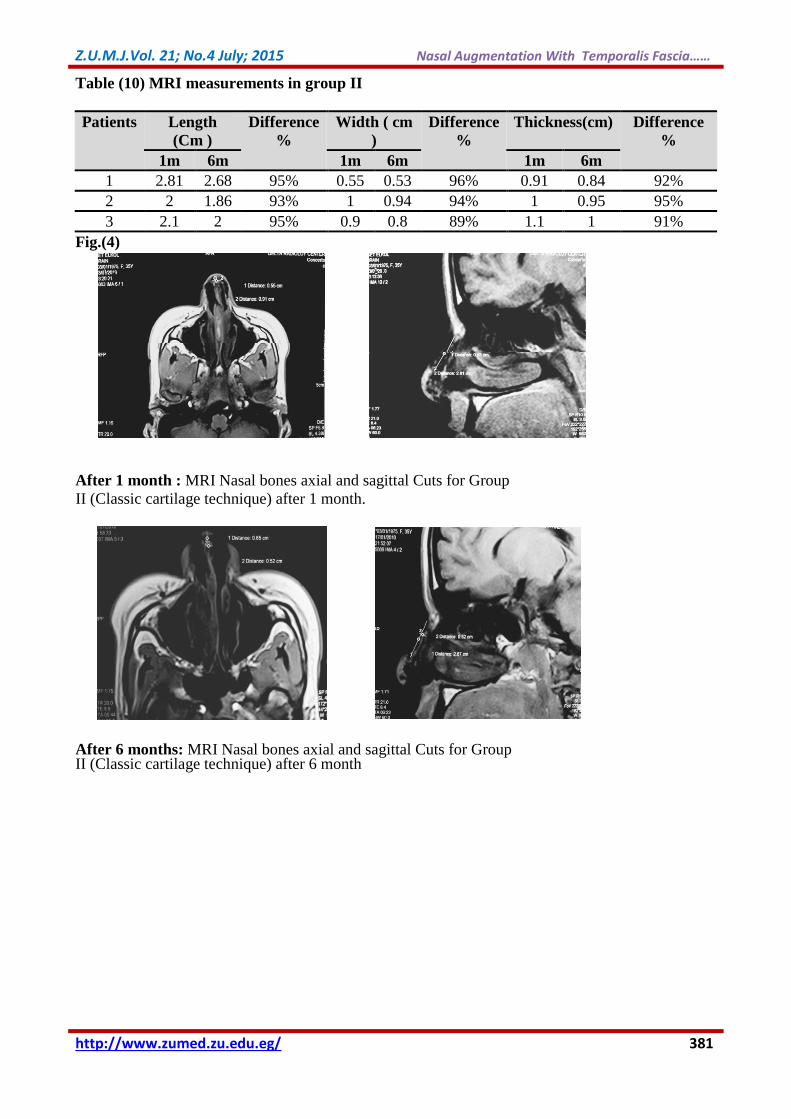
MRI measurements in group II. Table(10),Fig.(4).
The cartilage grafts maintained approximately 90% to 95% of their dimensions in MRI.
Z.U.M.J.Vol. 21; No.4 July; 2015 Nasal Augmentation With Temporalis Fascia……
http://www.zumed.zu.edu.eg/ 381
Table (10) MRI measurements in group II
Difference
%
Thickness(cm) Difference
%
Width ( cm
)
Difference
%
Length
(Cm )
Patients
6m 1m 6m 1m 6m 1m
92% 0.84 0.91 96% 0.53 0.55 95% 2.68 2.81 1
95% 0.95 1 94% 0.94 1 93% 1.86 2 2
91% 1 1.1 89% 0.8 0.9 95% 2 2.1 3
Fig.(4)
After 1 month : MRI Nasal bones axial and sagittal Cuts for Group
II (Classic cartilage technique) after 1 month.
After 6 months: MRI Nasal bones axial and sagittal Cuts for Group II (Classic cartilage technique) after 6 month
Pre-operative Post-operative (1month) Post-operative (6 month)
Pre-operative Post-operative (1month) Post-operative (6 month)
Pre-operative Post-operative (1month) Post-operative (6 month)

Z.U.M.J.Vol. 21; No.4 July; 2015 Nasal Augmentation With Temporalis Fascia……
http://www.zumed.zu.edu.eg/ 382
Aesthetic results:
A- Group I (DCFG):Fig.(5)
Group II (Classic Cartilage Graft Group):Fig.(6)
Pre-operative Post-operative (1month) Post-operative (6 month)
Pre-operative Post-operative (1month) Post-operative (6 month)
Pre-operative Post-operative (1month) Post-operative (6 month)
Pre-operative Post-operative (1month) Post-operative (6 month)
Pre-operative Post-operative (1month) Post-operative (6 month)
Pre-operative Post-operative (1month) Post-operative (6 month)

Z.U.M.J.Vol. 21; No.4 July; 2015 Nasal Augmentation With Temporalis Fascia……
http://www.zumed.zu.edu.eg/ 383
Table (11) The complications of both groups postoperatively
p-value Z test Group I I Group I Type of complication
<0.05 1.8 0 3 (20) Haematoma at Temporal Region
n.(%)
-- -- 0 0 Pneumo Thorax
n.(%)
>0.05 1.1 1(6.7) 3 (20) Post – operative infection
n.(%)
>0.05 1.1 1(6.7) 3 (20) Need for revision surgery
n.(%)
DISCUSSION Nasal augmentation presents a
significant challenge to the facial plastic
surgeon. The dual goals of nasal
augmentation were reestablishment of the
desired aesthetic nasal contour and restoration
of respiratory function (11)
.
Grafts used for reconstruction of
congenital deformities, traumatic saddle nose
deformities and secondary surgical defects
included different
augmentation materials, from autogenous
cartilage and bone to alloplastic materials (3)
.
Many techniques have been described
to avoid post-rhinoplasty dorsal irregularities.
Among them the use of diced cartilage grafts
wrapped with surgicel or fascia (6)
.
In 2000, EROL'S had described
(Turkish Delight) which is a modified
technique for using diced cartilage after its
wrapping in surgicel to create a moldable
cartilage graft for dorsal nasal augmentation
(7).
A series of clinical failures were noted
after the use of surgicel wrapped diced
cartilage, hypothesis for this failures was that
surgicel incited a foreign body reaction
leading to graft inflammation and subsequent
cartilage absorption (8)
.
In 2006, Clavert had proved that the
use of fascia wrapped diced cartilage is more
superior to surgicel wrapped diced cartilage
being an autologous tissue and more
histologically stable (10)
.
So, in this thesis, we aimed to
compare the effect of dorsal augmentation by
temporalis fascia-wrapped diced cartilage
versus dorsal augmentation by classic
cartilage technique. We used temporalis
fascia -wrapped diced cartilage grafts in 15
patients included in group I and we used
classic cartilage grafts in 15 patients included
in group II.
In group I, we observed that diced
grafts were 0.5 to 1.0 mm in size and wrapped
in temporalis fascia when placed beneath thin
skin. So the grafts were smooth and pliable
graft without visibility..
The temporalis fascia-wrapped diced
cartilage grafts offer more protection from
excessive graft mobility and shearing.
It allowed us to use all cartilage
fragments within the fascia grafts, which was
highly relevant in cases of insufficient septal
cartilage.
And also placement of the graft is achieved
by closed rhinoplasty (endonasal approach)
which is a simple surgical technique, more
over due to the time needed for
complete solidification of DC-F graft, it is
easily to be manipulated for one week post-
operatively.
In patients of group II, we used
conchal or costal cartilages in cases with
insufficient septal cartilage due to previous
septoplasty or rhinoplasty. We found that
conchal cartilage must be either scored or
specifically contoured to control its inherent
curves. Also, costal cartilage imposed a
thoracic scar on patients.
The edges of the graft had to be
shaved to bend with adjacent structures to
prevent sharp edges and visibility especially
in thin skinned patients. Then the graft had to
be stabilized by multiple sutures. Needle
fixation like k-wire were required for precise
positioning of the graft especially when using
rib grafts to prevent warpping. All of the
above added to the difficulties in classic

Z.U.M.J.Vol. 21; No.4 July; 2015 Nasal Augmentation With Temporalis Fascia……
http://www.zumed.zu.edu.eg/ 384
cartilage technique with more possibility of
warpping and visibility under the skin.
Comparison between the two groups
was assessed by the aesthetic results in
patients’ photography and the rate of
resorption of cartilage in each technique. As
regards the effect of augmentation materials
on the aesthetic results of patients’
photography: We measured the nasofrontal
angle and the nasofacial angle pre-operative
and post-operative after 6 months in the
lateral view of patients’ photography.
We found that dorsal augmentation
with temporalis fascia-wrapped diced
cartilage grafts had a statistical significant
effect on these mentioned angles improving
them towards the normal range in the same
manner as classic cartilage grafts. Concerning
these mentioned angles the difference
between patients in both groups was not
statistically significant.
In our photographic results, we
measured the dorsal projection at rhinion and
the mid dorsal projection (mid dorsal nasal
height) pre-operative and post-operative after
6 months in the lateral view of patients’
photography. Our goal was to assess the
effect of dorsal nasal augmentation with
fascia-wrapped diced cartilage grafts in
improving the aesthetic ratios in
correspondence to the nasal length.
Our results showed that temporalis
fascia-wrapped diced cartilage grafts had
altered both dorsal projection at rhinion and
the mid dorsal projection towards ideal
aesthetic ratios in correspondence to nasal
length and results were statistically
significant.
We compared the results of temporalis
fascia-wrapped diced cartilage grafts to the
results of classic cartilage grafts in improving
both dorsal projection at rhinion and the mid
dorsal projection which were not statistically
significant. That indicated the ability of
fascia-wrapped diced cartilage grafts to
augment large dorsal defects without
harvesting additional material as rib grafts so
they helped in preventing donor-site
morbidity.
As regards the radix projection, we
tried to see the effect fascia-wrapped diced
cartilage grafts in improving the radix/nasal
length ratio especially in patients with full
length dorsal grafts or partial length dorsal
grafts reaching the radix area. Our results
showed that fascia-wrapped diced cartilage
grafts improved the radix/nasal length ratio in
patients who needed radix augmentation in
the same manner as classic cartilage grafts.
Yet the results were statistically not
significant as not all patients needed radix
augmentation.
Our results showed that dorsal
augmentation with both grafts had a
statistically significant effect on tip
projection. Both grafts improved the aesthetic
proportion between the nasal length and the
tip projection.
Concerning the aesthetic results, we
found that temporalis fascia-wrapped diced
cartilage grafts improved patients’ aesthetic
profiles giving them a very smooth contour
plus preventing the visibility of the grafts on
the dorsum in a better manner than classic
cartilage grafts.
Our photographic results matched
those of Daniel (2006) (9)
. who used
temporalis fascia-wrapped diced cartilage in
546 patients over a period of 2 years
(2006&2007). Daniel Found that placement
of fascia wrapped diced cartilage under the
dorsal nasal skin was extremely satisfactory
in obtaining a smooth, straight, and proper
profile.
Regarding the rate of resorption of the
cartilage in our thesis, post operative MRI
results showed that fascia-wrapped diced
cartilage grafts maintained approximately
85% to 90% of their dimensions while classic
cartilage grafts maintained approximately
90% to 95% of their dimensions. Such finding
indicated that the degree of resorption is
higher in tempralis fascia -wrapped diced
grafts than classic cartilage graft. But this was
not a problem with our aesthetic results as we
overcorrected the volume by 20% to
compensate for this percentage of resorption.
Daniel and Calvert (2004) (8)
.studied
the Erol’s technique in a series of patients
without success. They sought to devise a wrap
envelope that could prevent cartilage graft
absorption, using autologous deep
temporoparietal fascia wrappers instead of
Surgicel.

Z.U.M.J.Vol. 21; No.4 July; 2015 Nasal Augmentation With Temporalis Fascia……
http://www.zumed.zu.edu.eg/ 385
They explained their findings on the
bases of surgicel-wrapped diced cartilage
grafts initially survived; then ultimate graft
absorption ensued by 6 months. They also
found that the hypothesis for this clinical
failure was that surgicel incited a foreign
body reaction, ultimately leading to graft
inflammation and subsequent cartilage
absorption.
In our results, we found that diced
cartilages wrapped in temporalis fascia grafts
didn’t show complete resorption after 6
months as they maintained the aesthetic
patients’ photographic profiles after 6 months
and also our MRI results show only 10-15%
of resorption after 6 months.
From our results, we found that
overcorrection of approximately 20% for
dorsal nasal augmentation would help to
overcome long term resorption of Surgicel
diced cartilage graft.
Also the nasal pocket should be larger
in volume than the graft to prevent distortion
during introduction into the nasal dorsum.
This allowed accurate graft placement, which
was essential for achieving good final results.
External molding of the graft and
fixation with tape applied to the nasal dorsum
were essential parts of the procedure, to
reinforce the position of the graft.
The principal advantages of the
temporalis fascia- wrapped diced cartilage
technique are the following: improving both
contour and volume of the dorsum, provides a
smooth and pliable graft without visibility,
avoidance of warping or distortion, wide
flexibility to correct irregular dorsal defects,
potential for external molding of the graft to
achieve the desired shape and maintenance of
dorsal augmentation without significant
resorption.
Regarding postoperative
complications, we reported hematoma of
temporal region in 3 cases(20%) of group I,
no pneumo thorax in both groups, but
postoperative infection in (20%) of group I a
and (6.7%) in group II and lastly need for
revision in 3 cases(20%) of group I and
(6.7%) in group II.
Conclusion: Use of diced cartilage grafts in
rhinoplasty surgery has recently undergone a
dramatic resurgence. However, it was Erol’s
“Turkish delight” modification of diced
cartilage grafting for dorsal nasal
augmentation that has recently popularized its
use.
Our work aimed to assess the usage
of Temporalis fascia-wrapped diced cartilage
in augmentation of the nose versus classic
cartilage technique and evaluate the efficiency
of this new technique versus the classic one.
We found that dorsal augmentation
with both grafts had a statistically significant
effect on nasofrontal angle, nasofacial angle,
dorsal projection at rhinion, mid dorsal
projection and tip projection by changing
their measurements toward the normal range;
yet concerning these latter mentioned
parameters the difference between the first
(Temporalis fascia -wrapped diced cartilage)
and the second group (Classic cartilage
technique) was not statistically significant.
According to the degree of resorption,
Temporalis fascia- wrapped diced cartilage
grafts maintained approximately 85% to 90%
of their dimensions in MRI. While the Classic
cartilage technique grafts maintained
approximately 90% to 95% of their
dimensions in MRI. This suggested that the
degree of resorption was present in both types
of grafts, but it was higher in Temporalis
fascia- wrapped diced cartilage grafts but the
grafts maintained approximately most of its
thickness after 6 months.
At the end we conclude that the
advantages of Temporalis fascia- wrapped
diced cartilage graft technique ‘Turkish
Delight’ are:
It is ease of preparation
Increase the volume of graft available for
use
Avoidance of contour irregularities
This technique shortens the operative time,
avoids donor site morbidity and avoid
open rhinoplasty which is time
consuming.
This technique is effective for dorsal nasal
augmentation and is good and effective
when closed rhinoplasty is chosen as the
surgical method.
This technique decrease the need for
revision rhinoplasty.

Z.U.M.J.Vol. 21; No.4 July; 2015 Nasal Augmentation With Temporalis Fascia……
http://www.zumed.zu.edu.eg/ 386
REFERENCES (1)Kim DW and Mau T (2006): Surgical anatomy
of the nose. In : Bailey B, Johnson J,
Newlands S, editors. 4th edition, Head and
Neck surgery - otolaryngology, vol.2.
Philadelphia: Lippincott Williams & Wilkins,
2006. p 2511-32.
(2)Davis R and Wayne I (2004): Rhinoplasty and
the nasal SMAS augmentation graft:
advantages and indications. Arch Facial Plast
Surg; 6(2): 124- 132.
(3)Parker J (2000): Grafts in rhinoplasty.
Alloplastic vs. autogenous. Arch Otolaryngol
Head Neck Surg; 126: 558-561
(4)Elahi M, Jackson I, Moreira-Gonzalez A, et al.
(2003): Nasal augmentation with Surgicel-
wrapped diced cartilage: a review of 67
consecutive cases. Plastic & Reconstructive
Surgery; 111(3): 1309-1321.
(5)Harel M and Margulis A (2013): Dorsal
augmentation with diced cartilage enclosed
with temporal fascia in secondary endonasal
rhinoplasty. Aesthetic Surg J.; 33(6):809-
816.
(6)Calvert J, Brenner K, Dacosta-Iyer M, Evans G
and Daniel R (2006): Histological analysis of
human diced cartilage grafts. Plast. Reconstr.
Surg. 118:230-236.
(7)Erol O (2000): The Turkish Delight: A pliable
graft for rhinoplasty. Plastic &
Reconstructive Surgery; 105: 2229-2243.
Bioplastique: Specific technical advice on its
use and possible complications. Aesthetic
Plast Surg; 16: 67-68.
(8)Daniel R and Calvert J (2004): Diced cartilage
grafts in rhinoplasty surgery. Plast. Reconstr.
Surg. 113: 2156-2171.
(9)Daniel RK (2006): The role of diced cartilage
grafts in rhinoplasty. Plast Reconstr Aesthet
Surg, 26: 209-13.
(10)Brenner K, McConnell M, Evans G and
Calvert J (2006): Survival of Diced Cartilage
Grafts: An Experimental Study. Plast.
Reconstr. Surg. 117: 105.
(11)Bateman N and Jones N (2000): Retrospective
review of augmentation rhinoplasties using
autologous cartilage grafts. Journal of
Laryngology .