nasolabial cyst
DESCRIPTION
thtTRANSCRIPT

121
Case Report
Nasolabial bilateral cyst as cause of the nasal obstruction: Case report andliterature review
Cisto nasolabial bilateral como causa de obstrução nasal: Relato de caso e revisão deliteratura
Alexandre Minoru Enoki 1, Gilberto Ulson Pizarro 2, Marcelo de Sampaio Morais 3, Danilo Pereira Pimentel Fernandes 4,Paulo Roberto Grimaldi Oliveira 5.
1) Fellow in Pharyngology at Clínicas Hospital of Medicine College at São Paulo University. Otorhinolaryngologist.2) PhD in Otorhinolaryngology by Paulista School of Medicine. Preceptor of medical residency in Otorhinolaryngology at Paulista Hospital, São Paulo - Brazil.3) Fellow in Plastic Surgery of the Face at Clínicas Hospital of Medicine College of São Paulo University. Otorhinolaryngologist.4) Otorhinolaryngologist.5) Master in Pathological Anatomy. Pathologist
Institution: Paulista Hospital of Otorhinolaryngology.São Paulo / SP - Brazil.
Mailing address: Alexandre Minoru Enoki - 369 Jaú, Alameda - Apto 610 - Jardim Paulista - São Paulo / SP - Brazil – Zip-code: 01420-000 - Telephones: (+55 11) 3262-2328, (+55 11) 7159-2131 - E-mail: [email protected]
Article received in 2009 November 26th. Article approved in 2010 April 25th.
RESUMO
Introdução: O cisto nasolabial é uma doença rara, normal-
mente unilateral, benigna, de origem embrionária, localizada
em partes moles da região do sulco nasolabial e asa nasal. O
diagnóstico é essencialmente clínico, levando em considera-
ção a topografia do cisto, que geralmente é assintomático.
Objetivo: Este artigo tem como objetivo principal à descrição
de um caso incomum de cisto nasolabial bilateral com obs-
trução nasal, seu tratamento, aspectos anatomopatológicos e
acompanhamento, além de revisão de literatura.
Relato do Caso: Paciente do sexo feminino, parda, 24 anos
de idade, apresentando abaulamento em região nasolabial e
obstrução nasal. Exames físico e complementares compatí-
veis com cisto nasolabial. Indicado tratamento cirúrgico para
exérese da lesão.
Considerações Finais: O cisto nasolabial bilateral, apesar de
raro, é uma possível causa de obstrução nasal, com boa res-
posta à terapia cirúrgica.
Palavras-chave: cistos não-odontogênicos, obstrução nasal,
cistos.
SUMMARY
Introduction: The nasolabial cyst is a rare disease, usually
unilateral, benign, of embryonic origin, located in soft parts
from the nasolabial folds and nasal wings. The diagnosis is
essentially clinic, take into consideration the cyst topography,
that is usually asymptomatic.
Objective: This article has as main goal the description of a
unusual case of nasolabial bilateral cyst with nasal obstruction,
its treatment, anatomic pathological and accompaniment,
besides the literature review.
Case Report: Female patient, brown, 24 years old, showing
bulging in nasolabial region and nasal obstruction. Physical
and complementary exams with nasolabial cyst. Indicate surgical
treatment of excision of the lesion.
Final Considerations: The nasolabial bilateral cyst, although
is rare, is a possible cause for the nasal obstruction, with good
response to surgical therapy.
Keywords: not odontogenic cyst, nasal obstruction, cysts.
Intl. Arch. Otorhinolaryngol., São Paulo - Brazil, v.16, n.1, p. 121-125, Jan/Feb/March - 2012.
122
INTRODUCTION
The nasoalveolar cyst is a rare benign lesion, located
in topography of nasolabial folds, anteroinferior of the
piriform rim of nasal cavity. It´s a lesion that is usually
unilateral (90% of the cases) (1,2), affecting mainly people
of black race, of feminine gender, in the age group which
comprises between the 4th and 5th decades of life (1,2).
The first to describe this pathology was ZUCKERKANDL
in 1882 (3). This embryonic, non-odontogenic, usually
asymptomatic, being diagnosed late, due to the facial
aesthetics changes and breathing. Although diverse
synonymy (nasoalveolar cyst, KLESTADT cyst, congenital
mucoid cyst of the nasal edge), the term considered most
appropriate, at the moment, is nasolabial cyst (4).
Besides literature review, this study has as objective,
report the case of a patient with bilateral nasoalveolar cyst,
presenting clinical aspects, surgical, histopathological and
radiographic.
CASE REPORT
Patient AWV, feminine gender, 24 year-old, mulatto,
coming and natural of São Paulo, housewife, sought for
treatment at Otorhinolaringoloy Paulista Hospital, SP, Brazil,
complaining about bulge region of bilateral nasolabial, of
progressive evolution for about 06 months, associated to
the nasal obstruction in the last 02 months. Patient denied
pain, rhinorrhea, nasal itching, sternutatory, epistaxis or
other nasal complaints.
At the otorhinolaryngological examination, it was
observed bulging without signs of inflammation at bilateral
nasolabial region and superior gingivolabial sulcus, especially
on the right, raising the nasal floor and erasing the bilateral
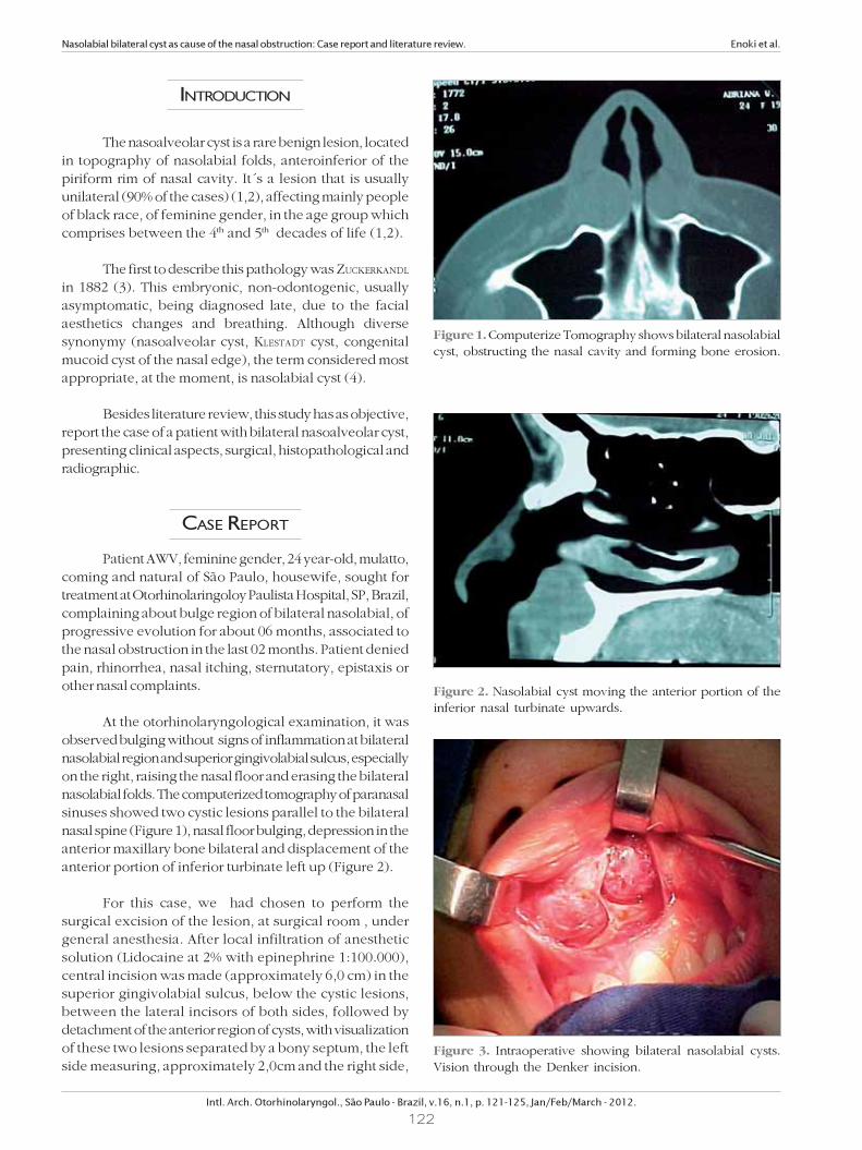
nasolabial folds. The computerized tomography of paranasal
sinuses showed two cystic lesions parallel to the bilateral
nasal spine (Figure 1), nasal floor bulging, depression in the
anterior maxillary bone bilateral and displacement of the
anterior portion of inferior turbinate left up (Figure 2).
For this case, we had chosen to perform the
surgical excision of the lesion, at surgical room , under
general anesthesia. After local infiltration of anesthetic
solution (Lidocaine at 2% with epinephrine 1:100.000),
central incision was made (approximately 6,0 cm) in the
superior gingivolabial sulcus, below the cystic lesions,
between the lateral incisors of both sides, followed by
detachment of the anterior region of cysts, with visualization
of these two lesions separated by a bony septum, the left
side measuring, approximately 2,0cm and the right side,
Figure 1. Computerize Tomography shows bilateral nasolabial
cyst, obstructing the nasal cavity and forming bone erosion.
Nasolabial bilateral cyst as cause of the nasal obstruction: Case report and literature review. Enoki et al.
Intl. Arch. Otorhinolaryngol., São Paulo - Brazil, v.16, n.1, p. 121-125, Jan/Feb/March - 2012.
Figure 2. Nasolabial cyst moving the anterior portion of the
inferior nasal turbinate upwards.
Figure 3. Intraoperative showing bilateral nasolabial cysts.
Vision through the Denker incision.

123
around 3,0cm (Figure 3). It was performed a dissection of
the right cyst, initially, preserving its contents, being
possible the detachment of the lateral walls. It was
located the nasal floor plane (nasal mucosa) at the
superior region of the cyst; at this point, for better
detachment, it was chosen by emptying the contents
serous, with yellowish color, of the cyst by needle and
syringe. After emptying the cyst, it was performed a
careful dissection in its upper portion, which kept contact
with nasal mucosa, region where it presented greater
adherence. The displacement was performed without
lesion of the nasal mucosa. At the back region of the cyst,
the displacement showed planes less adhered, facilitating
the removal. The same surgical technique was performed
at the left side of the cyst, being possible to indentify the
nasal floor mucosa and perform the removal without
lesion (Figure 4). Synthesis was performed at plans
dissected, with absorbable lines (Catgut 2-0, simple).
At immediate postoperative, patient complained of
paresthesia at anterior region of the upper lip and nasal
vestibule, which remained for 03 months.
Patient is at ambulatory accompaniment of
postoperative for 06 months, without evidences of
recurrence or other changes, presenting improvement of
nasal obstruction.
Anatomopathology
It was sent for histological study, two cystic structures
previously sectioned, conserved in formaldehyde, and
represented wall studs, smooth and bright, with shades of
brown color. The larger structure measured 3 x 2,5 x 1 cm
and the smallest, 2,5 x 1,5 x 0,5cm.
The sample was submitted to the processing
chemical pradronized, obtaining a block of paraffin to each
cystic structures, being made the respective histological
concoction, with thickness of 5 (five) micra and color by
the technique of hematoxylin-eosin and PAS (Schiff periodic
acid).
The histological study revealed identical aspect in
both lesions, being identified of cystic wall constituted by
loose connective tissue showing moderate edema, covered
by two different types of epithelium: the predominant
Nasolabial bilateral cyst as cause of the nasal obstruction: Case report and literature review. Enoki et al.
Intl. Arch. Otorhinolaryngol., São Paulo - Brazil, v.16, n.1, p. 121-125, Jan/Feb/March - 2012.
Figure 4. The operative field after removal of nasolabial cysts,
presenting bone erosion.
Figure 5. 400X – nasolabial cyst - hematoxylin-eosin. Observe
the pluri squamous epithelium formed by the poligonal cells,
abundant cytoplasm and acidophilic around the round
nuclei, isochromatic. Superficially, single layer of cylindrical
cells showing vacuolated and clear cytoplasm.
Figure 6. 400X – nasolabial cyst - PAS. Note accumulation of
PAS-positive granules in the cytoplasm of the cell located in
the center of the microscopic field.
124
was of the type stratified squamous, with preservation of
the polarity and absence of nuclear atypia, removing
away the suspicion of malignancy.
The other type of epithelium was constituted by
one or two layers of cylindrical cells sometimes massive
clear vacuoles in the cytoplasm (Figure 5), that special
color by the technique of PAS revealed weak positivity to
the mucopolysaccharide substances (Figure 6). At the
connective tissue wall, the histological sections revealed
nerve fibers and blood capillaries of ecstatic lights,
alongside to the moderate interstitial edema. Some muscle
striated fibers was also indentified at the region of surgical
region.
The anatomopathological diagnosis was of bilateral
nasolabial cyst.
DISCUSSION
The nasoalveolar cyst is an embryonic cyst, non-
odontogenic, which has its controversial origin , being the
theories based on:
1) cyst originated from the invagination of ectodermal
debris among the processes nasal side and media being
for that reason, considered as fissural cyst (Klestadt
Theory, 1913) (1,5,6);
2) cyst derived from the epithelium of the nasolacrimal
duct during the embryonic period (Bruggemann Theory,
1920) (5,7).
For reason of its poor symptomatology, this disease
is underdiagnosed (8), showing in the literature, an
incidence of 0,7% of all maxillofacial cysts and 2,5% of non-
odontogenic cysts. In the presented case, we observed a
mulatto patient, even being more common at black race,
according to the literature. Epidemiological data shows that
this cyst is more frequent in persons of feminine gender,
in the proportion of 4:1 (when unilateral) and 5,5:1 (when
bilateral) (9). The age group most affected includes the 4th
an 5th life decades (2,3,7). The cysts presentation is most
unilateral (90%), being only10% bilateral (1, 2, 8).
Clinically, this lesion presents as a bulging of slow
growing located at the portion ventral inferior of piriform
fossa region. In the course of time, the cyst leads to a facial
deformity with a deletion of nasolabial folds, nasal obstruction
by the elevation of nasal floor and superior displacement
of the anterior portion of the inferior turbinate. It is worth
to highlight that the dentition remains intact (6). Eventually,
it may occur cyst infection (50% of the cases), presenting
signs of inflammation. In this infection cases, it can occur
the cyst drainage to the oral cavity and /or to the nasal
vestibule (3,6).
The diagnosis of nasoalveolar cyst is clinical and
topographic, with visual and palpation of the affected
area (6). The workup done by the imaging examination
confirms the suspicion and the clinical examination,
being the Computerized Tomography the examination
of choice, which may show, in some cases, jawbone
erosion (10,11, 12). The nasolabial cyst consists of lesion
in the soft parts, and for this reason, the x-ray being
considered an obsolete examination, capable to show
few details; except in the cases in which the cyst
presents gigantic dimensions leading to the significant
erosion of the jawbone.
The differential diagnosis which must be done with
the nasolabial cyst include the dermoid , nasopalatine,
median palatal, median alveolar, globulomaxilar cysts
(which origin in the interior of the bone), besides of
furuncles at nasal floor, which resembles to infected nasolabial
cyst (13).
in the literature, it has been reported Just one case
(Arnold, 1929) of nasolabial cyst which evolves to carcino-
ma (9,14).
Although there are reports of treatment of nasolabial
cyst by sclerosing substances or marsupialization (15), the
most indicated therapy found in the literature is surgical
removal. Enucleation can be performed with local anesthesia
or general, being the best way to access the Denker
incision ( intra-oral incision, sublabial next to the incisive
fossa) which offers an ample exposition. During the
surgery, should take in account the cyst intimate adherence
with the nasal floor (3,16), detail that, constantly, leads to
the laceration of the mucosa in this region of the nose. This
was possible to avoid in this case, in which we had chosen
to empty the cystic contents to obtain a better dissection
of the cyst in relation to this region of nasal floor. The
closing of the planes should be complete, in order to avoid
possible oronasal fistulas. By the reason the extension of
the cysts related did not affected the region of nasal wing,
it was not necessary apply any technique to avoid retraction.
The surgery aims the facial esthetic restoration, the nasal
function (in case it is affected) and the prevention of
recurrent infections, which can be associated, and minimize
the patient anxiety
The surgical treatment present few complications,
among them may recede with deformity of the nasal ala,
mainly in Blacks and still, oronasal fistula. Recurrence of the
cyst is rare and the prognosis is very good (5,7).
The description of the surgical technique in this
case, aims to provide a foundation to help similar cases in
order to obtain surgical success, without submit the patient
to complications and disease recurrence.
Nasolabial bilateral cyst as cause of the nasal obstruction: Case report and literature review. Enoki et al.
Intl. Arch. Otorhinolaryngol., São Paulo - Brazil, v.16, n.1, p. 121-125, Jan/Feb/March - 2012.

125
BIBLIOGRAPHICAL REFERENCES
1. Barzilai M. Bilateral nasoalveolar cysts: case report. Clin
Radiol. 1994, 49(2):140-141.
2. Cohen MA, Hertzanu Y. Huge growth potential of the
nasolabial cyst. Oral Surg. 1985, 59:441-5.
3. Allard RHB. Nasolabial cyst. A review of the literature and
report of cases. Int J Oral Surg. 1982, 11:351-359.
4. Fanibunda KB. Bilateral nasolabial cysts: a case report.
Dent Pract. 1970, 20:249-250.
5. Graamans K. Nasolabial cysts: diagnosis mainly based on
topography?. Rhinology. 1983, 21:239-249.
6. Kuriloff DB. The nasolabial cyst-nasal hamartoma.
Otolaryngol Head Neck Surg. 1987, 96:268-272.
7. David VC, O´Connell JE. Nasolabial cyst. Clin Otolaryngol.
1986, 11:5-8.
8. Smith RA, Katibah RN, Merrell P. Nasolabial cyst: report
of a case. J Canad Dent Assoc. 1982, 11:727-729.
9. Roed-Peterson B. Nasolabial cysts: a presentation of five
patients with a review of literature. Br J Oral Surg. 1969,
7:84-95.
10. Adams A. Roentgeno-Oddities. Oral Surg. 1985, 60:118-
119.
11. Balfour RS. Nasoalveolar cyst. J MD State Dent Assoc.
1977, 20:92-94.
12. Seward GR. Nasolabial cysts and their radiology. Dent
Pract. 1962, 12:154-161.
13. Karmody CS, Gallagher JC. Nasoalveolar cysts. Ann Otol.
1972, 81:278-283.
14. Egervary G, Csiba A. Bilateral nasolabial cyst. Dental
Digest. 1969, 75:504-7.
15. Crowford W, Korchin L, Greskovich FJ. Nasolabial cysts:
report of two cases. J Oral Surg. 1968, 26:582-588.
16. Brandão GS, Ebling H, Souza IF. Bilateral nasolabial cyst.
Oral Surg. 1974, 37:480-484.
Nasolabial bilateral cyst as cause of the nasal obstruction: Case report and literature review. Enoki et al.
Intl. Arch. Otorhinolaryngol., São Paulo - Brazil, v.16, n.1, p. 121-125, Jan/Feb/March - 2012.