natural airway management - midmeds · natural airway management ... to facilitate fibre-optically...
TRANSCRIPT
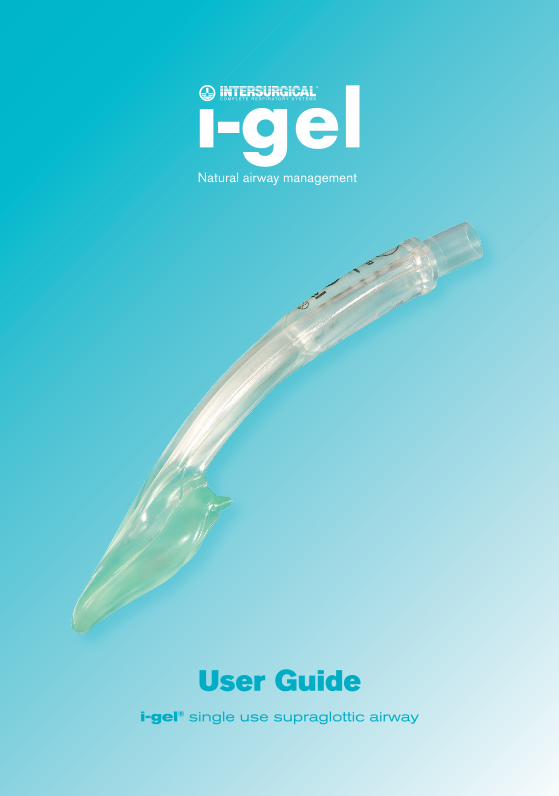
User Guidei-gel® single use supraglottic airway
Natural airway management
i-gelcover_guide_issue 2.indd 2 15/12/2006 13:33:29
i-gel
1.0 Introduction.........................................................................................................1
1.1 The i-gel design...............................................................................................................1
1.2 Key components and their function.................................................................................2
1.2.1Softnon-inflatablecuff............................................................................................3
1.2.2Gastricchannel.......................................................................................................3
1.2.3Epiglottisblocker.....................................................................................................3
1.2.4Buccalcavitystabiliser............................................................................................3
1.2.5 15mm connector......................................................................................................4
1.2.6Importantkeypoints...............................................................................................4
2.0 Indications...........................................................................................................5
3.0 Contraindications................................................................................................6
4.0 Warnings..............................................................................................................6
5.0 Preparationforuse..............................................................................................7
5.1Sizeselection..................................................................................................................7
5.2Pre-use checks...............................................................................................................7
5.3Pre-insertion preparation................................................................................................8
6.0 InductionofAnaesthesia....................................................................................9
6.1 Preferred technique.........................................................................................................9
6.2 Other techniques of induction.........................................................................................9
7.0 Insertion technique...........................................................................................10
7.1Recommendedinsertiontechnique..............................................................................10
7.2Importantnotestotherecommendedinsertiontechnique...........................................11
8.0 MaintenanceofAnaesthesia.............................................................................12
9.0 EmergencefromAnaesthesia...........................................................................12
10.0 RecoveryphaseofAnaesthesiaandi-gelremoval.......................................12
11.0 Howtousethegastricchannel........................................................................13
12.0 Problemsolving.................................................................................................14
13.0 Adverseoutcomes.............................................................................................15
14.0 Furtherreading..................................................................................................16
Contents
i-gelcover_guide_issue 2.indd 3 15/12/2006 13:33:29
i-gel �
�.0
�.� The i-gel design
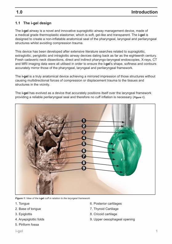
The i-gel airway is a novel and innovative supraglottic airway management device, made of a medical grade thermoplastic elastomer, which is soft, gel-like and transparent. The i-gel is designed to create a non-inflatable anatomical seal of the pharyngeal, laryngeal and perilaryngeal structures whilst avoiding compression trauma.
This device has been developed after extensive literature searches related to supraglottic, extraglottic, periglottic and intraglottic airway devices dating back as far as the eighteenth century. Fresh cadaveric neck dissections, direct and indirect pharyngo-laryngeal endoscopies, X-rays, CT and MRI imaging data were all utilised in order to ensure the i-gel’s shape, softness and contours accurately mirror those of the pharyngeal, laryngeal and perilaryngeal framework.
The i-gel is a truly anatomical device achieving a mirrored impression of those structures without causing multidirectional forces of compression or displacement trauma to the tissues and structures in the vicinity.
The i-gel has evolved as a device that accurately positions itself over the laryngeal framework providing a reliable perilaryngeal seal and therefore no cuff inflation is necessary (Figure 1).
Figure 1: View of the i-gel cuff in relation to the laryngeal framework
1. Tongue2. Base of tongue3. Epiglottis4. Aryepiglottic folds5. Piriform fossa
6. Posterior cartilages7. Thyroid Cartilage8. Cricoid cartilage9. Upper oesophageal opening
Introduction
12
345
67
89
igel user guide_1_081206.indd 1 15/12/2006 11:50:24
i-gel �
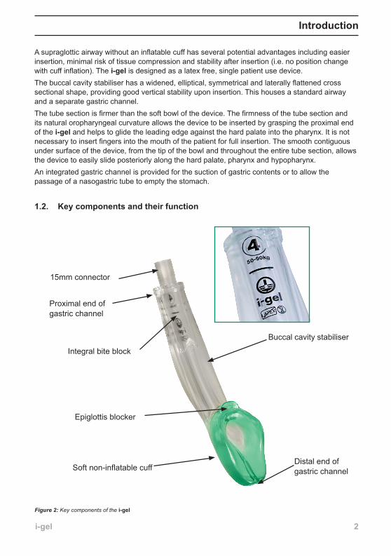
A supraglottic airway without an inflatable cuff has several potential advantages including easier insertion, minimal risk of tissue compression and stability after insertion (i.e. no position change with cuff inflation). The i-gel is designed as a latex free, single patient use device.The buccal cavity stabiliser has a widened, elliptical, symmetrical and laterally flattened cross sectional shape, providing good vertical stability upon insertion. This houses a standard airway and a separate gastric channel. The tube section is firmer than the soft bowl of the device. The firmness of the tube section and its natural oropharyngeal curvature allows the device to be inserted by grasping the proximal end of the i-gel and helps to glide the leading edge against the hard palate into the pharynx. It is not necessary to insert fingers into the mouth of the patient for full insertion. The smooth contiguous under surface of the device, from the tip of the bowl and throughout the entire tube section, allows the device to easily slide posteriorly along the hard palate, pharynx and hypopharynx. An integrated gastric channel is provided for the suction of gastric contents or to allow the passage of a nasogastric tube to empty the stomach.
�.�. Key components and their function
Introduction
Integral bite block
Distal end of gastric channelSoft non-inflatable cuff
Buccal cavity stabiliser
Epiglottis blocker
15mm connector
Proximal end of gastric channel
Figure 2: Key components of the i-gel
igel user guide_1_081206.indd 2 15/12/2006 11:50:39
i-gel �
Introduction
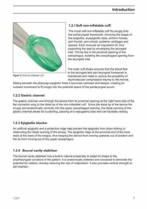
1.2.1Softnon-inflatablecuff:
The novel soft non-inflatable cuff fits snugly onto the perilaryngeal framework, mirroring the shape of the epiglottis, aryepiglottic folds, piriform fossae, peri-thyroid, peri-cricoid, posterior cartilages and spaces. Each receives an impression fit, thus supporting the seal by enveloping the laryngeal inlet. The tip lies in the proximal opening of the oesophagus, isolating the oesophageal opening from the laryngeal inlet.
The outer cuff shape ensures that the blood flow to the laryngeal and peri-laryngeal framework is maintained and helps to reduce the possibility of neurovascular compression trauma to the nerves.
Sliding beneath the pharyngo-epiglottic folds it becomes narrower and deeper, creating an outward movement to fit snugly into the potential space of the perilaryngeal pouch.
�.�.� Gastric channel
The gastric channel runs through the device from its proximal opening at the right hand side of the flat connector wing to the distal tip of the non-inflatable cuff. Since the distal tip of the device fits snugly and anatomically correctly into the upper oesophageal opening, the distal opening of the gastric channel allows for suctioning, passing of a nasogastric tube and can facilitate venting.
1.2.3Epiglottisblocker
An artificial epiglottis and a protective ridge help prevent the epiglottis from down-folding or obstructing the distal opening of the airway. The epiglottic ridge at the proximal end of the bowl rests at the base of the tongue, thus keeping the device from moving upwards out of position and the tip from moving out of the upper oesophagus.
1.2.4 Buccalcavitystabiliser
The buccal cavity stabiliser has a built-in natural propensity to adapt its shape to the oropharyngeal curvature of the patient. It is anatomically widened and concaved to eliminate the potential for rotation, thereby reducing the risk of malposition. It also provides vertical strength to aid insertion.
Figure 3: Soft non-inflatable cuff
igel user guide_1_081206.indd 3 15/12/2006 11:50:42
i-gel �
�.�.5 �5mm connector
The innovative connector serves a number of functions;
To provide a standard 15mm connection to the patient connection. A port of entry for the gastric channel – the port is independent of the main 15mm connection and is located on the right hand side of the connector wing. An integral bite block – this function is provided by the distal (below the wing) part of the connector, which runs through the centre of the proximal part of the buccal cavity stabiliser. To reduce the possibility of the airway channel occluding - the junction of the distal tip to the body of the connector is V-shaped, which significantly reduces the risk of kinking. As a guide to correct positioning - the integral part of the bite-block is marked with a horizontally placed black line, which signifies the optimum position of the teeth while the device is in situ. Easy visibility of key product information – this includes size and recommended weight. The information is located on the integrated bite block.
1.2.6ImportantkeypointsThe internal diameter of the connector is the same as the internal diameter of the airway channel to facilitate fibre-optically guided endotracheal intubation in cases of difficult or failed intubations.
The i-gel does not use aperture bars like some supraglottic airways. The cuff creates a deep tunnelling effect whilst in-situ, thus making it more difficult for a down-folded epiglottis to block the distal airway channel.
The softness of the i-gel is designed to match that of the pharyngeal, laryngeal and perilaryngeal structures whilst being able to retain its shape to facilitate ease of insertion.
•
•
•
•
•
•
Introduction
igel user guide_1_081206.indd 4 15/12/2006 11:50:42
i-gel 5
The i-gel is indicated in:
Securing and maintaining a patent airway in routine and emergency anaesthetics for operations of fasted patients during spontaneous or intermittent positive pressure ventilation (IPPV).
i-gelhasnottodatebeenevaluatedinalternativeapplicationsandtherearecurrentlynodata to support its use in such circumstances.
Howeveritisbelieved,asasupraglotticdevice,itmaybeappropriateforuseinareaswhereothersuchdeviceshaveprovedtobebeneficial.Someexamplesofotherpotentialapplicationsarebrieflydescribedbelow.
1. Establishing a clear airway in pre-hospital or intra-hospital cardio-respiratory arrest patients, where techniques to intubate the patient have failed or expertise to intubate the patient is not available.
2. Use by an ambulance crew in difficult or unexpectedly difficult intubations in a pre-hospital stage in order to achieve and maintain a clear airway.
3. Securing a clear airway in difficult or unexpectedly difficult intubations in airway management of a patient in the operating theatre.
4. In an elective, difficult or unexpectedly difficult intubation, for intubating the patient, by passing a cuffed endotracheal tube (CETT) through the device.
5. In a difficult or unexpectedly difficult intubation, to pass a gum-elastic bougie blindly, but gently, through the device in-situ, into the trachea and to rail-road the CETT over it.
6. In a known difficult or unexpectedly difficult intubation, to pass a fibre-optic scope through the device, to provide visualisation of the glottic opening to aid intubation.
7. In the Intensive Care patient, for weaning a certain category of the population, where an endotracheal tube is not well tolerated.
8. In difficult mouth opening situations, i-gel can also be inserted under direct vision with the help of a laryngoscope.
Indications�.0
i-gel size MaximumsizeofCuffedEndotrachaelTube
� 6.00mm
� 7.00mm
5 8.00mm
igel user guide_1_081206.indd 5 15/12/2006 11:50:42
i-gel 6
Warnings �.0
�.0 Contraindications
1. Non-fasted patients for routine and emergency anaesthetic procedures.2. Patients with an ASA or Mallampati score of III and above. 3. Trismus, limited mouth opening, pharyngo-perilaryngeal abscess or mass.4. Do not allow peak airway pressure of ventilation to exceed 40cm H2O.5. Do not use excessive force to insert the device or nasogastric tube.6. Inadequate levels of anaesthesia which may lead to coughing, bucking, excessive salivation,
retching, laryngospasm or breath holding thus complicating the anaesthetic outcome. 7. Do not leave the device in situ for more than 4 hours.8. Do not reuse or attempt to reprocess the i-gel.9. Patients with any condition which may increase the risk of a full stomach e.g. hiatus hernia,
sepsis, diabetes melitus, obesity, pregnancy or a history of upper gastro-intestinal surgery etc.
Warnings are provided throughout this User Guide in the section relevant to the issue involved. The user should familiarise themselves with this User Guide before attempting to use the i-gel.
Recommendations regarding anaesthetic technique are provided. These are intended as general recommendations only and it remains the responsibility of the user to ensure the procedures and techniques chosen are appropriate to the clinical situation, depending on their level of training and experience of using the device.
igel user guide_1_081206.indd 6 15/12/2006 11:50:42
i-gel 7
5.0 Preparation for use
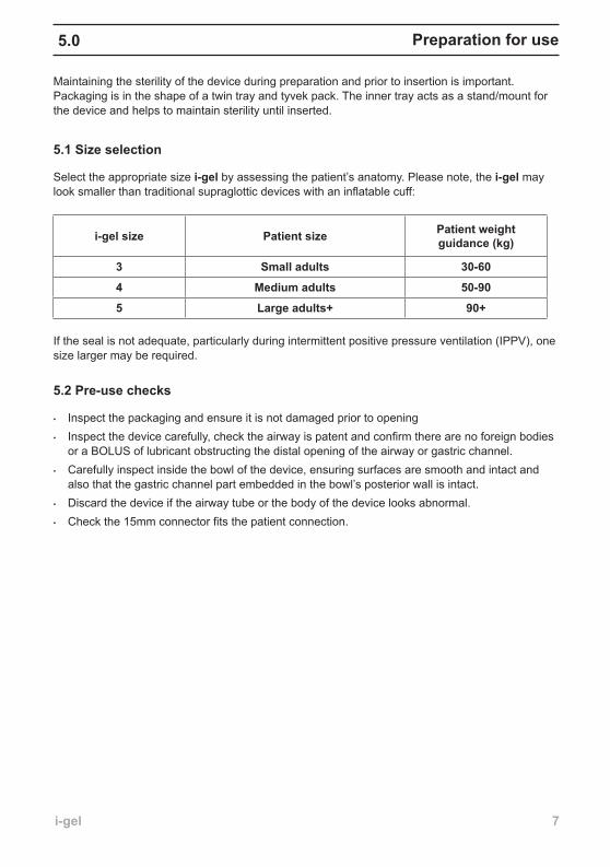
Maintaining the sterility of the device during preparation and prior to insertion is important. Packaging is in the shape of a twin tray and tyvek pack. The inner tray acts as a stand/mount for the device and helps to maintain sterility until inserted.
5.� Size selection
Select the appropriate size i-gel by assessing the patient’s anatomy. Please note, the i-gel may look smaller than traditional supraglottic devices with an inflatable cuff:
If the seal is not adequate, particularly during intermittent positive pressure ventilation (IPPV), one size larger may be required.
5.2Pre-usechecks
Inspect the packaging and ensure it is not damaged prior to opening Inspect the device carefully, check the airway is patent and confirm there are no foreign bodies or a BOLUS of lubricant obstructing the distal opening of the airway or gastric channel. Carefully inspect inside the bowl of the device, ensuring surfaces are smooth and intact and also that the gastric channel part embedded in the bowl’s posterior wall is intact.Discard the device if the airway tube or the body of the device looks abnormal. Check the 15mm connector fits the patient connection.
•
•
•
•
•
i-gel size Patient size Patient weight guidance(kg)
� Small adults �0-60
� Medium adults 50-90
5 Large adults+ 90+
igel user guide_1_081206.indd 7 15/12/2006 11:50:42
i-gel 8
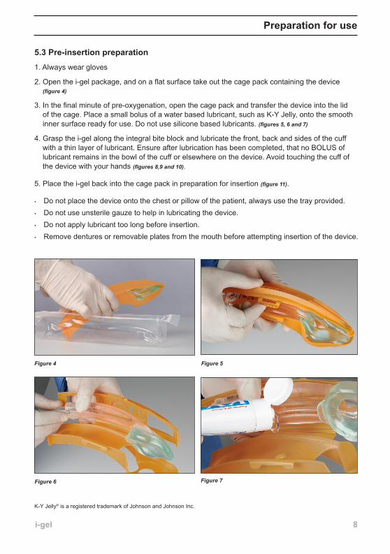
5.� Pre-insertion preparation
1. Always wear gloves
2. Open the i-gel package, and on a flat surface take out the cage pack containing the device (figure 4)
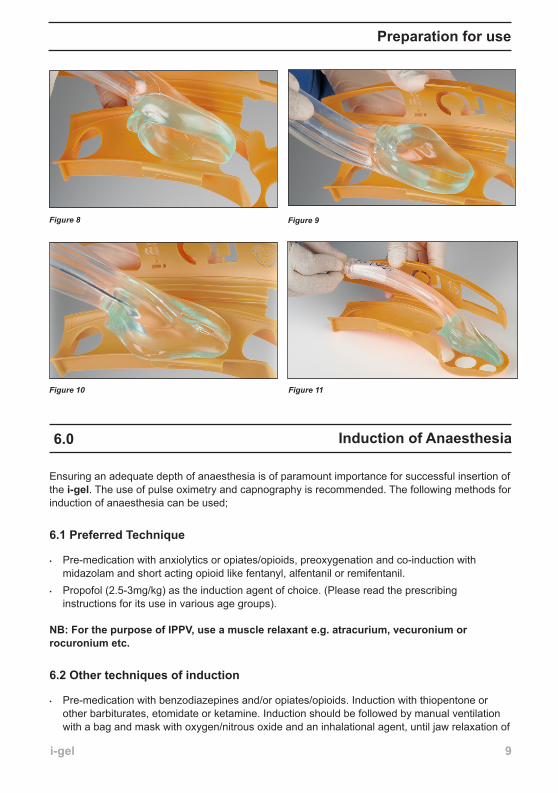
3. In the final minute of pre-oxygenation, open the cage pack and transfer the device into the lid of the cage. Place a small bolus of a water based lubricant, such as K-Y Jelly, onto the smooth inner surface ready for use. Do not use silicone based lubricants. (figures 5, 6 and 7)
4. Grasp the i-gel along the integral bite block and lubricate the front, back and sides of the cuff with a thin layer of lubricant. Ensure after lubrication has been completed, that no BOLUS of lubricant remains in the bowl of the cuff or elsewhere on the device. Avoid touching the cuff of the device with your hands (figures 8,9 and 10).
5. Place the i-gel back into the cage pack in preparation for insertion (figure 11).
Do not place the device onto the chest or pillow of the patient, always use the tray provided.Do not use unsterile gauze to help in lubricating the device.Do not apply lubricant too long before insertion.Remove dentures or removable plates from the mouth before attempting insertion of the device.
•
•
•
•
Figure 4
Figure 7Figure 6
K-Y Jelly® is a registered trademark of Johnson and Johnson Inc.
Preparation for use
Figure 5
igel user guide_1_081206.indd 8 15/12/2006 11:51:04
i-gel 9
Figure 8 Figure 9
Figure 10
6.0 Induction of Anaesthesia
Ensuring an adequate depth of anaesthesia is of paramount importance for successful insertion of the i-gel. The use of pulse oximetry and capnography is recommended. The following methods for induction of anaesthesia can be used;
6.� Preferred Technique
Pre-medication with anxiolytics or opiates/opioids, preoxygenation and co-induction with midazolam and short acting opioid like fentanyl, alfentanil or remifentanil. Propofol (2.5-3mg/kg) as the induction agent of choice. (Please read the prescribing instructions for its use in various age groups).
NB:ForthepurposeofIPPV,useamusclerelaxante.g.atracurium,vecuroniumorrocuronium etc.
6.� Other techniques of induction
Pre-medication with benzodiazepines and/or opiates/opioids. Induction with thiopentone or other barbiturates, etomidate or ketamine. Induction should be followed by manual ventilation with a bag and mask with oxygen/nitrous oxide and an inhalational agent, until jaw relaxation of
•
•
•
Figure 11
Preparation for use
igel user guide_1_081206.indd 9 15/12/2006 11:51:21
i-gel �0
7.0 Insertion technique
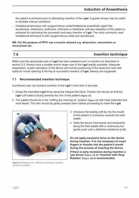
Make sure the appropriate size of i-gel has been prepared prior to insertion as described in section 5.0. Always have a smaller and/or larger size of the i-gel readily available. Adequate preparation, proper lubrication of the device and correct positioning of the head and neck with optimum mouth opening is the key to successful insertion of i-gel. Always pre-oxygenate.
7.� Recommended insertion technique
A proficient user can achieve insertion of the i-gel in less than 5 seconds.
Grasp the lubricated i-gel firmly along the integral bite block. Position the device so that the i-gel cuff outlet is facing towards the chin of the patient (Figure 12).
The patient should be in the ‘sniffing the morning air’ position (Figure 12) with head extended and neck flexed. The chin should be gently pressed down before proceeding to insert the i-gel.
Introduce the leading soft tip into the mouth of the patient in a direction towards the hard palate. Glide the device downwards and backwards along the hard palate with a continuous but gentle push until a definitive resistance is felt.
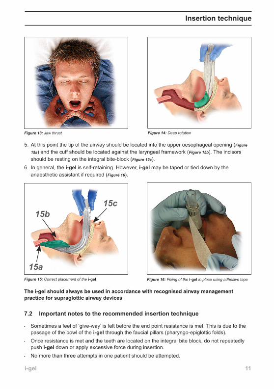
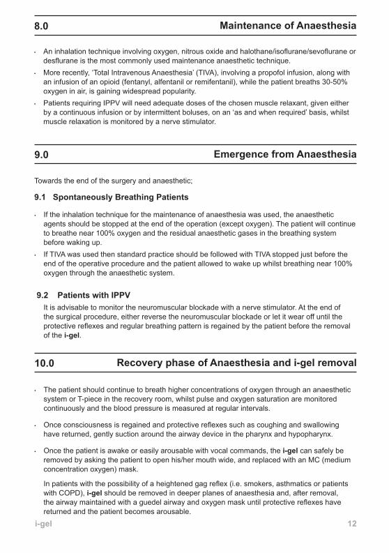
Donotapplyexcessiveforceonthedeviceduring insertion. It is not necessary to insert fingersorthumbsintothepatient’smouthduring the process of inserting the device.If there is early resistance during insertion a ‘jawthrust’(Figure13)or‘InsertionwithDeepRotation’(Figure14)is recommended.
1.
2.
3.
4.
Figure 12: Patient in the ‘sniffing the morning air’ position, just prior to insertion
the patient is achieved prior to attempting insertion of the i-gel. A guedel airway may be useful to facilitate manual ventilation. Inhalational technique with oxygen/nitrous oxide/inhalational anaesthetic agent like sevoflurane, desflurane, isoflurane, enflurane or halothane until jaw relaxation of the patient is achieved for optimising the successful and easy insertion of i-gel. The most commonly used inhalational technique is with oxygen/nitrous oxide and sevoflurane.
NB:ForthepurposeofIPPVuseamusclerelaxante.g.atracurium,vecuroniumorrocuronium etc.
•
Induction of Anaesthesia
igel user guide_1_081206.indd 10 15/12/2006 11:51:25
i-gel ��
Figure 13: Jaw thrust Figure 14: Deep rotation
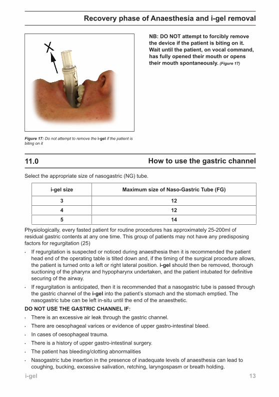
5. At this point the tip of the airway should be located into the upper oesophageal opening (Figure
15a) and the cuff should be located against the laryngeal framework (Figure 15b). The incisors should be resting on the integral bite-block (Figure 15c).
6. In general, the i-gel is self-retaining. However, i-gel may be taped or tied down by the anaesthetic assistant if required (Figure 16).
Figure 16: Fixing of the i-gel in place using adhesive tapeFigure 15: Correct placement of the i-gel
15a
15b15c
Insertion technique
Thei-gelshouldalwaysbeusedinaccordancewithrecognisedairwaymanagementpractice for supraglottic airway devices
7.� Important notes to the recommended insertion technique
Sometimes a feel of ‘give-way’ is felt before the end point resistance is met. This is due to the passage of the bowl of the i-gel through the faucial pillars (pharyngo-epiglottic folds). Once resistance is met and the teeth are located on the integral bite block, do not repeatedly push i-gel down or apply excessive force during insertion. No more than three attempts in one patient should be attempted.
•
•
•
igel user guide_1_081206.indd 11 15/12/2006 11:52:05
i-gel ��
Recovery phase of Anaesthesia and i-gel removal�0.0
9.0 Emergence from Anaesthesia
Towards the end of the surgery and anaesthetic;
9.� Spontaneously Breathing Patients
If the inhalation technique for the maintenance of anaesthesia was used, the anaesthetic agents should be stopped at the end of the operation (except oxygen). The patient will continue to breathe near 100% oxygen and the residual anaesthetic gases in the breathing system before waking up. If TIVA was used then standard practice should be followed with TIVA stopped just before the end of the operative procedure and the patient allowed to wake up whilst breathing near 100% oxygen through the anaesthetic system.
9.2 PatientswithIPPV It is advisable to monitor the neuromuscular blockade with a nerve stimulator. At the end of
the surgical procedure, either reverse the neuromuscular blockade or let it wear off until the protective reflexes and regular breathing pattern is regained by the patient before the removal of the i-gel.
•
•
The patient should continue to breath higher concentrations of oxygen through an anaesthetic system or T-piece in the recovery room, whilst pulse and oxygen saturation are monitored continuously and the blood pressure is measured at regular intervals.
Once consciousness is regained and protective reflexes such as coughing and swallowing have returned, gently suction around the airway device in the pharynx and hypopharynx.
Once the patient is awake or easily arousable with vocal commands, the i-gel can safely be removed by asking the patient to open his/her mouth wide, and replaced with an MC (medium concentration oxygen) mask.
In patients with the possibility of a heightened gag reflex (i.e. smokers, asthmatics or patients with COPD), i-gel should be removed in deeper planes of anaesthesia and, after removal, the airway maintained with a guedel airway and oxygen mask until protective reflexes have returned and the patient becomes arousable.
•
•
•
Maintenance of Anaesthesia8.0
An inhalation technique involving oxygen, nitrous oxide and halothane/isoflurane/sevoflurane or desflurane is the most commonly used maintenance anaesthetic technique. More recently, ‘Total Intravenous Anaesthesia’ (TIVA), involving a propofol infusion, along with an infusion of an opioid (fentanyl, alfentanil or remifentanil), while the patient breaths 30-50% oxygen in air, is gaining widespread popularity. Patients requiring IPPV will need adequate doses of the chosen muscle relaxant, given either by a continuous infusion or by intermittent boluses, on an ‘as and when required’ basis, whilst muscle relaxation is monitored by a nerve stimulator.
•
•
•
igel user guide_1_081206.indd 12 15/12/2006 11:52:05
i-gel ��
��.0 How to use the gastric channel
Select the appropriate size of nasogastric (NG) tube.
Physiologically, every fasted patient for routine procedures has approximately 25-200ml of residual gastric contents at any one time. This group of patients may not have any predisposing factors for regurgitation (25)
If regurgitation is suspected or noticed during anaesthesia then it is recommended the patient head end of the operating table is tilted down and, if the timing of the surgical procedure allows, the patient is turned onto a left or right lateral position. i-gel should then be removed, thorough suctioning of the pharynx and hypopharynx undertaken, and the patient intubated for definitive securing of the airway. If regurgitation is anticipated, then it is recommended that a nasogastric tube is passed through the gastric channel of the i-gel into the patient’s stomach and the stomach emptied. The nasogastric tube can be left in-situ until the end of the anaesthetic.
DONOTUSETHEGASTRICCHANNELIF:There is an excessive air leak through the gastric channel.There are oesophageal varices or evidence of upper gastro-intestinal bleed.In cases of oesophageal trauma.There is a history of upper gastro-intestinal surgery.The patient has bleeding/clotting abnormalities Nasogastric tube insertion in the presence of inadequate levels of anaesthesia can lead to coughing, bucking, excessive salivation, retching, laryngospasm or breath holding.
•
•
•
•
•
•
•
•
i-gel size MaximumsizeofNaso-GastricTube(FG)
� ��
� ��
5 ��
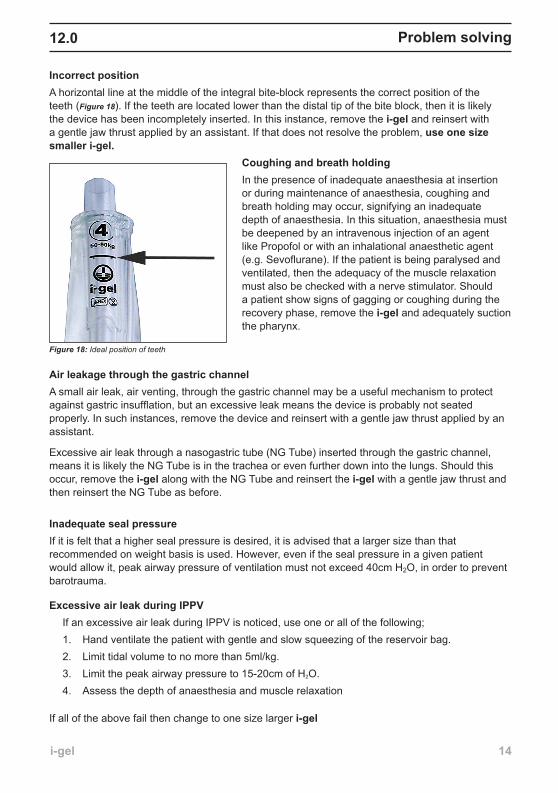
Figure 17: Do not attempt to remove the i-gel if the patient is biting on it
NB:DONOTattempttoforciblyremovethedeviceifthepatientisbitingonit.Waituntilthepatient,onvocalcommand,has fully opened their mouth or opens their mouth spontaneously. (Figure 17)
Recovery phase of Anaesthesia and i-gel removal
igel user guide_1_081206.indd 13 15/12/2006 11:52:12
i-gel ��
Problemsolving��.0
Incorrect positionA horizontal line at the middle of the integral bite-block represents the correct position of the teeth (Figure 18). If the teeth are located lower than the distal tip of the bite block, then it is likely the device has been incompletely inserted. In this instance, remove the i-gel and reinsert with a gentle jaw thrust applied by an assistant. If that does not resolve the problem, use one size smaller i-gel.
Coughingandbreathholding In the presence of inadequate anaesthesia at insertion or during maintenance of anaesthesia, coughing and breath holding may occur, signifying an inadequate depth of anaesthesia. In this situation, anaesthesia must be deepened by an intravenous injection of an agent like Propofol or with an inhalational anaesthetic agent (e.g. Sevoflurane). If the patient is being paralysed and ventilated, then the adequacy of the muscle relaxation must also be checked with a nerve stimulator. Should a patient show signs of gagging or coughing during the recovery phase, remove the i-gel and adequately suction the pharynx.
AirleakagethroughthegastricchannelA small air leak, air venting, through the gastric channel may be a useful mechanism to protect against gastric insufflation, but an excessive leak means the device is probably not seated properly. In such instances, remove the device and reinsert with a gentle jaw thrust applied by an assistant.
Excessive air leak through a nasogastric tube (NG Tube) inserted through the gastric channel, means it is likely the NG Tube is in the trachea or even further down into the lungs. Should this occur, remove the i-gel along with the NG Tube and reinsert the i-gel with a gentle jaw thrust and then reinsert the NG Tube as before.
Inadequate seal pressureIf it is felt that a higher seal pressure is desired, it is advised that a larger size than that recommended on weight basis is used. However, even if the seal pressure in a given patient would allow it, peak airway pressure of ventilation must not exceed 40cm H2O, in order to prevent barotrauma.
ExcessiveairleakduringIPPV If an excessive air leak during IPPV is noticed, use one or all of the following; 1. Hand ventilate the patient with gentle and slow squeezing of the reservoir bag. 2. Limit tidal volume to no more than 5ml/kg. 3. Limit the peak airway pressure to 15-20cm of H2O. 4. Assess the depth of anaesthesia and muscle relaxation If all of the above fail then change to one size larger i-gel
Figure 18: Ideal position of teeth
igel user guide_1_081206.indd 14 15/12/2006 11:52:14
i-gel �5
Adverse outcomes��.0
The anatomical design and soft material of the i-gel are less likely to cause adverse outcomes when compared with other supraglottic devices. As the i-gel is manufactured from a soft gel-like material, it is unlikely to cause any trauma during insertion or whilst in-situ, thereby reducing the risk of postoperative complications and co-morbidity.
Some of the known risks and complications of the use of supraglottic airway devices include laryngospasm, sore throat, trauma to the pharyngo-laryngeal framework, gastric insufflation, regurgitation and inhalation of the gastric contents, nerve injuries, vocal cord paralysis, lingual or hypoglossal nerve injuries, tongue numbness and cyanosis.
The risk of rotation and malpositioning leading to partial or complete airway obstruction are extremely low with the i-gel compared to other supraglottic devices. Down-folding of the epiglottis can occasionally occur, but the i-gel’s cuff and airway channel have been designed in such a way that the chances of obstruction to the fresh gas flow (FGF) are minimal.
If the i-gel is placed too high in the pharynx, this may result in a poor seal and cause excessive leakage. If the FGF is forced in too hard by squeezing the reservoir bag, this may cause gastric insufflation and distension, which will increase the risk of regurgitation and post-operative nausea and vomiting.
If the tip of the i-gel enters into the glottic opening, this can lead to an excessive air leak through the gastric channel, which may result in obstruction to the FGF. If a NG tube is then inserted through the i-gel, it will enter into the trachea and lungs. It is recommended that if this situation is suspected, the i-gel is removed and re-inserted with a gentle jaw thrust and correct placement checked.
•
•
•
•
igel user guide_1_081206.indd 15 15/12/2006 11:52:14
i-gel �6
Furtherreading
1. R. M. Levitan and W. C. Kinkle. Initial anatomic investigations of the I-gel airway: a novel supraglottic airway without inflatable cuff. Anaesthesia 2005 Oct;60(10):1022–6.
2. Miller, DM. A proposed classification and scoring system for supraglottic sealing airways: A brief review. Anesthesia and Analgesia 2004; 99: 1553-1559.
3. Brimacombe J, Berry A. The incidence of aspiration associated with the laryngeal mask airway-a meta-analysis of published literature. J Clin Anesth 1995; 7: 297-30.
4. Brimacombe J, Keller C. Aspiration of gastric contents during use of a ProSeal laryngeal mask airway secondary to unidentified foldover malposition. Anesthesia Analgesia 2003; 97: 1192-4 British Journal of Anaesthesia
5. Cook TM, Nolan JP, Verghese C, Strube PJ, Lees M, Millar JM, Baskett PJ: Randomized crossover comparison of the Proseal with the Classic laryngeal mask airway in unparalysed anaesthetized patients. British Journal of Anaesthesia 2002;88:527–33
6. Brimacombe J, Keller C, Fullekrug B, Agro F, Rosenblatt W, Dierdorf SF, Garcia de Lucas E, Capdevila X, Brimacombe N: A multicenter study comparing the ProSeal with the Classic laryngeal mask airway in anesthetized, nonparalyzed patients. Anesthesiology 2002; 96:289–95
7. Brimacombe J, Keller C: The ProSeal laryngeal mask airway: A randomized, crossover study with the standard laryngeal mask airway in paralyzed, anesthetized patients. Anesthesiology 2000; 93:104–9
8. Brain AIJ, Verghese C, Strube PJ: The LMA ‘ProSeal’: A laryngeal mask with an oesophageal vent. British Journal of Anaesthesia 2000; 84:650–4
9. Keller C, Brimacombe J, Keller K, Morris R. A comparison of four methods for assessing airway sealing pressure with the laryngeal mask airway in adult patients. British Journal of Anaesthesia 1999; 82: 286-7
10. Brimacombe J, Keller C, Giampalmo M, Sparr HJ, Berry A. Direct measurement of mucosal pressures exerted by cuff and non-cuff portions of tracheal tubes with different cuff volumes and head and neck positions. British Journal of Anaesthesia 1999; 82: 708-1 I
11. Brimacombe J, Keller C. A comparison of pharyngeal mucosal pressure and airway sealing pressure with the laryngeal mask airway in anesthetized adult patients. Anesthesia & Analgesia 1998; 87:1379-82.
12. Keller C, Puehringer F, Brimacombe J. The influence of cuff volume on oropharyngeal leak pressure and fibreoptic position with the laryngeal mask airway. British Journal of Anaesthesia 1998;81:186-7.
13. Brimacombe J, Keller C, Morris R, Mecklem D. A comparison of the disposable versus the reusable laryngeal mask airway in paralyzed adult patients. Anesthesia & Analgesia 1998;87:921-24.
14. Twigg S. Brown JM. Williams R. Swelling and cyanosis of the tongue associated with use of a laryngeal mask airway. Anaesthesia & Intensive Care. 2000; 28:449-50.
15. Stewart A. Lindsay WA. Bilateral hypoglossal nerve injury following the use of the laryngeal mask airway. Anaesthesia. 2002; 57:264-5.
16. Drummond GB. Influence of thiopentone on upper airway muscles. British Journal of Anaesthesia 1989; 63: 12-21
igel user guide_1_081206.indd 16 15/12/2006 11:52:14
i-gel
Furtherreading
17. LowingerD.BenjaminB.GaddL.Recurrentlaryngealnerveinjurycausedbyalaryngealmaskairway.Anaesthesia&IntensiveCare.1999;27:202-5.
18.OuelletteRG.Theeffectofnitrousoxideonlaryngealmaskcuffpressure.AmericanAssociationofNurseAnesthetistsJournal.2000;68:411-4.
19. AgroF,CataldoR,CarassitiM,CostaF.Theseeingstylet:Anewdevicefortrachealintubation.Resuscitation.2000;44:177–80.
20. LevitanRM,OchrochAE,HollanderJ,etal.AssessmentofAirwayVisualization:ValidationofthePercentofGlotticOpening(POGO)Scale.AcademicEmergencyMedicine.1998;5:919–23.
21. OchrochAE,KushS,StuartS,HollanderJE,LevitanRM.Assessmentoflaryngealviewindirectlaryngoscopy:Thepercentageofglotticopening(POGO)scorecomparedtoCormackandLehanegrading.CanadianJournalofAnesthesia.1999;46:987–90.
22. CooperRM.Useofanewvideolaryngoscope(GlideScope)inthemanagementofadifficultairway.CanadianJournalofAnesthesia.2003;50:611–3.
23. BrimacombeJ,BerryA.Aproposedfiber-opticscoringsystemtostandardizetheassessmentoflaryngealmaskairwayposition.AnesthesiaAnalgesia1993;76:45
24. KellerC,BrimacombeJ:MucosalpressureandoropharyngealleakpressurewiththeProSealversustheclassiclaryngealmaskairway.BritishJournalofAnaesthesia2000;85:262–6
25. BrimacombeJ,KellerC,BoehlerM,PuehringerF:PositivepressureventilationwiththeProSealversusClassiclaryngealmaskairway:A–3randomized,crossoverstudyofhealthyfemalepatients.AnesthesiaAnalgesia2001;93:1351
26. Preoperativefastingforadultstopreventperioperativecomplications.BradyM,KinnS,StuartP.CochraneDatabaseSystRev.2003;(4):CD004423
27. BrimacombeJ,LaryngealMaskAnaesthesia,PrincipesandPractice2ndedition2004
17
i-gelcover_guide_issue 2.indd 4 15/12/2006 13:33:29
9279 Issue 2 • 12.06
All rights reserved. No part of this publication may be reproduced, stored in a retrieval system or transmitted in any form or by any means electrical, mechanical, photocopying, recording or otherwise, without the prior written permission of the publisher.
i-gel is a registered trademark of Intersurgical. The information provided in this document are correct at the time of publication. Intersurgical reserves the right to improve or modify the product without prior notification
User Guidei-gel® single use supraglottic airway
Intersurgical LtdCrane House, Molly Millars LaneWokingham, Berkshire RG41 2RZT: +44 (0)118 9656 300F: +44 (0)118 9656 [email protected]
DeutschlandT: 02241 [email protected]
FranceT: 01 48 76 72 [email protected]
EspañaT: 91 665 73 [email protected]
PortugalT: 21 910 85 [email protected]
LietuvaT: +370 387 [email protected]
NederlandT: 0413 [email protected]
PoccияT: 095 771 [email protected]
Česká RepublikaT: 272 940 [email protected]
South AfricaT: 011 444 [email protected]
PhilippinesT: 632 820 [email protected]
i-gelcover_guide_issue 2.indd 1 15/12/2006 13:33:09