natural killer cells: angels and devils for immunotherapy
TRANSCRIPT

International Journal of
Molecular Sciences
Review
Natural Killer Cells: Angels and Devilsfor Immunotherapy
Beatriz Martín-Antonio 1,2,* ID , Guillermo Suñe 1,2, Lorena Perez-Amill 1, Maria Castella 1,2
and Alvaro Urbano-Ispizua 1,2
1 Department of Hematology, Hospital Clinic, Institut d’Investigacions Biomèdiques August Pi iSunyer (IDIBAPS), 08036 Barcelona, Spain; [email protected] (G.S.); [email protected] (L.P.-A.);[email protected] (M.C.); [email protected] (A.U.-I.)
2 Josep Carreras Leukaemia Research Institute, 08036 Barcelona, Spain* Correspondence: [email protected]; Tel.: +34-93-227-5400 (ext. 4528)
Received: 1 August 2017; Accepted: 19 August 2017; Published: 29 August 2017
Abstract: In recent years, the relevance of the immune system to fight cancer has led to thedevelopment of immunotherapy, including the adoptive cell transfer of immune cells, such asnatural killer (NK) cells and chimeric antigen receptors (CAR)-modified T cells. The discovery ofdonor NK cells’ anti-tumor activity in acute myeloid leukemia patients receiving allogeneic stemcell transplantation (allo-SCT) was the trigger to conduct many clinical trials infusing NK cells.Surprisingly, many of these studies did not obtain optimal results, suggesting that many different NKcell parameters combined with the best clinical protocol need to be optimized. Various parametersincluding the high array of activating receptors that NK cells have, the source of NK cells selected totreat patients, different cytotoxic mechanisms that NK cells activate depending on the target cell andtumor cell survival mechanisms need to be considered before choosing the best immunotherapeuticstrategy using NK cells. In this review, we will discuss these parameters to help improve currentstrategies using NK cells in cancer therapy. Moreover, the chimeric antigen receptor (CAR)modification, which has revolutionized the concept of immunotherapy, will be discussed in thecontext of NK cells. Lastly, the dark side of NK cells and their involvement in inflammation will alsobe discussed.
Keywords: natural killer (NK); immunotherapy; tumor cell survival mechanisms; inflammation
1. Natural Killer (NK) Cell Modulation Activity
Natural killer (NK) cells are cells of the innate immune system with high anti-tumor, antiviraland antimicrobial activity. In healthy individuals, 90% of NK cells in peripheral blood (PB) are matureand cytotoxic, and characterized by the expression of CD16bright and CD56dim. The remaining 10% ofNK cells represent the immature subset of NK cells that are cytokine producers, and express CD16dim
or CD16−, CD56bright and CD25+ [1]. The activity of NK cells is modulated by an array of inhibitoryand activating receptors, which are fundamental in controlling NK cell activity. Whereas inhibitoryKiller-cell immunoglobulin-like (KIR) receptors inhibit NK cell cytotoxicity by interacting with theHuman Leukocyte Antigen (HLA)-I in human cells, activating receptors activate NK cells by interactingwith their ligands in target cells induced upon tumor transformation, viral infection and cell stress.In physiological conditions, inhibitory KIR-HLA-I interaction holds NK cell cytotoxicity against normalcells in the body. However, when a KIR-HLA-I mismatch or HLA-I down-regulation occurs afterviral infection or in some tumor cells, inhibitory KIRs cannot interact with their ligands activatingNK cell cytotoxic function [2]. This finding observed by Ruggeri et al. in 2002 in acute myeloblasticleukemia (AML) patients after allogeneic stem cell transplantation (allo-SCT), allowed using the
Int. J. Mol. Sci. 2017, 18, 1868; doi:10.3390/ijms18091868 www.mdpi.com/journal/ijms

Int. J. Mol. Sci. 2017, 18, 1868 2 of 20
inhibitory KIR (donor)-HLA-I (patient) mismatch as a prognostic factor in AML patients receivingan allo-SCT [3]. This “missing self” recognition leads to allo-reactivity after allo-SCT, and consequently,NK cells lyse leukemia blasts, recipient dendritic cells (DCs) and recipient T cells, which translatesinto a reduction of relapse, prevention of Graft vs. Host Disease (GVHD), and prevention of graftrejection, respectively. The “missing self” recognition led to the proposal of the “missing ligandmodel” as a powerful algorithm to predict a potent anti-leukemia effect and, consequently, a favorableoutcome after allo-SCT [3,4]. However, in non-myeloid malignancies, such as acute lymphoblasticleukemia (ALL), the GVHD reduction and prevention of graft rejection was not always accompaniedby an increase of the graft vs. leukemia effect observed in AML [5]. These conflicting results led toapplying the “missing ligand model” mostly in AML [6], and in non-myeloid malignancies to usethis model just for NK-mediated killing of DCs and T cells to predict a reduced GVHD and reducedgraft rejection, respectively. The graft vs. leukemia effect occurring mostly in AML could be mediatedby HLA down-regulation, which does not occur in other malignancies such as ALL. To support this,clinical studies in ALL confirmed that low HLA-I expression levels in blasts conferred a beneficialeffect mediated by allo-reactive NK cells [7].
However, NK cells have a high variety of activating receptors, which also modulate their cytotoxicactivity. These receptors will play a fundamental role in the recognition of other types of tumor cellswhich do not down-regulate HLA-I. Some of these receptors are expressed only at given stages ofdifferentiation or by specific NK cell subsets [8]. Surprisingly, this high number of activating receptorsin NK cells is responsible for the existence of 6000–30,000 different phenotypic NK populationsin each healthy individual, which provide flexibility to respond to pathogens and tumor cells [9].Activating receptors include different families such as the: (i) activating KIRs which interact withTYRO protein tyrosine kinase binding protein (DAP-12); (ii) C-type lectin-like receptors, including theactivating CD94/NKG2C and NKG2D. NKP80, included in this family, exerts an autonomous controlof NK cells against excessive inflammatory response causing self-NK cells mediated cytolysis [10–12];(iii) natural cytotoxicity receptors (NCR) which include NKP30, NKP44 and NKP46 and interact withligands overexpressed on tumor cells and viral infected cells; and (iv) signaling lymphocyte activatingmolecule (SLAM) family of receptors which include SLAMF1, 2B4, NTB-A, CD48, CD84, Ly9, andCRACC. They transmit activating signals to mediate NK cytotoxicity. Moreover, NK cells express CD16,which is the Fc receptor that binds Ig-G mediating antibody dependent cellular cytotoxicity (ADCC).
2. NK Cell Classic Cytotoxicity Mechanisms
Upon recognition, NK cells eliminate target cells rapidly (within 30–60 min) by differentmechanisms. The two classic NK cell cytotoxic mechanisms include the death receptor pathwayand the granule dependent pathway. The death receptor pathway is activated by the tumor necrosisfactor (TNF)-related apoptosis inducing ligand family (TRAIL) and by Fas-Ligand (FASL) (CD95L)which are expressed on NK cells [13], and interact with their ligands in target cells. Interaction ofFASL with FAS and TRAIL with TRAIL receptors allows the formation of a death-inducing signalingcomplex that includes Fas-associated death domain protein (FADD), caspase-8, and caspase-10 [14].Activation of caspase-8 results either in direct activation of the other caspases or in the proteolysis ofBid, with release of cytochrome C and subsequent caspase activation. Even if it were believed thatthese receptors only activate apoptosis, new studies have revealed that other types of non-apoptoticinflammatory types of cell death can be activated [15,16]. The granule dependent pathway is initiatedafter NK cells adhere to the target cell, with subsequent delivery of cytotoxic granules containingperforin and proteases called Granzymes toward the bound target cell [17]. In humans, there arefive different types of Granzymes (A, B, H, K and M) and each one will activate different cell deathpathways, either apoptotic or non-apoptotic. Granzymes A and B are the most studied ones. WhereasGranzyme B activates apoptosis through activation of caspases or release of cytochrome C from themitochondria, apparently, Granzyme A activates non-apoptotic cell death [18]; however, there areconflicting results regarding the cytotoxic role of Granzyme A [19]. The other Granzymes have been

Int. J. Mol. Sci. 2017, 18, 1868 3 of 20
less studied, although it has been shown that Granzyme K is released by CD56bright cells mediatingnon-apoptotic tumor cell death [20]. Granzyme M shows anti-tumor properties after adoptive NKcell transfer [21]. Granzyme H helps Granzyme B kill adenovirus-infected cells [22]; and Granulysin,which is another NK cytotoxic molecule released in the cytotoxic granules, activates EndoplasmaticReticulum stress leading to cell death [23]. All these different cytotoxicity mechanisms that NK cellsshow, enable them to eliminate different types of tumor cells with their intrinsic characteristics.
3. Immunotherapy Strategies Using NK Cells
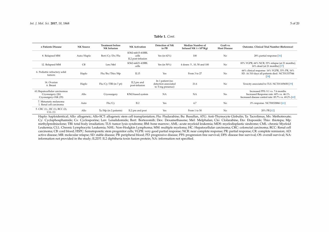
As previously mentioned, the anti-tumor NK cell activity described after allo-SCT added to thefinding that unlike donor T cells, NK cells do not induce GVHD, led to the development of manyclinical trials infusing NK cells in patients receiving an SCT. We have reviewed published studiesinfusing NK cells as an immunotherapy option. Results are not optimal; they are summarized inTable 1. In most of these studies, NK cells were activated in vitro and/or in vivo with Interleukin (IL)2,and administered after immunosuppressive treatment based on fludarabine and cyclophosphamide.Patient disease, disease status, number of NK cells infused and number of NK cell infusions differedfrom one study to another. All published studies agree that NK cell infusion is a safe and well-toleratedprocedure and does not associate to GVHD. Whereas, for myeloid malignancies, most studies used NKcells from haploidentical donors considering the KIR-HLA-I mismatch, in non-myeloid malignancies,NK cells were either autologous or allogeneic and expanded in vitro.
In myeloid malignancies, haploidentical NK cells with KIR-HLA-I mismatch have been used eitheras a consolidation therapy [24,25] or in high risk and refractory patients [26,27]. As a consolidationtherapy, in both children with AML and elderly AML patients, NK cells showed a clear benefit.After a median follow up of four years, pediatric patients remain in remission, and elderly patientswith high risk AML showed prolonged disease free survival (DFS) after NK cell infusion [24,25,28].These studies concluded that NK cells could be a promising consolidation therapy strategy in patientswho are not candidates for receiving an allo-SCT. Another novel consolidation therapy strategy inAML consisted on the infusion of NK cells derived from hematopoietic progenitor cells (HPC) obtainedfrom a cord blood (CB) unit. This technique allowed obtaining enough number of NK cells whichwere well tolerated. Unlike other procedures, this study did not administrate IL2 into patients, andresponses were detected, suggesting that this technique allows obtaining effective NK cells whichmight be used off-the-shelf when required [29].
In refractory patients, results are not good. However, it was noticed that infusion of haploidenticalNK cells combined with IL2 diphtheria toxin fusion protein (IL2DT) to deplete host T-reg cells ledto higher NK cell expansion, improved complete remission rate and disease-free survival with noincreased toxicity in AML patients [26]. Lee et al. also showed durable responses associated with CD56+cells delivered in myeloid malignancies [27]. Another study in AML and MDS found resolution ofdysplastic features in 50% of myelodisplastic sindrome (MDS) patients, and 16% of complete remission(CR) in AML; however, they could not find NK cells in peripheral blood (PB) [30]. In pediatricrefractory AML patients who received or did not receive an allo-SCT previously, responses higherthan 50% were detected after NK infusion, and combined with allo-SCT afterwards, 27% and 36% ofDFS were achieved, respectively, at six years [31]. Another approach tested in high risk AML patients,consisted in the infusion of haploidentical NK cells previously primed with tumor cell lysates. IL2 wasnot administrated to patients, and still NK cells seemed to exert anti-leukemia effect in 57% of thepatients. However, at 2 years 85.7% patients died [32]. Studies in myeloid malignancies indicated thatbetter responses were achieved when infusing NK cells in patients in remission suggesting that NKcells cannot overcome large tumor burdens. Therefore, more recent approaches aim at expanding NKcells in vitro to obtain a high number of NK for large tumor mass.

Int. J. Mol. Sci. 2017, 18, 1868 4 of 20
Table 1. Clinical studies performed with NK (natural killer) cells as immunotherapy treatment.
n Patients Disease NK Source Treatment beforeNK Infusion NK Activation Detection of NK
in PBMedian Number of
Infused NK (×106/Kg)Graft vs.
Host Disease Outcome. Clinical Trial Number (Reference)
10. AML in CR (pediatric) Haplo Flu/Cy IL2 post-infusion Yes 29 No All in remission at 964 days [24]
13. AML: 38.4% in AD, 15.3%MR, 46% CR Haplo Flu/Cy IL2 post-infusion Yes 2.74 No
AD: 20% achieved transient CRMR: 100% achieved CR
CR: 50% DFS after 34, 32, 18 months.NCT00799799 [25]
16. AML in CR Haplo Flu/Cy IL2 post-infusion Yes From 1.29 to 5.53 NoAt 22.5 months: 56% DFS, 44% relapse. Higher NK
cell number associated to higher DFS.NCT00799799 [28]
10. AML in CRAllo NK derived
from CD34+ HSPCfrom CB
Flu/Cy IL15 and IL2 Yes (in 21%) From 3 to 30 No 20% became MRD negative for 6 months [29]
57. Refractory AML (15received IL2DT) Haplo Flu/Cy IL2 post-infusion
Yes: in 10% ofpatients, and in 27%of patients receiving
IL2DT)
26 NoCR: 53% (IL2DT) vs. 21% (no IL2DT)DFS: 33% (IL2DT) vs. 5% (no IL2DT)
NCT00274846, NCT01106950 [26]
21. AML, MDS, CML Haplo Flu, Bu IL2 pre andpost-infusion NA From 0.22 to 8.32 No associated to NK
Survival associated with CD56+ cells delivered;24% durable CR (no association to KIR-HLAmismatch). NCT00402558. NCT01390402 [27]
Refractory 6: AML, 2: MDS Haplo Flu/Cy IL2 post-infusion No 10.6 No 16% CR; 83% Disease progression. NCT00871689[30]
29. Pediatric refractory AMLCohort 1: no prior allo-SCT (14)
Cohort 2: relapsed afterallo-SCT (15)
Haplo Clo/Eto/Cy IL2 post-infusion Yes From 3.5 to 103 No
Cohort 1: 71% response; 86% underwent allo-SCT;36% DFS at 6 years.
Cohort 2: 66.6% response and underwent allo-SCT;27% DFS at 6 years.
NCT00697671 and NCT00187096 [31]
7. High risk AML Haplo Flu/TBITumor-primed NK
cells with tumorlysate
Yes 3 doses: 1, 5, 10 No
At 6 months: 42.8% in CR remained in remission,14% in PR achieved CR, 28% relapse, 14% died.
At 1 year: 14% remained in CR.At 2 years: 85.7% died. Median OS: 400 days [32]
10. Relapsed MM Haplo Flu/Mel/Dx IL2 pre and postinfusion Yes (until day 14) 1.7 No 50% CR or near CR, 20% PR, 10% SD and 20% PD
[33]
17. Lymphoma (2), advancedsolid tumors (15) Allo
Nonimmunosuppressive
regimen
IL2 (MG4101method) Yes From 1 to 30 (1 and 3 doses) No
Lymphoma: 50% SD, 50% PDSolid tumors: 47% SD, 53% PD. PFS in SD: 4
months. NCT01212341 [34]
5. Relapsed MM Auto Len, BortIL2,
K562-mb15-41BBLcells
Yes (7.5)x2 No 80% disease stabilization; 40–50% reduction in BM.NCT02481934 [35]
Int. J. Mol. Sci. 2017, 18, 1868 5 of 20
Table 1. Cont.
n Patients Disease NK Source Treatment beforeNK Infusion NK Activation Detection of NK
in PBMedian Number of
Infused NK (×106/Kg)Graft vs.
Host Disease Outcome. Clinical Trial Number (Reference)
8. Relapsed MM Auto/Haplo Bort/Cy/Dx/FluK562-mb15-41BBL
cellsIL2 post-infusion
Yes (in 62%) 100 No 28% partial response [36]
12. Relapsed MM CB Len/Mel K562-mb21-41BBLcells Yes (in 50%) 4 doses: 5 , 10, 50 and 100 No 83% VGPR, 66% NCR; 33% relapse (at 21 months);
16% dead (at 21 months) [37]
6. Pediatric refractory solidtumors Haplo Flu/Bu/Thio/Mp IL15 Yes From 3 to 27 No
66% clinical response: 16% VGPR, 33% PR, 16%SD. At 310 days all patients died. NCT01337544
[38]
14. Ovarian6. Breast Haplo Flu/Cy/TBI (in 7 pt) IL2 pre and
post-infusion
In 1 patient (nodetection associated
to T-reg presence)21.6 No Toxicity associated to TLS. NCT01105650 [39]
61.Hepatocellular carcinomoaCryosurgery (26)
Cryosurgery+NK (35)Allo Cryosurgery K562-based system NA NA No
Increased PFS: 9.1 vs. 7.6 monthsIncreased Response rate: 60% vs. 46.1%
Increased disease control rate: 85.7% vs. 69.2% [40]
7. Metastatic melanoma1. Renal cell carcinoma Auto Flu/Cy IL2 Yes 4.7 No 0% response. NCT00328861 [41]
5. CRC (1), .HC (1), RCC (2),CLL (1) Allo Ta/Mp (in 2 patients) IL2 pre and post Yes From 1 to 50 No 20% PR [42]
Haplo: haploidentical; Allo: allogeneic; Allo-SCT: allogeneic stem cell transplantation; Flu: Fludarabine; Bu: Busulfan, ATG: Anti-Thymocyte Globulin; Ta: Tacrolimus, Mx: Methotrexate;Cy: Cyclophosphamide; Cs: Cyclosporine; Len: Lenalidomide; Bort: Bortezomib; Dex: Dexamethasone; Mel: Melphalan; Clo: Clofarabine, Eto: Etoposide; Thio: thiotepa; Mp:methylprednisolone; TBI: total body irradiation; TLS: tumor lysis syndrome; BM: bone marrow; AML: acute myeloid leukemia; MDS: myelodisplastic sindrome; CML: chronic MyeloidLeukemia; CLL: Chronic Lymphocytic Leukemia; NHL: Non-Hodgkin Lymphoma; MM: multiple myeloma; HC: Hepatocellular carcinoma; CRC: colorectal carcinoma; RCC: Renal cellcarcinoma; CB: cord blood; HSPC: hematopoietic stem progenitor cells; VGPR: very good partial response; NCR: near complete response; PR: partial response; CR: complete remission; AD:active disease; MR: molecular relapse; SD: stable disease; PB: peripheral blood; PD: progressive disease; PFS: progression free survival; DFS: disease free survival; OS: overall survival; NA:information not provided in the study; IL2DT: IL2 diphtheria toxin fusion protein; NA: information not specified.

Int. J. Mol. Sci. 2017, 18, 1868 6 of 20
In non-myeloid malignancies, both haploidentical NK cells and NK cells expanded in vitrofrom other sources have been used. Infusion of haploidentical NK cells in multiple myeloma(MM) relapsed patients, before an autologous-SCT (auto-SCT), obtained 50% of complete or nearcomplete responses [33]. Yang et al. expanded allogeneic NK cells in vitro allowing infusionof repetitive administrations of NK cells in advanced lymphoma and advanced solid tumors.They found that activated and expanded NK cells are also safe obtaining 47.1% of stable disease.Interestingly, they observed that T-reg cells and myeloid-derived suppressor cells were reducedafter NK administration [34]. In relapsed MM, different protocols infusing in vitro expanded NKcells with K562 artificial Antigen Presenting Cells (aAPCs) have been tested. For instance, Leivaset al. combining autologous expanded NK cells with anti-MM drugs (Lenalidomide/Bortezomib)showed 80% of disease stabilization and that the combination of NK cells with Lenalidomide wasthe best one. They did not administrate IL2 into the patients [35]. Szmania et al. infused eitherautologous or haploidentical expanded NK cells combined with Bortezomib-Dexamethasone basedanti-MM treatment, and IL2 in vivo administration, obtaining much lower responses (28% PR) [36].Shah et al. [37] infused expanded NK cells derived from a CB unit (CB-NK) and combined them withLenalidomide before auto-SCT. Eighty-three percent of patients showed very good partial responses;at 21 months, 33% of patients relapsed. These three studies showed that NK cell in vitro expansionusing aAPC K562-based system is an efficient technique to obtain a high number of activated NK cells,which are safe for patients. The NK cells in vitro expansion technique with aAPC K562-based systemcan be visualized in Figure 1.
Int. J. Mol. Sci. 2017, 18, 1868 6 of 20
In non-myeloid malignancies, both haploidentical NK cells and NK cells expanded in vitro from other sources have been used. Infusion of haploidentical NK cells in multiple myeloma (MM) relapsed patients, before an autologous-SCT (auto-SCT), obtained 50% of complete or near complete responses [33]. Yang et al. expanded allogeneic NK cells in vitro allowing infusion of repetitive administrations of NK cells in advanced lymphoma and advanced solid tumors. They found that activated and expanded NK cells are also safe obtaining 47.1% of stable disease. Interestingly, they observed that T-reg cells and myeloid-derived suppressor cells were reduced after NK administration [34]. In relapsed MM, different protocols infusing in vitro expanded NK cells with K562 artificial Antigen Presenting Cells (aAPCs) have been tested. For instance, Leivas et al. combining autologous expanded NK cells with anti-MM drugs (Lenalidomide/Bortezomib) showed 80% of disease stabilization and that the combination of NK cells with Lenalidomide was the best one. They did not administrate IL2 into the patients [35]. Szmania et al. infused either autologous or haploidentical expanded NK cells combined with Bortezomib-Dexamethasone based anti-MM treatment, and IL2 in vivo administration, obtaining much lower responses (28% PR) [36]. Shah et al. [37] infused expanded NK cells derived from a CB unit (CB-NK) and combined them with Lenalidomide before auto-SCT. Eighty-three percent of patients showed very good partial responses; at 21 months, 33% of patients relapsed. These three studies showed that NK cell in vitro expansion using aAPC K562-based system is an efficient technique to obtain a high number of activated NK cells, which are safe for patients. The NK cells in vitro expansion technique with aAPC K562-based system can be visualized in Figure 1.
Figure 1. Clinical expansion of Natural Killer (NK) cells from apheresis products or cord blood (CB) units. Activated NK cells can be generated starting either from mononuclear cells (MNC) or with magnetically selected NK cells. First, either CB or apheresis products are ficolled to get the MNC, and then they are either added directly to a bio-reactor or subjected to magnetic CD56+ selection. These CD56+ cells will be added to the bio-reactor. Then, they are expanded in vitro for seven days with artificial antigen presenting cells (aAPCs). IL2 is added exogenously every other day. aAPCs are K562-based aAPCs expressing 41BB ligand, CD64, CD86 and either membrane bound IL21 or IL15. They are co-cultured in a 2:1 aAPC:MNC or NK cells ratio. On Day 7, fresh aAPCs are added again in the same ratio and co-cultured in the same conditions for an additional seven days. On Day 7 and Day 14, cells are CD3-depleted, only in case the expansion was started with MNC. Expansion can be continued for a total of 4 weeks repeating the same procedure. PB: peripheral blood.
In the context of non-hematological malignancies, haploidentical NK cells have been used with no optimal results. In pediatric patients with refractory solid tumors, haploidentical NK cells were infused after haplo-SCT. No toxic effects were observed, and 66% of the patients showed a clinical
Figure 1. Clinical expansion of Natural Killer (NK) cells from apheresis products or cord blood (CB)units. Activated NK cells can be generated starting either from mononuclear cells (MNC) or withmagnetically selected NK cells. First, either CB or apheresis products are ficolled to get the MNC,and then they are either added directly to a bio-reactor or subjected to magnetic CD56+ selection.These CD56+ cells will be added to the bio-reactor. Then, they are expanded in vitro for seven dayswith artificial antigen presenting cells (aAPCs). IL2 is added exogenously every other day. aAPCs areK562-based aAPCs expressing 41BB ligand, CD64, CD86 and either membrane bound IL21 or IL15.They are co-cultured in a 2:1 aAPC:MNC or NK cells ratio. On Day 7, fresh aAPCs are added againin the same ratio and co-cultured in the same conditions for an additional seven days. On Day 7 andDay 14, cells are CD3-depleted, only in case the expansion was started with MNC. Expansion can becontinued for a total of 4 weeks repeating the same procedure. PB: peripheral blood.

Int. J. Mol. Sci. 2017, 18, 1868 7 of 20
In the context of non-hematological malignancies, haploidentical NK cells have been used withno optimal results. In pediatric patients with refractory solid tumors, haploidentical NK cells wereinfused after haplo-SCT. No toxic effects were observed, and 66% of the patients showed a clinicalresponse. However, all patients had died after 310 days [38]. In adults with recurrent ovarian andbreast cancer, the beneficial effect of NK cells could not be differentiated from the chemotherapyregimen [39]. Another approach in hepatocellular carcinoma combined allogeneic in vitro expandedNK cells showing KIR-HLA-I mismatch with cryosurgery. Beneficial effects were detected in termsof enhanced immune function, increased progression free survival (PFS) and improved of patients’quality of life [40]. In metastatic melanoma and renal cell carcinoma, autologous NK cells did notshow any anti-tumor activity. Although NK cells persisted in PB they had lost NKG2D expressionand needed to be re-activated with IL2 [41]. In 2008, Alici et al. also developed a technique to expandNK cells without the need of feeder cells. They managed to obtain a high number of activated NKcells starting from Peripheral Blood Mononuclear Cells (PBMC), by adding anti-CD3 antibody forthe first five days and IL2 [43]. Afterwards, they compared different expansion systems by usingflasks, cell culture bags and bioreactors and showed that bioreactors without the need of feedercells obtained the best results [44]. Afterwards, they used this technique in a clinical trial with fiverefractory cancer patients who received donor-derived expanded NK cells after allo-SCT. In one patient(20%) with hepatocellular carcinoma partial responses were observed with markedly decreased serumalpha-fetoprotein levels [42]. Of interest, other type of feeder cells (Epstein-Barr virus immortalizedlymphoblastoid B-cell lines: EBV-LCL) have also been tested to expand NK cells, also obtaining a highnumber of activated NK cells with efficient anti-tumor activity in mouse melanoma models [45].More information about clinical studies infusing activated NK cells into patients has been addressedin other reviews [46].
All these studies demonstrated that: (1) there is a lack of expansion and persistence of NKcells in PB at long term, which is due to allo-reactive T cells eliminating NK cells [47,48]; (2) thenegative immunosuppressive effect of T-reg cells might be improved with the use of IL2DT [26];(3) NK cells might be a better choice for consolidation therapy rather than for refractory patients; (4) innon-myeloid refractory malignancies, NK cells do not achieve durable responses and the KIR-HLA-Iligand mismatch might not be always efficient. Moreover, after NK in vitro expansion, the KIR-HLA-Imismatch effect in some occasions can be bypassed, and the expression of NK receptors becomehomogenous because of the expansion. Therefore, in non-myeloid malignancies, activating receptorscould be more relevant [49,50]; (5) different expansion techniques such as the aAPC K562-based system,NK cells derived from HPC and NK-92 cell line have been developed to overcome the limitation inobtaining large number of NK cells for the treatment of large tumor masses. These techniques allowobtaining a high number of NK cells ready to infuse off-the-shelf [29].
4. Chimeric Antigen Receptors (CAR) Modified NK Cells
Whereas CAR-T cell therapy has appeared in the last years as a revolutionary immunotherapyoption for the treatment of hematological malignancies, CAR-modified NK cells is a field still underdevelopment. A CAR is a chimeric molecule composed of three regions: (1) an extracellular domainderived from the single chain variable fragment (scFV) of a monoclonal antibody (mAb), whichredirects the specificity of T cells towards a specific target expressed in tumor cells without the needof antigen presentation; (2) a transmembrane domain; and (3) an intracellular domain derived fromthe ζ chain of the T cell receptor (TCR)/CD3 complex which activates the lytic pathway of T cells.Moreover, co-stimulatory signaling endodomains (CD28, 4-1BB or OX40) activate T cell proliferationafter encounter of the target cell. The number of co-stimulatory domains can differ between thedifferent CAR [51]. First clinical studies infusing CAR-T cells showed the efficacy of these cells inrefractory patients [52]; consequently, an increasing number of clinical studies infusing CAR-T cellsare currently being executed.

Int. J. Mol. Sci. 2017, 18, 1868 8 of 20
The intrinsic anti-tumor activity of NK cells added to the high number of activating receptorsthat initiate their cytotoxic activity would lead us to hypothesize that NK cells do not need a CAR.However, the negative clinical results infusing NK cells, especially in refractory patients, indicate thatother options are needed. The addition of a CAR into NK cells might add an additional mechanism oftumor cell recognition, specifically useful in patients with down-regulation of the ligands required foractivation of NK receptors. Furthermore, after recognizing the tumor cell, the CAR will induce NK cellproliferation increasing NK cell persistence in patients.
However, up to date, only preclinical studies using CAR-NK cells have been published.These include CAR-NK targeting CD19 and CD20 for B cell malignancies [53–55], CD5 for T cellmalignancies [56], and CD138 and CS1 for MM [57,58]. In solid tumors, many preclinical studieshave also been published targeting among others Her2, GD2 and EGFR for breast cancer, renal cellcarcinoma, ovarian cancer, melanoma, neuroblastoma and glioblastoma [59–62]. Most of these studieshave used NK-92 cells. However, other NK cell sources tested include NK cells previously obtainedfrom HPCs [63], and NK cells from CB and expanded in vitro with aAPC K562-based systems [53].More detailed information about current pre-clinical studies on-going with CAR-NK cells can be foundin other reviews [64].
Some advances made in these studies indicate that the addition of IL15 into the CAR constructincreases NK cell persistence in vivo [53]. Moreover, the inability of NK cells to traffic to tumorsites has been eliminated by the addition of C-X-X motif chemokine receptor 4 (CXCR4) in the CARconstruct [65]. Clinical studies infusing CAR-NK are very scarce up to date and they are still recruitingpatients. These studies target CD19 for B cell malignancies, CD33 for CD33+ AML and CD7 forleukemias and lymphomas. Most of these studies use either NK-92 cells or NK cells expanded in vitrowith aAPC K562-based systems. They are summarized in Table 2.
Table 2. Clinical studies on-going infusing CAR-NK cells in cancer patients.
NCT Number Institution Type of NK/CAR-Co-Stimulatory Domains Disease Treatment/Doses
NCT02892695PersonGen BioTherapeutics
NK-92Anti-CD19-CD28, 4-1BB
Relapsed/refractoryALL, CLL, FL, BCL,
DLBCLNK before SCT
NCT02944162PersonGen BioTherapeutics
NK-92Anti-CD33-CD28, CD137
Relapsed/refractoryCD33+ AML NK on Days 0, 3 and 5
NCT02742727PersonGen BioTherapeutics
NK92Anti-CD7- CD28, 4-1BB
Relapsed/refractory CD7positive leukemias and
lymphomasNK
NCT03056339M.D.Anderson Cancer Center
CB-NK expanded with K562-mb21Anti-CD19, 4-1BB, CD28, iCasp9,
IL15
B-cell malignancies: ALL,CLL, NHL
Day-5 to -3: Flu, Cy, MesnaDay 0: NK
AP1903 in case of CRSor GVHD
NCT02839954PersonGen BioTherapeutics NA
Relapsed/refractoryMuc1 positivesolid tumors
NA
NCT01974479National University Health
system, Singapore
Haploidentical NK expanded withK562-mb15-41BBLAnti-CD19, 4-1BB
Refractory ALL NK
NCT00995137St. Jude Children‘s Research
Hospital
Haploidentical NK expanded withK562-mb15-41BBLAnti-CD19, 4-1BB
Refractory ALL NK
ALL: acute lympoblastic leukemia; CLL: chronic lymphocytic leukemia; FL: follicular lymphoma; BCL: B celllymphoma; MCL: mantle cell lymphoma; DLBCL: diffuse large cell lymphoma; AML: acute myeloid leukemia;NHL: non Hodgkin Lymphoma; Flu: fludarabine; Cy: cyclophosphamide; NA: information not specified
5. CB Derived NK Cells (CB-NK): A Source of Highly Activated NK Cells Which Initiate aTransmissible Cytotoxicity
The aAPC K562-based system used to expand NK cells appears as one of the preferred techniquesto expand NK cells in all published studies. These cells can have either membrane-bound IL15 or IL21,

Int. J. Mol. Sci. 2017, 18, 1868 9 of 20
which are required for NK cell differentiation [66]. Therefore, this system, starting from mononuclearcells, allows obtaining a large number of differentiated NK cells. We have used this technique toexpand NK cells from a CB unit. These NK cells are termed CB derived NK cells (CB-NK). At the end ofthe in vitro expansion, a large number of highly activated NK cells are obtained. Therefore, fully testedand HLA-typed NK cells are available off-the-shelf [50]. Interestingly, in physiological conditions, theCD56 bright NK cells are the immature population, with longer telomeres than CD56dim NK cells [67].However, after in vitro expansion, CB-NK become a CD56bright cell population with a homogenousphenotype in terms of inhibitory KIRs and activating receptors, such as NKG2D and the NCR family ofreceptors [50]. Moreover, CB-NK show longer telomeres than freshly obtained NK cells from CB [68],and are highly activated in terms of both cytokine production and cytotoxicity [43,64].
We have studied the specific cytotoxicity of CB-NK against MM cells, which express HLA-I, andcompared it against that of tumor cells with HLA-I down-regulation (K562 cells) [69]. We observedthat CB-NK cytotoxicity differs for each type of tumor cell, in terms of NK cell receptors, cytotoxicmolecules and types of cell death activated. Whereas NKG2D and NKP30 activating NK cell receptorsdo not have any impact in CB-NK cytotoxicity against K562, for MM cells, these receptors as wellas NKG2D ligands have a significant role. Moreover, whereas Granzyme B is involved in CB-NKcytotoxicity against K562 cells mediating a Caspase-3 dependent cytotoxicity; in MM cells, Granzyme Bdoes not impact the CB-NK cytotoxicity, which, in addition, is Caspase-3 independent. Moreover, thiscytotoxicity against MM cells involves cathepsin release from lysosomes, which mediate a type of celldeath termed “lysosomal cell death”. This dependence on cathepsins occurs only in MM cells and notin K562 cells [69], indicating the variety of cytotoxic mechanisms that NK cells can activate dependingon each type of tumor cell.
Interestingly, when CB-NK and MM cells become in contact, both NKG2D and NKP30 receptorsare transferred to MM cells. This transfer of NK cell receptors co-localizes with lipid structures.When Filipin-III—a lipid raft inhibitor—is added, this NK cell receptor transfer to MM cells decreases,suggesting a role of lipid metabolism in controlling the stability of these receptors in NK cells.NK cell activating receptors are continuously being recycled and degraded by endocytic pathway [70].More specifically, the transfer of NKG2D and NKP30 co-localizing with proteins of the endocyticpathway (Rab1, Rab7 and Rab11) was confirmed [69]. Importantly, the transfer and degradationof these receptors in MM cells, might explain why cancer patients have NK cells that show withdown-regulation of activating receptors [71,72].
Moreover, after CB-NK contact, MM cells become stressed and increase cell–cell communicationbetween them. This increased cell–cell contact enables a secondary transfer of NKG2D and NKP30NK cell receptors from MM cells exposed to CB-NK to neighboring MM cells non-exposed toCB-NK. Interestingly, this secondary transfer between MM cells of CB-NK proteins translates intoa transmissible cytotoxicity mediated by MM cells, as initial MM cells exposed to CB-NK are able totransfer lipid-protein vesicles to neighboring MM cells non-exposed to CB-NK, causing cytotoxicityinto a proportion of these neighboring MM cells (Figure 2) [69].

Int. J. Mol. Sci. 2017, 18, 1868 10 of 20Int. J. Mol. Sci. 2017, 18, 1868 10 of 20
Figure 2. Transmissible cytotoxicity mediated by cord blood-derived NK cells (CB-NK) against multiple myeloma (MM) cells. When MM cells and CB-NK become in contact, NKG2D and NKP30 receptors are transferred into MM cells through lipid structures. A decrease in reactive oxygen species (ROS) and lysosome (Lys) levels is observed in MM cells exposed to CB-NK. When neighboring MM cells contact MM cells exposed to CB-NK, NK cell receptors are transferred into these neighboring MM cells, and Lys and ROS levels also decrease into neighboring MM cells. Consequently, a fraction of neighboring MM cells dies because of this CB-NK cytotoxicity. At the same time, ROS and Lys levels in MM cells exposed to CB-NK return to their original levels which could be a recovery of the initial damage caused by CB-NK.
Remarkably, in addition to this novel transmissible cytotoxicity mechanism, we also observed that MM cells by establishing cell–cell communication among them; a diluent effect of the toxicity appears to be initiated. In particular, after CB-NK exposure, MM cells decrease their Reactive Oxygen Species (ROS) and Lysosome (Lys) levels. After contacting neighboring MM cells, this decrease in ROS and Lys is subsequently also observed in the neighboring MM cells, and surprisingly, the initial MM cells which contacted CB-NK manage to recover their original ROS and Lys levels (Figure 2) [69]. These events suggest a mechanism used by MM cells to recover from the damage caused by CB-NK. In this regard, increased cell–cell communication is a common event observed in cells after toxic events, such as cytotoxic drug administration, where damaged cells, by communicating with neighboring cells, propagate the damage across the cell population, and, therefore, they manage to recover [73–75]. Therefore, this transmissible effect mediated by tumor cells is a double edged-sword: on the one side, there is a transmissible cytotoxicity between MM cells; and, on the other side, it could be a tumor cell survival mechanism. This mechanism might decrease the final cytotoxic impact of NK cells, a finding that might have negative implications when using expanded NK cells as an immunotherapy option.
Figure 2. Transmissible cytotoxicity mediated by cord blood-derived NK cells (CB-NK) against multiplemyeloma (MM) cells. When MM cells and CB-NK become in contact, NKG2D and NKP30 receptorsare transferred into MM cells through lipid structures. A decrease in reactive oxygen species (ROS)and lysosome (Lys) levels is observed in MM cells exposed to CB-NK. When neighboring MM cellscontact MM cells exposed to CB-NK, NK cell receptors are transferred into these neighboring MMcells, and Lys and ROS levels also decrease into neighboring MM cells. Consequently, a fraction ofneighboring MM cells dies because of this CB-NK cytotoxicity. At the same time, ROS and Lys levelsin MM cells exposed to CB-NK return to their original levels which could be a recovery of the initialdamage caused by CB-NK.
Remarkably, in addition to this novel transmissible cytotoxicity mechanism, we also observedthat MM cells by establishing cell–cell communication among them; a diluent effect of the toxicityappears to be initiated. In particular, after CB-NK exposure, MM cells decrease their Reactive OxygenSpecies (ROS) and Lysosome (Lys) levels. After contacting neighboring MM cells, this decrease inROS and Lys is subsequently also observed in the neighboring MM cells, and surprisingly, the initialMM cells which contacted CB-NK manage to recover their original ROS and Lys levels (Figure 2) [69].These events suggest a mechanism used by MM cells to recover from the damage caused by CB-NK.In this regard, increased cell–cell communication is a common event observed in cells after toxic events,such as cytotoxic drug administration, where damaged cells, by communicating with neighboringcells, propagate the damage across the cell population, and, therefore, they manage to recover [73–75].Therefore, this transmissible effect mediated by tumor cells is a double edged-sword: on the one side,there is a transmissible cytotoxicity between MM cells; and, on the other side, it could be a tumor cellsurvival mechanism. This mechanism might decrease the final cytotoxic impact of NK cells, a findingthat might have negative implications when using expanded NK cells as an immunotherapy option.
6. Tumor Cell Survival Mechanisms after Immunotherapy
Tumor cells develop many different types of immune evasion mechanisms that make themunrecognizable by NK cells. These mechanisms include down-regulation of MICA/B and ULBP1/3,the ligands of NKG2D [76,77]. Moreover, high levels of soluble ULBP [78], and B7-H6 [79], the ligandsof NKG2D and NKP30, induce down-regulation of NK cell activating receptors, which correlate with

Int. J. Mol. Sci. 2017, 18, 1868 11 of 20
metastases in patients [79]. In fact, it is common that cancer patients have hypo-responsive NK cellswith down-regulation of NKG2D [71] and NKP30 [72]. Interestingly, high plasma level of solubleMICA/B, in Head and neck squamous cell carcinoma (HNSCC) patients correlates with NK cellinhibition, inability of NK cells to infiltrate HNSCC tumors, and disease progression, which wasreverted in vitro after removal of soluble MICA/B from HNSCC patients’ plasma [80]. Moreover,the authors of this study tested successfully in rhesus monkeys an approach to remove solubleMICA from plasma by adsorption apheresis, a technique that could be used to improve cancerimmunotherapy using NK cells [80]. A pro-inflammatory environment also contributes to NKG2Ddown-regulation, as release of the pro-inflammatory macrophage migration inhibitory factor (MIF)after inflammatory stimuli [81,82], contributes to NKG2D down-regulation and impaired NK cellcytotoxicity in cancer patients [83]. Interestingly, NKG2D down-regulation has been also observedafter NK cell immunotherapy, leading to hypo-responsive NK cells [41]. This down-regulation ofNK cell activating receptors associate with progressive disease [71], and also it has been suggestedto occur due to chronic engagement of these receptors by their ligands expressed on tumor cells [72].Another mechanism explaining this observed down-regulation of NK cell activating receptors couldbe due to the transfer of NK cell receptors between tumor cells after immune cell–tumor cell contact,as we have observed for CB-NK and MM cells. This transfer, occurring in part through lipid structures,could be avoided by previously inhibiting lipid metabolism in tumor cells. An interesting clinicalapproach would be to test approved and not toxic drugs involved in lipid metabolism inhibition, andto use them as co-adjuvants just before patients receive the immunotherapy treatment.
CAR-immunotherapy requires continuous engagement of the CAR with its target antigen ontumor cells, which unfortunately can lead to development of tumor cell immune evasion mechanisms,such as loss of expression of the target antigen. Actually, this loss of expression is one of the mainproblems after CAR-T cell therapy with concomitant patient relapse [84,85], which unfortunately willlead to 2nd and 3rd lines of CAR treatment targeting different antigens. Researchers have tried to solvethis problem by using dual CARs, which target two antigens at the same time. However, even thougha dual CAR might show superior activity [86], there is a risk for a relapse that will be resistant to twodifferent antigens instead of one. Even though this phenomenon with CAR-NK has not been detectedyet due to lack of studies, it is a possibility that needs to be analyzed. Moreover, after CAR-NK celltherapy, other types of tumor cell survival mechanisms have been detected, such as up-regulation bytumor cells of the immunosuppressive ligand HLA-G, which render CAR-NK unresponsive to tumorcells [87], and showing the variety of immune evasion mechanisms that tumor cells can develop afterimmunotherapy. The previously explained phenomenon of receptor transfer between cells, occurringalso after CAR immunotherapy, could explain why some patients relapse. In particular, we observedthat BCMA, a target antigen in MM cells and currently being tested for CAR immunotherapy [84], istransferred to CB-NK and to neighboring MM cells (Personal Communication: [88]). Avoiding thisreceptor transfer between cells as previously mentioned, by previously inhibiting lipid metabolism intumor cells, could be also a strategy to improve CAR-NK immunotherapy.
The “immune checkpoints” which consist in pairs of receptors-ligands present in immuneand tumor cells are also among the tumor cell survival mechanisms developed against NK cells.The interaction of these receptors in tumor cells with their ligands in immune cells inhibits theimmune activity. Lymphocyte activation gene-3 (LAG-3), cytotoxic T lymphocyte antigen-4 (CTLA-4),programmed death-1 (PD-1) and T cell immunoglobulin and ITIM domain (TIGIT) are among the moststudied immune checkpoints. Whereas immune checkpoints have been mainly studied in T cells, NKcells are also affected by these interactions. For instance, studies have shown that NK cells from MMpatients express PD-1 whereas normal NK cells do not [89], and interestingly, monoclonal antibodiesagainst PDL1 enhanced NK cell cytotoxicity in MM patients [90]. B7-H3, which is over-expressedin tumor cells, inhibits NK cell activity correlating with poor prognosis; and its inhibition improvedNK cell activity controlling tumor growth [91]. PD-1 expression correlates with poor prognosis indigestive cancers, and its blockade promoted NK cell functions and suppressed tumor growth in

Int. J. Mol. Sci. 2017, 18, 1868 12 of 20
hepatocellular carcinoma models [92]. TIM-3, which correlates with poor survival in melanoma,inhibits NK cell activity, and its blockade improved NK cell anti-melanoma activity in vitro [93].Up-regulation of HLA-G in Ewing Sarcoma cells induced up-regulation of its receptor CD85j inhibitingNK cell activity [87]. Inhibiting all these interactions with monoclonal Antibodies, such as Ipilimumab(anti-CTLA-4), nivolumab (anti-PD-1) and others that are already being used in the clinic might alsobe useful in immunotherapy strategies infusing NK cells. These tumor cell survival mechanisms aresummarized in Table 3.
Table 3. Tumor cell survival mechanisms developed by tumor cells against NK cells.
Tumor Cell Survival Mechanism Effect (Reference)
Down-regulation of MICA/B and ULBP1/3 (NKG2D ligands) NK cell inhibition [76,77]Increased levels of soluble ULBP (NKG2D ligand) NKG2D down-regulation, NK cell inhibition [78]Increased levels of soluble B7-H6 (NKP30 ligand) NKP30 down-regulation, NK cell inhibition [79]
Increased levels of soluble MICA/B (NKG2D ligand) NK cell inhibition [80]Release of pro-inflammatory molecules (MIF) NKG2D down-regulation [81,82]Transfer of NKG2D and NKP30 to tumor cells NKG2D and NKP30 down-regulation in NK cells [69]
Up-regulation of inhibitory HLA-G CAR-NK unresponsive to tumor cells [87]PDL1 over-expression PD-1 interaction with subsequent NK cell inhibition [90]B7-H3 over-expression NK cell inhibition [91]TIM-3 over-expression NK cell inhibition [93]
NKG2D: also known as KLRK1 (killer cell lectin like receptor K1); NK: natural killer; NKP30: also known as NCR3(natural cytotoxicity triggering receptor 3); CAR: chimeric antigen receptors; HLA: Human leukocyte antigen; MIF:migration inhibitory factor; PD-1: programmed cell death-1; PDL1: PD ligand 1; TIM-3: also known as PD-1.
7. Inflammatory Response: A Double-Edged Sword in Cancer
In addition to their anti-tumor role, NK cells are also well-known by their antimicrobial propertiesagainst fungal [94] and bacterial pathogens [95]. Granzymes and Granulysin are important mediatorsof this activity. In detail, Granzymes A and B are involved in the resolution of bacterial infection afterEscherichia coli (E. coli)-induced peritonitis [96] and Klebsiella-induced pneumosepsis [97]. Granzyme Mis involved in the antimicrobial NK cell activity against Listeria monocytogenes [98]; and Granulysinalso show antimicrobial properties [99–101].
Unfortunately, the antimicrobial activity of these Granzymes correlates also with the productionof pro-inflammatory mediators required to mount a general inflammatory response to eliminatepathogens. An inflammatory response prolonged and not resolved can lead to sepsis, but also to otherinflammatory complications such as autoimmunity. Of interest, CD56 bright cells are associated toautoimmunity [102]. In more detail, Granzyme A and M are involved in the production of IL-1α,IL-1β, TNF, and IFNγ after bacterial stimuli, and also in the development of sepsis [103]. Granzyme Minduces the production of MIP-1α after Listeria monocytogenes infection [98]. Granzyme K promotesa pro-inflammatory response with production of TNF-α, IL6 and monocyte chemotactic protein 1,is elevated sepsis, and also is released against activated T cells in multiple sclerosis [104–106].Granzyme A is associated to autoimmune diseases such as arthritis [107]; and Granulysin acts asa chemoattractant activating monocytes to produce cytokines [108]. This pro-inflammatory activityof NK cells is also activated by the other cytotoxic mechanism that they can employ, the deathreceptors; as both, TRAIL receptors [109] and FASL [110] activate inflammation through TNF andNF-κB pathway [111,112].
Moreover, a prolonged and not resolved inflammatory response also leads to tumor cellproliferation. In fact, almost 2000 years ago, Galenus described the possibility that cancers evolvedfrom inflammatory lesions. Therefore, the pro-inflammatory mediators released after NK cellimmunotherapy in cancer patients is a factor that needs to be considered. On the one side, an acuteinflammatory response of the immune system is needed to eliminate tumor cells. Actually, after CAR-Tcell immunotherapy, the patients with the best clinical outcomes are the ones who develop the highestcytokine release syndrome (CRS) with the highest IL6 levels detected [84]. However, if this response isnot resolved and prolonged, pro-inflammatory cytokines (IL1b, IL18, and IL6) might have a prejudicialeffect activating inflammation and tissue damage [14,113], and, moreover, will lead to tumor cell

Int. J. Mol. Sci. 2017, 18, 1868 13 of 20
proliferation. In fact, the same inflammatory molecules associated to NK cell activity, such as TNF-α,IL-1β and IL-6, are also elevated in advanced stage cancer patients correlating with risk of death [114].Moreover, TNF and NF-κB pathway activated by TRAIL receptors and FASL, in addition to inducingtumor cell death, eventually will promote proliferation of tumor cells [111,112].
As previously mentioned, in vitro expanded NK cells used for immunotherapy, such as CB-NK,acquire a highly activated and inflammatory phenotype with bright CD56 expression, high productionof Granzymes and other inflammatory mediators. We have performed proteomic studies to define theproteome released from CB-NK to MM cells and have observed a high variety of pro-inflammatorymediators including Granzyme A and Granulysin (Personal communication: [88]). Moreover, CAR-NKcells also require a process of in vitro expansion, indicating the pro-inflammatory potential of thesecells. These pro-inflammatory properties acquired after in vitro expansion, suggest that a properstrategy for cancer immunotherapy, should also target inflammation in the long term to avoid thedetrimental consequences of this response. Clinical protocols should include co-adjuvants to avoidchronic inflammation without eliminating the anti-tumor response. Future clinical studies performedwith in vitro expanded NK cells will provide with more information about this issue.
8. Concluding Remarks
To summarize, clinical results infusing allogeneic NK cells have shown a clear benefit wheninfused as a consolidation therapy in AML patients. On the contrary, in refractory cancer patients withnon-myeloid malignancies, NK cells have not yet been successful. NK cells do not proliferate in vivoand, moreover, many patients show down-regulation of NK cell activating receptors. In addition,tumor cells develop immune evasion mechanisms, including down-regulation of the ligands of NKcell activating receptors. Similarly, tumor cells are able to acquire NK cell activating receptors in partthrough lipid structures, which might explain the down-regulation of these receptors in NK cells.CAR-NK cell therapy will add an additional tumor cell recognition mechanism to compensate thisimbalance in NK cell activating receptors, and, furthermore, will induce NK cell proliferation in vivo.In addition, a dilution of the NK cell damage performed by cell–cell communication between tumorcells has been observed, which might allow them to recover after NK cell treatment. Drugs inhibitingthis tumor cell survival mechanism will improve clinical results using NK cells. Lastly, most NK cellsources infused into patients have been expanded in vitro acquiring high inflammatory properties.Therefore, clinical protocols will need to find the best protocol that target the inflammatory responseafter NK cell immunotherapy without impacting in the NK cell anti-tumor activity.
Conflicts of Interest: The authors declare no conflict of interest.
References
1. Morice, W.G. The immunophenotypic attributes of NK cells and NK-cell lineage lymphoproliferativedisorders. Am. J. Clin. Pathol. 2007, 127, 881–886. [CrossRef] [PubMed]
2. Lanier, L.L. Up on the tightrope: Natural killer cell activation and inhibition. Nat. Immunol. 2008, 9, 495–502.[CrossRef] [PubMed]
3. Bertaina, A.; Locatelli, F.; Moretta, L. Transplantation and innate immunity: The lesson of natural killer cells.Ital. J. Pediatr. 2009, 35, 1–5. [CrossRef] [PubMed]
4. Moretta, L.; Locatelli, F.; Pende, D.; Marcenaro, E.; Mingari, M.C.; Moretta, A. Killer Ig-like receptor-mediatedcontrol of natural killer cell alloreactivity in haploidentical hematopoietic stem cell transplantation. Blood2011, 17, 764–771. [CrossRef] [PubMed]
5. Ruggeri, L.; Capanni, M.; Urbani, E.; Perruccio, K.; Shlomchik, W.D.; Tosti, A.; Posati, S.; Rogaia, D.;Frassoni, F.; Aversa, F.; et al. Effectiveness of donor natural killer cell alloreactivity in mismatchedhematopoietic transplants. Science 2002, 295, 2097–2100. [CrossRef] [PubMed]

Int. J. Mol. Sci. 2017, 18, 1868 14 of 20
6. Cooley, S.; Weisdorf, D.J.; Guethlein, L.A.; Klein, J.P.; Wang, T.; Le, C.T.; Marsh, S.G.; Geraghty, D.;Spellman, S.; Haagenson, M.D.; et al. Donor selection for natural killer cell receptor genes leads to superiorsurvival after unrelated transplantation for acute myelogenous leukemia. Blood 2010, 116, 2411–2419.[CrossRef] [PubMed]
7. Feuchtinger, T.; Pfeiffer, M.; Pfaffle, A.; Teltschik, H.M.; Wernet, D.; Schumm, M.; Lotfi, R.; Handgretinger, R.;Lang, P. Cytolytic activity of NK cell clones against acute childhood precursor-B-cell leukaemia is influencedby HLA class I expression on blasts and the differential KIR phenotype of NK clones. Bone Marrow Transplant.2009, 43, 875–881. [CrossRef] [PubMed]
8. Del Zotto, G.; Marcenaro, E.; Vacca, P.; Sivori, S.; Pende, D.; Della Chiesa, M.; Moretta, F.; Ingegnere, T.;Mingari, M.C.; Moretta, A.; et al. Markers and function of human NK cells in normal and pathologicalconditions. Cytom. B Clin. Cytom. 2017, 92, 100–114. [CrossRef] [PubMed]
9. Horowitz, A.; Strauss-Albee, D.M.; Leipold, M.; Kubo, J.; Nemat-Gorgani, N.; Dogan, O.C.; Dekker, C.L.;Mackey, S.; Maecker, H.; Swan, G.E.; et al. Genetic and environmental determinants of human NK celldiversity revealed by mass cytometry. Sci. Transl. Med. 2013, 5. [CrossRef] [PubMed]
10. Foley, B.; Cooley, S.; Verneris, M.R.; Pitt, M.; Curtsinger, J.; Luo, X.; Lopez-Verges, S.; Lanier, L.L.; Weisdorf, D.;Miller, J.S. Cytomegalovirus reactivation after allogeneic transplantation promotes a lasting increase ineducated NKG2C+ natural killer cells with potent function. Blood 2012, 119, 2665–2674. [CrossRef] [PubMed]
11. Lopez-Verges, S.; Milush, J.M.; Schwartz, B.S.; Pando, M.J.; Jarjoura, J.; York, V.A.; Houchins, J.P.; Miller, S.;Kang, S.M.; Norris, P.J.; et al. Expansion of a unique CD57+ NKG2Chi natural killer cell subset during acutehuman cytomegalovirus infection. Proc. Natl. Acad. Sci. USA 2011, 108, 14725–14732. [CrossRef] [PubMed]
12. Foley, B.; Cooley, S.; Verneris, M.R.; Curtsinger, J.; Luo, X.; Waller, E.K.; Anasetti, C.; Weisdorf, D.; Miller, J.S.Human cytomegalovirus (CMV)-induced memory-like NKG2C+ NK cells are transplantable and expandin vivo in response to recipient CMV antigen. J. Immunol. 2012, 189, 5082–5088. [CrossRef] [PubMed]
13. Takeda, K.; Hayakawa, Y.; Smyth, M.J.; Kayagaki, N.; Yamaguchi, N.; Kakuta, S.; Iwakura, Y.; Yagita, H.;Okumura, K. Involvement of tumor necrosis factor-related apoptosis-inducing ligand in surveillance oftumor metastasis by liver natural killer cells. Nat. Med. 2001, 7, 94–100. [CrossRef] [PubMed]
14. Vanden Berghe, T.; Linkermann, A.; Jouan-Lanhouet, S.; Walczak, H.; Vandenabeele, P. Regulated necrosis:The expanding network of non-apoptotic cell death pathways. Nat. Rev. Mol. Cell Biol. 2014, 15, 135–147.[CrossRef] [PubMed]
15. Chavez-Galan, L.; Arenas-Del Angel, M.C.; Zenteno, E.; Chavez, R.; Lascurain, R. Cell death mechanismsinduced by cytotoxic lymphocytes. Cell. Mol. Immunol. 2009, 6, 15–25. [CrossRef] [PubMed]
16. Nikoletopoulou, V.; Markaki, M.; Palikaras, K.; Tavernarakis, N. Crosstalk between apoptosis, necrosis andautophagy. Biochim. Biophys. Acta 2013, 1833, 3448–3459. [CrossRef] [PubMed]
17. Mace, E.M.; Zhang, J.; Siminovitch, K.A.; Takei, F. Elucidation of the integrin LFA-1-mediated signalingpathway of actin polarization in natural killer cells. Blood 2010, 116, 1272–1279. [CrossRef] [PubMed]
18. Susanto, O.; Stewart, S.E.; Voskoboinik, I.; Brasacchio, D.; Hagn, M.; Ellis, S.; Asquith, S.; Sedelies, K.A.;Bird, P.I.; Waterhouse, N.J.; et al. Mouse granzyme A induces a novel death with writhing morphologythat is mechanistically distinct from granzyme B-induced apoptosis. Cell Death Differ. 2013, 20, 1183–1193.[CrossRef] [PubMed]
19. Joeckel, L.T.; Bird, P.I. Are all granzymes cytotoxic in vivo? Biol. Chem. 2014, 395, 181–202. [CrossRef][PubMed]
20. Hua, G.; Wang, S.; Zhong, C.; Xue, P.; Fan, Z. Ignition of p53 bomb sensitizes tumor cells to granzymeK-mediated cytolysis. J. Immunol. 2009, 182, 2152–2159. [CrossRef] [PubMed]
21. Wang, H.; Sun, Q.; Wu, Y.; Wang, L.; Zhou, C.; Ma, W.; Zhang, Y.; Wang, S.; Zhang, S. Granzyme M expressedby tumor cells promotes chemoresistance and EMT in vitro and metastasis in vivo associated with STAT3activation. Oncotarget 2015, 6, 5818–5831. [CrossRef] [PubMed]
22. Waterhouse, N.J.; Trapani, J.A. H is for helper: Granzyme H helps granzyme B kill adenovirus-infected cells.Trends Immunol. 2007, 28, 373–375. [CrossRef] [PubMed]
23. Saini, R.V.; Wilson, C.; Finn, M.W.; Wang, T.; Krensky, A.M.; Clayberger, C. Granulysin delivered by cytotoxiccells damages endoplasmic reticulum and activates caspase-7 in target cells. J. Immunol. 2011, 186, 3497–3504.[CrossRef] [PubMed]

Int. J. Mol. Sci. 2017, 18, 1868 15 of 20
24. Rubnitz, J.E.; Inaba, H.; Ribeiro, R.C.; Pounds, S.; Rooney, B.; Bell, T.; Pui, C.H.; Leung, W. NKAML: A pilotstudy to determine the safety and feasibility of haploidentical natural killer cell transplantation in childhoodacute myeloid leukemia. J. Clin. Oncol. 2010, 28, 955–959. [CrossRef] [PubMed]
25. Curti, A.; Ruggeri, L.; D’Addio, A.; Bontadini, A.; Dan, E.; Motta, M.R.; Trabanelli, S.; Giudice, V.; Urbani, E.;Martinelli, G.; et al. Successful transfer of alloreactive haploidentical KIR ligand-mismatched naturalkiller cells after infusion in elderly high risk acute myeloid leukemia patients. Blood 2011, 118, 3273–3279.[CrossRef] [PubMed]
26. Bachanova, V.; Cooley, S.; Defor, T.E.; Verneris, M.R.; Zhang, B.; McKenna, D.H.; Curtsinger, J.;Panoskaltsis-Mortari, A.; Lewis, D.; Hippen, K.; et al. Clearance of acute myeloid leukemia by haploidenticalnatural killer cells is improved using IL-2 diphtheria toxin fusion protein. Blood 2014, 123, 3855–3863.[CrossRef] [PubMed]
27. Lee, D.A.; Denman, C.J.; Rondon, G.; Woodworth, G.; Chen, J.; Fisher, T.; Kaur, I.; Fernandez-Vina, M.; Cao, K.;Ciurea, S.; et al. Haploidentical Natural Killer Cells Infused before Allogeneic Stem Cell Transplantationfor Myeloid Malignancies: A Phase I Trial. Biol. Blood Marrow Transplant. 2016, 22, 1290–1298. [CrossRef][PubMed]
28. Curti, A.; Ruggeri, L.; Parisi, S.; Bontadini, A.; Dan, E.; Motta, M.R.; Rizzi, S.; Trabanelli, S.; Ocadlikova, D.;Lecciso, M.; et al. Larger Size of Donor Alloreactive NK Cell Repertoire Correlates with Better Response toNK Cell Immunotherapy in Elderly Acute Myeloid Leukemia Patients. Clin. Cancer Res. 2016, 22, 1914–1921.[CrossRef] [PubMed]
29. Dolstra, H.; Roeven, M.W.H.; Spanholtz, J.; Hangalapura, B.N.; Tordoir, M.; Maas, F.; Leenders, M.; Bohme, F.;Kok, N.; Trilsbeek, C.; et al. Successful Transfer of Umbilical Cord Blood CD34+ Hematopoietic Stem andProgenitor-derived NK Cells in Older Acute Myeloid Leukemia Patients. Clin. Cancer Res. 2017. [CrossRef][PubMed]
30. Shaffer, B.C.; le Luduec, J.B.; Forlenza, C.; Jakubowski, A.A.; Perales, M.A.; Young, J.W.; Hsu, K.C. Phase IIStudy of Haploidentical Natural Killer Cell Infusion for Treatment of Relapsed or Persistent MyeloidMalignancies Following Allogeneic Hematopoietic Cell Transplantation. Biol. Blood Marrow Transplant. 2016,22, 705–709. [CrossRef] [PubMed]
31. Rubnitz, J.E.; Inaba, H.; Kang, G.; Gan, K.; Hartford, C.; Triplett, B.M.; Dallas, M.; Shook, D.; Gruber, T.;Pui, C.H.; et al. Natural killer cell therapy in children with relapsed leukemia. Pediatr. Blood Cancer 2015, 62,1468–1472. [CrossRef] [PubMed]
32. Kottaridis, P.D.; North, J.; Tsirogianni, M.; Marden, C.; Samuel, E.R.; Jide-Banwo, S.; Grace, S.; Lowdell, M.W.Two-Stage Priming of Allogeneic Natural Killer Cells for the Treatment of Patients with Acute MyeloidLeukemia: A Phase I Trial. PLoS ONE 2015, 10, e0123416. [CrossRef] [PubMed]
33. Shi, J.; Tricot, G.; Szmania, S.; Rosen, N.; Garg, T.K.; Malaviarachchi, P.A.; Moreno, A.; Dupont, B.; Hsu, K.C.;Baxter-Lowe, L.A.; et al. Infusion of haplo-identical killer immunoglobulin-like receptor ligand mismatchedNK cells for relapsed myeloma in the setting of autologous stem cell transplantation. Br. J. Haematol. 2008,143, 641–653. [CrossRef] [PubMed]
34. Yang, Y.; Lim, O.; Kim, T.M.; Ahn, Y.O.; Choi, H.; Chung, H.; Min, B.; Her, J.H.; Cho, S.Y.; Keam, B.; et al.Phase I Study of Random Healthy Donor-Derived Allogeneic Natural Killer Cell Therapy in Patients withMalignant Lymphoma or Advanced Solid Tumors. Cancer Immunol. Res. 2016, 4, 215–224. [CrossRef][PubMed]
35. Leivas, A.; Perez-Martinez, A.; Blanchard, M.J.; Martin-Clavero, E.; Fernandez, L.; Lahuerta, J.J.;Martinez-Lopez, J. Novel treatment strategy with autologous activated and expanded natural killer cellsplus anti-myeloma drugs for multiple myeloma. Oncoimmunology 2016, 5, e1250051. [CrossRef] [PubMed]
36. Szmania, S.; Lapteva, N.; Garg, T.; Greenway, A.; Lingo, J.; Nair, B.; Stone, K.; Woods, E.; Khan, J.;Stivers, J.; et al. Ex vivo-expanded natural killer cells demonstrate robust proliferation in vivo in high-riskrelapsed multiple myeloma patients. J. Immunother. 2015, 38, 24–36. [CrossRef] [PubMed]
37. Shah, N.; Li, L.; McCarty, J.; Kaur, I.; Yvon, E.; Shaim, H.; Muftuoglu, M.; Liu, E.; Orlowski, R.Z.;Cooper, L.; et al. Phase I study of cord blood-derived natural killer cells combined with autologousstem cell transplantation in multiple myeloma. Br. J. Haematol. 2017, 177, 457–466. [CrossRef] [PubMed]

Int. J. Mol. Sci. 2017, 18, 1868 16 of 20
38. Perez-Martinez, A.; Fernandez, L.; Valentin, J.; Martinez-Romera, I.; Corral, M.D.; Ramirez, M.; Abad, L.;Santamaria, S.; Gonzalez-Vicent, M.; Sirvent, S.; et al. A phase I/II trial of interleukin-15–stimulated naturalkiller cell infusion after haplo-identical stem cell transplantation for pediatric refractory solid tumors.Cytotherapy 2015, 17, 1594–1603. [CrossRef] [PubMed]
39. Geller, M.A.; Cooley, S.; Judson, P.L.; Ghebre, R.; Carson, L.F.; Argenta, P.A.; Jonson, A.L.;Panoskaltsis-Mortari, A.; Curtsinger, J.; McKenna, D.; et al. A phase II study of allogeneic natural killer celltherapy to treat patients with recurrent ovarian and breast cancer. Cytotherapy 2011, 13, 98–107. [CrossRef][PubMed]
40. Lin, M.; Liang, S.; Wang, X.; Liang, Y.; Zhang, M.; Chen, J.; Niu, L.; Xu, K. Cryoablation combinedwith allogenic natural killer cell immunotherapy improves the curative effect in patients with advancedhepatocellular cancer. Oncotarget 2017. [CrossRef]
41. Parkhurst, M.R.; Riley, J.P.; Dudley, M.E.; Rosenberg, S.A. Adoptive transfer of autologous natural killer cellsleads to high levels of circulating natural killer cells but does not mediate tumor regression. Clin. Cancer Res.2011, 17, 6287–6297. [CrossRef] [PubMed]
42. Barkholt, L.; Alici, E.; Conrad, R.; Sutlu, T.; Gilljam, M.; Stellan, B.; Christensson, B.; Guven, H.;Bjorkstrom, N.K.; Soderdahl, G.; et al. Safety analysis of ex vivo-expanded NK and NK-like T cellsadministered to cancer patients: A phase I clinical study. Immunotherapy 2009, 1, 753–764. [CrossRef][PubMed]
43. Alici, E.; Sutlu, T.; Bjorkstrand, B.; Gilljam, M.; Stellan, B.; Nahi, H.; Quezada, H.C.; Gahrton, G.;Ljunggren, H.G.; Dilber, M.S. Autologous antitumor activity by NK cells expanded from myeloma patientsusing GMP-compliant components. Blood 2008, 111, 3155–3162. [CrossRef] [PubMed]
44. Sutlu, T.; Stellan, B.; Gilljam, M.; Quezada, H.C.; Nahi, H.; Gahrton, G.; Alici, E. Clinical-grade, large-scale,feeder-free expansion of highly active human natural killer cells for adoptive immunotherapy using anautomated bioreactor. Cytotherapy 2010, 12, 1044–1055. [CrossRef] [PubMed]
45. Granzin, M.; Stojanovic, A.; Miller, M.; Childs, R.; Huppert, V.; Cerwenka, A. Highly efficient IL-21 andfeeder cell-driven ex vivo expansion of human NK cells with therapeutic activity in a xenograft mousemodel of melanoma. Oncoimmunology 2016, 5, e1219007. [CrossRef] [PubMed]
46. Gao, X.; Mi, Y.; Guo, N.; Xu, H.; Xu, L.; Gou, X.; Jin, W. Cytokine-Induced Killer Cells As PharmacologicalTools for Cancer Immunotherapy. Front. Immunol. 2017, 8, 774. [CrossRef] [PubMed]
47. Bishara, A.; de Santis, D.; Witt, C.C.; Brautbar, C.; Christiansen, F.T.; Or, R.; Nagler, A.; Slavin, S. The beneficialrole of inhibitory KIR genes of HLA class I NK epitopes in haploidentically mismatched stem cell allograftsmay be masked by residual donor-alloreactive T cells causing GVHD. Tissue Antigens 2004, 63, 204–211.[CrossRef] [PubMed]
48. Lowe, E.J.; Turner, V.; Handgretinger, R.; Horwitz, E.M.; Benaim, E.; Hale, G.A.; Woodard, P.; Leung, W. T-cellalloreactivity dominates natural killer cell alloreactivity in minimally T-cell-depleted HLA-non-identicalpaediatric bone marrow transplantation. Br. J. Haematol. 2003, 123, 323–326. [CrossRef] [PubMed]
49. Kim, S.; Poursine-Laurent, J.; Truscott, S.M.; Lybarger, L.; Song, Y.J.; Yang, L.; French, A.R.; Sunwoo, J.B.;Lemieux, S.; Hansen, T.H.; et al. Licensing of natural killer cells by host major histocompatibility complexclass I molecules. Nature 2005, 436, 709–713. [CrossRef] [PubMed]
50. Shah, N.; Martin-Antonio, B.; Yang, H.; Ku, S.; Lee, D.A.; Cooper, L.J.N.; Decker, W.K.; Li, S.; Robinson, S.N.;Sekine, T.; et al. Antigen presenting cell-mediated expansion of human umbilical cord blood yields log-scaleexpansion of natural killer cells with anti-myeloma activity. PLoS ONE 2013, 8, e76781. [CrossRef] [PubMed]
51. Dotti, G.; Savoldo, B.; Brenner, M. Fifteen years of gene therapy based on chimeric antigen receptors: “Arewe nearly there yet?”. Human Gene Ther. 2009, 20, 1229–1239. [CrossRef] [PubMed]
52. Maude, S.L.; Frey, N.; Shaw, P.A.; Aplenc, R.; Barrett, D.M.; Bunin, N.J.; Chew, A.; Gonzalez, V.E.; Zheng, Z.;Lacey, S.F.; et al. Chimeric antigen receptor T cells for sustained remissions in leukemia. N. Engl. J. Med.2014, 371, 1507–1517. [CrossRef] [PubMed]
53. Liu, E.; Tong, Y.; Dotti, G.; Shaim, H.; Savoldo, B.; Mukherjee, M.; Orange, J.; Wan, X.; Lu, X.;Reynolds, A.; et al. Cord blood NK cells engineered to express IL-15 and a CD19-targeted CAR showlong-term persistence and potent anti-tumor activity. Leukemia 2017. [CrossRef] [PubMed]
54. Shimasaki, N.; Fujisaki, H.; Cho, D.; Masselli, M.; Lockey, T.; Eldridge, P.; Leung, W.; Campana, D. A clinicallyadaptable method to enhance the cytotoxicity of natural killer cells against B-cell malignancies. Cytotherapy2012, 14, 830–840. [CrossRef] [PubMed]

Int. J. Mol. Sci. 2017, 18, 1868 17 of 20
55. Chu, Y.; Hochberg, J.; Yahr, A.; Ayello, J.; van de Ven, C.; Barth, M.; Czuczman, M.; Cairo, M.S. TargetingCD20+ Aggressive B-cell Non-Hodgkin Lymphoma by Anti-CD20 CAR mRNA-Modified Expanded NaturalKiller Cells In Vitro and in NSG Mice. Cancer Immunol. Res. 2015, 3, 333–344. [CrossRef] [PubMed]
56. Chen, K.H.; Wada, M.; Pinz, K.G.; Liu, H.; Lin, K.W.; Jares, A.; Firor, A.E.; Shuai, X.; Salman, H.;Golightly, M.; et al. Preclinical targeting of aggressive T-cell malignancies using anti-CD5 chimeric antigenreceptor. Leukemia 2017. [CrossRef] [PubMed]
57. Jiang, H.; Zhang, W.; Shang, P.; Zhang, H.; Fu, W.; Ye, F.; Zeng, T.; Huang, H.; Zhang, X.; Sun, W.; et al.Transfection of chimeric anti-CD138 gene enhances natural killer cell activation and killing of multiplemyeloma cells. Mol. Oncol. 2014, 8, 297–310. [CrossRef] [PubMed]
58. Chu, J.; Deng, Y.; Benson, D.M.; He, S.; Hughes, T.; Zhang, J.; Peng, Y.; Mao, H.; Yi, L.; Ghoshal, K.; et al.CS1-specific chimeric antigen receptor (CAR)-engineered natural killer cells enhance in vitro and in vivoantitumor activity against human multiple myeloma. Leukemia 2014, 28, 917–927. [CrossRef] [PubMed]
59. Schonfeld, K.; Sahm, C.; Zhang, C.; Naundorf, S.; Brendel, C.; Odendahl, M.; Nowakowska, P.;Bonig, H.; Kohl, U.; Kloess, S.; et al. Selective inhibition of tumor growth by clonal NK cells expressingan ErbB2/HER2-specific chimeric antigen receptor. Mol. Ther. 2015, 23, 330–338. [CrossRef] [PubMed]
60. Zhang, C.; Burger, M.C.; Jennewein, L.; Genssler, S.; Schonfeld, K.; Zeiner, P.; Hattingen, E.; Harter, P.N.;Mittelbronn, M.; Tonn, T.; et al. ErbB2/HER2-Specific NK Cells for Targeted Therapy of Glioblastoma. J. Natl.Cancer Inst. 2016, 108. [CrossRef] [PubMed]
61. Seidel, D.; Shibina, A.; Siebert, N.; Wels, W.S.; Reynolds, C.P.; Huebener, N.; Lode, H.N.Disialoganglioside-specific human natural killer cells are effective against drug-resistant neuroblastoma.Cancer Immunol. Immunother. 2015, 64, 621–634. [CrossRef] [PubMed]
62. Genssler, S.; Burger, M.C.; Zhang, C.; Oelsner, S.; Mildenberger, I.; Wagner, M.; Steinbach, J.P.; Wels, W.S.Dual targeting of glioblastoma with chimeric antigen receptor-engineered natural killer cells overcomesheterogeneity of target antigen expression and enhances antitumor activity and survival. Oncoimmunology2016, 5, e1119354. [CrossRef] [PubMed]
63. Lowe, E.; Truscott, L.C.; de Oliveira, S.N. In Vitro Generation of Human NK Cells Expressing ChimericAntigen Receptor Through Differentiation of Gene-Modified Hematopoietic Stem Cells. Methods Mol. Biol.2016, 1441, 241–251. [PubMed]
64. Rezvani, K.; Rouce, R.; Liu, E.; Shpall, E. Engineering Natural Killer Cells for Cancer Immunotherapy.Mol. Ther. 2017, 25, 1769–1781. [CrossRef] [PubMed]
65. Muller, N.; Michen, S.; Tietze, S.; Topfer, K.; Schulte, A.; Lamszus, K.; Schmitz, M.; Schackert, G.; Pastan, I.;Temme, A. Engineering NK Cells Modified With an EGFRvIII-specific Chimeric Antigen Receptor toOverexpress CXCR4 Improves Immunotherapy of CXCL12/SDF-1α-secreting Glioblastoma. J. Immunother.2015, 38, 197–210. [CrossRef] [PubMed]
66. Sivori, S.; Cantoni, C.; Parolini, S.; Marcenaro, E.; Conte, R.; Moretta, L.; Moretta, A. IL-21 induces bothrapid maturation of human CD34+ cell precursors towards NK cells and acquisition of surface killer Ig-likereceptors. Eur. J. Immunol. 2003, 33, 3439–3447. [CrossRef] [PubMed]
67. Romagnani, C.; Juelke, K.; Falco, M.; Morandi, B.; D’Agostino, A.; Costa, R.; Ratto, G.; Forte, G.; Carrega, P.;Lui, G.; et al. CD56brightCD16− killer Ig-like receptor− NK cells display longer telomeres and acquirefeatures of CD56dim NK cells upon activation. J. Immunol. 2007, 178, 4947–4955. [CrossRef] [PubMed]
68. Denman, C.J.; Senyukov, V.V.; Somanchi, S.S.; Phatarpekar, P.V.; Kopp, L.M.; Johnson, J.L.; Singh, H.;Hurton, L.; Maiti, S.N.; Huls, M.H.; et al. Membrane-bound IL-21 promotes sustained ex vivo proliferationof human natural killer cells. PLoS ONE 2012, 7, e30264. [CrossRef] [PubMed]
69. Martin-Antonio, B.; Najjar, A.; Robinson, S.N.; Chew, C.; Li, S.; Yvon, E.; Thomas, M.W.; Mc Niece, I.;Orlowski, R.; Urbano-Ispizua, A.; et al. Transmissible cytotoxicity of Multiple Myeloma cells by NK cellsmediated by vesicle trafficking. Cell Death Differ. 2015, 22, 96–107. [CrossRef] [PubMed]
70. Masilamani, M.; Peruzzi, G.; Borrego, F.; Coligan, J.E. Endocytosis and intracellular trafficking of humannatural killer cell receptors. Traffic 2009, 10, 1735–1744. [CrossRef] [PubMed]
71. Huergo-Zapico, L.; Acebes-Huerta, A.; Gonzalez-Rodriguez, A.P.; Contesti, J.; Gonzalez-Garcia, E.;Payer, A.R.; Villa-Alvarez, M.; Fernandez-Guizan, A.; Lopez-Soto, A.; Gonzalez, S. Expansion of NK cellsand reduction of NKG2D expression in chronic lymphocytic leukemia. Correlation with progressive disease.PLoS ONE 2014, 9, e108326. [CrossRef] [PubMed]

Int. J. Mol. Sci. 2017, 18, 1868 18 of 20
72. Pesce, S.; Tabellini, G.; Cantoni, C.; Patrizi, O.; Coltrini, D.; Rampinelli, F.; Matta, J.; Vivier, E.; Moretta, A.;Parolini, S.; et al. B7-H6-mediated downregulation of NKp30 in NK cells contributes to ovarian carcinomaimmune escape. Oncoimmunology 2015, 4, e1001224. [CrossRef] [PubMed]
73. Rogers, R.S.; Bhattacharya, J. When cells become organelle donors. Physiology 2013, 28, 414–422. [CrossRef][PubMed]
74. Islam, M.N.; Das, S.R.; Emin, M.T.; Wei, M.; Sun, L.; Westphalen, K.; Rowlands, D.J.; Quadri, S.K.;Bhattacharya, S.; Bhattacharya, J. Mitochondrial transfer from bone-marrow-derived stromal cells topulmonary alveoli protects against acute lung injury. Nat. Med. 2012, 18, 759–765. [CrossRef] [PubMed]
75. Yasuda, K.; Khandare, A.; Burianovskyy, L.; Maruyama, S.; Zhang, F.; Nasjletti, A.; Goligorsky, M.S. Tunnelingnanotubes mediate rescue of prematurely senescent endothelial cells by endothelial progenitors: Exchangeof lysosomal pool. Aging 2011, 3, 597–608. [CrossRef] [PubMed]
76. Xie, J.; Liu, M.; Li, Y.; Nie, Y.; Mi, Q.; Zhao, S. Ovarian tumor-associated microRNA-20a decreases naturalkiller cell cytotoxicity by downregulating MICA/B expression. Cell. Mol. Immunol. 2014, 11, 495–502.[CrossRef] [PubMed]
77. Zhang, X.; Rao, A.; Sette, P.; Deibert, C.; Pomerantz, A.; Kim, W.J.; Kohanbash, G.; Chang, Y.; Park, Y.;Engh, J.; et al. IDH mutant gliomas escape natural killer cell immune surveillance by downregulation ofNKG2D ligand expression. Neuro Oncol. 2016, 18, 1402–1412. [CrossRef] [PubMed]
78. Song, H.; Kim, J.; Cosman, D.; Choi, I. Soluble ULBP suppresses natural killer cell activity viadown-regulating NKG2D expression. Cell. Immunol. 2006, 239, 22–30. [CrossRef] [PubMed]
79. Semeraro, M.; Rusakiewicz, S.; Minard-Colin, V.; Delahaye, N.F.; Enot, D.; Vely, F.; Marabelle, A.; Papoular, B.;Piperoglou, C.; Ponzoni, M.; et al. Clinical impact of the NKp30/B7-H6 axis in high-risk neuroblastomapatients. Sci. Transl. Med. 2015, 7. [CrossRef] [PubMed]
80. Weil, S.; Memmer, S.; Lechner, A.; Huppert, V.; Giannattasio, A.; Becker, T.; Muller-Runte, A.; Lampe, K.;Beutner, D.; Quaas, A.; et al. Natural Killer Group 2D Ligand Depletion Reconstitutes Natural Killer CellImmunosurveillance of Head and Neck Squamous Cell Carcinoma. Front. Immunol. 2017, 8, 387. [CrossRef][PubMed]
81. Lue, H.; Dewor, M.; Leng, L.; Bucala, R.; Bernhagen, J. Activation of the JNK signalling pathway bymacrophage migration inhibitory factor (MIF) and dependence on CXCR4 and CD74. Cell Signal. 2011, 23,135–144. [CrossRef] [PubMed]
82. Shi, X.; Leng, L.; Wang, T.; Wang, W.; Du, X.; Li, J.; McDonald, C.; Chen, Z.; Murphy, J.W.; Lolis, E.; et al.CD44 is the signaling component of the macrophage migration inhibitory factor-CD74 receptor complex.Immunity 2006, 25, 595–606. [CrossRef] [PubMed]
83. Krockenberger, M.; Dombrowski, Y.; Weidler, C.; Ossadnik, M.; Honig, A.; Hausler, S.; Voigt, H.; Becker, J.C.;Leng, L.; Steinle, A.; et al. Macrophage migration inhibitory factor contributes to the immune escape ofovarian cancer by down-regulating NKG2D. J. Immunol. 2008, 180, 7338–7348. [CrossRef] [PubMed]
84. Ali, S.A.; Shi, V.; Maric, I.; Wang, M.; Stroncek, D.F.; Rose, J.J.; Brudno, J.N.; Stetler-Stevenson, M.;Feldman, S.A.; Hansen, B.G.; et al. T cells expressing an anti-B-cell maturation antigen chimeric antigenreceptor cause remissions of multiple myeloma. Blood 2016, 128, 1688–1700. [CrossRef] [PubMed]
85. Grupp, S.A.; Kalos, M.; Barrett, D.; Aplenc, R.; Porter, D.L.; Rheingold, S.R.; Teachey, D.T.; Chew, A.;Hauck, B.; Wright, J.F.; et al. Chimeric antigen receptor-modified T cells for acute lymphoid leukemia.N. Engl. J. Med. 2013, 368, 1509–1518. [CrossRef] [PubMed]
86. Ruella, M.; Barrett, D.M.; Kenderian, S.S.; Shestova, O.; Hofmann, T.J.; Perazzelli, J.; Klichinsky, M.;Aikawa, V.; Nazimuddin, F.; Kozlowski, M.; et al. Dual CD19 and CD123 targeting prevents antigen-lossrelapses after CD19-directed immunotherapies. J. Clin. Investig. 2016, 126, 3814–3826. [CrossRef] [PubMed]
87. Kailayangiri, S.; Altvater, B.; Spurny, C.; Jamitzky, S.; Schelhaas, S.; Jacobs, A.H.; Wiek, C.; Roellecke, K.;Hanenberg, H.; Hartmann, W.; et al. Targeting Ewing sarcoma with activated and GD2-specificchimeric antigen receptor-engineered human NK cells induces upregulation of immune-inhibitory HLA-G.Oncoimmunology 2017, 6, e1250050. [CrossRef] [PubMed]
88. Martín-Antonio, B.; Suñe, G.; Najjar, A.; Perez-Amill, L.; Velasco-de Andrés, M.; Lozano, F.; Lozano, E.;Bueno, C.; Estanyol, J.-M.; Muñoz-Pinedo, C.; et al. Natural killer cells transfer antimicrobial and antitumoralHistone H2AZ to kill multiple myeloma cells contributing to transmissible cytotoxicity. Blood 2016, 128, 2115.

Int. J. Mol. Sci. 2017, 18, 1868 19 of 20
89. Benson, D.M., Jr.; Bakan, C.E.; Mishra, A.; Hofmeister, C.C.; Efebera, Y.; Becknell, B.; Baiocchi, R.A.; Zhang, J.;Yu, J.; Smith, M.K.; et al. The PD-1/PD-L1 axis modulates the natural killer cell versus multiple myelomaeffect: A therapeutic target for CT-011, a novel monoclonal anti-PD-1 antibody. Blood 2010, 116, 2286–2294.[CrossRef] [PubMed]
90. Ray, A.; Das, D.S.; Song, Y.; Richardson, P.; Munshi, N.C.; Chauhan, D.; Anderson, K.C. Targeting PD1-PDL1immune checkpoint in plasmacytoid dendritic cell interactions with T cells, natural killer cells and multiplemyeloma cells. Leukemia 2015, 10. [CrossRef] [PubMed]
91. Lee, Y.H.; Martin-Orozco, N.; Zheng, P.; Li, J.; Zhang, P.; Tan, H.; Park, H.J.; Jeong, M.; Chang, S.H.; Kim, B.S.;et al. Inhibition of the B7-H3 immune checkpoint limits tumor growth by enhancing cytotoxic lymphocytefunction. Cell Res. 2017, 27, 1034–1045. [CrossRef] [PubMed]
92. Liu, Y.; Cheng, Y.; Xu, Y.; Wang, Z.; Du, X.; Li, C.; Peng, J.; Gao, L.; Liang, X.; Ma, C. Increased expression ofprogrammed cell death protein 1 on NK cells inhibits NK-cell-mediated anti-tumor function and indicatespoor prognosis in digestive cancers. Oncogene 2017. [CrossRef] [PubMed]
93. Tallerico, R.; Cristiani, C.M.; Staaf, E.; Garofalo, C.; Sottile, R.; Capone, M.; Pico de Coana, Y.; Madonna, G.;Palella, E.; Wolodarski, M.; et al. IL-15, TIM-3 and NK cells subsets predict responsiveness to anti-CTLA-4treatment in melanoma patients. Oncoimmunology 2017, 6, e1261242. [CrossRef] [PubMed]
94. Schmidt, S.; Tramsen, L.; Hanisch, M.; Latge, J.P.; Huenecke, S.; Koehl, U.; Lehrnbecher, T. Human naturalkiller cells exhibit direct activity against Aspergillus fumigatus hyphae, but not against resting conidia.J. Infect. Dis. 2011, 203, 430–435. [CrossRef] [PubMed]
95. Sporri, R.; Joller, N.; Albers, U.; Hilbi, H.; Oxenius, A. MyD88-dependent IFN-γ production by NK cells iskey for control of Legionella pneumophila infection. J. Immunol. 2006, 176, 6162–6171. [CrossRef] [PubMed]
96. Garcia-Laorden, M.I.; Stroo, I.; Terpstra, S.; Florquin, S.; Medema, J.P.; de Vos, A.F.; van der Poll, T. Expressionand Function of Granzymes A and B in Escherichia coli Peritonitis and Sepsis. Mediat. Inflamm. 2017, 2017,4137563. [CrossRef] [PubMed]
97. Garcia-Laorden, M.I.; Stroo, I.; Blok, D.C.; Florquin, S.; Medema, J.P.; de Vos, A.F.; van der Poll, T.Granzymes A and B Regulate the Local Inflammatory Response during Klebsiella pneumoniae Pneumonia.J. Innate Immun. 2016, 8, 258–268. [CrossRef] [PubMed]
98. Baschuk, N.; Wang, N.; Watt, S.V.; Halse, H.; House, C.; Bird, P.I.; Strugnell, R.; Trapani, J.A.; Smyth, M.J.;Andrews, D.M. NK cell intrinsic regulation of MIP-1α by granzyme M. Cell Death Dis. 2014, 5, e1115.[CrossRef] [PubMed]
99. Wei, H.M.; Lin, L.C.; Wang, C.F.; Lee, Y.J.; Chen, Y.T.; Liao, Y.D. Antimicrobial Properties ofan Immunomodulator—15 kDa Human Granulysin. PLoS ONE 2016, 11, e0156321. [CrossRef] [PubMed]
100. Stenger, S.; Hanson, D.A.; Teitelbaum, R.; Dewan, P.; Niazi, K.R.; Froelich, C.J.; Ganz, T.; Thoma-Uszynski, S.;Melian, A.; Bogdan, C.; et al. An antimicrobial activity of cytolytic T cells mediated by granulysin. Science1998, 282, 121–125. [CrossRef] [PubMed]
101. Lu, C.C.; Wu, T.S.; Hsu, Y.J.; Chang, C.J.; Lin, C.S.; Chia, J.H.; Wu, T.L.; Huang, T.T.; Martel, J.;Ojcius, D.M.; et al. NK cells kill mycobacteria directly by releasing perforin and granulysin. J. Leukoc. Biol.2014, 96, 1119–1129. [CrossRef] [PubMed]
102. Dalbeth, N.; Callan, M.F. A subset of natural killer cells is greatly expanded within inflamed joints.Arthritis Rheumatol. 2002, 46, 1763–1772. [CrossRef] [PubMed]
103. Anthony, D.A.; Andrews, D.M.; Chow, M.; Watt, S.V.; House, C.; Akira, S.; Bird, P.I.; Trapani, J.A.; Smyth, M.J.A role for granzyme M in TLR4-driven inflammation and endotoxicosis. J. Immunol. 2010, 185, 1794–1803.[CrossRef] [PubMed]
104. Sharma, M.; Merkulova, Y.; Raithatha, S.; Parkinson, L.G.; Shen, Y.; Cooper, D.; Granville, D.J. Extracellulargranzyme K mediates endothelial activation through the cleavage of protease-activated receptor-1. FEBS J.2016, 283, 1734–1747. [CrossRef] [PubMed]
105. Wensink, A.C.; Kemp, V.; Fermie, J.; Garcia Laorden, M.I.; van der Poll, T.; Hack, C.E.; Bovenschen, N.Granzyme K synergistically potentiates LPS-induced cytokine responses in human monocytes. Proc. Natl.Acad. Sci. USA 2014, 111, 5974–5979. [CrossRef] [PubMed]

Int. J. Mol. Sci. 2017, 18, 1868 20 of 20
106. Jiang, W.; Chai, N.R.; Maric, D.; Bielekova, B. Unexpected role for granzyme K in CD56bright NKcell-mediated immunoregulation of multiple sclerosis. J. Immunol. 2011, 187, 781–790. [CrossRef] [PubMed]
107. Santiago, L.; Menaa, C.; Arias, M.; Martin, P.; Jaime-Sanchez, P.; Metkar, S.; Comas, L.; Erill, N.;Gonzalez-Rumayor, V.; Esser, E.; et al. Granzyme A Contributes to Inflammatory Arthritis in Mice ThroughStimulation of Osteoclastogenesis. Arthritis Rheumatol. 2017, 69, 320–334. [CrossRef] [PubMed]
108. Deng, A.; Chen, S.; Li, Q.; Lyu, S.C.; Clayberger, C.; Krensky, A.M. Granulysin, a cytolytic molecule, is alsoa chemoattractant and proinflammatory activator. J. Immunol. 2005, 174, 5243–5248. [CrossRef] [PubMed]
109. Falschlehner, C.; Emmerich, C.H.; Gerlach, B.; Walczak, H. TRAIL signalling: Decisions between life anddeath. Int. J. Biochem. Cell Biol. 2007, 39, 1462–1475. [CrossRef] [PubMed]
110. Barnhart, B.C.; Legembre, P.; Pietras, E.; Bubici, C.; Franzoso, G.; Peter, M.E. CD95 ligand induces motilityand invasiveness of apoptosis-resistant tumor cells. EMBO J. 2004, 23, 3175–3185. [CrossRef] [PubMed]
111. Qadir, A.S.; Ceppi, P.; Brockway, S.; Law, C.; Mu, L.; Khodarev, N.N.; Kim, J.; Zhao, J.C.; Putzbach, W.;Murmann, A.E.; et al. CD95/Fas Increases Stemness in Cancer Cells by Inducing a STAT1-Dependent Type IInterferon Response. Cell Rep. 2017, 18, 2373–2386. [CrossRef] [PubMed]
112. Hartwig, T.; Montinaro, A.; von Karstedt, S.; Sevko, A.; Surinova, S.; Chakravarthy, A.; Taraborrelli, L.;Draber, P.; Lafont, E.; Arce Vargas, F.; et al. The TRAIL-Induced Cancer Secretome Promotesa Tumor-Supportive Immune Microenvironment via CCR2. Mol. Cell. 2017, 65, 730–742. [CrossRef][PubMed]
113. Su, Z.; Yang, Z.; Xie, L.; DeWitt, J.P.; Chen, Y. Cancer therapy in the necroptosis era. Cell Death Differ. 2016,23, 748–756. [CrossRef] [PubMed]
114. Del Fabbro, E.; Skoro, N.; Brian, C. The prevalence of cachexia among patients with solid and hematologicmalignancies at a National Cancer Institute (NCI)-designated cancer center in the 12 months prior to death.J. Clin. Oncol. 2014, 32. [CrossRef]
© 2017 by the authors. Licensee MDPI, Basel, Switzerland. This article is an open accessarticle distributed under the terms and conditions of the Creative Commons Attribution(CC BY) license (http://creativecommons.org/licenses/by/4.0/).