natural killer cells protect against mucosal and systemic infection
TRANSCRIPT

Natural Killer Cells Protect against Mucosal and Systemic Infectionwith the Enteric Pathogen Citrobacter rodentium
Lindsay J. Hall,a,b Carola T. Murphy,a Grainne Hurley,a Aoife Quinlan,a Fergus Shanahan,a Kenneth Nally,a Silvia Melgara
Alimentary Pharmabiotic Centre, University College Cork, National University of Ireland, Cork, Irelanda; Norwich Medical School, University of East Anglia, Norwich, UnitedKingdomb
Natural killer (NK) cells are traditionally considered in the context of tumor surveillance and viral defense, but their role in bac-terial infections, particularly those caused by enteric pathogens, is less clear. C57BL/6 mice were orally gavaged with Citrobacterrodentium, a murine pathogen related to human diarrheagenic Escherichia coli. We used polyclonal anti-asialo GM1 antibody toactively deplete NK cells in vivo. Bioluminescent imaging and direct counts were used to follow infection. Flow cytometry andimmunofluorescence microscopy were used to analyze immune responses. During C. rodentium infection, NK cells were re-cruited to mucosal tissues, where they expressed a diversity of immune-modulatory factors. Depletion of NK cells led to higherbacterial loads but less severe colonic inflammation, associated with reduced immune cell recruitment and lower cytokine levels.NK cell-depleted mice also developed disseminated systemic infection, unlike control infected mice. NK cells were also cytotoxicto C. rodentium in vitro.
Natural killer (NK) cells are a subset of lymphocytes with acritical function in innate immune surveillance against the
development of tumors and viral infection. NK cell-mediated cy-totoxicity is controlled by complex interactions of inhibitory andactivating receptors which trigger specialized downstream effectorsignaling pathways (1). NK cells also produce a wide range ofcytokines and immune modulators, with tumor necrosis factoralpha (TNF-�) and gamma interferon (IFN-�) being the mostprominent cytokines (2). During inflammation, bidirectionalcross talk with accessory cells, such as dendritic cells (DCs) ormacrophages, is required for the full activation and antimicrobialactivity of NK cells. Specifically, during systemic bacterial infec-tion, NK cell-derived IFN-� can enhance phagocytosis of extra-cellular bacteria or infected host cells by macrophages (3). In ad-dition, NK cells can also modulate both the DC and macrophageantigen presentation function, promoting the generation of thesubsequent adaptive immune response (4–6).
The attaching-and-effacing (A/E) pathogens enteropathogenicEscherichia coli (EPEC) and enterohemorrhagic E. coli (EHEC)attach to and colonize the host epithelium, causing diarrhea, co-lon inflammation, and epithelial cell hyperplasia (7). Both of theseenteric pathogens are major contributors to the global burden ofinfectious disease associated with substantial annual mortality,particularly at the extremes of life (8). As EPEC and EHEC arehuman specific, a related mouse enteric pathogen, Citrobacter ro-dentium, is widely used as an in vivo model of A/E bacterial infec-tion (9). Employing similar pathogenic strategies as noninvasiveEPEC and EHEC, C. rodentium contains the 35-kb pathogenicityisland called the locus of enterocyte effacement, which encodes�41 essential virulence factors required for the formation of A/Elesions (7). C. rodentium infection leads to acute colitis, mucosalhyperplasia, and diarrhea, which resolves in C57BL/6 mice by 2 to3 weeks postinfection (10). Adaptive immune responses, both Th1and Th17, are required for clearance of this pathogen (11–15).While the role of the adaptive immune system in the host responseto C. rodentium infection is well-known, the contribution of theinnate response, in particular, NK cells, remains unclear.
To determine the contribution of NK cells to the overall im-
mune response to A/E bacterial infection, we studied the responseof NK cell-depleted mice after oral infection with bioluminescentC. rodentium. Our results demonstrate that NK cells play an im-portant integrating role during the immune response to entericbacterial infection by promoting C. rodentium clearance. NK cellsare directly cytotoxic to C. rodentium, provide important signalsrequired for the recruitment and activation of other innate andadaptive immune populations, and crucially, also prevent bacte-rial dissemination to extracolonic tissues.
MATERIALS AND METHODSMice and NK cell depletion. Female C57BL/6JOlasHsD mice (age, 8 to 10weeks) from Harlan UK were used. Animal husbandry and experimentalprocedures were approved by the University College Cork Animal Exper-imentation Ethics Committee. Mice received intraperitoneal (i.p.) injec-tions (every 3 to 4 days) of 50 �g anti-asialo GM1 (anti-AGM1) to depleteNK cells or an appropriate rabbit IgG isotype control (6). Less than 1%splenic NK cells were consistently observed in NK cell-depleted animalsthroughout the study, with no significant reductions in other cellularpopulations (data not shown).
C. rodentium infection and bioluminescent imaging of mice. Thebioluminescent C. rodentium derivative strain ICC180 expresses the lux-CDABE operon from the entemopathogenic nematode symbiont Photo-rhabdus luminescens (10). C. rodentium was grown in LB medium at 37°Cwith nalidixic acid at 50 �g/ml and kanamycin at 100 �g/ml. Groups of 6to 8 mice were inoculated orally by a 200-�l gavage with approximately1 � 109 CFU C. rodentium at 1 day after antibody injection. Fecal samples
Received 10 September 2012 Returned for modification 15 October 2012Accepted 21 November 2012
Published ahead of print 3 December 2012
Editor: B. A. McCormick
Address correspondence to Lindsay J. Hall, [email protected].
K.N. and S.M. are joint senior authors.
Supplemental material for this article may be found at http://dx.doi.org/10.1128/IAI.00953-12.
Copyright © 2013, American Society for Microbiology. All Rights Reserved.
doi:10.1128/IAI.00953-12
460 iai.asm.org Infection and Immunity p. 460–469 February 2013 Volume 81 Number 2
Dow
nloa
ded
from
http
s://j
ourn
als.
asm
.org
/jour
nal/i
ai o
n 22
Feb
ruar
y 20
22 b
y 19
1.53
.236
.135
.

were recovered aseptically at various time points after inoculation, and thenumber of viable bacteria per mg of feces was determined by plating ontoLB agar containing the appropriate antibiotic. At the end of the study,mice were culled and their small intestine, cecum, colon, kidneys, spleens,and livers were aseptically removed, homogenized, and plated onto LBagar with nalidixic acid at 50 �g/ml and kanamycin at 100 �g/ml. Colo-nies were enumerated after overnight incubation at 37°C. On days 7 and14 postinfection, in vivo bioluminescence imaging was performed as pre-viously described (15) with an IVIS 100 charge-coupled-device imagingsystem (Xenogen, Alameda, CA). Emission images of whole bodies werecollected with 5-min integration times, and organs were washed withsterile phosphate-buffered saline (PBS) and imaged for 5 min. Biolumi-nescent signals were quantified by the creation of regions of interest(ROIs). To standardize the data, light emission from the same surface area(ROI) was quantified for each organ type. In addition, background lightemission, taken from ROIs created on organs of uninfected control ani-mals, was subtracted from test organs. Imaging data were analyzed andquantified with Living Image software (version 2.50; Xenogen) and ex-pressed as the numbers of photons/second/cm2.
Histology and immunofluorescent staining. Six-micrometer distalcolon sections were stained with hematoxylin-eosin (H&E) or Alcian blue(counterstained with hematoxylin) according to standard histologicalprocedures or stained with primary monoclonal antibody (MAb), as spec-ified in Table S1 in the supplemental material. Where required, purifiedMAbs were revealed using the appropriate Alexa Fluor 488- or 568-con-jugated anti-Ig antibodies, with Hoechst (Invitrogen) used as a nuclearcounterstain. Colon sections were evaluated and in a blinded fashion wereassigned scores for evidence of inflammatory damage, such as goblet cellloss, crypt elongation, mucosal thickening, and epithelial injury, includ-ing hyperplasia and enterocyte shedding into the gut lumen. Scores weredetermined on a scale of 0 to 3 (0, none; 1, mild; 2, moderate; 3, severe). Amean inflammatory score was then assigned per mouse distal colon (3 to4 mice per group) (15). For goblet cell enumeration, the average numberof Alcian blue-positive cells per mm2 was based on 6 mice per time point,with the numbers of cells in 5 to 10 fields being measured.
Flow cytometry. Single-cell suspensions from spleens and mesentericlymph nodes (MLNs) of individual mice were prepared as described pre-viously (6). For colonic lamina propria (cLP) cell isolation, colons wereremoved, cut longitudinally, and washed in PBS to remove debris. Colonswere cut into 1-cm pieces and incubated at 37°C with gentle shaking indigestion buffer (Hanks balanced salt solution [HBSS], 1 mM EDTA) for15 min. Samples were then washed with HBSS and incubated in RPMImedium plus 0.2 mg/ml collagenase type IV and 0.04 mg/ml DNase I at37°C with gentle shaking for 1.5 h. Cells were suspended in 44% Percollunderlaid with 70% Percoll and centrifuged for 20 min, collected at theinterface, and washed twice with cold PBS. Cells were added at a concen-tration of 2 � 105 cells/well (96-well plate) in blocking buffer (1� PBS,1% bovine serum albumin, 0.05% sodium azide, 1% rat, hamster, andmouse serum). To this, 50 �l of each MAb dye mix plus 5 �l of amine-reactive viability UV dye (Invitrogen) was added, and the mixture wasincubated in the dark at 4°C for 30 min to determine dead cells (16). TheMAbs used for flow cytometry are listed in Table S1 in the supplementalmaterial. Cells were washed and resuspended in 200 �l 3% formalin. Toperform flow cytometric analyses, a FACSLSRII 5 laser (UV/violet/blue/yellow-green/red) cytometer and BD Diva software (Becton, Dickinson)were used. For each sample, 50,000 to 200,000 events were recorded.Background staining was controlled by the use of labeled isotype controlsand fluorescence minus one. The results represent the percentage of pos-itively stained cells in the total cell population with a signal exceeding thebackground staining signal. For determination of intracellular cytokineproduction by leukocytes, cells were incubated for 6 h at 37°C with BDactivation cocktail plus GolgiPlug (phorbol myristate acetate [PMA],ionomycin, and brefeldin A [BD Biosciences]); unstimulated controlswere also set up for each cytokine study. Cells were then washed withstaining buffer and stained at 4°C for 30 min with the appropriate surface
MAbs. Controls were stained with the appropriate isotype-matched con-trol MAbs. Cells were then fixed, permeabilized with saponin (Perm/fixsolution; BD Biosciences), and incubated with the MAbs listed in Table S1in the supplemental material or isotype control MAbs. After 30 min, cellswere twice washed in permeabilization buffer (BD Biosciences) and thenanalyzed by flow cytometry as described above. NK cells were identified asNK1.1�/CD3�, neutrophils were defined as Ly6G�, DCs were defined asCD11c�, macrophages were defined as F4/80�, B cells were defined asCD19�, and T cells were defined as CD3�.
Oxidative burst analysis. To analyze respiratory burst activity in neu-trophils, intracellular reactive oxygen species (ROS) production was as-sessed with dihydrorhodamine 123 (DHR; Invitrogen) by flow cytometry.This primarily nonfluorescent dye becomes fluorescent upon oxidation torhodamine by ROS produced during the respiratory burst. DHR (10 �M)was added to ex vivo cultures, this mixture was incubated at 37°C for 5min, and then cells were incubated for a further 25 min with or without100 nM PMA before quenching on ice. Cells were then prepped for flowcytometry as described above.
Analysis of total cytokine levels. Colonic homogenates were analyzedusing Th1/Th2 9-plex kits (IFN-�, TNF-�, interleukin-10 [IL-10], IL-1�,IL-12 total, IL-2, IL-4, IL-5, and keratinocyte chemoattractant [KC];Meso Scale Discovery) and enzyme-linked immunosorbent assays (IL-22and IL-17; BenderMedSystems) All assays were performed per the man-ufacturers’ instructions. Cytokine levels are expressed as pg cytokine/mgcolonic tissue (sensitivities of assays, 0.5 to 11 pg/ml).
NK cell-C. rodentium killing assay. NK cells were isolated fromspleens and lymph nodes of naïve mice, pooled, and sorted by magneticcell sorting (MACS) with a mouse NK cell isolation kit (Miltenyi Biotec).The purity of sorted populations ranged from 86% to 92%. Sortednon-NK cells were also retained for studies to compare against NK-spe-cific function. C. rodentium bacteria were cultured overnight in LB brothand serially diluted in PBS. Bacteria (�1 � 106 CFU) were added to 1 �105 NK cells or non-NK cells in RPMI medium without antibiotics. After1, 2, or 3 h, the bacterium-cell culture was diluted 1:10 in water for 10 minto lyse the cells, and duplicate serial dilutions were plated on LB agar.
Statistical analysis. Experimental results were plotted and analyzedfor statistical significance with Prism4 software (GraphPad Software Inc.,CA). A P value of 0.05 was considered significant.
RESULTSNK cells are recruited to the colon and express immune-modu-latory factors after C. rodentium infection. To determine if NKcells play a role during C. rodentium infection, we first examinedtheir characteristics over a number of infection time points (Fig. 1;see Fig. S1 in the supplemental material). Kinetic analysis of theNK cell population revealed significant increases (P 0.05) in thepercentage and total number of NK cells within the MLN andspleen at day 7 and day 14 postinfection compared to uninfectedcontrol mice (Fig. 1A; see Fig. S1 in the supplemental material).We also observed significant (P 0.001) accumulation of NK cellsfrom day 7 onwards in the colons of these mice (Fig. 1B and C).Functional aspects of the NK cell response to C. rodentium infec-tion were addressed by flow cytometric analysis of intracellularcytokine and cytotoxic molecules in colonic and MLN NK cellsfrom C. rodentium-infected mice (Fig. 1D and E; see Fig. S1 in thesupplemental material). On day 7 and day 14 postinfection, weobserved a significant increase (P 0.05) in cLP (Fig. 1D and E)and MLN (see Fig. S1 in the supplemental material) NK cells pos-itive for IFN-�, IL-17, IL-10, and CD107a (degranulation marker)in infected compared to uninfected mice. Perforin (pf)-positiveMLN NK cells were significantly elevated (P 0.01) on day 7, andthe number of granzyme B (gzmB)-positive MLN NK cells wassignificantly higher (P 0.01) at the end of the study (day 14).
NK Cells Protect against C. rodentium Infection
February 2013 Volume 81 Number 2 iai.asm.org 461
Dow
nloa
ded
from
http
s://j
ourn
als.
asm
.org
/jour
nal/i
ai o
n 22
Feb
ruar
y 20
22 b
y 19
1.53
.236
.135
.

These data show that the numbers of NK cells are persistentlyincreased in gut mucosal tissues and that they express a wide rangeof cytokine and cytotoxic factors during C. rodentium infection.
NK cells reduce C. rodentium burden in colon and cecum. To
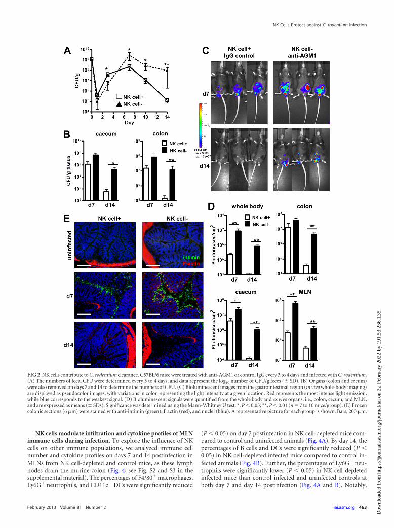
determine the impact of these changes within the NK cell popula-tion on the course of C. rodentium infection in mice, polyclonalanti-AGM1 antibody was used to actively deplete the NK cell pop-ulation from murine tissues (6). We observed that NK cell-de-pleted mice had significantly higher (P 0.05) numbers of C.rodentium organisms than C. rodentium-infected control mice byday 7 and until the study end in feces and by day 14 in the colonand cecum (Fig. 2A and B). Bioluminescence imaging analysisconfirmed these observations. NK cell-depleted mice infectedwith C. rodentium had a significantly higher (P 0.01) C. roden-tium bioluminescence signal from the lower abdomen on days 7and 14 than control infected mice (Fig. 2C and D). When organswere excised and imaged, NK cell-depleted infected mice had sig-nificantly higher bioluminescence signals (P 0.05) in the cecumand MLN on days 7 and 14 and the colon on day 14 than controlmice infected with C. rodentium. Notably, at the end of the study(day 14), NK cell-depleted mice demonstrated delayed C. roden-tium clearance (Fig. 2). Furthermore, colonic staining for the A/Evirulence factor intimin was observed in control and NK cell-depleted mice on day 7 but only NK cell-depleted mice on day 14(Fig. 2E). Taken together, these data indicate that NK cells arerequired to reduce the C. rodentium burden.
NK cells influence colonic pathology after C. rodentium in-fection. C. rodentium infection is associated with colonic crypthyperplasia, goblet cell depletion, mucosal inflammation, anderosion. Epithelial hyperplasia can be indirectly assessed bychanges in distal colonic weights. Infected mice had significantlyheavier (P 0.05) colons on days 7 and 14 postinfection thanuninfected mice (Fig. 3A). Notably, distal colons from NK cell-depleted mice infected with C. rodentium were significantlyheavier (P 0.05) on day 14 than colons from control infectedmice, suggesting increased hyperplasia during infection (Fig. 3A).However, histological analysis and scoring revealed that NKcell-depleted infected mice had significantly less (P 0.01) in-flammation, as exemplified by shorter colonic crypts and reducedleukocyte infiltration, especially at day 14, than control C. roden-tium-infected mice (Fig. 3B and C). Interestingly, NK cell-de-pleted mice had larger and significantly greater (P 0.001) num-bers of goblet cells at day 14 postinfection, in contrast to thecharacteristic goblet cell depletion in control infected mice (Fig.3D to F). Consistently, analysis of cytokine levels in colon homog-enates revealed significantly reduced (P 0.05) levels of IFN-�,IL-10, murine KC (mKC), and IL-17 in NK cell-depleted mice atboth 7 and 14 days postinfection than control mice (Fig. 3G andH). TNF-� was significantly reduced (P 0.01) on day 7 but notday 14 in NK cell-depleted mice compared to the levels in controlinfected mice. Increased levels of IL-12 were found in NK cell-depleted mice at 7 days postinfection but not on day 14 comparedto the levels in control C. rodentium-infected mice (Fig. 3G andH). Significantly lower levels (P 0.001) of IL-1� were detected inNK cell-depleted mice on day 14 after C. rodentium infection butnot on day 7 compared to the levels in control infected mice (Fig.3G and H). No major changes in colonic IL-2, IL-4, IL-5, andIL-22 were found between the different groups at the differenttime points analyzed (Fig. 3G and H and data not shown). Thus,the absence of NK cells appears to result in less inflammation andenhanced epithelial function, suggesting that NK cells contributeto the pathology (possibly via recruitment and/or activation ofother cell types) associated with infection.
FIG 1 NK cells are recruited and express immune-modulatory factors after C.rodentium infection. Cells were isolated from the MLNs and spleens of C.rodentium-infected or uninfected control C57BL/6 mice at various time pointsand analyzed by flow cytometry. (A) Mean NK cell percentage � SD. (B)Frozen colonic sections (6 �m) were stained with anti-NKp46 (green) andnuclei (blue) from uninfected control and infected mice at days 7 and 14. Arepresentative picture for each group is shown. Bars, 200 �m. (C) Total num-ber of positive cells per mm2. Values are based on 6 mice/time point, withvalues in 5 to 10 fields being measured. Arrows highlight the locations of theNK cells. cLP cells were stimulated for 6 h with BD leukocyte activation cock-tail plus GolgiPlug and stained with NK1.1 and CD3, permeabilized, andstained with an anti-cytokine fluorochrome-labeled MAb. Data represent thepercentage of cytokine-positive NK cells out of the total NK cell population onday 7 (D) and day 14 (E). Data are means � SDs (n � 7; 2 independentexperiments). *, P 0.05 versus uninfected control mice; **, P 0.01 versusuninfected control mice; ***, P 0.001 versus uninfected control mice; NS,not significant; d7, day 7; d14, day 14.
Hall et al.
462 iai.asm.org Infection and Immunity
Dow
nloa
ded
from
http
s://j
ourn
als.
asm
.org
/jour
nal/i
ai o
n 22
Feb
ruar
y 20
22 b
y 19
1.53
.236
.135
.
NK cells modulate infiltration and cytokine profiles of MLNimmune cells during infection. To explore the influence of NKcells on other immune populations, we analyzed immune cellnumber and cytokine profiles on days 7 and 14 postinfection inMLNs from NK cell-depleted and control mice, as these lymphnodes drain the murine colon (Fig. 4; see Fig. S2 and S3 in thesupplemental material). The percentages of F4/80� macrophages,Ly6G� neutrophils, and CD11c� DCs were significantly reduced
(P 0.05) on day 7 postinfection in NK cell-depleted mice com-pared to control and uninfected animals (Fig. 4A). By day 14, thepercentages of B cells and DCs were significantly reduced (P 0.05) in NK cell-depleted infected mice compared to control in-fected animals (Fig. 4B). Further, the percentages of Ly6G� neu-trophils were significantly lower (P 0.05) in NK cell-depletedinfected mice than control infected and uninfected controls atboth day 7 and day 14 postinfection (Fig. 4A and B). Notably,
FIG 2 NK cells contribute to C. rodentium clearance. C57BL/6 mice were treated with anti-AGM1 or control IgG every 3 to 4 days and infected with C. rodentium.(A) The numbers of fecal CFU were determined every 3 to 4 days, and data represent the log10 number of CFU/g feces (� SD). (B) Organs (colon and cecum)were also removed on days 7 and 14 to determine the numbers of CFU. (C) Bioluminescent images from the gastrointestinal region (in vivo whole-body imaging)are displayed as pseudocolor images, with variations in color representing the light intensity at a given location. Red represents the most intense light emission,while blue corresponds to the weakest signal. (D) Bioluminescent signals were quantified from the whole body and ex vivo organs, i.e., colon, cecum, and MLN,and are expressed as means (� SDs). Significance was determined using the Mann-Whitney U test: *, P 0.05; **, P 0.01 (n � 7 to 10 mice/group). (E) Frozencolonic sections (6 �m) were stained with anti-intimin (green), F actin (red), and nuclei (blue). A representative picture for each group is shown. Bars, 200 �m.
NK Cells Protect against C. rodentium Infection
February 2013 Volume 81 Number 2 iai.asm.org 463
Dow
nloa
ded
from
http
s://j
ourn
als.
asm
.org
/jour
nal/i
ai o
n 22
Feb
ruar
y 20
22 b
y 19
1.53
.236
.135
.

Hall et al.
464 iai.asm.org Infection and Immunity
Dow
nloa
ded
from
http
s://j
ourn
als.
asm
.org
/jour
nal/i
ai o
n 22
Feb
ruar
y 20
22 b
y 19
1.53
.236
.135
.

when total cell numbers were examined, we observed significantlyreduced levels of all immune populations analyzed in NK cell-depleted mice compared to control infected animals (see Fig. S2 inthe supplemental material).
We also analyzed cytokine profiles from MLN immune cellsduring C. rodentium infection from both NK cell-depleted andcontrol mice (see Fig. S3 in the supplemental material). We ob-served significantly lower (P 0.001) percentages of T cells ex-pressing IL-17 in NK cell-depleted mice than both control anduninfected mice on days 7 and 14 postinfection and IFN-�-posi-tive T cells on day 14 (P 0.05). IFN-�- and TNF-�-positiveneutrophils, DCs, and macrophages were also significantly re-duced (P 0.05) in NK cell-depleted mice compared to controland uninfected mice at both infection time points. At day 7, IL-10-positive macrophages were significantly lower (P 0.01) InNK cell-depleted infected mice than uninfected and control in-fected mice. As a marker of antimicrobial activity, ROS produc-tion from phagocytes was assessed (Fig. 4C and D). On days 7 and14 postinfection, NK cell-depleted mice had significantly lowerpercentages (P 0.05) of macrophages and neutrophils produc-ing ROS than control or uninfected mice. These data indicate thatNK cells modulate the activation and function of other immunecell populations during C. rodentium infection.
NK cells recruit colonic effector cells during infection. C. ro-dentium induces a localized inflammatory response in the colon,characterized by infiltration of immune cells such as neutrophilsand lymphocytes. At day 7 postinfection, NK cell-depleted micehad significantly lower (P 0.001) counts of Ly6G� neutrophilsthan control mice (Fig. 5A and B). Notably, by day 14, all immunepopulations examined, including CD3� T cells, CD19� B cells,CD11c� DCs, F4/80� macrophages, and neutrophils, were signif-icantly reduced (P 0.001) in the colons of NK cell-depleted micecompared to the colons of control C. rodentium-infected mice(Fig. 5C and D). These data suggest that NK cells are crucial for theregulated recruitment of other immune cell populations to theinflamed colon during infection.
NK cells limit extracolonic dissemination of C. rodentium.C. rodentium infection is restricted to the cecum and colon ofC57BL/6 mice. To determine if NK cells had a role in preventingdisseminated infection, we measured the bacterial loads in varioussystemic tissues (liver, kidney, and spleen) following infection.NK cell-depleted mice had high numbers of bacterial CFU in allsystemic organs on days 7 and 14 postinfection, whereas no suchincrement was detected in control infected mice (Fig. 6A). Theseobservations suggest that the presence of NK cells prevents dis-semination of C. rodentium from the colon to peripheral tissues.
NK cells can directly kill C. rodentium in vitro. Our observa-tion that bacterial loads in NK cell-depleted mice were greaterthan those in control animals suggested the possibility that NKcells might have a direct cytotoxic effect on C. rodentium. To testwhether NK cells directly kill C. rodentium, live bacteria weremixed with cultured NK cells and bacterial survival was quanti-tated. NK cells exhibited significant efficiency (P 0.05) in killingC. rodentium after 2 h of coculture compared to medium alone(Fig. 6B). Notably, we did not observe any significant killing in
FIG 3 NK cells impact colonic pathology after C. rodentium infection. Anti-AGM1- and IgG-treated control mice had colons removed at days 7 and 14postinfection. (A) Mean colon weight (mg/cm � SD). (B) Representative histology of H&E-stained colon. Bars, 200 �m. (C) Mean inflammatory scores of IgGcontrol or anti-AGM1-treated mice on days 7 and 14 postinfection determined by histological analysis of distal colonic sections. (D) Sections of H&E-stainedcolonic tissue on day 14, with differences in goblet cells indicated by arrows. Bars, 100 �m. (E) Alcian blue, counterstained with hematoxylin, indicating (in blue)mucus-producing goblet cells. Bars, 100 �m. (F) Means � SDs of the total number of goblet cell per mm2 at day 14 postinfection, based on 6 mice/time point (5to 10 fields). (G and H) Mean cytokine levels in whole colonic homogenates � SD on day 7 (G) and day 14 (H). Data are for 7 mice per group and arerepresentative of two experiments. P values were determined using the Mann-Whitney U test: *, P 0.05; **, P 0.01; ***, P 0.001; NS; not significant.
FIG 4 NK cells modulate infiltration and cytokine profiles of MLN immunecells during infection. Cells were isolated from MLNs of C57BL/6 mice at 7days (A) and 14 days (B) after C. rodentium infection, stained with fluoro-chrome-labeled MAb, and analyzed by flow cytometry. Columns represent themean percentage � SD of seven mice per group from two independent exper-iments. MLN cells were incubated with DHR (a marker for ROS) before addi-tion of PMA. Cells were then analyzed by flow cytometry to determine thepercentage of DHR/ROS-positive cells in the Ly6G� and F4/80� populationson day 7 (C) and day 14 (D). Data represent the percentage of ROS-positivecells out of the total specific cell population � SD. P values were determinedusing one-way analysis of variance, followed by Bonferroni’s multiple-com-parison test: *, P 0.05; **, P 0.01; ***, P 0.001.
NK Cells Protect against C. rodentium Infection
February 2013 Volume 81 Number 2 iai.asm.org 465
Dow
nloa
ded
from
http
s://j
ourn
als.
asm
.org
/jour
nal/i
ai o
n 22
Feb
ruar
y 20
22 b
y 19
1.53
.236
.135
.

FIG 5 NK cells induce recruitment of immune populations to the colons of infected mice. Anti-AGM1- and IgG-treated control mice had colons removed at days7 and 14 postinfection for immunofluorescent staining of cellular populations. (A) Neutrophils (Ly6G, green) and nuclei (blue); (B) mean of the total numberof positive cells per mm2 � SD at day 7 postinfection; (C) neutrophils (Ly6G, green), T cells (CD3, red), B cells (CD19, red), DCs (CD11c, red), macrophages(F4/80, red), and nuclei (blue); (D) mean of the total number of positive cells per mm2 � SD at day 14 postinfection. A representative picture for each group (n �7) is shown. Bars, 200 �m. P values were determined using one-way analysis of variance, followed by Bonferroni’s multiple-comparison test: *, P 0.05; **, P 0.01; ***, P 0.001.
466 iai.asm.org Infection and Immunity
Dow
nloa
ded
from
http
s://j
ourn
als.
asm
.org
/jour
nal/i
ai o
n 22
Feb
ruar
y 20
22 b
y 19
1.53
.236
.135
.

non-NK cell–C. rodentium cultures compared to medium aloneover 3 h. These data indicate that NK cells are directly cytotoxic toC. rodentium.
DISCUSSION
Enteroadherent pathogens such as EPEC, EHEC, and C. roden-tium cause disease characterized by colonic inflammation and di-arrhea. While previous studies have elucidated the pathogenicstrategies employed by these pathogens, the role of the host im-mune system, particularly the innate arm of the immune re-sponse, is still poorly understood. We provide evidence that NKcells protect the host from prolonged mucosal and systemic infec-tion by generating direct cytokine and antimicrobial cytotoxic fac-tors and by providing signals regulating infiltration and increasedactivation of other immune cell populations. Therefore, while NKcells contribute to colonic inflammation, this response is also cru-cial for reducing the bacterial burden.
After oral infection with C. rodentium, the distal colon is pref-erentially colonized by the bacterium through intimate attach-ment to the gut epithelium. Breaches in the intestinal barrier afterA/E lesion formation lead to acute inflammation, peaking at 1week postinfection in C57BL/6 mice (9). During C. rodentiuminfection, we observed NK cell recruitment, in tandem with strongexpression of cytokine and cytotoxic factors, into mucosal andsystemic tissues, suggesting that NK cells play an important roleduring C. rodentium infection. Data from bioluminescencewhole-body and organ-specific imaging and viable bacterialcounts revealed that NK cell-depleted mice had delayed bacterialclearance. In addition to a higher bacterial burden, reduced im-mune cell infiltration and diminished cytokine levels within the
MLNs and colons of infected mice were observed in NK cell-de-pleted mice.
Previous studies have highlighted the importance of neutro-phils in defense against C. rodentium (17). IL-17 coordinates earlyrecruitment and activation of neutrophils to infection sitesthrough chemokine expression, including the induction of mKC(18). It is noteworthy that within the colons of NK cell-depletedinfected mice we observed lower levels of IL-17 and mKC and,correspondingly, diminished neutrophil infiltration, potentiallydue to the absence of NK cell and/or NK cell-activated T cell-derived IL-17. While only neutrophil infiltration was impaired onday 7 postinfection in NK cell-depleted mice, by day 14 all otherimmune populations examined were markedly reduced withinNK cell-depleted colons. IFN-� is readily produced by NK cellsafter activation, which we also observed during C. rodentium in-fection. IFN-� is known to regulate chemokine production and,with regard to bacterial infection, has been shown to determinethe outcome of Staphylococcus aureus infection through chemo-kine-induced immune cell recruitment (19). Furthermore, NKcell-derived cytokine production, particularly IFN-� production,is required for maturation and activation of other immune cellpopulations for clearance of infection (3). These data support theevidence for the role of NK cell-derived signals in recruitment ofimmune cells to resolve infection with C. rodentium. Several stud-ies have identified a new and distinct population of IL-22-produc-ing, NKp46-positive (NKp46�) transcription factor ROR�t-pos-itive (ROR�t�) gut-associated mucosal cells (20, 21). These cellshave been shown to play an IL-22-protective role in host defenseagainst gut pathogens (22). Initially, these cells were thought to bea new subset of classical NK cells, as they express the activationreceptor NKp46. However, these cells do not express other well-characterized NK cell markers, such as NK1.1 or DX5, are notcytotoxic, and do not produce IFN-�. Importantly, we did notdetect any significant differences between infected groups for IL-22, suggesting that IL-22 production from the resident NKp46�
ROR�t� subset population and/or the presence of activated Tcells cannot explain why NK cell-deficient mice have increasedenteric infection in our model (23, 24)
DCs and macrophages require help from NK cells for matura-tion, proper antigen presentation function, and priming of T cellresponses (25). This bidirectional cross talk during the earlyphases of immunity influences the following type and magnitudeof adaptive immune response (2). It is noteworthy that in ourstudy, mucosal infiltration and cytokine expression of both T andB cells were significantly impaired in NK cell-depleted mice. Pre-vious work in RAG1-knockout mice has highlighted the impor-tance of T and B cells for the clearance of C. rodentium (11). In-terestingly, these mice also had transient, inflammatory, and crypthyperplastic responses that subsided after 2 weeks, similar to ourobservations in NK cell-depleted mice during infection (11).
C. rodentium growth is restricted to the colon, with little bac-terial translocation except in highly susceptible mouse strains,such as C3H (26). In NK cell-depleted mice, we observed signifi-cant dissemination of the bacterium to systemic organs, includingthe spleen, kidneys, and liver, in stark contrast to control mice,which exhibited no extracolonic spread. Previous work has alsoshown a role for NK cells in preventing systemic bacterial spreadfrom initial mucosal colonization of the lungs during Mycobacte-rium tuberculosis infection (27). Our data reveal a mechanism bywhich NK cells may protect the host from systemic bacterial
FIG 6 NK cells limit systemic spread of C. rodentium and have a direct anti-microbial effect in vitro. C57BL/6 mice were treated with anti-AGM1 or con-trol IgG every 3 to 4 days and infected with C. rodentium. (A) The bacterial load(numbers of CFU) in systemic organs was determined on days 7 and 14, anddata represent the log10 number of CFU/g tissue (� SD; n � 6/time point).ND, not detectable. (B) NK cells or non-NK cells (both at 1 � 105 cells) ormedium alone was coincubated with C. rodentium (1 � 106 CFU of bacteria)for 1, 2, and 3 h of culture (multiplicity of infection, 10:1). Data are the meannumbers of CFU at each time point and are representative of two independentexperiments. P values were determined using the Kruskal-Wallis test, followedby Dunn’s multiple-comparison test: *, P 0.05; **, P 0.01; ***, P 0.001.
NK Cells Protect against C. rodentium Infection
February 2013 Volume 81 Number 2 iai.asm.org 467
Dow
nloa
ded
from
http
s://j
ourn
als.
asm
.org
/jour
nal/i
ai o
n 22
Feb
ruar
y 20
22 b
y 19
1.53
.236
.135
.

spread. ROS are involved in host defense to bacterial pathogensand are induced in response to proinflammatory cytokines such asIFN-� (28). We observed diminished ROS production from bothneutrophils and macrophages in NK cell-depleted mice, suggest-ing that NK cell-derived cytokines may stimulate ROS productionby granulocytes, thereby increasing bacterial killing both withinthe colon and also within distal sites. Another mechanism bywhich NK cells may protect the host from systemic spread of bac-teria is to kill C. rodentium directly. NK cells may mediate antibac-terial effects through the indirect action of secreted mediators or,in the case of infected host cells, by direct cytolytic activity. Weobserved that NK cells also have significant antibacterial activityagainst extracellular C. rodentium. Indeed, NK cells may kill bac-teria in the colon as well as systemically, where they are present inlarger numbers, and thereby prevent dissemination of bacteria tosystemic organs.
The signals which activate NK cells to mediate direct damage offree bacteria are poorly understood. Many NK cell receptors areknown to bind several viral as well as host cell proteins, and recentstudies have shown that NKp44 directly binds ligands on the sur-face of M. tuberculosis and Pseudomonas aeruginosa (29). Unde-fined ligands from C. rodentium might be recognized by NK cellreceptors, although studies examining NKp46 suggest that thisparticular activating receptor is dispensable for defense against C.rodentium (30). Secreted NK-derived factors, other than cyto-kines, could also play a role in bacterial clearance. Defensins areantimicrobial peptides, expressed mainly by epithelial cells andimmune cells, that disrupt the bacterial membrane, leading topathogen death (31). Previous studies have shown that NK cellscan directly recognize pathogen-associated molecular patterns onthe surface of bacteria and respond by production of �-defensins(32). This may also represent a direct cytotoxic pathway involvedin NK cell-mediated protection against C. rodentium.
Goblet cells are highly specialized secretory cells present in theintestinal tract that produce mucins which form a dynamic defen-sive mucus barrier against enteric pathogens (33). Previous workhas shown that infection by several enteric pathogens, includingC. rodentium, leads to a marked reduction in the number of gobletcells, and this may represent a mechanism for subversion of thishost defense (34). In contrast to infected murine controls, wherewe observed goblet cell depletion, NK cell-depleted mice had asignificant increase in the number and size of goblet cells. Previousstudies have demonstrated that while colonic goblet cells can besubject to direct infection and potential subversion by A/E patho-gens in vivo, it is the host immune system that primarily modulatesthe function of these cells during infection (35). Concurrent withthis idea, the reduced immune cell infiltration observed in NKcell-depleted mice during C. rodentium infection may account forthe absence of characteristic goblet cell depletion in our study.Indeed, goblet cell hyperplasia may represent a compensatorymechanism in the absence of normal immune cell infiltration andmay also account for the increased colonic mass observed withinNK cell-depleted mice.
In summary, we show the complex nature of NK cell involve-ment in protection against the A/E enteric pathogen C. rodentium.NK cells are necessary for reducing pathogen burden. They medi-ate their effects through cytokine production and the subsequentregulation of innate immune populations, leading to the genera-tion of robust adaptive immunity. NK cells are also able to directlykill C. rodentium while regulating the antimicrobial activity of
granulocytes and protect the host from bacterial dissemination.Indeed, this comprehensive anti-infection NK cell response mayrepresent a promising therapeutic focus, particularly for ap-proaches against enteric bacterial infections.
ACKNOWLEDGMENT
This work was supported by Science Foundation Ireland through the IrishGovernment’s National Development Plan (02/CE/B124 and 07/CE/B1368).
REFERENCES1. Pegram HJ, Andrews DM, Smyth MJ, Darcy PK, Kershaw MH. 2011.
Activating and inhibitory receptors of natural killer cells. Immunol. CellBiol. 89:216 –224.
2. Vivier E, Raulet DH, Moretta A, Caligiuri MA, Zitvogel L, Lanier LL,Yokoyama WM, Ugolini S. 2011. Innate or adaptive immunity? Theexample of natural killer cells. Science 331:44 – 49.
3. Horowitz A, Stegmann KA, Riley EM. 2011. Activation of natural killer cellsduring microbial infections. Front. Immunol. 2:88. doi:10.3389/fimmu.2011.00088.
4. Degli-Esposti MA, Smyth MJ. 2005. Close encounters of different kinds:dendritic cells and NK cells take centre stage. Nat. Rev. 5:112–124.
5. Nedvetzki S, Sowinski S, Eagle RA, Harris J, Vely F, Pende D, Trows-dale J, Vivier E, Gordon S, Davis DM. 2007. Reciprocal regulation ofhuman natural killer cells and macrophages associated with distinct im-mune synapses. Blood 109:3776 –3785.
6. Hall LJ, Clare S, Dougan G. 2010. NK cells influence both innate andadaptive immune responses after mucosal immunization with antigenand mucosal adjuvant. J. Immunol. 184:4327– 4337.
7. Croxen MA, Finlay BB. 2010. Molecular mechanisms of Escherichia colipathogenicity. Nat. Rev. Microbiol. 8:26 –38.
8. Nataro JP, Kaper JB. 1998. Diarrheagenic Escherichia coli. Clin. Micro-biol. Rev. 11:142–201.
9. Mundy R, MacDonald TT, Dougan G, Frankel G, Wiles S. 2005.Citrobacter rodentium of mice and man. Cell. Microbiol. 7:1697–1706.
10. Wiles S, Clare S, Harker J, Huett A, Young D, Dougan G, Frankel G.2004. Organ specificity, colonization and clearance dynamics in vivo fol-lowing oral challenges with the murine pathogen Citrobacter rodentium.Cell. Microbiol. 6:963–972.
11. Vallance BA, Deng W, Knodler LA, Finlay BB. 2002. Mice lacking T andB lymphocytes develop transient colitis and crypt hyperplasia yet sufferimpaired bacterial clearance during Citrobacter rodentium infection. In-fect. Immun. 70:2070 –2081.
12. Bry L, Brigl M, Brenner MB. 2006. CD4�-T-cell effector functions andcostimulatory requirements essential for surviving mucosal infection withCitrobacter rodentium. Infect. Immun. 74:673– 681.
13. Simmons CP, Clare S, Ghaem-Maghami M, Uren TK, Rankin J, HuettA, Goldin R, Lewis DJ, MacDonald TT, Strugnell RA, Frankel G,Dougan G. 2003. Central role for B lymphocytes and CD4� T cells inimmunity to infection by the attaching and effacing pathogen Citrobacterrodentium. Infect. Immun. 71:5077–5086.
14. Ishigame H, Kakuta S, Nagai T, Kadoki M, Nambu A, Komiyama Y,Fujikado N, Tanahashi Y, Akitsu A, Kotaki H, Sudo K, Nakae S,Sasakawa C, Iwakura Y. 2009. Differential roles of interleukin-17A and-17F in host defense against mucoepithelial bacterial infection and allergicresponses. Immunity 30:108 –119.
15. Murphy CT, Hall LJ, Hurley G, Quinlan A, Macsharry J, Shanahan F,Nally K, Melgar S. 2012. The sphingosine-1-phosphate analogue FTY720impairs mucosal immunity and clearance of the enteric pathogen Citro-bacter rodentium. Infect. Immun. 80:2712–2723.
16. Perfetto SP, Chattopadhyay PK, Lamoreaux L, Nguyen R, Ambrozak D,Koup RA, Roederer M. 2006. Amine reactive dyes: an effective tool todiscriminate live and dead cells in polychromatic flow cytometry. J. Im-munol. Methods 313:199 –208.
17. Lebeis SL, Bommarius B, Parkos CA, Sherman MA, Kalman D. 2007.TLR signaling mediated by MyD88 is required for a protective innateimmune response by neutrophils to Citrobacter rodentium. J. Immunol.179:566 –577.
18. Hartupee J, Liu C, Novotny M, Li X, Hamilton T. 2007. IL-17 enhanceschemokine gene expression through mRNA stabilization. J. Immunol.179:4135– 4141.
Hall et al.
468 iai.asm.org Infection and Immunity
Dow
nloa
ded
from
http
s://j
ourn
als.
asm
.org
/jour
nal/i
ai o
n 22
Feb
ruar
y 20
22 b
y 19
1.53
.236
.135
.

19. McLoughlin RM, Lee JC, Kasper DL, Tzianabos AO. 2008. IFN-gammaregulated chemokine production determines the outcome of Staphylococ-cus aureus infection. J. Immunol. 181:1323–1332.
20. Cella M, Fuchs A, Vermi W, Facchetti F, Otero K, Lennerz JK, Doherty JM,Mills JC, Colonna M. 2009. A human natural killer cell subset provides aninnate source of IL-22 for mucosal immunity. Nature 457:722–725.
21. Sanos SL, Bui VL, Mortha A, Oberle K, Heners C, Johner C, DiefenbachA. 2009. RORgammat and commensal microflora are required for thedifferentiation of mucosal interleukin 22-producing NKp46� cells. Nat.Immunol. 10:83–91.
22. Zheng Y, Valdez PA, Danilenko DM, Hu Y, Sa SM, Gong Q, Abbas AR,Modrusan Z, Ghilardi N, de Sauvage FJ, Ouyang W. 2008. Interleu-kin-22 mediates early host defense against attaching and effacing bacterialpathogens. Nat. Med. 14:282–289.
23. Colonna M. 2009. Interleukin-22-producing natural killer cells and lym-phoid tissue inducer-like cells in mucosal immunity. Immunity 31:15–23.
24. Wilson NJ, Boniface K, Chan JR, McKenzie BS, Blumenschein WM,Mattson JD, Basham B, Smith K, Chen T, Morel F, Lecron JC, KasteleinRA, Cua DJ, McClanahan TK, Bowman EP, de Waal Malefyt R. 2007.Development, cytokine profile and function of human interleukin 17-producing helper T cells. Nat. Immunol. 8:950 –957.
25. Vitale M, Della Chiesa M, Carlomagno S, Pende D, Arico M, MorettaL, Moretta A. 2005. NK-dependent DC maturation is mediated by TNFal-pha and IFNgamma released upon engagement of the NKp30 triggeringreceptor. Blood 106:566 –571.
26. Vallance BA, Deng W, Jacobson K, Finlay BB. 2003. Host susceptibilityto the attaching and effacing bacterial pathogen Citrobacter rodentium.Infect. Immun. 71:3443–3453.
27. Feng CG, Kaviratne M, Rothfuchs AG, Cheever A, Hieny S, Young HA,
Wynn TA, Sher A. 2006. NK cell-derived IFN-gamma differentially reg-ulates innate resistance and neutrophil response in T cell-deficient hostsinfected with Mycobacterium tuberculosis. J. Immunol. 177:7086 –7093.
28. Fang FC. 2011. Antimicrobial actions of reactive oxygen species. mBio2:e00141–11. doi:10.1128/mBio.00141-11.
29. Esin S, Batoni G, Counoupas C, Stringaro A, Brancatisano FL, ColoneM, Maisetta G, Florio W, Arancia G, Campa M. 2008. Direct binding ofhuman NK cell natural cytotoxicity receptor NKp44 to the surfaces ofmycobacteria and other bacteria. Infect. Immun. 76:1719 –1727.
30. Satoh-Takayama N, Dumoutier L, Lesjean-Pottier S, Ribeiro VS, Man-delboim O, Renauld JC, Vosshenrich CA, Di Santo JP. 2009. The naturalcytotoxicity receptor NKp46 is dispensable for IL-22-mediated innate in-testinal immune defense against Citrobacter rodentium. J. Immunol. 183:6579 – 6587.
31. Doss M, White MR, Tecle T, Hartshorn KL. 2010. Human defensins andLL-37 in mucosal immunity. J. Leukoc. Biol. 87:79 –92.
32. Chalifour A, Jeannin P, Gauchat JF, Blaecke A, Malissard M, N=GuyenT, Thieblemont N, Delneste Y. 2004. Direct bacterial protein PAMPrecognition by human NK cells involves TLRs and triggers alpha-defensinproduction. Blood 104:1778 –1783.
33. Kim YS, Ho SB. 2010. Intestinal goblet cells and mucins in health anddisease: recent insights and progress. Curr. Gastroenterol. Rep. 12:319 –330.
34. McGuckin MA, Linden SK, Sutton P, Florin TH. 2011. Mucin dynamicsand enteric pathogens. Nat. Rev. Microbiol. 9:265–278.
35. Bergstrom KS, Guttman JA, Rumi M, MA C, Bouzari S, Khan MA,Gibson DL, Vogl AW, Vallance BA. 2008. Modulation of intestinalgoblet cell function during infection by an attaching and effacing bacterialpathogen. Infect. Immun. 76:796 – 811.
NK Cells Protect against C. rodentium Infection
February 2013 Volume 81 Number 2 iai.asm.org 469
Dow
nloa
ded
from
http
s://j
ourn
als.
asm
.org
/jour
nal/i
ai o
n 22
Feb
ruar
y 20
22 b
y 19
1.53
.236
.135
.