natural killer t cells in adipose tissue are activated in...
TRANSCRIPT
—Original—
Natural Killer T Cells in Adipose Tissue Are Activated in Lean Mice
Taisuke Kondo, Yujiro ToYoshima, Yoshiyuki ishii, and shigeru KYuwa
Department of Biomedical Science, Graduate School of Agricultural and Life Sciences, The University of Tokyo, 1–1–1 Yayoi, Bunkyo-ku, Tokyo 113-8657, Japan
Abstract: Adipose tissues are closely connected with the immune system. It has been suggested that metabolic syndromes such as type 2 diabetes, arteriosclerosis and liver steatosis can be attributed to adipose tissue inflammation characterized by macrophage infiltration. To understand a physiological and pathological role of natural killer T (NKT) cells on inflammation in adipose tissue, we characterized a subset of NKT cells in abdominal and subcutaneous adipose tissues in C57BL/6J mice fed normal or high-fat diets. NKT cells comprised a larger portion of lymphocytes in adipose tissues compared with the spleen and peripheral blood, with epididymal adipose tissue having the highest number of NKT cells. Furthermore, some NKT cells in adipose tissues expressed higher levels of CD69 and intracellular interferon-γ, whereas the Vβ repertoires of NKT cells in adipose tissues were similar to other cells. In obese mice fed a high-fat diet, adipose tissue inflammation had little effect on the Vβ repertoire of NKT cells in epididymal adipose tissues. We speculate that the NKT cells in adipose tissues may form an equivalent subset in other tissues and that these subsets are likely to participate in adipose tissue inflammation. Additionally, the high expression level of CD69 and intracellular IFN-γ raises the possibility that NKT cells in adipose tissue may be stimulated by some physiological mechanism.Key words: adipose tissue, diet induced obesity, NKT cell, Vβ repertoire
Introduction
The world health organization (who) recently es-timated that more than one billion adults were over-weight and 500 million were obese [34]. numerous re-cent studies have shown that not only are metabolic syndromes such as type 2 diabetes, dyslipidemia, liver steatosis and arteriosclerosis caused by obesity, but also are cancers, strokes and other noncommunicable dis-eases [10].
over the past 10 years, a large number of studies have established that chronic adipose tissue inflammation is closely associated with metabolic syndromes. macro-phages resident in adipose tissues activated by the innate
immune response are thought to induce adipocyte death and lead to the secretion of several inflammatory me-diators, resulting in inflammation in adipose tissues. Studies have shown that tumor necrosis factor α (TNF-α), IL-6, and IL-1β are produced in adipose tissues and the liver, and are exposed to inflammatory mediators. A long-term, low-dose exposure to these cytokines is connected to adipose tissue insulin resistance [7]. other immune cell types, such as T helper cells, cytotoxic T cells, B cells, and mast cells, also cause adipose tissue inflam-mation [19, 24, 32, 33]. This makes it necessary to un-derstand the role of each of the immune cells involved in adipose tissue inflammation, to develop new immu-nological therapies for metabolic syndrome. although
(Received 18 November 2012 / Accepted 7 May 2013)Address corresponding: S. Kyuwa, Department of Biomedical Science, Graduate School of Agricultural and Life Sciences, The University of Tokyo, 1–1–1 Yayoi, Bunkyo-ku, Tokyo 113-8657, Japan
Exp. Anim. 62(4), 319–328, 2013
©2013 Japanese association for Laboratory animal science

T. Kondo, ET AL.320
previous studies have reported a role for nKT cells in adipose tissue inflammation [17, 21, 27], this is contro-versial and little is known of the nKT cells resident in adipose tissue in lean mice.
nKT cells have the properties of nK and T cells. un-like T cells, which recognize peptides, nKT cells are unique in recognizing glycolipids as antigens. These antigens are presented by the major histocompatibility complex (mhC) class 1-like molecule, Cd1d [12]. Re-cent research suggests that nKT cells comprise various subsets. a major one of these, invariant nKT (inKT) (also known type 1 nKT) cells, expresses an invariant T cell antigen receptor (TCR), the α-chain of which is formed by a variable region 14 α-chain and joint region 18 α-chain (Vα14Jα18) in mice and Vα24Jα18 in hu-mans, and the variable β-chain (Vβ) of which has the following variants: Vβ8, Vβ7, and Vβ2. Moreover, NKT cells are classified into CD4-positive and CD4-negative groups [12].
nKT cells may be involved in both defense against microbial pathogens and the anti-tumor response; this protective effect may be associated with the production of T helper 1 (Th1) cytokines, including interferon-gamma (IFN-γ) [8]. Antitumor NKT cells resident in the liver are primarily Cd4-negative [6]. additionally, nKT cells are thought to be involved in the regulation of sev-eral autoimmune diseases including type 1 diabetes, bronchial asthma and experimental autoimmune en-cephalomyelitis [22, 25, 26, 27]. For example, produc-tion of Th2 cytokines, including iL-4 and iL-13, by nKT cells not only protects against experimental autoimmune encephalomyelitis by suppressing the Th1 response, but also induces airway hyperreactivity, a prime feature of the Th2 response, which promotes asthma [22, 27].
The immunological functions of nKT cells are thought to change within the subsets and according to environ-ment [4, 29]. Therefore, identification of NKT cell sub-sets resident in adipose tissue may provide an important clue to the physiological and pathological roles of nKT cells. white adipose tissues are present in the abdominal cavity and subcutaneous areas, and are anatomically and physiologically distinct [15]. in this study, we evaluated the proportions of nKT, nK and T cells in abdominal and subcutaneous adipose tissue, the spleen and periph-eral blood and identified a major subset of NKT cells resident within adipose tissue. moreover, we show that nKT cells in adipose tissue were more active than those in the spleen and peripheral blood, and we investigated
the TCR Vβ repertoire and the change in the main subset of nKT cells in obesity using diet-induced obese mice.
Materials and Methods
Animalswe used male C57BL/6J mice. mice were housed
under a 12-h light/12-h dark cycle and fed a normal CRF-1 diet (6% fat, oriental Yeast Co., Ltd., Tokyo, Japan) or a high-fat diet (d12492, 60 Kcal% fat, Research di-ets, new Brunswick, nJ, usa) ad libitum. For the diet-induced obese (dio) mice, C57BL/6J mice were ob-tained at 7 weeks, acclimated for 1 week and then fed a high-fat diet from 8 until 20 weeks. mice were main-tained under an SPF environment, and all animal ex-periments were approved by the institutional animal Care and use Committee of the university of Tokyo, and performed in accordance with the guidelines for animal experiments.
Flow cytometrywe referred to adipose tissue protocols described by
Brake et al. [2]. Epididymal adipose tissue and the spleen were removed from 20-week-old C57BL/6J male mice. Stromal vascular fraction (SVF) from adipose tissues was isolated by digesting adipose tissue with 2 mg/ml type 2 collagenase (worthington Biochemical Corpora-tion, Lakewood, nJ, usa), removing adipocytes. sple-nocytes were isolated by physically dissociating spleens between the frosted ends of two glass slides. Red blood cells in SVF and splenocytes were removed with ACK lysing buffer.
The following monoclonal antibodies which were conjugated with biotin, FITC, PE, PE-Cy7, PerCP-Cy5.5, aPC, aPC-Cy7, and PE-Cy7 were purchased from eBioscience (san diego, Ca, usa), miltenyi Bio-tec (Bergisch Gladbach, Germany) and BioLegend (san Diego, CA, USA): anti-CD3ε (145-2C11), anti-NK1.1 (PK136), anti-CD69 (H1.2F3) and anti-IFN-γ (XMG1.2) antibodies, and rat IgG isotype control (eBRG1). Purified anti-FcγRII/III (2.4G2) antibody for FcR blocking was purchased from ancell (Bayport, mn, usa). streptavi-din PerCP-Cy5.5 was purchased from eBioscience.
For intracellular cytokines staining, SVF was cultured in RPmi-1640 (Life Technologies, Carlsbad, Ca, usa) supplemented with 10% FBS, and stimulated with 50 ng/ml phorbol 12-myristate 13-acetate (Pma) (Life Technologies) and 500 ng/ml ionomycin (Life Tech-
NKT CELLS ARE ACTIVATED IN ADIPOSE TISSUES 321
nologies) for 5 h, and added to 10 µg/ml brefeldin a (eBioscience) at 2 h after stimulation at 37°C. single-cell suspensions were incubated with anti-FcγRII/III anti-body for 20 min, incubated with anti-cell surface mark-er mabs for 30 min on ice and then washed. subse-quently, the cells were fixed with 4% paraformaldehyde for 10 min at room temperature, and permeabilized by PBs with 0.5% saponin (Life technologies) and 0.5% Bsa, and incubated with anti-cytokine mabs overnight at 4°C. Data were acquired by flow cytometry with; FACSCaliber and FACSCanto II systems (Becton, Dick-inson and Co., Franklin Lakes, NJ, USA). The obtained data were analyzed with the Flowjo software (Tree Star, inc., ashland, oR, usa) and CellQuest software (Bec-ton, Dickinson and Co.). SVF was gated according to cell size (FSC-H) and granularity (SSC-H) criteria as described elsewhere [5].
HistologyMice were sacrificed by cervical dislocation. Epi-
didymal adipose tissues were dissected, fixed in 4% paraformaldehyde, embedded in paraffin and sectioned at 4 µm. sections were stained with hematoxylin and eosin.
Adipose tissue imagingThree-dimensional adipose tissue imaging was per-
formed with a confocal laser scanning microscope (Lsm 510 mETa, Carl Zeiss, oberkochen, Germany), and all images were scanned with <5 µm. Epididymal adipose tissue was removed from 20-week-old C57BL/6J mice and minced into small pieces <1 mm. The fat pieces were fixed with 4% paraformaldehyde for 45 min, permeabi-lized with 1% Triton-X 100 for 10 min and incubated with hoechst 33342 (Life Technologies, Carlsbad, Ca, USA), BODIPY FLC12 (Life Technologies), and purified anti-CD3ε (145-2C11) and anti-NK1.1 (PK136) antibod-ies. dyLight 649-conjugated anti-hamster igG (BioLe-gend, san diego, Ca, usa) and alexa 568-conjugated anti-mouse igG (Life Technologies) antibodies were used as secondary antibodies.
Glucose and insulin tolerance testsGlucose and insulin tolerance tests were performed as
reported previously [13]. Twenty-week-old lean and dio mice were fasted for 16 h but had access to drinking water at all times. on the following day, intraperitoneal glucose or insulin tolerance tests were performed. Blood
glucose concentration was determined using an accu-Chek active blood glucose monitor (Roche diagnostics, Basel, switzerland). mice were administered 2 g/kg glucose intraperitoneally, and the blood glucose concen-tration was measured at 0, 15, 30, 60, 90, 120, 150, and 180 min post injection. For the intraperitoneal insulin tolerance test, mice were administered intraperitoneally 0.5 iu/kg of insulin, and the blood glucose concentration was determined at 0, 30, 45, 60, 75, and 90 min post injection.
Statistical analysisall data were evaluated using a two-tailed student’s
t-test to determine statistical significance, unless other-wise specified. The two-tailed Student’s t-test was per-formed using microsoft Excel (microsoft Corporation, Redmond, washington, usa). a value of P<0.05 was considered to indicate statistical significance.
Results
Correlation between adipose tissue weightswe assessed the relationship between body weight and
adipose tissue weight in 20-week-old male mice. First, we extirpated epididymal and perirenal adipose tissue, and detached femoral subcutaneous adipose tissue from the femoral dermal, and then weighed the tissues. Body weight was not correlated with abdominal adipose tissue weights (data not shown). Both correlation coefficients (body weight vs. epididymal and perirenal adipose tis-sue) were <0.4 (data not shown). in contrast, adipose tissue weight was correlated with other adipose tissue weights, especially that of perirenal adipose tissue, which correlated highly with subcutaneous adipose tis-sue weight (Fig. 1). subcutaneous and visceral adipose tissues are thought to have different metabolic functions, and these results suggested that the quantities of these adipose tissues were equal [15], whereas body weight did not provide an accurate indication of the quantity of adipose tissue in lean mice.
Fraction of lymphocytes in adipose tissues, the spleen and peripheral blood
Both epididymal and subcutaneous adipose tissues contained many primitive lymphocytes, nK cells and NKT cells [5]. To confirm the fraction of lymphocytes present in adipose tissue, we enumerated nK cells (CD3ε−nK1.1+), T cells (CD3ε+nK1.1−), and nKT cells

T. Kondo, ET AL.322
(CD3ε+nK1.1+). Their numbers were compared with those in the spleen (a major lymphoid tissue) and in peripheral blood. adipose tissue consists primarily of adipocytes and stromal and vascular cells; we assessed only stromal and vascular cells by flow cytometry. The percentage of T cells present in epididymal and perirenal adipose tissue was equal to that in peripheral blood, but significantly less than in the spleen. However, the per-centage of T cells in subcutaneous adipose tissue was equal to that in the spleen but slightly more than in pe-ripheral blood (Fig. 2B). The proportions of nK and nKT cells in adipose tissues were higher than those in the spleen and peripheral blood (Fig. 2A and 2C). These results suggest that both nK and nKT cells were found more often in adipose tissue than in other tissues. in addition, these findings suggest that NKT cells play a pivotal role in the physiology of adipose tissue.
Expression of cell-surface markers and intracellular IFN-γ of NKT cells
CD3ε and NK1.1 are cell-surface molecules on NKT
cells [3, 20, 31]. To characterize nKT cells in adipose tissues, the expression levels of CD3ε and NK1.1 were determined. CD3ε expression on NKT cells in subcuta-neous adipose tissue was significantly higher than in the spleen and peripheral blood (Fig. 3A), whereas expres-sion of nK1.1 in epididymal and subcutaneous adipose tissues was significantly higher than in the spleen and peripheral blood (Fig. 3B). We also examined the expres-sion level of Cd69, an activation marker of lymphocytes. high expression of Cd69 was observed in nKT cells in adipose tissue in comparison with nKT cells in the spleen (Fig. 3C). Furthermore, we investigated whether nKT cells in adipose tissue of nonobese young mice retained the potential to produce cytokines. surpris-ingly, some nKT cells in adipose tissue produced a large amount of IFN-γ in comparison with those in the spleen (Fig. 3D). These results suggest that NKT cells in adi-pose tissue are functionally activated compared with nKT cells in the spleen.
Fig. 1. Correlations among adipose tissue weights. (a–C) Epididymal, perirenal and subcutaneous adipose tissues from 20-week-old mice. Epididymal adipose tissue weight was correlated with those of perire-nal and subcutaneous adipose tissues. Perirenal adipose tissue weight was strongly correlated with that of subcutaneous adipose tissue (n=20).

NKT CELLS ARE ACTIVATED IN ADIPOSE TISSUES 323
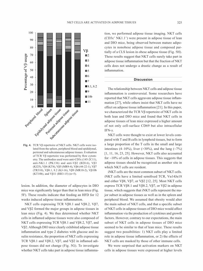
TCR Vβ repertoire of NKT cellsAlmost all iNKT cells have an invariant TCR α-chain,
Vα14Jα18 in mice, and limited β-chains, which consist primarily of either Vβ8, Vβ7, or Vβ2. However, the high levels of expression of nKT cell-surface markers in adipose tissues and the potency with regard to producing massive cytokines of nKT cells in adipose tissues sug-gest that some endogenous antigens of nKT cells are present in nonobese adipose tissue and that nKT cells in adipose tissues express a distinct TCR Vβ repertoire compared with that of nKT cells in peripheral tissues. We therefore assessed the TCR Vβ repertoire of NKT cells in adipose tissues, the spleen and peripheral blood by flow cytometry. NKT cells expressing TCR Vβ8.1 and Vβ8.2 formed the largest group, followed by those expressing Vβ2 and Vβ7, which also accounted for a considerable proportion of cells, particularly in the spleen, peripheral blood and epididymal adipose tissue, whereas nKT cells in subcutaneous adipose tissue showed a different pattern of Vβ repertoire compared with NKT cells in other adipose tissue (Fig. 4). These
results indicate that nKT cells in abdominal adipose tissue are composed of similar subsets of nKT cells in the spleen and peripheral circulation, whereas nKT cells in subcutaneous adipose tissue are a distinct subset.
Effects of obesity on NKT cell subsets in adipose tissueAdipose tissue inflammation, in particular that of
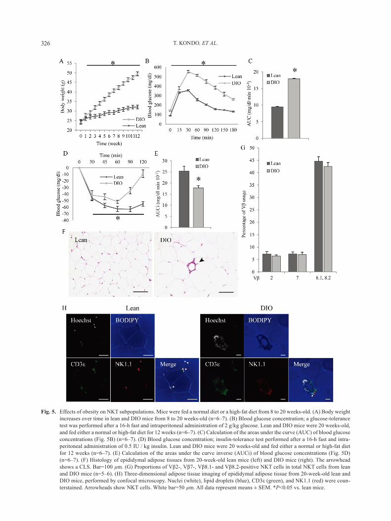
epididymal adipose tissue, is an important subject area in immunology [28]. we determined whether adipose tissue inflammation influenced NKT cell subsets in epi-didymal adipose tissue. we prepared dio mice as an obesity and type 2 diabetes model by feeding 8-week-old mice a high-fat diet (HFD) for 12 weeks. After 12 weeks, the body weights of the dio mice reached 49.45 ± 1.33 g, while those of the lean mice reached 32.08 ± 0.67 g. DIO mice gained significant weight compared with lean mice from the first week following the start of the HFD (Fig. 5A).
To confirm resistance to glucose and insulin, we per-formed glucose- and insulin-tolerance tests in lean and dio mice. Blood glucose concentrations in dio mice
Fig. 2. Lymphocytes fractions in adipose tissues, the spleen and peripheral blood. (a) nK cells (CD3ε−nK1.1+), (B) T cells (CD3ε+nK1.1−), and (C) NKT cells (CD3ε+nK1.1+) in SVFs from the spleen, peripheral blood and epididymal, perirenal and subcutaneous adipose tis-sues were evaluated by flow cytometry. SVFs derived from adipose tissues were obtained by removal of adipocytes from 20-week-old mice. data represent means ± sEm (n=5).*P<0.05 vs. peripheral blood, †P<0.05 vs. spleen.

T. Kondo, ET AL.324
were significantly higher than those in lean mice in terms of both the glucose- and insulin-tolerance tests (Fig. 5B and 5d). The area under the curve for the glucose-toler-ance test and the incremental area under the curve for the insulin-tolerance test were significantly larger than those in lean mice (Fig. 5C and 5E). These results indi-
cate that DIO mice were sufficiently resistant to glucose and insulin.
additionally, histopathological examination indicated induction of adipose tissue inflammation in DIO mice. adipose tissue macrophages aggregated around dead adipocytes to form a distinct crown-like structure (CLs)
Fig. 3. Flow cytometric analysis of cell-surface markers and intracellular cytokine of NKT cells. (A) Mean fluorescence intensity (MFI) of CD3ε in NKT cells, and (B) MFI of NK1.1 in NKT cells from periph-eral blood, the spleen, and epididymal and subcutaneous adipose tissues. MFI determinations were performed by flow cytometry using 20-week-old mice. (C) Flow cytometric analysis of CD69 expres-sion in NKT cells of spleen and epididymal adipose tissue. (D) Flow cytometric analysis of intracel-lular IFN-γ in NKT cells of spleen and epididymal adipose tissue. The fraction of NKT cells was gated by CD3ε+ and nK1.1+ cells. mice were 8 weeks-old. data represent means ± sEm (n=5).*P<0.05 vs. peripheral blood; †P<0.05 vs. spleen.
NKT CELLS ARE ACTIVATED IN ADIPOSE TISSUES 325
lesion. in addition, the diameter of adipocytes in dio mice was significantly larger than that in lean mice (Fig. 5F). These results indicate that feeding an HFD for 12 weeks induced adipose tissue inflammation.
NKT cells expressing TCR Vβ8.1 and Vβ8.2, Vβ7, and Vβ2 formed the major groups in adipose tissues in lean mice (Fig. 4). We thus determined whether NKT cells in inflamed adipose tissues were also composed of NKT cells expressing TCR Vβ8.1 and Vβ8.2, Vβ7, and Vβ2. Although DIO mice clearly exhibited adipose tissue inflammation and type 2 diabetes with glucose and in-sulin resistance, the proportion of nKT cells expressing TCR Vβ8.1 and Vβ8.2, Vβ7, and Vβ2 in inflamed adi-pose tissues did not change (Fig. 5G). To investigate whether NKT cells take part in adipose tissue inflamma-
tion, we performed adipose tissue imaging. nKT cells (CD3ε+ nK1.1+) were present in adipose tissue of lean and dio mice, being observed between mature adipo-cytes in nonobese adipose tissue and composed par-tially of a CLS lesion in obese adipose tissue (Fig. 5H). These results suggest that nKT cells surely take part in adipose tissue inflammation but that the fraction of NKT cells does not undergo a drastic change as a result of inflammation.
Discussion
The relationship between nKT cells and adipose tissue inflammation is controversial. Some researchers have reported that NKT cells aggravate adipose tissue inflam-mation [27], while others insist that nKT cells have no effect on adipose tissue inflammation [21]. In this paper, we characterized the TCR Vβ repertoire of NKT cells in both lean and dio mice and found that nKT cells in adipose tissues of lean mice expressed a higher amount of not only cell-surface Cd69 but also intracellular IFN-γ.
nKT cells were thought to exist at lower levels com-pared with T and B cells in lymphoid tissues, but to form a large proportion of the T cells in the small and large intestines (4–10%), liver (>30%), and the lung (~7%) [1, 11, 16, 23, 25]. however, nKT cells also accounted for ~10% of cells in adipose tissues. This suggests that adipose tissues should be recognized as another site in which nKT cells are resident.
inKT cells are the most common subset of nKT cells. iNKT cells have a limited semifixed TCR, Vα14Jα18 and either Vβ8, Vβ7, or Vβ2 [12, 25]. Most NKT cells express TCR Vβ8.1 and Vβ8.2, Vβ7, or Vβ2 in adipose tissue, which suggests that inKT cells represent the ma-jor subset in adipose tissues as well as in the spleen and peripheral blood. we assumed that obesity would alter the main subset of NKT cells, and that a specific subset of nKT cells in adipose tissues of dio mice would affect inflammation via the production of cytokines and growth factors. however, contrary to our expectations, the main subset of nKT cells in adipose tissues of dio mice seemed to be similar to that of lean mice. These results suggest two possibilities: 1) nKT cells play a limited role in adipose tissue inflammation, or 2) the effects of nKT cells are masked by those of other immune cells.
we were surprised that activation markers on nKT cells in adipose tissues were expressed at higher levels
Fig. 4. TCR Vβ repertoire of NKT cells. NKT cells were iso-lated from the spleen, peripheral blood and epididymal, perirenal and subcutaneous adipose tissues. Evaluation of TCR Vβ repertoire was performed by flow cytom-etry. The antibodies used were anti-CD3ε (145-2C11), anti-NK1.1 (PK136) and anti-Vβ2 (B20.6), Vβ3 (KJ25), Vβ4 (KT4), Vβ5 (MR9-4), Vβ6 (44-22-1), Vβ7 (TR310), Vβ8.1, 8.2 (KJ-16), Vβ9 (MR10-2), Vβ10b (KT10b), and Vβ11 (RR3-15) (n=5).
T. Kondo, ET AL.326
Fig. 5. Effects of obesity on nKT subpopulations. mice were fed a normal diet or a high-fat diet from 8 to 20 weeks-old. (a) Body weight increases over time in lean and dio mice from 8 to 20 weeks-old (n=6–7). (B) Blood glucose concentration; a glucose-tolerance test was performed after a 16-h fast and intraperitoneal administration of 2 g/kg glucose. Lean and dio mice were 20 weeks-old, and fed either a normal or high-fat diet for 12 weeks (n=6–7). (C) Calculation of the areas under the curve (auC) of blood glucose concentrations (Fig. 5B) (n=6–7). (D) Blood glucose concentration; insulin-tolerance test performed after a 16-h fast and intra-peritoneal administration of 0.5 iu / kg insulin. Lean and dio mice were 20 weeks-old and fed either a normal or high-fat diet for 12 weeks (n=6–7). (E) Calculation of the areas under the curve inverse (AUCi) of blood glucose concentrations (Fig. 5D) (n=6–7). (F) Histology of epididymal adipose tissues from 20-week-old lean mice (left) and DIO mice (right). The arrowhead shows a CLs. Bar=100 µm. (G) Proportions of Vβ2-, Vβ7-, Vβ8.1- and Vβ8.2-positive NKT cells in total NKT cells from lean and dio mice (n=5–6). (h) Three-dimensional adipose tissue imaging of epididymal adipose tissue from 20-week-old lean and DIO mice, performed by confocal microscopy. Nuclei (white), lipid droplets (blue), CD3ε (green), and NK1.1 (red) were coun-terstained. arrowheads show nKT cells. white bar=50 µm. all data represent means ± sEm. *P<0.05 vs. lean mice.
NKT CELLS ARE ACTIVATED IN ADIPOSE TISSUES 327
than those in the spleen and peripheral blood. it is well-known that the activation via TCR signaling, for ex-ample, by treatment with anti-CD3ε antibody, down-regulates CD3ε expression in T cells [14], and that stimulation by an exogenous antigen also induced down-regulation of cell-surface receptors on inKT cells [3, 31]. Therefore, we thought that this result may suggest the functions of nKT cells in adipose tissue were sup-pressed in comparison with peripheral nKT cells. how-ever, nKT cells in adipose tissue were unpredictably potent with regard to secretion of large amounts of Th1 cytokines compared with nKT cells in the spleen, and in fact, the expression of Cd69, an activation marker, was high in nKT cells in adipose tissue in comparison with nKT cells in the spleen. These data suggests that nKT cells were physiologically activated in adipose tis-sues even in the absence of inflammation. In lean mice, adipose and other tissues are thought to be maintained in an anti-inflammatory and insulin-sensitive state [28]. NKT cells are stimulated by the proinflammatory cyto-kines iL-12 and iL-18 and by TCR signaling. it is like-ly that in lean mice, these proinflammatory cytokines are produced at very low concentrations in adipose tissues [30]. hence, antigen recognition via Cd1d may be as-sociated with the activation of nKT cells in normal adipose tissue. Recently, it has been reported that gram-positive-bacteria-derived antigens can activate nKT cells, but that adipose tissue presents a sanctuary from infectious diseases, since to date, few infectious organ-isms have been reported in adipose tissues [18]. There-fore, the probability of exogenous antigens being pre-sented to NKT cells without inflammation of the adipose tissue is extremely slim. Our findings suggest the pre-sentation of novel endogenous antigens to nKT cells situated in normal adipose tissue. Endogenous antigens of nKT cells are also present in the thymus, and one is known to promote production of iL-4 [9]. Endogenous antigens in adipose tissues, as well as in the thymus, also regulate NKT cells in the anti-inflammatory state. In this study, the activated nKT cells we described were stim-ulated by an unknown endogenous antigen and contrib-uted to maintenance of adipose tissue homeostasis.
Acknowledgments
we thank dr. Takashi sekiya for technical assistance. This work was supported by JsPs KaKEnhi Grant number 23650230.
References
1. Bannai, m., Kawamura, T., naito, T., Kameyama, h., abe, T., Kawamura, h., Tsukada, C., watanabe, h., hatakeya-ma, K., hamada, h., nishiyama, Y., ishikawa, h., Takeda, K., okumura, K., Taniguchi, m., and abo, T. 2001. abun-dance of unconventional Cd8(+) natural killer T cells in the large intestine. Eur. J. Immunol. 31: 3361–3369. [medline] [CrossRef]
2. Brake, D.K. and Smith, C.W. 2008. Flow cytometry on the stromal-vascular fraction of white adipose tissue. Methods Mol. Biol. 456: 221–229. [medline] [CrossRef]
3. Braun, N.A., Mendez-Fernandez, Y.V., Covarrubias, R., Por-celli, S.A., Savage, P.B., Yagita, H., Van Kaer, L., and Major, a.s. 2010. development of spontaneous anergy in invari-ant natural killer T cells in a mouse model of dyslipidemia. Arterioscler. Thromb. Vasc. Biol. 30: 1758–1765. [medline] [CrossRef]
4. Brutkiewicz, R.R. and Sriram, V. 2002. Natural killer T (nKT) cells and their role in antitumor immunity. Crit. Rev. Oncol. Hematol. 41: 287–298. [medline] [CrossRef]
5. Caspar-Bauguil, s., Cousin, B., Galinier, a., segafredo, C., nibbelink, m., andré, m., Casteilla, L., and Pénicaud, L. 2005. adipose tissues as an ancestral immune organ: site-specific change in obesity. FEBS Lett. 579: 3487–3492. [medline] [CrossRef]
6. Crowe, n.Y., Coquet, J.m., Berzins, s.P., Kyparissoudis, K., Keating, R., Pellicci, d.G., hayakawa, Y., Godfrey, d.i., and smyth, m.J. 2005. differential antitumor immunity medi-ated by nKT cell subsets in vivo. J. Exp. Med. 202: 1279–1288. [medline] [CrossRef]
7. donath, m.Y. and shoelson, s.E. 2011. Type 2 diabetes as an inflammatory disease. Nat. Rev. Immunol. 11: 98–107. [med-line] [CrossRef]
8. duwaerts, C.C., and Gregory, s.h. 2011. Targeting the di-verse immunological functions expressed by hepatic nKT cells. Expert. Opin. Ther. Targets 15: 973–988. [medline] [CrossRef]
9. Facciotti, F., Ramanjaneyulu, G.S., Lepore, M., Sansano, S., Cavallari, M., Kistowska, M., Forss-Petter, S., Ni, G., Co-lone, A., Singhal, A., Berger, J., Xia, C., Mori, L., and De Libero, G. 2012. Peroxisome-derived lipids are self antigens that stimulate invariant natural killer T cells in the thymus. Nat. Immunol. 13: 474–480. [medline] [CrossRef]
10. Faulds, M.H. and Dahlman-Wright, K. 2012. Metabolic dis-eases and cancer risk. Curr. Opin. Oncol. 24: 58–61. [med-line] [CrossRef]
11. Godfrey, d.i., hammond, K.J.L., Poulton, L.d., smyth, m.J., and Baxter, a.G. 2000. nKT cells: facts, functions and fallacies. Immunol. Today 21: 573–583. [medline] [Cross-Ref]
12. Godfrey, d.i., stankovic, s., and Baxter, a.G. 2010. Raising the nKT cell family. Nat. Immunol. 11: 197–206. [medline] [CrossRef]
13. Heikkinen, S., Argmann, C.A., Champy, M.F., and Auwerx, J. 2007. Evaluation of glucose homeostasis. Curr. Protoc. Mol. Biol. Chapter 29: unit 29B.3.
14. hirsch, R., Eckhaus, m., auchincloss, h., sachs, d.h., and

T. Kondo, ET AL.328
Bluestone, J.a. 1988. Effects of in vivo administration of anti-T3 monoclonal antibody on T cell function in mice. i. immunosuppression of transplantation responses. J. Immu-nol. 140: 3766–3772. [medline]
15. ibrahim, m.m. 2010. subcutaneous and visceral adipose tissue: structural and functional differences. Obes. Rev. 11: 11–18. [medline] [CrossRef]
16. ishimoto, Y., Tomiyama-miyaji, C., watanabe, h., Yokoya-ma, h., Ebe, K., Tsubata, s., aoyagi, Y., and abo, T. 2004. age-dependent variation in the proportion and number of in-testinal lymphocyte subsets, especially natural killer T cells, double-positive Cd4(+) Cd8(+) cells and B220(+) T cells, in mice. Immunology 113: 371–377. [medline] [CrossRef]
17. Ji, Y., Sun, S., Xu, A., Bhargava, P., Yang, L., Lam, K.S., Gao, B., Lee, C.h., Kersten, s., and Qi, L. 2012. activation of natural killer T cells promotes m2 macrophage polariza-tion in adipose tissue and improves systemic glucose toler-ance via the iL-4/sTaT6 signaling axis in obesity. J. Biol. Chem. 287: 13561–13571. [medline] [CrossRef]
18. Kinjo, Y., Illarionov, P., Vela, J.L., Pei, B., Girardi, E., Li, X., Li, Y., Imamura, M., Kaneko, Y., Okawara, A., Yesilka-ya, H., Andrew, P.W., Wong, C.H., Kawakami, K., Nizet, V., Besra, G.s., Tsuji, m., Zajonc, d.m., and Kronenberg, m. 2011. invariant natural killer T cells recognize glycolipids from pathogenic Gram-positive bacteria. Nat. Immunol. 12: 966–974. [medline] [CrossRef]
19. Liu, J., divoux, a., sun, J., Zhang, J., Clement, K., Glick-man, J.n., sukhova, G.K., wolters, P.J., du, J., Gorgun, C.Z., doria, a., Libby, P., Blumberg, R.s., Kahn, B.B., hota-misligil, G.S., and Shi, G.P. 2009. Genetic deficiency and pharmacological stabilization of mast cells reduce diet-in-duced obesity and diabetes in mice. Nat. Med. 15: 940–945. [medline] [CrossRef]
20. Ljutic, B., Carlyle, J.R., Filipp, D., Nakagawa, R., Julius, M., and Zuniga-Pflucker, J.C. 2005. Functional require-ments for signaling through the stimulatory and inhibitory mouse nKR-P1 (Cd161) nK cell receptors. J. Immunol. 174: 4789–4796. [medline]
21. Mantell, B.S., Stefanovic-Racic, M., Yang, X., Dedousis, n., sipula, i.J., and o’doherty, R.m. 2011. mice Lacking nKT Cells but with a Complete Complement of Cd8(+) T-Cells are not Protected against the metabolic abnormalities of diet-induced obesity. PLoS ONE 6: e19831. [medline] [CrossRef]
22. meyer, E.h., deKruyff, R.h., and umetsu, d.T. 2008. T cells and nKT cells in the pathogenesis of asthma. Annu. Rev. Med. 59: 281–292. [medline] [CrossRef]
23. middendorp, s., and nieuwenhuis, E.E.s. 2009. nKT cells in mucosal immunity. Mucosal Immunol. 2: 393–402. [med-line] [CrossRef]
24. nishimura, s., manabe, i., nagasaki, m., Eto, K., Yamashita, h., ohsugi, m., otsu, m., hara, K., ueki, K., sugiura, s., Yoshimura, K., Kadowaki, T., and nagai, R. 2009. Cd8(+)
effector T cells contribute to macrophage recruitment and adipose tissue inflammation in obesity. Nat. Med. 15: 914–920. [medline] [CrossRef]
25. novak, J., Griseri, T., Beaudoin, L., and Lehuen, a. 2007. Regulation of type 1 diabetes by nKT cells. Int. Rev. Immu-nol. 26: 49–72. [medline] [CrossRef]
26. oh, s.J. and Chung, d.h. 2011. invariant nKT Cells Pro-ducing IL-4 or IL-10, But Not IFN-gamma, Inhibit the Th1 Response in Experimental autoimmune Encephalomyelitis, whereas none of These Cells inhibits the Th17 Response. J. Immunol. 186: 6815–6821. [medline] [CrossRef]
27. ohmura, K., ishimori, n., ohmura, Y., Tokuhara, s., no-zawa, A., Horii, S., Andoh, Y., Fujii, S., Iwabuchi, K., Onoe, K., and Tsutsui, h. 2010. natural Killer T Cells are involved in Adipose Tissues Inflammation and Glucose Intolerance in diet-induced obese mice. Arterioscler. Thromb. Vasc. Biol. 30: 193–199. [medline] [CrossRef]
28. osborn, o. and olefsky, J.m. 2012. The cellular and signal-ing networks linking the immune system and metabolism in disease. Nat. Med. 18: 363–374. [medline] [CrossRef]
29. Rhost, s., sedimbi, s., Kadri, n., and Cardell, s.L. 2012. immunomodulatory type ii natural killer T (nKT) lympho-cytes in health and disease. Scand. J. Immunol. 76: 246–255. [medline] [CrossRef]
30. Strissel, K.J., DeFuria, J., Shaul, M.E., Bennett, G., Green-berg, a.s., and obin, m.s. 2010. T-Cell Recruitment and Th1 Polarization in adipose Tissue during diet-induced obesity in C57BL/6 mice. Obesity (Silver Spring) 18: 1918–1925. [medline] [CrossRef]
31. Wilson, M.T., Johansson, C., Olivares-Villagómez, D., singh, a.K., stanic, a.K., wang, C.R., Joyce, s., wick, M.J., and Van Kaer, L. 2003. The response of natural killer T cells to glycolipid antigens is characterized by surface recep-tor down-modulation and expansion. Proc. Natl. Acad. Sci. U.S.A. 100: 10913–10918. [medline] [CrossRef]
32. winer, d.a., winer, s., shen, L., wadia, P.P., Yantha, J., Paltser, G., Tsui, h., wu, P., davidson, m.G., alonso, m.n., Leong, H.X., Glassford, A., Caimol, M., Kenkel, J.A., Ted-der, T.F., McLaughlin, T., Miklos, D.B., Dosch, H.M., and Engleman, E.G. 2011. B cells promote insulin resistance through modulation of T cells and production of pathogenic igG antibodies. Nat. Med. 17: 610–617. [medline] [Cross-Ref]
33. winer, s., Chan, Y., Paltser, G., Truong, d., Tsui, h., Bah-rami, J., dorfman, R., wang, Y., Zielenski, J., mastronardi, F., Maezawa, Y., Drucker, D.J., Engleman, E., Winer, D., and dosch, h.m. 2009. normalization of obesity-associated insulin resistance through immunotherapy. Nat. Med. 15: 921–929. [medline] [CrossRef]
34. World Health Organisation Fact sheet: obesity and over-weight. available at: http://www.who.int/mediacentre/fact-sheets/fs311/en/print.html. accessed 15 october 2012.