nccn task force report: management of dermatologic and other
TRANSCRIPT

JNCCN Volume 7 Supplement 1 Journal of the National Comprehensive Cancer Network
NCCN.org
S U P P L E M E N T
NCCN Task Force Report: Management of Dermatologic and Other Toxicities Associated With EGFR Inhibition in Patients With CancerBarbara Burtness, MD; Milan Anadkat, MD; Surendra Basti, MD; Miranda Hughes, PhD; Mario E. Lacouture, MD; Patricia L. Myskowski, MD; Jennifer Paul, PA-C; Clifford S. Perlis, MD, MBe; Leonard Saltz, MD; and Sharon Spencer, MD
NCCN appreciates that supporting companies recognize NCCN’s need for autonomy in the development of the content of NCCN resources. All NCCN content is produced completely independently. The distribution of this task force report is supported by an educational grant from Bristol-Myers Squibb Company.
CE Provided by NCCN

Volume 7 Supplement 1 Journal of the National Comprehensive Cancer Network
JNCCNEditorial
Editor-in-Chief:Harold J. Burstein, MD, PhD
National Comprehensive Cancer Network
Director of NCCN Publications/Managing Editor:Kimberly A. Callan, MS, ELS
Assistant Managing Editor:Kerrin Robinson, MA
Editorial Associate:Genevieve Emberger Hartzman, MA
National Comprehensive Cancer Network
Chairman of the Board:Al B. Benson III, MD
Vice Chair of the Board: Thomas A. D’Amico, MD
Chief Executive Officer: William T. McGivney, PhD
Executive Vice President/Chief Operating Officer:
Patricia J. Goldsmith
Senior VP, Finance/Chief Financial Officer:Lisa Kimbro, CPA, MBA
Clinical Practice Guidelines
Senior VP, Clinical Information and Publications: Joan S. McClure, MS
VP, Clinical Information Operations:Kristina M. Gregory, RN, MSN
Associate Director, Clinical Information:Dorothy A. Shead, MS
Guidelines Coordinators: Nicole R. McMillian, MS Mary Dwyer Rosario, MS
Oncology Scientists/Sr. Medical Writers: Miranda Hughes, PhD Hema Sundar, PhD Susan J. Moench, PhD Rashmi Kumar, PhD Maria Ho, PhD
Administrative Coordinators:Mary Anne Bergman Jean Marie Dougherty
Business Development and Marketing
Sr. VP, Strategic Development:Alana L.K. Brody, MBA
Advertising
Cold Spring Publishing
Director of Business Development:David Horowitz
Publishing and Design
Cold Spring Publishing
Executive Editor: Conor Lynch
Production Coordinator: Sarah McGullam
Publisher: John A. Gentile, Jr.
Masthead Postal and Contact Information
JNCCN (ISSN 1540-1405), the official journal of the National Comprehensive Cancer Network, is published 10 times annually by Cold Spring Publishing, 147 Main Street, Cold Spring Harbor, NY 11724.
Copyright © 2009 by the National Comprehensive Cancer Network. All rights reserved. No part of this publication may be reproduced or transmitted in any form or by any means now or hereafter known, electronic or mechanical, including photocopy, recording, or any information storage and retrieval system, without permission in writing from NCCN. Subscriptions: Prices for yearly subscriptions (10 issues plus supplements) are: Individual: Print only or online only, US $440; Can/Mex + Int’l $545; print and online, US $485; Can/Mex + Int’l $610. Institutional: Print only or online only, US $685; Can/Mex + Int’l $790; print and online, US $750; Can/Mex + Int’l $865. Single Copy: US $70.00; Can/Mex $85.00; Int’l $95.00. Subscription Inquiries should be directed to Sarah McGullam, Cold Spring Publishing, at: 631-692-0800 x317 or www.cspubs.com/jnccn.html. Online access is available to subscribers through IngentaConnect (www.ingentaconnect.com).Contact Information Editorial Office: Manuscripts, correspondence, and commentaries to be considered for publication should be sent to Kimberly Callan, Director of NCCN Publications, JNCCN, 275 Commerce Drive, Suite 300, Fort Washington, PA 19034; or e-mail [email protected]. Correspondence can also be faxed: 215-690-0283 (attn: JNCCN). Questions about requirements for publication or topic suitability can be directed as above or to Harold J. Burstein, MD, PhD, Editor-in-Chief, JNCCN, 275 Commerce Drive, Suite 300, Fort Washington, PA 19034; or e-mail [email protected].
Instructions for authors are published in JNCCN as space allows and can be found on-line at www.nccn.org/jnccn. They can also be requested by calling 215-690-0270 or e-mailing [email protected] purchase advertising space: Contact David Horowitz, Director of Business Development, Cold Spring Publishing, 147 Main Street, Cold Spring Harbor, NY 11724; phone 631-692-0800 x304; fax 631-692-0805; or e-mail [email protected] send film or digital ad materials: Ship to Cold Spring Publishing, Attn: Sarah McGullam, (JNCCN, Vol ___ Issue ___), 147 Main Street, Cold Spring Harbor, NY 11724; phone 631-692-0800 x317; fax 631-692-0805; or e-mail [email protected] send pre-printed inserts: Ship to Publishers Press, Inc., Attn: Jamie Baugh, 13487 South Preston Highway, Lebanon Junction, KY 40150.ProductionReprints: Reprints of individual articles are available. Orders must be for a minimum of 100 copies. Please contact David Horowitz, Director of Business Development, Cold Spring Publishing, 147 Main Street, Cold Spring Harbor, NY 11724; phone 631-692-0800 x304; fax 631-692-0805; or e-mail [email protected] information about photocopying, republishing, reprinting, or adapting material, please go online to www.nccn.org/about/permissions/default.asp.IndexingJNCCN is indexed by MEDLINE/PUBMED®, Chemical Abstracts, EMBASE, EmCare, and Scopus. This paper meets the requirements of ANSI/NISO Z39.48-1992 (Permanence of Paper) effective with Volume 1, Issue 1, 2003.JNCCN is a member of the Medscape Publisher’s Circle®, an alliance of leading medical publishers whose content is featured on Medscape (http://www.medscape.com). Medscape is part of the WebMD Medscape Health Network, a leading online healthcare resource for professionals and consumers.
DisclaimerThe treatment algorithms presented in JNCCN and its supplements are a statement of consensus of the authors regarding their views of currently accepted approaches to treatment. Any clinician seeking to apply or consult these guidelines is expected to use independent medical judgment in the context of individual circumstances to determine any patient’s care or treatment. The research articles, reviews, and other individually authored papers presented herein are the work of the authors listed. Furthermore, the reader is advised that, except where specifically stated, all of the ideas and opinions expressed in JNCCN are the authors’ own and do not necessarily reflect those of NCCN, the member organizations, the editor, or the publisher. Publication of an advertisement or other product mention in JNCCN should not be construed as an endorsement of the product or the manufacturer’s claims.
The information contained in JNCCN is presented for the purpose of educating our readership on cancer treatment and management. The information should not be relied on as complete or accurate, nor should it be relied on to suggest a course of treatment for a particular individual. It should not be used in place of a visit, call, consultation, or the advice of a licensed physician or other qualified health care provider. Patients with health care-related questions or concerns are advised to contact a physician or other qualified health care provider promptly.
Although every attempt has been made to verify that information presented within is complete and accurate, the information is provided “AS IS” without warranty, express or implied. NCCN hereby excludes all implied warranties of merchantability and fitness for a particular use or purpose with respect to the Information. Furthermore, NCCN makes no warranty as to the reliability, accuracy, timeliness, usefulness, adequacy, completeness, or suitability of the information.
Volume 7 Supplement 1 Journal of the National Comprehensive Cancer Network
JNCCNCME AcceditationThe National Comprehensive Cancer Network (NCCN) is accredited by the Accreditation Council for Continuing Medical Education (ACCME) to provide continuing medical education for physicians.
The NCCN designates this educational activity for a maximum of 1 AMA PRA Category 1 Credits™. Physicians should only claim credit commensurate with the extent of their participation on the activity.
This educational activity was planned and produced in accordance with ACCME Essential Areas and Policies.
The NCCN adheres to the ACCME Standards for Commercial Support of Continuing Medical Education.
This activity is approved for 1 contact hour. NCCN is an approved provider of continuing nursing education by the PA State Nurses Association, an accredited approver by the American Nurses Credentialing Center’s Commission on Accreditation.
Approval as a provider refers to recognition of educational activities only and does not imply ANCC Commission Accreditation of PA Nurses approval or endorsement of any product. Kristina M. Gregory, RN, MSN, OCN, is our nurse planner for this educational activity.
Continuing Education Information
Target AudienceThis educational program is designed to meet the needs of oncologists, advanced practice nurses, and other clinical professionals treat and manage patients with cancer whose treatment includes EGFR inhibitors.
Educational Objectives After completion of this CME activity, participants should be able to:• Identify epidermal growth factor receptor (EGFR) inhibitors.• List specific toxicities that may occur in patients being treated with EGFR inhibitor
therapy.• Identify which patients are more likely to experience skin rash associated with
EGFR inhibitor therapy.• Identify symptoms that should prompt a referral for an ophthalmologic examination
in patients treated with EGFR inhibitor therapy.• Recommend management options in patients with toxicities caused by EGFR
inhibitor therapy.The opinions expressed in this publication are those of the participating faculty
and not those of the National Comprehensive Cancer Network, Bristol-Myers Squibb Company, or the manufacturers of any products mentioned herein.
This publication may include the discussion of products for indications not approved by the FDA.
Participants are encouraged to consult the package inserts for updated information and changes regarding indications, dosages, and contraindications. This recommendation is particularly important with new or infrequently used products.
Activity InstructionsParticipants will read all portions of this monograph, including all tables, figures, and references. A post-test and an evaluation form follow this activity, both of which require completion. To receive your continuing education certificate, you will need a score of at least 70% on the post-test. The post-test and evaluation form must be completed and returned by May 29, 2010. It should take approximately 1 hour to complete this activity as designed.
There are no registration fees for this activity. Certificates will be mailed within 3 to 4 weeks of receipt of the post-test.
Copyright 2009, National Comprehensive Cancer Network (NCCN). All rights reserved. No part of this publication may be reproduced or transmitted in any other form or by any means, electronic or mechanical, without first obtaining written permission from the NCCN.
Volume 7 Supplement 1 Journal of the National Comprehensive Cancer Network
JNCCNNCCN Task Force: Management of Dermatologic and Other Toxicities Associated with EGFR Inhibition in Patients With Cancer
Faculty*CBarbara Burtness, MD†
Fox Chase Cancer Center*PMilan Anadkat, MDϖ
Siteman Cancer Center at Barnes-Jewish Hospital and Washington University School of Medicine
*PSurendra Basti, MDλRobert H. Lurie Comprehensive Cancer Center and Northwestern University Feinberg School of Medicine
*Miranda Hughes, PhDNational Comprehensive Cancer Network
*PMario E. Lacouture, MDϖRobert H. Lurie Comprehensive Cancer Center of Northwestern University
Joan S. McClure, MSNational Comprehensive Cancer Network
*PPatricia L. Myskowski, MDϖMemorial Sloan-Kettering Cancer Center
*Jennifer Paul, PA-C†Fox Chase Cancer Center
*Clifford S. Perlis, MD, MBeϖFox Chase Cancer Center
*PLeonard Saltz, MD‡Þ†Memorial Sloan-Kettering Cancer Center
*PSharon Spencer, MD§University of Alabama at BirminghamComprehensive Cancer Center
KEY:*Writing Committee Member; PPresenter; CChair
Specialties: †Medical Oncology; ϖDermatology, Including Surgery; λOphthalmology; ‡Hematology; ÞInternal Medicine; §Radiotherapy/Radiation Oncology
Disclosure of Affiliations and Significant RelationshipsDr. Burtness has disclosed that she has financial interests, arrangements, or affiliations with the manufacturer of products and devices discussed in this report or who may financially support the educational activity. She has received grant or research support from Array Pharmaceuticals and Genentech, Inc., and has received drug supply for research from Bristol-Myers Squibb Company. She is also a consultant for Rexahn Pharmaceuticals, Inc.
Dr. Anadkat has disclosed that he has financial interests, arrangements, or affiliations with the manufacturer of products and devices discussed in this report or who may financially support the educational activity. He is a principal investigator for a study sponsored by Hana Biosciences, Inc.; is on speakers’ bureau for ImClone Systems Incorporated and Genentech, Inc.; and has received speakers’ honoraria from ImClone Systems Incorporated (that totals $10,000 or more).
Dr. Basti has disclosed that he has no financial interests, arrangements, or affiliations with the manufacturer of products and devices discussed in this report or who may financially support the educational activity. Dr. Basti’s work is supported in part by an unrestricted grant from Research to Prevent Blindness to the department of ophthalmology.
Dr. Hughes has disclosed that she has financial interests, arrangements, or affiliations with the manufacturer of products and devices discussed in this report or who may financially support the educational activity. She owns stock in Qiagen NV, Affymetrix, and Myriad Genetic Laboratories, Inc. She is also an employee of the National Comprehensive Cancer Network.
Dr. Lacouture has disclosed that he has financial interests, arrangements, or affiliations with the manufacturer of products and devices discussed in this report or who may financially support the educational activity. He is a consultant for OSI Pharmaceuticals Inc., Amgen Inc., and ImClone Systems Incorporated; is on the speakers’ bureau for Bristol-Myers Squibb Company and ImClone Systems Incorporated; and has received grant or research support from Hana Biosciences, Inc. He has also received honoraria as a speaker for ImClone Systems Incorporated (that totals $10,000 or more).
Ms. McClure has disclosed that she has no financial interests, arrangements, or affiliations with the manufacturer of products and devices discussed in this report or who may financially support the educational activity. She is an employee of the National Comprehensive Cancer Network.
Dr. Myskowski has disclosed that she has financial interests, arrangements, or affiliations with the manufacturer of products and devices discussed in this report or who may financially support the educational activity. She is an investigator for Bristol-Myers Squibb Company and ImClone Systems Incorporated, and has received grant or research support from Ortho Biotech, L.P.; Allos Therapeutics, Inc.; Ligand Pharmaceuticals, Inc.; Schering-Plough Corporation; MedImmune, Inc.; Bristol-Myers Squibb Company; Merck & Co., Inc.; Genmab; and ImClone Systems Incorporated.
Ms. Paul has disclosed that she has financial interests, arrangements, or affiliations with the manufacturer of products and devices discussed in this report or who may financially support the educational activity. A member of her immediate family is employed by Bristol-Myers Squibb Company.
Dr. Perlis has disclosed that he has financial interests, arrangements, or affiliations with the manufacturer of products and devices discussed in this report or who may financially support the educational activity. He is a consultant for and has received gifts in kind (totaling $10,000 or more) from Lucid, Inc.
Dr. Saltz has disclosed that he has financial interests, arrangements, or affiliations with the manufacturer of products and devices discussed in this report or who may financially support the educational activity. He is a consultant for Abbott Laboratories; Celgene Corporation; Exelixis Inc.; Genentech, Inc.; ImClone Systems Incorporated; Merck & Co., Inc.; Alechemia; Delcath; Pfizer Inc.; Roche Laboratories, Inc.; and YM BioScience Inc. He has received research funding from Amgen Inc.; Bayer Healthcare; Bristol-Myers Squibb Company; Genentech, Inc.; ImClone Systems Incorporated; Merck & Co., Inc.; Pfizer Inc.; Roche Laboratories, Inc.; and Taiho Pharmaceuticals Co., Ltd.
Dr. Spencer has disclosed that she has no financial interests, arrangements, or affiliations with the manufacturer of products and devices discussed in this report or who may financially support the educational activity.

Volume 7 Supplement 1 Journal of the National Comprehensive Cancer Network
JNCCNNCCN Member InstitutionsCity of Hope Comprehensive
Cancer Center Los Angeles, California
Dana-Farber/Brigham and Women’s Cancer Center| Massachusetts General Hospital Cancer Center Boston, Massachusetts
Duke Comprehensive Cancer CenterDurham, North Carolina
Fox Chase Cancer Center Philadelphia, Pennsylvania
Huntsman Cancer Institute at the University of Utah Salt Lake City, Utah
Fred Hutchinson Cancer Research Center/ Seattle Cancer Care Alliance Seattle, Washington
The Sidney Kimmel Comprehensive Cancer Center at Johns Hopkins Baltimore, Maryland
Robert H. Lurie Comprehensive Cancer Center of Northwestern University Chicago, Illinois
Memorial Sloan-Kettering Cancer Center New York, New York
H. Lee Moffitt Cancer Center & Research Institute Tampa, Florida
The Ohio State University Comprehensive Cancer Center – James Cancer Hospital and Solove Research Institute Columbus, Ohio
Roswell Park Cancer Institute Buffalo, New York
Siteman Cancer Center at Barnes-Jewish Hospital and Washington University School of Medicine St. Louis, Missouri
St. Jude Children’s Research Hospital/University of Tennessee Cancer Institute Memphis, Tennessee
Stanford Comprehensive Cancer Center Stanford, California
University of Alabama at Birmingham Comprehensive Cancer Center Birmingham, Alabama
UCSF Helen Diller Family Comprehensive Cancer Center San Francisco, California
University of Michigan Comprehensive Cancer Center Ann Arbor, Michigan
UNMC Eppley Cancer Center at The Nebraska Medical Center Omaha, Nebraska
The University of Texas M. D. Anderson Cancer Center Houston, Texas
Vanderbilt-Ingram Cancer Center Nashville, Tennessee
For more information, visit www.nccn.org
JNCCN is dedicated to improving the quality of cancer care locally, nationally, and internationally while enhancing the collaboration between academic medicine and the community physician. JNCCN is further committed to disseminating information across the cancer care continuum by publishing clinical practice guidelines and reporting rigorous outcomes data collected and analyzed by experts from the world’s leading care centers. JNCCN also provides a forum for original research and review papers focusing on clinical and translational research and applications of the NCCN Guidelines in everyday practice, as well as correspondence and commentary.
Mission Statement
The National Comprehensive Cancer Network (NCCN), a not-for-profit alliance of 21 of the world’s leading cancer centers, is dedicated to improving the quality and effectiveness of care provided to patients with cancer. Through the leadership and expertise of clinical professionals at NCCN Member Institutions, NCCN develops resources that present valuable information to the numerous stakeholders in the health care delivery system. As the arbiter of high-quality cancer care, NCCN promotes the importance of continuous quality improvement and recognizes the significance of creating clinical practice guidelines appropriate for use by patients, clinicians, and other health care decision-makers. The primary goal of all NCCN initiatives is to improve the quality, effectiveness, and efficiency of oncology practice so patients can live better lives. For more information, visit www.nccn.org.
About the NCCN
NCCN
275 Commerce Drive
Suite 300
Fort Washington, PA 19034
215–690–0300
www.nccn.org
Fred Hutchinson Cancer Research Center/Seattle Cancer Care Alliance
UCSF Helen Diller FamilyComprehensive Cancer Center
Stanford Comprehensive Cancer Center
City of Hope Comprehensive Cancer Center
Huntsman Cancer Institute at the U. of Utah
UNMC Eppley CancerCenter at The NebraskaMedical Center
St. Jude Children’s Research Hospital/U. of Tennessee Cancer Institute
U. of Alabama at Birmingham Comprehensive Cancer Center
Duke Comprehensive Cancer Center
H. Lee Moffitt Cancer Center & Research Institute
Memorial Sloan-Kettering Cancer Center
Fox Chase Cancer Center
The Sidney KimmelComprehensive CancerCenter at Johns Hopkins
Roswell Park Cancer Institute
U. of Michigan Comprehensive Cancer Center
Robert H. Lurie Comprehensive Cancer Center of Northwestern U.
The Ohio State University Comprehensive Cancer Center - James Cancer Hospital and Solove Research Institute
The University of Texas M. D. Anderson Cancer Center
Siteman Cancer Center at Barnes-Jewish Hospitaland Washington U. School of Medicine
Dana-Farber/Brigham and Women’s Cancer CenterMassachusetts General Hospital Cancer Center
Vanderbilt-IngramCancer Center

Supplement
© Journal of the National Comprehensive Cancer Network | Volume 7 Supplement 1 | May 2009
S-5
Key WordsDermatologic toxicities, ocular toxicities, rash, acne-like rash, papulopustular rash, dry eye, paronychia, radiation dermatitis, epidermal growth factor receptor, EGFR, EGFR inhibitor, tyrosine kinase inhibitors, TKI, cetuximab, panitumumab, erlotinib, gefitinib, colorectal cancer, head and neck cancer, non–small cell lung cancer
AbstractThis NCCN Task Force Report describes the management of der- der-matologic and ocular toxicities that occur in patients treated with epidermal growth factor receptor (EGFR) inhibitors. Task force members are from NCCN member institutions and include oncologists, dermatologists, an ophthalmologist, and a mid-level oncology provider. This report describes commonly used therapies that the task force agreed are appropriate standards of care for dermatologic and ophthalmologic toxicities associated with EGFR inhibitors, which generally are supported only by anecdotal evi-dence. Few recommendations are evidence based; however, some commonly used therapies have data supporting their use. Conclu-sions from completed clinical trials are generally limited by the small numbers of patients enrolled. The information in this report is based on available published data on treating toxicities associ-ated with EGFR inhibitors, data from treatment of clinically similar toxicities from different etiologies, and expert opinion among the NCCN Task Force members. (JNCCN 2009;7[Suppl 1]:5–21)
types. Classes of EGFR inhibitors include monoclonal antibodies, such as cetuximab and panitumumab, and small molecular weight tyrosine kinase inhibitors, such as erlotinib, gefitinib, and lapatinib (which is a dual EGFR and human epidermal growth factor receptor 2 [HER2] inhibitor).2 The monoclonal antibodies target the extracellular domain of EGFR and are given intra-venously. The small molecule inhibitors of EGFR in-hibit the ATP-binding site of the kinase domain and are given orally. These EGFR inhibitors are currently approved by the FDA for certain types of metastatic cancer, such as breast, colon, head and neck, NSCLC, and pancreatic cancers. In addition, clinical trials are in progress for EGFR inhibitors in patients with skin cancer and other solid tumors.3
Cetuximab is used for metastatic colorectal cancer in patients with wild-type K-ras tumors either as mono-therapy or combined with irinotecan.4–7 In selected pa-tients, cetuximab is also indicated for advanced NSCLC when combined with cisplatin and vinorelbine,8 and for advanced or metastatic/recurrent head and neck can-cer.9,10 Panitumumab is indicated for metastatic, che-motherapy-refractory colorectal cancer in patients with wild-type K-ras tumors.11,12 Determination of K-ras mu-tational status is important in colorectal cancer because patients with mutated activated K-ras do not respond to treatment with cetuximab or panitumumab.12,13
Erlotinib is indicated as second- and third-line therapy for patients with recurrent or metastatic NSCLC. Patients with NSCLC who have adenocarci-noma histology and never smoked often benefit from erlotinib therapy, because many of these patients have EGFR mutations (e.g., exon-19 deletion) that are as-sociated with sensitivity to erlotinib or gefitinib.14
Indications for Epidermal Growth Factor Receptor Inhibitor TherapyEpidermal growth factor receptor (EGFR) is a tyrosine kinase receptor from a larger family of ErbB receptors that mediate cell survival, proliferation, angiogenesis, and invasiveness; thus, EGFR is associated with many carcinogenic processes.1 Inhibitors of EGFR increase overall survival in patients with colorectal, head and neck, pancreatic, and non–small cell lung cancers (NSCLC) and are under investigation in other tumor
NCCN Task Force Report: Management of Dermatologic and Other Toxicities Associated With EGFR Inhibition in Patients With CancerBarbara Burtness, MD; Milan Anadkat, MD; Surendra Basti, MD; Miranda Hughes, PhD; Mario E. Lacouture, MD; Joan S. McClure, MS; Patricia L. Myskowski, MD; Jennifer Paul, PA-C; Clifford S. Perlis, MD, MBe; Leonard Saltz, MD; and Sharon Spencer, MD

Supplement
NCCN Task Force Report
© Journal of the National Comprehensive Cancer Network | Volume 7 Supplement 1 | May 2009
S-6
impediment to EGFR inhibitor use, familiarity with management strategies to avoid dose reduction may be beneficial. NCCN convened a task force of ex-perts in EGFR inhibitors and their associated tox-icities to assess the biomedical literature and clinical experience in managing these toxicities. This report presents the conclusions.
Dermatologic ToxicitiesEGFR inhibitors are associated with dermatologic toxicities, such as skin rash, paronychia (suppurative inflammation around the nails), dry skin (xerosis), dry mucus membranes, pruritus, urticaria, superinfec-tion of the skin, increased growth of eyelashes (tri-chomegaly), facial hirsutism, and alopecia.29 Some patients experience mucositis, which rarely includes aphthous ulcers. Some patients treated with gefitinib experience a purpuric drug reaction and necrolytic migratory erythema-like lesions.
Skin RashThe skin rash associated with EGFR inhibitors oc-curs more often than other dermatologic toxicities and may be severe; it affects mainly the face, scalp, neck, upper chest, and back. Patients often refer to the rash as acne, and the term acneiform was used for initial descriptions of the rash. However, the EGFR inhibitor–associated skin rash is actually a papulo-pustular rash distinct from acne, but is often referred to as an acne-like or acneiform rash because of the in-flammatory follicular appearance of the lesions.5,30,31 Although the EGFR inhibitor–associated skin rash has the papules and pustules of acne vulgaris, it lacks comedones, which are the primary lesions of classic acne. Despite a similar appearance to acne vulgaris, however, the etiology, pathophysiology, and thera-peutic approaches to EGFR inhibitor–associated exanthems are entirely different. More recently, the rash has been described as follicular rash, folliculi-tis, macular/papular eruption, pustular eruption, and monomorphic pustular lesions.30,32
Quality-of-life assessments using the Skindex-16 score have shown that this skin rash has a significant negative emotional component.33 Patients also state that the skin rash causes irritation, burning, and sting-ing. In a survey conducted among oncology providers, 80% indicated that patients complain of pruritus, 57% said patients also complain of tenderness, and 32% had to prescribe pain medications for the skin rash.27
Patients who have NSCLC with an exon-19 dele-tion and are treated with erlotinib have a 1-year survival rate of 80% and a median survival of 34 months.14 Thus, molecular selection can be used to determine which patients with NSCLC will ben-efit from therapy with erlotinib and exclude those with little chance of benefit. Erlotinib in combina-tion with gemcitabine is also indicated for patients with pancreatic cancer.15 Gefitinib is indicated for patients with metastatic NSCLC who benefit from this therapy.16,17 Lapatinib in combination with capecitabine is indicated for patients with HER2-positive advanced breast cancer.18–20
EGFR inhibitors are associated with reproduc-ible side effects, including dermatologic toxicities (e.g., skin rash, xerosis, paronychia), mucositis, sto-matitis, diarrhea, infusion reactions, ocular abnor-malities, and hypomagnesemia.21 Skin rash is one of the most frequently occurring side effects and occurs with many EGFR inhibitors, including cetuximab, panitumumab, erlotinib, and gefitinib, whether the agent is a monoclonal antibody or small molecule; therefore, skin rash seems to be a class effect.
Skin rash caused by cetuximab, panitumumab, and erlotinib is associated with a higher response rate and increased survival;5,22–24 severity of the skin rash is directly related to increasing survival.23,25,26 For example, patients treated with cetuximab and radiation therapy (RT) for head and neck cancer with a more prominent skin rash have longer me-dian survival compared with those who have a less prominent rash (56.7 vs. 24.4 months; P = .02).26 Patients treated with erlotinib for NSCLC who have a grade 2 or higher rash have a median survival of 11.1 months compared with 3.3 months for those with no rash (P < .001).23 The association between skin rash and response/survival observed in the erlo-tinib trials suggests that administering this agent at its highest tolerated dose may improve the possibility of a clinical response. Several studies are investigat-ing the feasibility of escalating the dose of EGFR in-hibitors until a tolerable rash occurs in patients with various malignancies.
Oncologists sometimes discontinue therapy or reduce the dose of EGFR inhibitor because the rash is disfiguring or painful.27 Because of the association between rash and survival, current recommenda-tions favor treating the rash over dose reduction, whenever possible.28 Although the rash can be an

Supplement
Dermatologic Toxicities
© Journal of the National Comprehensive Cancer Network | Volume 7 Supplement 1 | May 2009
S-7
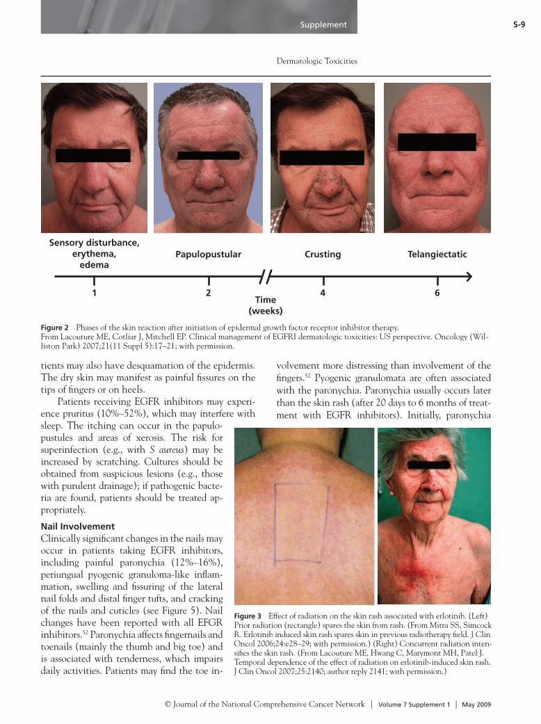
The skin rash typically begins within 8 to 10 days after initiation of EGFR therapy, peaks at ap-proximately 2 weeks, and often tapers within 8 weeks of therapy cessation. If patients continue receiving EGFR inhibitors, the skin rash persists; however, its severity often waxes and wanes over time. Postin-flammatory side effects, such as telangiectasias, ery-thema, and hyperpigmentation, tend to occur 5 to 9 weeks after initiation of EGFR inhibitor therapy.34 This section discusses biopsy findings, incidence, grading, and description of skin rash (including in-field toxicity) associated with EGFR inhibitors; management is discussed later.Biopsy Findings: The papulopustular rash mainly occurs in areas with many sebaceous glands and fol-licles, such as the face, neck, shoulders, upper trunk, and scalp.35 Histopathologic findings also indicate that the papulopustular rash is a suppurative inflam-mation and not acne vulgaris, because no comedones or other signs of classic acne are present. The rash is presumed to occur because inhibition of EGFR signaling affects the epithelium, hair follicles, and sebaceous glands. The initial skin rash is caused by inflammation, although infection can occur later (Staphylococcus aureus is typically found).30,36 In pa-tients with papulopustular rash, the lesions initially show a T-lymphocyte infiltrate followed by a neutro-philic inflammatory infiltrate. Skin biopsies from day 8 show a dermal inflammatory cell infiltrate around follicular infundibula and a suppurative folliculitis with rupture of the epithelial lining.36
EGFR is expressed in the basal layer of kerati-nocytes in the epithelium, follicles, and sebaceous glands.37,38 Inhibition of EGFR decreases cell growth and alters differentiation of keratinocytes in the epithelium and hair follicle.39,40 The abnormal epi-dermal differentiation leads to follicular obstruc-tion and subsequent inflammation. The epidermis becomes thin (leading to xerosis), and the skin becomes fragile.41
Incidence: Approximately 90% of patients treated with cetuximab or panitumumab will experience skin toxicity.27 The papulopustular rash occurs in 67% to 75% of patients treated with erlotinib.27,42 Patients treated with cetuximab, panitumumab, er-lotinib, or gefitinib are more likely to have a rash than those receiving lapatinib.43 The severity of the skin rash also varies depending on the patient and type of EGFR inhibitor used. Patients treated with
monoclonal antibodies, such as cetuximab, often have a more severe skin reaction on the face.9,44 In addition, patients with a better performance status or a more robust immune system may experience a more severe rash. However, in patients with breast cancer treated with lapatinib (the dual HER2 and EGFR inhibitor), the skin rash is typically only mild to moderate in severity and occurs mainly on the upper chest, not face; many patients do not develop any rash.43,45
Ultraviolet light seems to trigger the rash in some patients perhaps by inducing EGFR expression and activation in the skin; thus, patients treated with EGFR inhibitors should consider protecting their skin from the sun by using sunscreen.46,47 Although both fair- and dark-skinned patients experience the rash, it is more likely to be severe in those with fair skin.48 A retrospective review of 42 patients treated with erlotinib found that 63% of patients (n = 16) with fair skin (a lower Fitzpatrick skin phototype [I/II]) experienced a more severe rash (grade 3–4) than those (n = 7) with darker skin (higher phototype [V/VI]), who did not have severe rash (P = .0006).48 African-American and Hispanic patients also expe-rience skin rash (see Figure 1).Grading: The severity of the acne/acneiform rash, rash/desquamation, and dermatitis associated with radiation can be graded using the National Cancer Institute’s (NCI) common terminology criteria for adverse events ([CTCAE] see version 3).31,49 Ad-verse events are graded on a scale of 1 to 5, with grade 1 being the mildest and least symptomatic. A grade 3 acneiform rash is defined as a “symptomatic and disfiguring” rash, which can be interpreted dif-ferently by various researchers. Thus, comparing the severity of rash among different studies is difficult, because it is difficult to accurately grade. The NCI’s CTCAE (see version 3) does not adequately reflect the potential for dose limitation that can arise from skin rash associated with EGFR inhibitors.50 The rash typically occurs on a limited extent of skin but may be dose-limiting when painful, confluent, or su-perinfected. The NCI’s CTCAE can also be used to grade other dermatologic problems that may occur with EGFR inhibitors (e.g., hair loss/alopecia, dry skin, nail changes, pruritus/itching, telangiectasias).Description: Patients experience the rash as follows: during the first week of EGFR inhibitor therapy, some patients may have a sensation of erythema and

Supplement
NCCN Task Force Report
© Journal of the National Comprehensive Cancer Network | Volume 7 Supplement 1 | May 2009
S-8
In-Field Toxicity: Distinguishing in-field from systemic skin toxicity is important in patients undergoing external-beam RT together with an EGFR inhibitor. Curative doses of ionizing radiation used for head and neck cancer result in a high incidence of grade 3 and 4 in-field skin injury, including radiation dermatitis, mucositis and hemorrhagic mucositis, severe pain, dysphagia, and odynophagia.9,55–57 A few case reports indicate that patients irradiated 6 months to 1 year before the administration of EGFR inhibitors (e.g., er-lotinib), may have sparing of skin rash in the site that was previously irradiated (Figure 3).58–60 However, in patients treated simultaneously with EGFR inhibitors and RT, a more confluent area of the papulopustules may occur in the irradiated area; the worsening of in-field toxicity may be a higher grade of dermatitis/mu-cositis (see Figure 3).61 A recent meta-analysis showed that patients (most with head and neck cancer) treat-ed with EGFR inhibitors combined with RT have an increased risk for radiation dermatitis, skin rash, and mucositis compared with those just receiving RT.62 The radiation dermatitis can be severe (see Figure 4).
A recent survey by the EORTC of 125 patients with head and neck cancer from 15 institutions found that grade 3 to 4 dermatitis occurs in approx-imately 49% of patients treated with cetuximab and concurrent RT.57 In another group of 14 patients with head and neck cancer treated with cetuximab and concurrent RT, 5 developed grade 3 to 4 derma-titis, and 10 (71%) also developed superinfection with S aureus.53 Grade 4 dermatitis has also been described when cetuximab is administered in the setting of reirradiation, when using radiation to a recurrence or second primary within a previously radiated field.63,64 In 5 patients who were reirradi-ated, 2 experienced complete necrosis of the skin in the area of radiation. In the histologic specimen, complete apoptosis of the keratinocytes throughout the epidermis and evidence of superinfection were seen.63,65 In patients who must undergo reirradia-tion in the same field, whether they can also receive concurrent EGFR inhibitors is not currently clear. A phase I study of reirradiation and erlotinib has been completed recently.
Xerosis and PruritusXerosis may be observed in patients treated with EGFR inhibitors.30 Histologic specimens from pa-tients with xerosis show miniaturized sweat (eccrine) glands and a compacted stratum corneum.66,67 Pa-
edema on the central face with a sensation of sun-burn (termed sensory disturbance). During the second week of therapy, many patients have a papulopus-tular eruption that eventually dries out and forms crusts, which are not always indicative of infection (see Figure 2). Later, patients may develop telangi-ectasias and diffuse erythema on areas previously af-fected by the skin rash.
Although the pustules are usually sterile, some patients may develop a secondary bacterial infection at the site of the dermatologic toxicity (i.e., face, nail bed).30,51–53 For example, of 231 patients treated with EGFR inhibitors, 36% had infections.54 These super-infections can be bacterial, fungal, or viral. Thus, it is important to culture suspicious lesions (e.g., those with purulent drainage) so that patients can undergo early treatment and sensitivity-directed antibiotic therapy.
Figure 1 Skin rash associated with epidermal growth fac-tor receptor (EGFR) inhibitor therapy. (Top) Rash associated with cetuximab. (Bottom) Rash associated with panitumumab. Courtesy of Patricia L. Myskowski, MD.
Supplement
Dermatologic Toxicities
© Journal of the National Comprehensive Cancer Network | Volume 7 Supplement 1 | May 2009
S-9
tients may also have desquamation of the epidermis. The dry skin may manifest as painful fissures on the tips of fingers or on heels.
Patients receiving EGFR inhibitors may experi-ence pruritus (10%–52%), which may interfere with sleep. The itching can occur in the papulo-pustules and areas of xerosis. The risk for superinfection (e.g., with S aureus) may be increased by scratching. Cultures should be obtained from suspicious lesions (e.g., those with purulent drainage); if pathogenic bacte-ria are found, patients should be treated ap-propriately.
Nail InvolvementClinically significant changes in the nails may occur in patients taking EGFR inhibitors, including painful paronychia (12%–16%), periungual pyogenic granuloma-like inflam-mation, swelling and fissuring of the lateral nail folds and distal finger tufts, and cracking of the nails and cuticles (see Figure 5). Nail changes have been reported with all EFGR inhibitors.52 Paronychia affects fingernails and toenails (mainly the thumb and big toe) and is associated with tenderness, which impairs daily activities. Patients may find the toe in-
volvement more distressing than involvement of the fingers.52 Pyogenic granulomata are often associated with the paronychia. Paronychia usually occurs later than the skin rash (after 20 days to 6 months of treat-ment with EGFR inhibitors). Initially, paronychia
Figure 3 Effect of radiation on the skin rash associated with erlotinib. (Left) Prior radiation (rectangle) spares the skin from rash. (From Mitra SS, Simcock R. Erlotinib induced skin rash spares skin in previous radiotherapy field. J Clin Oncol 2006;24:e28–29; with permission.) (Right) Concurrent radiation inten-sifies the skin rash. (From Lacouture ME, Hwang C, Marymont MH, Patel J. Temporal dependence of the effect of radiation on erlotinib-induced skin rash. J Clin Oncol 2007;25:2140; author reply 2141; with permission.)
Figure 2 Phases of the skin reaction after initiation of epidermal growth factor receptor inhibitor therapy. From Lacouture ME, Cotliar J, Mitchell EP. Clinical management of EGFRI dermatologic toxicities: US perspective. Oncology (Wil-liston Park) 2007;21(11 Suppl 5):17–21; with permission.
Sensory disturbance, erythema,
edemaPapulopustular Crusting Telangiectatic
1 2Time
(weeks)
4 6

Supplement
NCCN Task Force Report
© Journal of the National Comprehensive Cancer Network | Volume 7 Supplement 1 | May 2009
S-10
important factor in the prevention of healing.69–71 Pa-tients with paronychia can be infected with S aureus, methicillin-resistant S aureus (MRSA), Enterococcus, and Pseudomonas.52 Paronychia may wax and wane in intensity during therapy or in response to dose inter-ruption or reduction of EGFR inhibitors.
Hair AbnormalitiesEGFR inhibitors affect the skin and hair, because EGFR is highly expressed in the basal and suprabasal layers of keratinocytes in the epidermis and in the outer root sheath of the hair follicle, and is implicat-ed in differentiation and regulation of skin, nail, and hair follicle development. Treatment with EGFR inhibitors results in hair changes that vary with the type and location of the hair, and among individuals. After about 100 days of treatment with EGFR inhib-itors, approximately 30% of patients report hair ab-normalities, including scalp and body alopecia; con-versely, patients may also experience increased hair growth along with hair curling after erlotinib.30,67 Pa-tients often report a change in hair texture (curlier, finer, and brittle), which may reverse after treatment. Men have slower growth of their beards and andro-genic-pattern alopecia; however, increased growth of the eyelashes has been reported.30,67 After prolonged therapy on cetuximab, some patients have very thin, sparse eyebrows and brittle hair.
Skin lesions on the scalp can be associated with inflammation and alopecia.72 In areas with signifi-cant inflammation and consequent scarring, the hair may not grow back (termed scarring alopecia).73 Thus, it is important to treat scalp inflammation promptly to avoid permanent hair loss. Non-scarring alopecia can develop after 2 to 3 months of treatment; the hair that eventually grows back can be brittle and curly.74 Approximately 20% of patients have facial hypertrichosis and trichomegaly.67,75 The eyelashes grow long and curly, and may turn inward.76,77 Dur-ing treatment with EGFR inhibitors, hyperpigmen-tation of the hair (yielding very black hair) has been described in a small number of patients.
Other Toxicities
Ocular ChangesEGFR is widely distributed on the eye surface in the conjunctival and corneal epithelium; it is also pres-ent in the eyelid skin, lash follicles, tear glands, and
appears predominantly inflammatory, with soft tissue edema visible on MRI; the late course is often com-plicated by superinfection.52,68 Inflamed tissue may be colonized with bacteria, leading to positive wound cultures even in the absence of clinically significant infection. The presence of purulent discharge or col-lection in association with paronychia may indicate a clinically relevant infection. Although some believe very heavily colonized skin will not heal, others be-lieve that the presence of certain pathogens is a more
Figure 4 Severe radiation dermatitis associated with epider-mal growth factor receptor (EGFR) inhibitor therapy. Courtesy of Mario E. Lacouture, MD.
Figure 5 Tender paronychia and friable pyogenic granuloma-like lesion of fingernail after cetuximab therapy. Courtesy of Patricia L. Myskowski, MD.

Supplement
Dermatologic Toxicities
© Journal of the National Comprehensive Cancer Network | Volume 7 Supplement 1 | May 2009
S-11
sebaceous and sweat glands.78 Inhibitors of EGFR are known to have ocular side effects, such as dry eye, in-flammation of the lid margin (blepharitis), dysfunc-tion of the sebaceous glands of the eyelid (meibomi-tis), long eyelashes (trichomegaly), corneal erosion, and inversion or eversion of the eyelid margin (entro-pion or ectropion).79 Most ocular side effects are not vision-threatening; however, many require prompt ex-amination by an ophthalmologist to quickly treat the sometimes severe discomfort and prevent ocular injury. Many ocular symptoms resolve with discontinuation of the EGFR inhibitor, but symptomatic management of ocular toxicity, or dose reduction or interruption, may be preferable to discontinuation in patients who are deriving clinical benefit from the treatment.
Ocular side effects encompass 1) changes in the tear film, 2) changes in the eyelid, and 3) other mis-cellaneous eye conditions. Tear film changes cause dysfunctional tear syndrome, which is the most common ocular symptom in patients taking EGFR inhibitors.79 Eyelid changes include blepharitis, mei-bomitis, and changes to the eyelashes (including trichomegaly, patchy eyelash loss, and misdirected, thickened, or hyperpigmented eyelashes). Miscella-neous changes include corneal epithelial defects and ectropion or entropion.80,81
Dysfunctional tear syndrome is frequently asso-ciated with decreased tear production that leads to keratitis sicca; patients complain of burning or gritti-ness in their eyes, red eye, and vision fluctuation with blinking. The tears may also have altered composition in patients with blepharitis and meibomitis, who can have symptoms similar to patients with decreased tear production. The onset of dry eye may occur within a week or less of EGFR inhibitor initiation. Dysfunc-tional tear syndrome is easily treated (see section on “Management of Ocular Toxicities”).79
The diagnosis and management of blepharitis are straightforward. Patients taking EGFR inhibitors report that their eyelids become sore and irritated, and the discomfort can be severe. The changes to the eyelid margin range from mild redness to significant edema and soreness of the eyelid margin, with small pustules at the base of the eyelashes.79 Crusts of debris (crusting) can also collect at the base of the eyelashes. Matting of eyelashes and crusting are especially evi-dent on waking. Because severe blepharitis can af-fect the tear film, these patients may also note visual fluctuation. Chronic blepharitis can cause ectropion
or entropion of the eyelid margin, thus causing mis-directed eyelashes, which are associated with red eye, irritation, and abrasion. If inflammation of the eyelid margin is adequately treated, eyelash orientation may normalize. Patients with meibomitis typically com-plain of discomfort, burning, and visual fluctuation. Although these symptoms can all resemble blepha-ritis, meibomitis does not cause crusts on the eyelid margin; meibomitis causes pouting meibomian gland orifices with thick secretions.79
Trichomegaly sometimes occurs after months of exposure to an EGFR inhibitor. The long eyelashes may brush the cornea, leading to corneal erosions; these microabrasions of the cornea can lead to vi-sion-threatening conditions, such as corneal ulcer-ation and vision loss.82 The eyelashes of patients who are taking EGFR inhibitors long-term may also be excessively thick and hyperpigmented and may fall out or become brittle and break.
The package inserts for several EGFR inhibitors report a low incidence of eye symptoms, principally conjunctivitis (cetuximab, 7%–15% of patients; er-lotinib, 12%; panitumumab, 4%). Gefitinib is asso-ciated with eye pain, corneal erosion or ulcer, and aberrant eyelash growth, and rarely with ocular isch-emia or hemorrhage (see package insert). In 20% of patients treated with gefitinib, eye symptoms have been reported (including conjunctivitis, blepharitis, dry eye, corneal erosion, trichiasis).83,84 Recent data from the SERIES (Skin and Eye Reactions to Inhibi-tors of EGFR and Kinases) clinic indicate a higher incidence of ocular events than previously reported, with ocular symptoms occurring in at least one third of patients.82 The origin of ocular inflammation in patients treated with EGFR inhibitors is not well established but may, to some extent, result from dys-functional tear syndrome and abrasions from misdi-rected eyelash growth. The incidence of infectious conjunctivitis is actually less than 5% of patients with conjunctivitis taking EGFR inhibitors.
Symptoms that require prompt referral to an oph-thalmologist include 1) sustained eye pain and/or loss of vision; 2) severe eye redness and/or sensitivity to light; 3) no response within 1 week of initiation of treatment for squamous blepharitis, meibomitis, or dysfunctional tear syndrome; and 4) misdirected eye-lashes, especially recurrent misdirected eyelashes. In addition, if a topical steroid eye drop is used to treat ocular inflammation, ophthalmologic examination is

Supplement
NCCN Task Force Report
© Journal of the National Comprehensive Cancer Network | Volume 7 Supplement 1 | May 2009
S-12
in the case of monoclonal antibodies) is often fea-sible. The role of dose reduction remains uncertain. Pivotal trials of cetuximab used dose modification for managing high-grade skin rash. However, the repro-ducible relationship between rash and survival for all EGFR antagonists suggests, but does not prove, that maintaining full dose in patients with rash may be beneficial. Until clinical trials with improved patient selection prospectively test the role of higher dose, maintaining full dose seems preferable but should be guided by the patient’s tolerance of skin rash. Treat-ment of skin and ocular toxicities reduces the need for dose modification.
Treatment of Skin ToxicityMuch of the information about management of the papulopustular rash associated with EGFR in-hibitor therapy in the biomedical literature is anec-dotal.49,85 In addition, treatment varies in different countries.85–87 Because the skin rash waxes and wanes (over weeks or months), whether therapies are effi-cacious has been difficult to determine. Some studies of topical agents use a split-face application to allow for intrapatient control.88
Commonly Used Topical Therapies: Initial clinical descriptions of the skin eruption caused by EGFR in-hibitors compared it to acne. Biopsy and culture data show that the skin eruption is an inflammatory process completely distinct from acne.35,36 Most conventional topical antiacne medications, including topical reti-noids and benzoyl peroxide, are not indicated to treat the papulopustular skin rash resulting from EGFR in-hibitors.49,51 These agents are drying and can increase the sensations of burning and irritation, and no reports suggest they improve either rash or symptoms.44,89 A small randomized trial of tazarotene (a topical reti-noid) showed that it had no clinical benefit in treating cetuximab-related skin rash.88 A recent split-face study assessing topical pimecrolimus (a calcineurin inhibi-tor with anti-inflammatory properties) for cetuximab-related skin rash found that it did not improve patients’ assessments of their symptoms, and dermatologists agreed that it did not improve symptoms.90
Topical steroids and antibiotics (e.g., clindamy-cin, erythromycin) may be useful for treating the papulopustular skin rash.44,49,85,91–93 Some task force members routinely use low-strength topical steroids on the face, or medium-strength topical steroids on the body, if the patient is symptomatic (see Figure 6).44 However, the use of topical steroids and anti-
indicated to rule out an infectious cause and to moni-tor intraocular pressure in patients expected to have long-term survival.
Management of Skin ToxicitiesThis section describes commonly used therapies that task force members agreed are appropriate approach-es to care. Given the paucity of prospective data on managing skin and ocular toxicity associated with EGFR inhibitors, no evidence-based standards can be strongly recommended. Conclusions from com-pleted clinical trials are limited by the small numbers of patients enrolled.
Before treating the patient, what side effects are bothering the patient the most must be determined. Some patients may be very distressed because the rash affects their appearance, whereas others may be more concerned about the painful paronychia affecting fingers and toes, limiting their mobility. The experi-ence of the task force members is that reducing the dose of EGFR inhibitors is required more commonly for paronychia than for skin rash. Some patients may have pain, some may have pruritis, and others inflam-mation. Task force members recommend initiating treatment for mild or moderate side effects, lest they become dose-limiting. Table 1 provides a summary of the management for toxicities associated with EGFR inhibitor therapy.
In many cases, the patient’s symptoms and side effects are managed by mid-level practitioners. Prac-titioners caring for these patients must be aware of these expected toxicities and appropriate manage-ment options and must provide extensive patient teaching. Many of the current treatment options are based on anecdotal rather than evidence-based medicine. Further clinical trials are needed to better define the best treatment options for managing these toxicities. Given their integral role in managing these patients, mid-level practitioners should be di-rectly involved in clinical trials and educational ef-forts to better manage the toxicities associated with EGFR inhibitors.
Modifying EGFR Inhibitor TherapyBrief dosing interruptions can be helpful in manag-ing high-grade EGFR inhibitor–associated skin and ocular toxicities. These toxicities may lessen over the course of 1 to 2 weeks, and then reintroduction of the EGFR inhibitor (without a repeat loading dose

Supplement
Dermatologic Toxicities
© Journal of the National Comprehensive Cancer Network | Volume 7 Supplement 1 | May 2009
S-13
biotics to treat skin rash is based on expert prefer-ence and clinical experience rather than data from randomized clinical trials. Severe skin rash may be associated with extensive formation of yellow crusts and debris. These may be removed with petrolatum jelly, ammonium lactate, or dilute hydrogen peroxide soaks and with gentle débridement. However, hydro-gen peroxide should be avoided or used cautiously in areas with hair because of possible bleaching.
Some patients may develop culture-positive in-fections at the site of the dermatologic toxicity (i.e., face, nail bed).30,51 If superinfection is suspected (be-cause of the extent of inflammation and edema, the presence of a dominant lesion that appears larger and more inflamed than the remainder of the lesions, or purulent drainage), the site should be cultured to determine the organism and sensitivity, particularly if the patient has already been treated with topical or oral antibiotics.30 Long-term prophylactic topi-cal mupirocin ointment can be used in the nose to prevent S aureus colonization, especially for patients with recurrent infection.51
Most skin rashes related to EGFR inhibitors do not cause scarring after treatment, but telangiecta-sias (dilated blood vessels) and postinflammatory hyperpigmentation may occur. African-Americans may be especially prone to postinflammatory hyper-
pigmentation after EGFR inhibitors, which may re-solve or significantly improve within 3 months after treatment is discontinued. Patients who develop sig-nificant skin rashes during EGFR inhibitor therapy may be more sensitive to sunlight after treatment.
Based on experience treating telangiectasias from other etiologies, the pulsed dye laser and in-tense pulsed light may effectively decrease the ery-thema and prominence of telangiectatic vessels. Postinflammatory hyperpigmentation, again based on treating the condition resulting from other etiol-ogies, may fade through the use of hydroquinone, az-elaic acid, topical retinoids, or laser-based therapies.Commonly Used Systemic Therapies: Systemic therapy is an option for skin rash associated with EGFR inhibitors in certain settings, including 1) se-vere rash (grade 3 or 4), 2) rash shown to be or looks infected, 3) rash refractory to topical agents, or 4) rash that is recurrent despite dose modification (see Table 2). Although systemic steroids are not typi-cally used to treat skin rash associated with EGFR inhibitors, published case reports suggest they may be appropriate in some settings with careful supervi-sion, usually in the inpatient setting.Oral Antibiotics: Oral antibiotics for skin rash in-clude tetracycline, doxycycline, or minocycline.35,91 As previously described, oral antibiotics have been
Table 1 Management of Toxicities Associated With Epidermal Growth Factor Receptor Inhibitor Therapy
Trichomegaly with Eye Irritation
• Referral to ophthalmologist because of risk for trichiasis
• Clip long eyelashes
Paronychia • Bacterial and fungal culture; appropriate oral antibiotics
• Monsel’s (ferric subsulfate) solution or silver nitrate applied to bleeding overgrown tissue
• 4% thymol in alcohol, aluminum acetate (Burrows solution) soaks, white vinegar soaks (1:10), bleach soaks (1/4 cup bleach:3 gallons water)
• Topical steroids for noninfected paronychia
• Nail clipping or possible removal of nail plate
Fissuring • Monsel’s solution, zinc oxide cream
• Protective coverings, cyanoacrylate glue to fissures to relieve pain and promote healing
Desquamation • Petroleum jelly, other thick emollients (e.g., Bag Balm)
• Mild (neutral pH) soap
• 12% ammonium lactate, 6% salicylic acid, 20% urea
Pruritus • Cool compresses, sedating antihistamines at evening/bedtime, topical steroids, topical menthol lotions
• Gabapentin or pregabalin
Courtesy of Patricia L. Myskowski, MD.

Supplement
NCCN Task Force Report
© Journal of the National Comprehensive Cancer Network | Volume 7 Supplement 1 | May 2009
S-14
oral acitretin (10 mg/d) were used and the skin rash improved after 4 weeks.96 Whether this improve-ment was related to the cyclical nature of this rash or reflected a therapeutic effect is unclear.
Oral retinoids have anti-inflammatory effects and improve cellular differentiation. However, muco-cutaneous dryness (especially lip dryness) is a prob-lem with patients receiving oral retinoids (especially isotretinoin) at higher doses, and this may exacerbate the xerosis caused by EGFR inhibitors. Higher doses of oral retinoids are associated with desquamation and paronychial inflammation after longer durations of therapy, which also occur with EGFR inhibitors.52 In addition, because retinoids are photosensitizing, the concern exists that if patients are on concomitant ra-diation, the skin rash may worsen if they are also on retinoids. Thus, when oral retinoids are used, the low-est dose should be prescribed. However, the relative worth of this strategy, compared with dose modifica-tion or interruption, is not known.Novel Treatments: Topical Menadione: A topical vi-tamin K3 analog, menadione, is being investigated in a phase I trial for use in reducing the skin rash as-sociated with EGFR inhibitors. Menadione inhibits phosphatases that would usually inactivate EGFR in the skin. In vitro experiments suggest that menadi-one maintains or even increases EGFR phosphoryla-tion and activity in the skin. It is hypothesized that menadione may reverse skin changes caused by sys-temic EGFR inhibitors.97,98
In-Field Toxicity: In-field skin toxicity can occur when EGFR inhibitors are given concurrent with RT; it requires management of the radiation dermatitis component. Randomized clinical trials have shown a benefit for using topical mometasone to treat in-field radiation dermatitis.99 For patients with radiation dermatitis who are also superinfected, both topical antibiotics and steroids can be used (e.g., topical an-tibiotic to the eroded area and a topical steroid to the noneroded, but still-inflamed, areas). Oral anti-biotics and topical steroids can also be used in this setting. Some have suggested that systemic doxycy-cline should not be used in patients with grade 2 to 3 radiation dermatitis (e.g., in patients undergoing RT with cetuximab for head and neck squamous cell cancer); however, no data are available for or against using systemic doxycycline in this setting.100
Grade 4 in-field dermatologic toxicity, with ex-tensive desquamation, has been described with con-
studied in small trials of prophylaxis against the emergence of rash associated with EGFR inhibition. The enteric-coated form of doxycycline is less toxic to the gastrointestinal tract. If suspicious skin lesions yield positive cultures (which are caused by infection and not simply colonization), treatment according to standard of care is appropriate.Oral Retinoids: Isolated case reports suggest a limited role for oral retinoids in patients with high-grade dermatologic toxicity from EGFR inhibitors.94–96 In patients with skin rash from erlotinib, low doses of
Figure 6 Treatment of skin rash associated with cetuximab. (Left) Rash before treatment. (Right) Appearance 1 week after débridement and treatment with 0.2% hydrocortisone valerate on the right side of the face and with 0.1% tazarotene on the left side of the face. The forehead was not treated with either the topical steroid or retinoid. From Moss JE, Burtness B. Cetuximab-associated acneiform eruption. N Engl J Med 2005;353:e17; with permission.
Table 2 Systemic Therapies for Rash Associated With Epidermal Growth Factor Receptor Inhibitor TherapyProphylactic/Mitigating Treatments (i.e., to decrease severity of rash)
• Tetracycline, minocycline, doxycycline
Reactive Treatments (based on anecdotal or nonrandomized studies)
• Tetracyclines: minocycline, doxycycline, tetracycline
• Retinoids: isotretinoin (problem with paronychia), acitretin
Reactive Treatment for Infection
• Importance of bacterial culture, especially around nose, abscesses, pustules on body
• Anti-Staphylococcal antibiotics: cephalexin, dicloxacillin
• Anti–methicillin-resistant Staphylococcus aureus antibiotics: sulfamethoxazole/trimethoprim, linezolid
Courtesy of Patricia L. Myskowski, MD.

Supplement
Dermatologic Toxicities
© Journal of the National Comprehensive Cancer Network | Volume 7 Supplement 1 | May 2009
S-15
current cetuximab and radiation; this is often an in-dication for discontinuing the EGFR inhibitor and/or interrupting the radiation course. Hospitalization is sometimes indicated for pain management, hydra-tion, and wound care. Cautious and supervised use of potent topical steroids (e.g., clobetasol ointment) can be effective. Systemic steroids are not typically used to treat skin rash associated with EGFR inhibitors; how-ever, they may be appropriate for severe radiation der-matitis with careful supervision (usually inpatient). If indicated, a prednisone-equivalent dose of 40 mg dai-ly tapered slowly over 1 to 2 weeks is usually sufficient to calm a severe flare. Surveillance for possible super-infection has been discussed but requires increased vigilance during systemic immunosuppressive therapy.Prophylactic Approaches: Prophylactic oral tetracy-clines (e.g., minocycline, doxycycline) may be useful for decreasing the severity of the skin rash. A recent randomized trial of 48 patients showed that prophylax-is with oral minocycline (100 mg daily) diminished the severity of the cetuximab-related cutaneous adverse ef-fects during the first month of cetuximab treatment; facial lesions and moderate-to-severe itching were sig-nificantly decreased with minocycline prophylaxis (le-sion count with placebo = 110 vs. minocycline = 61; P = .008).88 Another recent placebo-controlled, double-blind study in 61 patients found that prophylactic oral tetracycline (500 mg, twice daily) did not prevent skin rash in patients taking EGFR inhibitors.101 However, tetracycline seemed to decrease severity of the skin rash and improve quality of life, because patients had less burning and itching. At 4 weeks (but not 8 weeks), fewer patients treated with tetracycline had moderate-severe skin rash (≥ grade 2) compared with those treat-ed with placebo (4 vs. 16 patients; P = .04). Using a less-photosensitizing agent (e.g., minocycline) may be prudent in certain settings (e.g., EGFR inhibitor com-bined with RT), although no studies have compared tetracycline antibiotics in this setting.
A randomized study (Skin Toxicity Evaluation Protocol with Panitumumab [STEPP]) of 95 patients found that multiagent prophylactic skin treatment, involving oral doxycycline (100 mg, twice daily), topical corticosteroids (1% hydrocortisone), skin moisturizer, and sunscreen, decreased (from 62% to 29%) the incidence of skin toxicities (≥ grade 2) compared with reactive skin treatment, which also used the same regimen during the first 6 weeks based on investigator assessment of skin toxicity.102 Pa-
tients were instructed to use the topical steroid and doxycycline rigorously, and the sunscreen and mois-turizer as they saw fit for dry areas.
When used prophylactically, a randomized study showed that topical steroids were beneficial in de-creasing, but not preventing, radiation dermatitis.103 Results of 2 large randomized studies using an oil-in-water topical trolamine emulsion to prevent radia-tion dermatitis have been negative.104,105
Ultraviolet radiation has been reported to trig-ger the rash in some cases; thus, patients treated with EGFR inhibitors should consider using sunscreen.47 Non–alcohol-based sunscreens will be less irritat-ing. In general, the task force members suggest using physical sunblocks (e.g., zinc oxide, titanium diox-ide) with 30 SPF that block UVA and UVB and ap-plying them thickly.
Management of Other Dermatologic ProblemsIn addition to the skin rash, patients being treated with EGFR inhibitors can have other dermatologic side effects, such as xerosis, pruritus, paronychia, fis-suring, desquamation, and hair abnormalities. A man-agement summary for these cutaneous problems is shown in Table 1. Brief dosing interruption of EGFR inhibitors may be necessary, but dose modification or cessation of therapy should be avoided if possible (see “Modifying EGFR Inhibitor Therapy”).
Xerosis and FissuresTreatment for xerosis relies on the use of emollients, such as zinc oxide (30%), petroleum jelly, and oth-er thick emollients (e.g., Aquaphor, Aveeno, Bag Balm, Cetaphil, Cutemol, Eucerin, Vanicream).106,107 Alcohol-based lotions, antibacterial soaps, and long, hot showers should be avoided.
Fissures on the heels and fingertips can be treated with Monsel’s solution (ferric subsulfate), silver nitrate, aluminum chloride solution, zinc ox-ide (20%–30%), or cyanoacrylate glue (Krazy Glue, Super Glue).108 Ferric subsulfate solution does not sting or stain as much as silver nitrate, is hemostatic, shrinks excess vascular tissue, and does not support the growth of bacteria.109,110 However, some task force members believe that ferric subsulfate solu-tion increases the size of the fissures and stains tissue. Ferric subsulfate solution should not be used on the face. Bleach soaks (10 min/d) are especially useful to

Supplement
NCCN Task Force Report
© Journal of the National Comprehensive Cancer Network | Volume 7 Supplement 1 | May 2009
S-16
prevent infection; an effective dilution is one-quar-ter cup of bleach in 3 gallons of water.
Cyanoacrylate preparations relieve pain and pro-mote healing. Some patients and health care providers prefer cyanoacrylate glue, because liquid cyanoacrylate coverings may increase the sensation of burning and delay healing. In Europe, clinicians use 50% propyl-ene glycol in an aqueous solution. Preparations of 10% salicylic acid with cyanoacrylate have also been used.
PruritusPruritus associated with xerosis or papulopustular rash can be distressing and interfere with sleep. Every effort should be made to maximize therapy for xerosis. Many dry skin care measures (i.e., minimizing the use of soap, increased use of emollients, avoiding alcohol-based agents) and topical antipruritics (e.g., Aveeno Anti-Itch, Sarna Ultra) are helpful, cheap, easy to use, and simple. Topical agents for the scalp include fluocinonide 0.05%, clobetasol foam, or steroid shampoo.106 To de-crease itching at night, cold compresses and antihista-mines with sedative effects (e.g., diphenhydramine) can be used in the evening and at bedtime. Some clinicians expressed concern that antihistamines are not useful when the itch is mediated by nonhistamine pathways. Because patients with pruritus often scratch, vigilance for evidence of superinfection is important.
Pregabalin (100 mg, twice daily) has been re-ported to be useful for pruritus caused by cetuximab in patients for whom antihistamine and topical anti-inflammatory therapy failed.111 Pregabalin inhibits the release of calcitonin gene-related peptide, which mediates itching through increase in gamma-ami-nobutyric acid. For patients with intractable itch, pregabalin seems to be more useful than antihista-mines.112 Ongoing clinical trials of interventions for pruritus in this population include 1) topical lido-caine, 2) fusidic acid combined with erythromycin, and 3) 1% metronidazole cream.
ParonychiaTo prevent paronychia, patients taking EGFR inhibi-tors should avoid frequent water immersion or contact with harsh chemicals, and should apply petroleum jelly to the periungual soft tissue frequently. Patients with paronychia may be superinfected with S aureus, MRSA, Enterococcus, and Pseudomonas.52 Therefore, suspicious sites should be cultured and infections treated with appropriate oral antibiotics.113 Yeast (e.g., Candida sp.) can occur on the finger and toe nails.
Patients with recurrent infections of the same strain should be urged to dispose of old slippers or shoes be-cause of the risk for reinfection from fomites. Trauma may play a role, especially in the paronychia of the great toes; patients may have neuropathy from other chemotherapy drugs and should be counseled to wear well-fitting shoes or sandals that minimize further trauma. Lesions may persist despite antibiotics. Heal-ing may take as long as several months after EGFR inhibitor treatment is stopped.
Silver nitrate or ferric subsulfate solution can be used to treat paronychia. Silver nitrate is hemostatic and is especially useful for patients with potential bleeding problems (i.e., those on anticoagulants or aspirin). Nails can be clipped (embedded nails can be removed) and cellulose sponge (Surgifoam) can be packed in the area. Paper tape (not Band-Aids) should be used to hold the cellulose sponge in. The nail should be kept clean and dry so it can grow out.
Patients with paronychia can also use daily soaks and cushioning to provide symptomatic relief.30,36,52 White vinegar soaks (1:10) are especially good for Pseudomonas. Other topical agents include alumi-num acetate (Burrows solution) soaks, silver nitrate (which stains around the nails), intralesional triam-cinolone, 4% thymol in alcohol, or bleach soaks (see Table 1).52,67 Bleach soaks (10 min/d) are especially useful to prevent infection (¼ cup of bleach:3 gal-lons of water). Some task force members recommend topical corticosteroid cream (e.g., methylpredniso-lone) for inflammatory paronychia.52,114 Because isotretinoin is associated with desquamation, xerosis, and paronychial inflammation, it should be avoided in patients with paronychia.52
Skin Lesions in Areas with HairScarring alopecia can develop in patients taking EGFR inhibitors.115 Thus, skin lesions in the scalp, beard, or chest must be treated to avoid permanent hair loss. Pa-tients can be treated with 0.2% hydrocortisone valer-ate, steroid shampoos (e.g., fluocinolone acetonide), or class 1 topical steroid lotions or solutions (e.g., clobeta-sol, betamethasone dipropionate). However, patients prefer not to use ointments or creams in these areas.85
Management of Ocular ToxicitiesThe most common eye condition seen in patients who are on EGFR inhibitors is a dysfunctional tear syndrome leading to dry eye, sensation of grittiness,

Supplement
Dermatologic Toxicities
© Journal of the National Comprehensive Cancer Network | Volume 7 Supplement 1 | May 2009
S-17
and complaints of vision fluctuation. Artificial tears may relieve these symptoms; however, many patients will not experience relief with artificial tears alone. A 2-week course of topical loteprednol or fluorometho-lone, under the supervision of an ophthalmologist, can help resolve these symptoms. Topical cyclospo-rine drops can be started concurrently with steroid drops, and cyclosporine can be continued alone after the steroid drops are discontinued.
Blepharitis can be managed with warm com-presses and careful eyelid hygiene using an eye-lid cleanser. The eyelid cleanser cleans the eyelid margin, softening the crusts and making it easy to remove them from the base of the eyelid margins. Anti-inflammatory eye ointment is often necessary in patients with an inflamed eyelid margin. A combi-nation of a topical steroid and an antibiotic eye oint-ment can be used initially (especially if pustules are present) and, after the first week, the steroid (e.g., fluorometholone ophthalmic ointment) can be used alone if necessary or to prevent recurrent symptoms. Patients taking topical ophthalmic steroids who are expected to have long-term survival must have their intraocular pressure measured by an ophthalmologist to assess for glaucoma.79 Meibomitis is managed with hot compresses to the eyelid margin (twice daily with a clean wash cloth). Severe meibomitis may respond to systemic doxycycline (100 mg/d for 6 weeks).
Patients with misdirected eyelashes are at risk for microabrasions on the surface of the cornea, which can become infected. Thus, it is important to clip long eyelashes or remove the misdirected eyelashes before they scratch the cornea.76,77 Patients whose eyelash-es need clipping may prefer to have this done by an ophthalmologist, although self-care or help from a caregiver is also an option; patients should keep their eyes closed during this procedure. In patients with re-current misdirected eyelashes, referral to an ophthal-mologist for diathermy in a focal manner at the base of the eyelashes can be considered as a means to per-manently remove the misdirected lashes.79
Future DirectionsPatients who are potential candidates for EGFR inhibitor therapy should be tested to confirm they have appropriate biomarkers (e.g., EGFR, K-Ras) so patients who will not benefit from therapy are not exposed to the discomforts of the skin toxicity.12–14
If biomarkers are developed that indicate which pa-tients are likely to get a skin rash, then prophylactic approaches to prevent rash will be more attractive.88 Clinical trials assessing different treatments for der-matologic toxicities have been underpowered; there-fore, future trials must have larger sample sizes.99,101 Patients undergoing topical therapy must be care-fully followed to determine whether they experience any systemic effects from these agents. In addition, it is essential to ascertain that novel interventions for dermatologic toxicities do not interfere with the anticancer efficacy of the EGFR inhibitors.
ConclusionsEGFR inhibitors have been shown to increase overall survival in patients with many types of cancer; however, toxicity can limit their use. Therefore, clinicians must manage these side effects in an appropriate and timely manner to avoid discontinuation and dosage reduction of EGFR inhibitors. Many of the current treatment op-tions for toxicity associated with EGFR inhibitors are based on anecdotal rather than evidence-based medi-cine. Further clinical trials are needed to better define the best treatment options for managing these toxicities.
Symptom-guided management with topical agents is generally appropriate for the papulopustu-lar eruption associated with EGFR inhibitors. Rec-ommendations are largely based on the experience of the task force members and case reports because this area has not been studied extensively. Topical steroids reduce inflammation in many patients; topi-cal clindamycin and erythromycin may have similar effects, albeit sometimes with more irritation.
Providers should remain vigilant for evidence of superinfection, particularly purulent drainage, domi-nant lesions, or excessive induration and erythema. Positive cultures may be evidence of infection or colonization, and clinical judgment is required in evaluating culture results.
Systemic therapy is an option for skin rash asso-ciated with EGFR inhibitors in certain settings, in-cluding severe, infected, refractory, or recurrent rash. Prophylactic oral tetracyclines may reduce the sever-ity of rash, based on small randomized trials. The use of systemic steroids is reserved for grade 4 rash and in-field toxicity and requires careful supervision, usu-ally in an inpatient setting.
To prevent paronychia and xerosis, patients

Supplement
NCCN Task Force Report
© Journal of the National Comprehensive Cancer Network | Volume 7 Supplement 1 | May 2009
S-18
taking EGFR inhibitors should avoid frequent wa-ter immersion or contact with harsh chemicals, and should apply petrolatum ointment frequently. Ferric subsulfate solution or silver nitrate can be used to treat the paronychia. Infections should be treated with appropriate oral antibiotics. Soaks and cush-ioning can provide symptomatic relief.30,36,52 Topical agents include aluminum acetate (Burrows solution) soaks, silver nitrate, intralesional triamcinolone, 4% thymol in alcohol, or white vinegar or bleach soaks. Topical corticosteroid cream (e.g., methylpredniso-lone) may be useful for inflammatory paronychia.52,114
Xerosis can be treated with zinc oxide (30%), am-monium lactate, petroleum jelly, and other thick emol-lients. Alcohol-based lotions, antibacterial soaps, and long hot showers should be avoided. To treat pruritus, many dry skin care measures (e.g., less soap, more emol-lients) and topical antipruritics are very helpful. Skin lesions in areas with hair can be treated with 2% hydro-cortisone valerate, steroid shampoos (e.g., fluocinolone acetonide), or topical steroid lotions or solutions.
Fissures on the heels and fingertips can be treat-ed with ferric subsulfate solution, silver nitrate, alu-minum chloride solution, or zinc oxide (30%). Many task force members recommend cyanoacrylate glue, because it is inexpensive, relieves pain, and seems to permit healing. Oral antibiotics (e.g., doxycycline) are useful for infected fissures.
Artificial tears can be used to treat dry eye, which is the most common eye condition in patients taking EGFR inhibitors. However, many patients with dry eye will not experience relief with artificial tears alone. Topical cyclosporine and loteprednol or fluorometholone can help resolve these symptoms.
Blepharitis can be managed using warm com-presses, careful eyelid hygiene using an eyelid cleans-er, and anti-inflammatory eye ointment. A combina-tion of an antibiotic eye ointment and/or a topical steroid can be used. Patients must be assessed for eye infection and those who are on topical steroids need to be assessed for glaucoma.79 Long eyelashes must be clipped or misdirected eyelashes removed before they scratch the cornea.76,77
References 1. Mendelsohn J. Targeting the epidermal growth factor receptor for
cancer therapy. J Clin Oncol 2002;20:1s–13s. 2. Castillo L, Etienne-Grimaldi MC, Fischel JL, et al. Pharmacological
background of EGFR targeting. Ann Oncol 2004;15:1007–1012.
3. Weber RS, Lustig R, Glisson B, et al. A phase II trial of ZD 1869 for advanced cutaneous squamous cell carcinoma of the head and neck [Abstract]. J Clin Oncol 2007;25(Suppl 1):Abstract 6038.
4. Saltz L, Rubin M, Hochster H, et al. Cetuximab (IMC-C225) plus irinotecan (CPT-11) is active in CPT-11-refractory colorectal cancer (CRC) that expresses epidermal growth factor receptor (EGFR) [Abstract]. Proc Am Soc Clin Oncol 2001;20:Abstract 7.
5. Cunningham D, Humblet Y, Siena S, et al. Cetuximab monotherapy and cetuximab plus irinotecan in irinotecan-refractory metastatic colorectal cancer. N Engl J Med 2004;351:337–345.
6. Saltz LB, Meropol NJ, Loehrer PJ Sr, et al. Phase II trial of cetuximab in patients with refractory colorectal cancer that expresses the epidermal growth factor receptor. J Clin Oncol 2004;22:1201–1208.
7. Van Cutsem E, Lang I, D’haens G, et al. KRAS status and efficacy in the first-line treatment of patients with metastatic colorectal cancer (mCRC) treated with FOLFIRI with or without cetuximab: the CRYSTAL experience [Abstract]. J Clin Oncol 2008;26(Suppl 1):Abstract 2.
8. Pirker R, Szczesna A, von Pawel J, et al. FLEX: a randomized, multicenter, phase III study of cetuximab in combination with cisplatin/vinorelbine (CV) versus CV alone in the first-line treatment of patients with advanced non-small cell lung cancer (NSCLC) [Abstract]. J Clin Oncol 2008;26(Suppl 1):Abstract 3.
9. Bonner JA, Harari PM, Giralt J, et al. Radiotherapy plus cetuximab for squamous-cell carcinoma of the head and neck. N Engl J Med 2006;354:567–578.
10. Vermorken JB, Mesia R, Rivera F, et al. Platinum-based chemotherapy plus cetuximab in head and neck cancer. N Engl J Med 2008;359:1116–1127.
11. Van Cutsem E, Peeters M, Siena S, et al. Open-label phase III trial of panitumumab plus best supportive care compared with best supportive care alone in patients with chemotherapy-refractory metastatic colorectal cancer. J Clin Oncol 2007;25:1658–1664.
12. Amado RG, Wolf M, Peeters M, et al. Wild-type KRAS is required for panitumumab efficacy in patients with metastatic colorectal cancer. J Clin Oncol 2008;26:1626–1634.
13. Bokemeyer C, Bondarenko I, Hartmann JT, et al. KRAS status and efficacy of first-line treatment of patients with metastatic colorectal cancer (mCRC) with FOLFOX with or without cetuximab: the OPUS experience [Abstract]. J Clin Oncol 2008;26(Suppl 1): Abstract 4000.
14. Riely GJ, Pao W, Pham D, et al. Clinical course of patients with non-small cell lung cancer and epidermal growth factor receptor exon 19 and exon 21 mutations treated with gefitinib or erlotinib. Clin Cancer Res 2006;12(3 Pt 1):839–844.
15. Moore MJ, Goldstein D, Hamm J, et al. Erlotinib plus gemcitabine compared with gemcitabine alone in patients with advanced pancreatic cancer: a phase III trial of the National Cancer Institute of Canada Clinical Trials group. J Clin Oncol 2007;25:1960–1966.
16. Dudek AZ, Kmak KL, Koopmeiners J, Keshtgarpour M. Skin rash and bronchoalveolar histology correlates with clinical benefit in patients treated with gefitinib as a therapy for previously treated advanced or metastatic non-small cell lung cancer. Lung Cancer 2006;51:89–96.
17. Chang A, Parikh P, Thongprasert S, et al. Gefitinib (IRESSA) in patients of Asian origin with refractory advanced non-small cell lung cancer: subset analysis from the ISEL study. J Thorac Oncol 2006;1:847–855.
18. Ryan Q, Ibrahim A, Cohen MH, et al. FDA drug approval summary: lapatinib in combination with capecitabine for

Supplement
Dermatologic Toxicities
© Journal of the National Comprehensive Cancer Network | Volume 7 Supplement 1 | May 2009
S-19
previously treated metastatic breast cancer that overexpresses HER-2. Oncologist 2008;13:1114–1119.
19. Cameron D, Casey M, Press M, et al. A phase III randomized comparison of lapatinib plus capecitabine versus capecitabine alone in women with advanced breast cancer that has progressed on trastuzumab: updated efficacy and biomarker analyses. Breast Cancer Res Treat 2008;112:533–543.
20. Geyer CE, Forster J, Lindquist D, et al. Lapatinib plus capecitabine for HER2-positive advanced breast cancer. N Engl J Med 2006;355:2733–2743.
21. Myskowski PL, Halpern AC. Cutaneous adverse reactions to therapeutic monoclonal antibodies for cancer. Curr Allergy Asthma Rep 2008;8:63–68.
22. Saltz L, Kies M, Abbruzzese JL, et al. The presence and intensity of the cetuximab-induced acne-like rash predicts increased survival in studies across multiple malignancies [Abstract]. Proc Am Soc Clin Oncol 2003;22:Abstract 817.
23. Wacker B, Nagrani T, Weinberg J, et al. Correlation between development of rash and efficacy in patients treated with the epidermal growth factor receptor tyrosine kinase inhibitor erlotinib in two large phase III studies. Clin Cancer Res 2007;13:3913–3921.
24. Hecht JR, Patnaik A, Berlin J, et al. Panitumumab monotherapy in patients with previously treated metastatic colorectal cancer. Cancer 2007;110:980–988.
25. Van Cutsem E, Nowacki M, Lang I, et al. Randomized phase III study of irinotecan and 5-FU/FA with or without cetuximab in the first-line treatment of patients with metastatic colorectal cancer (mCRC): the CRYSTAL trial [Abstract]. J Clin Oncol 2007;25(Suppl 1):Abstract 4000.
26. Bonner JA, Harari PM, Giralt J, et al. The relationship of cetauximab-induced rash and survival in patients with head and neck cancer treated with radiotherapy and cetuximab [Abstract]. Int J Radiat Oncol Biol Phys 2005;63:S73.
27. Boone SL, Rademaker A, Liu D, et al. Impact and management of skin toxicity associated with anti-epidermal growth factor receptor therapy: survey results. Oncology 2007;72:152–159.
28. Wagner LI, Lacouture ME. Dermatologic toxicities associated with EGFR inhibitors: the clinical psychologist’s perspective. Impact on health-related quality of life and implications for clinical management of psychological sequelae. Oncology (Williston Park) 2007;21(11 Suppl 5):34–36.
29. Lacouture ME, Lai SE. The PRIDE (papulopustules and/or paronychia, regulatory abnormalities of hair growth, itching, and dryness due to epidermal growth factor receptor inhibitors) syndrome. Br J Dermatol 2006;155:852–854.
30. Agero AL, Dusza SW, Benvenuto-Andrade C, et al. Dermatologic side effects associated with the epidermal growth factor receptor inhibitors. J Am Acad Dermatol 2006;55:657–670.
31. National Cancer Institute Common Terminology Criteria for Adverse Events (CTCAE) version 3.0. Available from: (http://ctep.cancer.gov/protocolDevelopment/electronic_applications/docs/ctcaev3.pdf). Accessed March 16, 2009.
32. Herbst RS, Maddox AM, Rothenberg ML, et al. Selective oral epidermal growth factor receptor tyrosine kinase inhibitor ZD1839 is generally well-tolerated and has activity in non–small-cell lung cancer and other solid tumors: results of a phase I trial. J Clin Oncol 2002;20:3815–3825.
33. Witherspoon JN, Wagner L, Rademaker A, et al. Correlation of patient characteristics and NCI-Common Terminology Criteria for Adverse Events (CTCAE) v 3.0 grading with dermatology-related quality of life (QoL) in patients with EGFR inhibitor-
induced rash [Abstract]. J Clin Oncol 2008;26(Suppl 1):Abstract 9559.
34. Van Cutsem E. Challenges in the use of epidermal growth factor receptor inhibitors in colorectal cancer. Oncologist 2006;11:1010–1017.
35. Segaert S, Van Cutsem E. Clinical signs, pathophysiology and management of skin toxicity during therapy with epidermal growth factor receptor inhibitors. Ann Oncol 2005;16:1425–1433.
36. Busam KJ, Capodieci P, Motzer R, et al. Cutaneous side-effects in cancer patients treated with the antiepidermal growth factor receptor antibody C225. Br J Dermatol 2001;144:1169–1176.
37. Yano S, Kondo K, Yamaguchi M, et al. Distribution and function of EGFR in human tissue and the effect of EGFR tyrosine kinase inhibition. Anticancer Res 2003;23:3639–3650.
38. Piepkorn M, Predd H, Underwood R, Cook P. Proliferation-differentiation relationships in the expression of heparin-binding epidermal growth factor-related factors and erbB receptors by normal and psoriatic human keratinocytes. Arch Dermatol Res 2003;295:93–101.
39. Lacouture ME. Mechanisms of cutaneous toxicities to EGFR inhibitors. Nat Rev Cancer 2006;6:803–812.
40. Albanell J, Rojo F, Averbuch S, et al. Pharmacodynamic studies of the epidermal growth factor receptor inhibitor ZD1839 in skin from cancer patients: histopathologic and molecular consequences of receptor inhibition. J Clin Oncol 2002;20:110–124.
41. Baselga J, Rischin D, Ranson M, et al. Phase I safety, pharmacokinetic, and pharmacodynamic trial of ZD1839, a selective oral epidermal growth factor receptor tyrosine kinase inhibitor, in patients with five selected solid tumor types. J Clin Oncol 2002;20:4292–4302.
42. Shepherd FA, Rodrigues Pereira J, Ciuleanu T, et al. Erlotinib in previously treated non–small-cell lung cancer. N Engl J Med 2005;353:123–132.
43. Lacouture ME, Laabs SM, Koehler M, et al. Analysis of dermatologic events in patients with cancer treated with lapatinib. Breast Cancer Res Treat 2009;114:485–493.
44. Moss JE, Burtness B. Cetuximab-associated acneiform eruption. N Engl J Med 2005;353:e17.
45. Laux I, Jain A, Singh S, Agus DB. Epidermal growth factor receptor dimerization status determines skin toxicity to HER-kinase targeted therapies. Br J Cancer 2006;94:85–92.
46. El-Abaseri TB, Putta S, Hansen LA. Ultraviolet irradiation induces keratinocyte proliferation and epidermal hyperplasia through the activation of the epidermal growth factor receptor. Carcinogenesis 2006;27:225–231.
47. Luu M, Lai SE, Patel J, et al. Photosensitive rash due to the epidermal growth factor receptor inhibitor erlotinib. Photodermatol Photoimmunol Photomed 2007;23:42–45.
48. Lai SE, Minnelly L, O’Keeffe P, et al. Influence of skin color in the development of erlotinib-induced rash: a report from the SERIES clinic [Abstract]. J Clin Oncol 2007;25(Suppl 1):Abstract 9127.
49. Gridelli C, Maione P, Amoroso D, et al. Clinical significance and treatment of skin rash from erlotinib in non-small cell lung cancer patients: results of an Experts Panel Meeting. Crit Rev Oncol Hematol 2008;66:155–162.
50. Edgerly M, Fojo T. Is there room for improvement in adverse event reporting in the era of targeted therapies? J Natl Cancer Inst 2008;100:240–242.
51. Pérez-Soler R, Delord JP, Halpern A, et al. HER1/EGFR

Supplement
NCCN Task Force Report
© Journal of the National Comprehensive Cancer Network | Volume 7 Supplement 1 | May 2009
S-20
inhibitor-associated rash: future directions for management and investigation outcomes from the HER1/EGFR inhibitor rash management forum. Oncologist 2005;10:345–356.
52. Fox LP. Nail toxicity associated with epidermal growth factor receptor inhibitor therapy. J Am Acad Dermatol 2007;56:460–465.
53. Lord HK, Junor E, Ironside J. Cetuximab is effective, but more toxic than reported in the Bonner trial. Clin Oncol (R Coll Radiol) 2008;20:96.
54. Eilers RE, West DP, Ortiz S, et al. Dermatologic infections complicate epidermal growth factor receptor inhibitor (EGFRI) therapy in cancer patients. J Am Acad Dermatol 2009;60(Suppl 3):AB3. Abstract P201.
55. Pryor DI, Porceddu SV, Burmeister BH, et al. Enhanced toxicity with concurrent cetuximab and radiotherapy in head and neck cancer. Radiother Oncol 2009;90:172–176.
56. Vano-Galvan S, de las Heras E, Harto A, Jaen P. Severe cutaneous toxicity during concomitant radiotherapy and cetuximab treatment of head and neck cancer. Eur J Dermatol 2008;18:471–472.
57. Giro C, Berger B, Bölke E, et al. High rate of severe radiation dermatitis during radiation therapy with concurrent cetuximab in head and neck cancer: results of a survey in EORTC institutes. Radiother Oncol 2009;90:166–171.
58. Mitra SS, Simcock R. Erlotinib induced skin rash spares skin in previous radiotherapy field. J Clin Oncol 2006;24:e28–29.
59. Acharya J, Lyon C, Bottomley DM. Folliculitis-perifolliculitis related to erlotinib therapy spares previously irradiated skin. J Am Acad Dermatol 2009;60:154–157.
60. Gerber PA, Enderlein E, Homey B, et al. Radiation-induced prevention of erlotinib-induced skin rash is transient: a new aspect toward the understanding of epidermal growth factor receptor inhibitor associated cutaneous adverse effects. J Clin Oncol 2007;25:4697–4698; author reply 4698–4699.
61. Lacouture ME, Hwang C, Marymont MH, Patel J. Temporal dependence of the effect of radiation on erlotinib-induced skin rash. J Clin Oncol 2007;25:2140; author reply 2141.
62. Tejwani A, Wu S, Jia Y, et al. Increased risk of high-grade dermatologic toxicities with radiation plus epidermal growth factor receptor inhibitor therapy. Cancer 2009;115:1286–1299.
63. Budach W, Bölke E, Homey B. Severe cutaneous reaction during radiation therapy with concurrent cetuximab. N Engl J Med 2007;357:514–515.
64. Bölke E, Gerber PA, Lammering G, et al. Development and management of severe cutaneous side effects in head-and-neck cancer patients during concurrent radiotherapy and cetuximab. Strahlenther Onkol 2008;184:105–110.
65. Bonner JA, Ang K, Budach W, et al. More on severe cutaneous reaction with radiotherapy and cetuximab. N Engl J Med 2007;357:1872–1873.
66. Tan AR, Yang X, Hewitt SM, et al. Evaluation of biologic end points and pharmacokinetics in patients with metastatic breast cancer after treatment with erlotinib, an epidermal growth factor receptor tyrosine kinase inhibitor. J Clin Oncol 2004;22:3080–3090.
67. Roé E, Muret M, Marcuello E, et al. Description and management of cutaneous side effects during cetuximab or erlotinib treatments: a prospective study of 30 patients. J Am Acad Derm 2006;55:429–437.
68. Konheim A, Brebach E, Samuel J, et al. Magnetic resonance imaging of paronychia induced by cetuximab. Clin Exp Dermatol 2009; in press.
69. Bowler PG, Duerden BI, Armstrong DG. Wound microbiology and associated approaches to wound management. Clin Microbiol Rev 2001;14:244–269.
70. Edwards R, Harding KG. Bacteria and wound healing. Curr Opin Infect Dis 2004;17:91–96.
71. Wysocki AB. Evaluating and managing open skin wounds: colonization versus infection. AACN Clin Issues 2002;13:382–397.
72. Costa DB, Kobayashi S, Schumer ST. Erlotinib-associated alopecia in a lung cancer patient. J Thorac Oncol 2007;2:1136–1138.
73. Donovan JC, Ghazarian DM, Shaw JC. Scarring alopecia associated with use of the epidermal growth factor receptor inhibitor gefitinib. Arch Dermatol 2008;144:1524–1525.
74. Graves JE, Jones BF, Lind AC, Heffernan MP. Nonscarring inflammatory alopecia associated with the epidermal growth factor receptor inhibitor gefitinib. J Am Acad Dermatol 2006;55:349–353.
75. Carser JE, Summers YJ. Trichomegaly of the eyelashes after treatment with erlotinib in non-small cell lung cancer. J Thorac Oncol 2006;1:1040–1041.
76. Braiteh F, Kurzrock R, Johnson FM. Trichomegaly of the eyelashes after lung cancer treatment with the epidermal growth factor receptor inhibitor erlotinib. J Clin Oncol 2008;26:3460–3462.
77. Lane K, Goldstein SM. Erlotinib-associated trichomegaly. Ophthal Plast Reconstr Surg 2007;23:65–66.
78. Liu Z, Carvajal M, Carraway CA, et al. Expression of the receptor tyrosine kinases, epidermal growth factor receptor, ErbB2 and ErbB3 in human ocular surface epithelia. Cornea 2001;20:81–85.
79. Basti S. Ocular toxicities of epidermal growth factor receptor inhibitors and their management. Cancer Nurs 2007;30(4 Suppl 1):S10–16.
80. Foerster CG, Cursiefen C, Kruse FE. Persisting corneal erosion under cetuximab (Erbitux) treatment (epidermal growth factor receptor antibody). Cornea 2008;27:612–614.
81. Methvin AB, Gausas RE. Newly recognized ocular side effects of erlotinib. Ophthal Plast Reconstr Surg 2007;23:63–65.
82. Zhang G, Basti S, Jampol LM. Acquired trichomegaly and symptomatic external ocular changes in patients receiving epidermal growth factor receptor inhibitors: case reports and a review of literature. Cornea 2007;26:858–860.
83. Shah NT, Kris MG, Pao W, et al. Practical management of patients with non–small-cell lung cancer treated with gefitinib. J Clin Oncol 2005;23:165–174.
84. Fukuoka M, Yano S, Giaccone G, et al. Multi-institutional randomized phase II trial of gefitinib for previously treated patients with advanced non–small-cell lung cancer. J Clin Oncol 2003;21:2237–2246.
85. Segaert S, Van Cutsem E. Clinical management of EGFRI dermatologic toxicities: the European perspective. Oncology (Williston Park) 2007;21(11 Suppl 5):22–26.
86. Lacouture ME, Cotliar J, Mitchell EP. Clinical management of EGFRI dermatologic toxicities: US perspective. Oncology (Williston Park) 2007;21(11 Suppl 5):17–21.
87. Yamazaki N, Muro K. Clinical management of EGFRI dermatologic toxicities: the Japanese perspective. Oncology (Williston Park) 2007;21(11 Suppl 5):27–28.
88. Scope A, Agero AL, Dusza SW, et al. Randomized double-blind trial of prophylactic oral minocycline and topical tazarotene for cetuximab-associated acne-like eruption. J Clin Oncol 2007;25:5390–5396.
89. Fox LP. Pathology and management of dermatologic toxicities associated with anti-EGFR therapy. Oncology (Williston Park)

Supplement
Dermatologic Toxicities
© Journal of the National Comprehensive Cancer Network | Volume 7 Supplement 1 | May 2009
S-21
2006;20(Suppl 2):26–34. 90. Scope A, Lieb JA, Dusza SW, et al. A prospective randomized
trial of topical pimecrolimus for cetuximab-associated acne-like eruption. J Am Acad Dermatol 2009; in press.
91. Lynch TJ Jr, Kim ES, Eaby B, et al. Epidermal growth factor receptor inhibitor-associated cutaneous toxicities: an evolving paradigm in clinical management. Oncologist 2007;12:610–621.
92. Hu JC, Sadeghi P, Pinter-Brown LC, et al. Cutaneous side effects of epidermal growth factor receptor inhibitors: clinical presentation, pathogenesis, and management. J Am Acad Dermatol 2007;56:317–326.
93. Racca P, Fanchini L, Caliendo V, et al. Efficacy and skin toxicity management with cetuximab in metastatic colorectal cancer: outcomes from an oncologic/dermatologic cooperation. Clin Colorectal Cancer 2008;7:48–54.
94. Vezzoli P, Marzano AV, Onida F, et al. Cetuximab-induced acneiform eruption and the response to isotretinoin. Acta Derm Venereol 2008;88:84–86.
95. Gutzmer R, Werfel T, Mao R, et al. Successful treatment with oral isotretinoin of acneiform skin lesions associated with cetuximab therapy. Br J Dermatol 2005;153:849–851.
96. Pomerantz RG, Chirinos RE, Falo LD Jr, Geskin LJ. Acitretin for treatment of EGFR inhibitor-induced cutaneous toxic effects. Arch Dermatol 2008;144:949–950.
97. Perez-Soler R, Zou Y, Li T, et al. Topical vitamin K3 (Vit K3, Menadione) prevents erlotinib and cetuximab-induced EGFR inhibition in the skin [Abstract]. J Clin Oncol 2006;24(Suppl 1):Abstract 3036.
98. Perez-Soler R, Zou Y, Li T, Ling Y. Steroids and immunosuppressive agents potentiate the cytotoxicity of the EGFR inhibitor erlotinib (E) in human skin keratinocytes whereas Vit K3 exerts a protective effect: implications for the management of the skin rash [Abstract]. J Clin Oncol 2007;25(Suppl 1):Abstract 9124.
99. Boström A, Lindman H, Swartling C, et al. Potent corticosteroid cream (mometasone furoate) significantly reduces acute radiation dermatitis: results from a double-blind, randomized study. Radiother Oncol 2001;59:257–265.
100. Bernier J, Bonner J, Vermorken JB, et al. Consensus guidelines for the management of radiation dermatitis and coexisting acne-like rash in patients receiving radiotherapy plus EGFR inhibitors for the treatment of squamous cell carcinoma of the head and neck. Ann Oncol 2008;19:142–149.
101. Jatoi A, Rowland K, Sloan JA, et al. Tetracycline to prevent epidermal growth factor receptor inhibitor-induced skin rashes: results of a placebo-controlled trial from the North Central Cancer Treatment Group (N03CB). Cancer 2008;113:847–853.
102. Lacouture ME, Mitchell EP, Shearer H, et al. Impact of pre-
emptive skin toxicity (ST) treatment (tx) on panitumumab (pmab)-related skin toxicities and quality of life (QOL) in patients (pts) with metastatic colorectal cancer (mCRC): results from STEPP [Abstract]. ASCO Gastrointestinal Cancers Symposium 2009;Abstract 291.
103. Schmuth M, Wimmer MA, Hofer S, et al. Topical corticosteroid therapy for acute radiation dermatitis: a prospective, randomized, double-blind study. Br J Dermatol 2002;146:983–991.
104. Elliott EA, Wright JR, Swann RS, et al. Phase III trial of an emulsion containing trolamine for the prevention of radiation dermatitis in patients with advanced squamous cell carcinoma of the head and neck: results of Radiation Therapy Oncology Group trial 99-13. J Clin Oncol 2006;24:2092–2097.
105. Pommier P, Gomez F, Sunyach MP, et al. Phase III randomized trial of Calendula officinalis compared with trolamine for the prevention of acute dermatitis during irradiation for breast cancer. J Clin Oncol 2004;22:1447–1453.
106. Eaby B, Culkin A, Lacouture ME. An interdisciplinary consensus on managing skin reactions associated with human epidermal growth factor receptor inhibitors. Clin J Oncol Nurs 2008;12:283–290.
107. Oishi K. Clinical approaches to minimize rash associated with EGFR inhibitors. Oncol Nurs Forum 2008;35:103–111.
108. Shuhaiber JH, Lipnick S, Teresi M, et al. More on Monsel’s solution... Surgery 2005;137:263–264.
109. Jetmore AB, Heryer JW, Conner WE. Monsel’s solution: a kinder, gentler hemostatic. Dis Colon Rectum 1993;36:866–867.
110. Rupp ME, Medcalf SJ, Fey PD, et al. Monsel’s solution: a potential vector for nosocomial infection? Infect Control Hosp Epidemiol 2003;24:142–144.
111. Porzio G, Aielli F, Verna L, et al. Efficacy of pregabalin in the management of cetuximab-related itch. J Pain Symptom Manage 2006;32:397–398.
112. Ehrchen J, Ständer S. Pregabalin in the treatment of chronic pruritus. J Am Acad Dermatol 2008;58(2 Suppl):S36–37.
113. Shu KY, Kindler HL, Medenica M, Lacouture M. Doxycycline for the treatment of paronychia induced by the epidermal growth factor receptor inhibitor cetuximab. Br J Dermatol 2006;154:191–192.
114. Tosti A, Piraccini BM, Ghetti E, Colombo MD. Topical steroids versus systemic antifungals in the treatment of chronic paronychia: an open, randomized double-blind and double dummy study. J Am Acad Dermatol 2002;47:73–76.
115. Donovan JC, Ghazarian DM, Shaw JC. Scarring alopecia associated with use of the epidermal growth factor receptor inhibitor gefitinib. Arch Dermatol 2008;144:1524–1525.
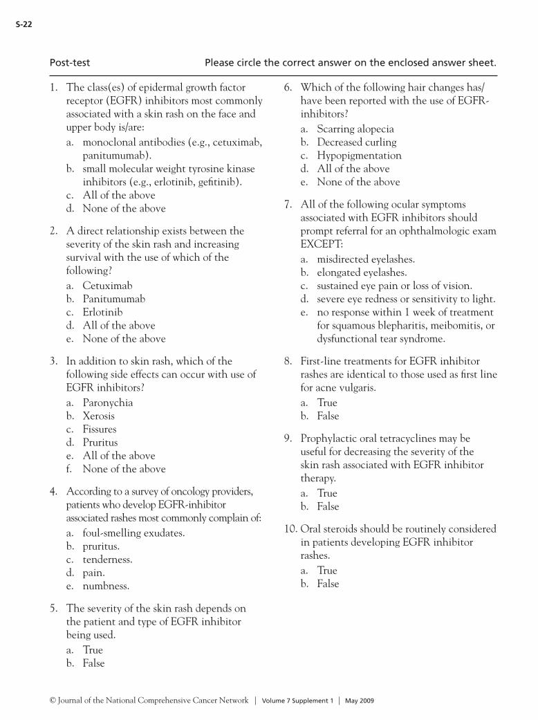
© Journal of the National Comprehensive Cancer Network | Volume 7 Supplement 1 | May 2009
S-22
1. The class(es) of epidermal growth factor receptor (EGFR) inhibitors most commonly associated with a skin rash on the face and upper body is/are:a. monoclonal antibodies (e.g., cetuximab,
panitumumab).b. small molecular weight tyrosine kinase
inhibitors (e.g., erlotinib, gefitinib).c. All of the aboved. None of the above
2. A direct relationship exists between the severity of the skin rash and increasing survival with the use of which of the following?a. Cetuximabb. Panitumumabc. Erlotinibd. All of the abovee. None of the above
3. In addition to skin rash, which of the following side effects can occur with use of EGFR inhibitors?a. Paronychiab. Xerosisc. Fissuresd. Prurituse. All of the abovef. None of the above
4. According to a survey of oncology providers, patients who develop EGFR-inhibitor associated rashes most commonly complain of:a. foul-smelling exudates.b. pruritus.c. tenderness.d. pain.e. numbness.
5. The severity of the skin rash depends on the patient and type of EGFR inhibitor being used.a. Trueb. False
6. Which of the following hair changes has/have been reported with the use of EGFR-inhibitors?a. Scarring alopeciab. Decreased curlingc. Hypopigmentationd. All of the abovee. None of the above
7. All of the following ocular symptoms associated with EGFR inhibitors should prompt referral for an ophthalmologic exam EXCEPT:a. misdirected eyelashes.b. elongated eyelashes.c. sustained eye pain or loss of vision.d. severe eye redness or sensitivity to light.e. no response within 1 week of treatment
for squamous blepharitis, meibomitis, or dysfunctional tear syndrome.
8. First-line treatments for EGFR inhibitor rashes are identical to those used as first line for acne vulgaris.a. Trueb. False
9. Prophylactic oral tetracyclines may be useful for decreasing the severity of the skin rash associated with EGFR inhibitor therapy.a. Trueb. False
10. Oral steroids should be routinely considered in patients developing EGFR inhibitor rashes.a. Trueb. False
Post-test Please circle the correct answer on the enclosed answer sheet.

© Journal of the National Comprehensive Cancer Network | Volume 7 Supplement 1 | May 2009
S-23
Post-Test Answer SheetPlease circle one answer per question. A score of at least 70% on the post-test is required.
1. a b c d 6. a b c d e
2. a b c d e 7. a b c d e
3. a b c d e f 8. a b
4. a b c d e 9. a b
5. a b 10. a b
The activity content helped me to achieve the following objectives:(1 = Strongly disagree; 3 = Not sure; 5 = Strongly agree)
Identify the epidermal growth factor receptor (EGFR) inhibitors
1 2 3 4 5
Recognize specific toxicities that may occur in patients treated with EGFR inhibitor therapy
1 2 3 4 5
Identify which patients are more likely to experience skin rash associated with EGFR inhibitor therapy
1 2 3 4 5
Recognize symptoms that should prompt a referral for an ophthalmologic examination in patients treated with EGFR inhibitor therapy
1 2 3 4 5
Recommend management options in patients with toxicities caused by EGFR inhibitor therapy
1 2 3 4 5
Please indicate the extent to which you agree or disagree with the following statements:
You were satisfied with the overall quality of this activity.
Strongly agree Agree Undecided Disagree Strongly disagree
Participation in this activity changed your knowledge/attitudes
Strongly agree Agree Undecided Disagree Strongly disagree
You will make a change in your practice as s result of participation in this activity.
Strongly agree Agree Undecided Disagree Strongly disagree
The activity presented scientifically rigorous, unbiased, and balanced information.
Strongly agree Agree Undecided Disagree Strongly disagree
Individual presentations were free of commercial bias.
Strongly agree Agree Undecided Disagree Strongly disagree

© Journal of the National Comprehensive Cancer Network | Volume 7 Supplement 1 | May 2009
S-24
NCCN Task Force Report: Management of Dermatologic and Other
Toxicities Associated With EGFR Inhibition in Patients With Cancer
Release Date: May 29, 2009Expiration Date: May 29,2010
Registration for Credit
To receive credit, please complete this page, the post-test, and the evaluation, and mail to the following address:
Continuing Education DepartmentNCCN275 Commerce Drive, Suite 300Fort Washington, PA 19034
There is no fee for participating in this activity.
Please print clearly.
Name Degree
Title/Position
Affiliation (University or Hospital)
Business Address
City State Zip
Business Telephone Business Fax
Email Address
I am claiming credits (maximum 1)
I certify that I have participated in this activity as designed.
Signature Date
TO RECEIVE CREDIT, YOU MUST SUBMIT THIS FORM BY MAY 29, 2010.
Comments and suggestions:
