nci-pbcf-ccl228 (sw480) colon adenocarcinoma · contamination and culture has not reached...
TRANSCRIPT
NCI-PBCF-CCL228 (SW480) Colon Adenocarcinoma
trade (ATCC
regCCL-228 )
February 27 2012 Version 16
Table of Contents
1 BACKGROUND INFORMATION ON SW480 CELL LINE 3
2 GENERAL INFORMATION FOR THE THAWING PROPAGATING AND CRYOPRESERVING OF NCI-PBCF- CCL228 (SW480) 3
3 REAGENTS 5
A PREPARATION OF COMPLETE GROWTH MEDIUM (L-15 + 10 (VV) FBS) 6
4 THAWING AND PROPAGATION OF CELLS 6
A THAWING CELLS 6 B PROPAGATING CELLS 7 C SUBCULTURING CELLS 8
5 HARVESTING OF CELLS FOR CRYOPRESERVATION 9
6 CRYOPRESERVATION OF CELLS 10
A CRYOPRESERVATION USING A RATE-CONTROLLED PROGRAMMABLE FREEZER 11 i Using the Cryomed 11
B CRYOPRESERVATION USING ldquoMR FROSTYrdquo 12
7 STORAGE 13
APPENDIX 1 PHOTOMICROGRAPHS OF NCI-PBCF-CCL228 (SW480) CELLS 14
APPENDIX 2 GROWTH PROFILE OF NCI-PBCF-CCL228 (SW480) CELLS 16
APPENDIX 3 CYTOGENETIC ANALYSIS OF NCI-PBCF-CCL228 SW480 CELLS 17
APPENDIX 4 GLOSSARY OF TERMS 20
APPENDIX 5 REFERENCE 21
APPENDIX 6 REAGENT LOT TRACEABILITY AND CELL EXPANSION TABLES 23
APPENDIX 7 CALCULATION OF POPULATION DOUBLING LEVEL (PDL) 25
APPENDIX 8 SAFETY PRECAUTIONS 25
SOP Thawing Propagation and Cryopreservation of NCI-PBCF-CCL228 (SW480)
Protocol for Thawing Propagation and Cryopreservation of NCI-PBCF-CCL228 (SW480) (ATCCregCCL-228 trade )
colon adenocarcinoma
1 Background Information on SW480 cell line
Designations SW480 [SW-480]
Biosafety Level 1
Shipped frozen (in dry ice)
Growth Properties Adherent (see Appendix 1)
Organism Homo sapiens
Organ colon
Source Disease Dukes type B
Derived from metastatic
site
colorectal adenocarcinoma
For more information visit the ATCC webpage
httpwwwatccorgATCCAdvancedCatalogSearchProductDetailstabid452DefaultaspxATC
CNum=CCL-228ampTemplate=cellBiology
2 General Information for the thawing propagating and
cryopreserving of NCI-PBCF- CCL228 (SW480)
Culture Initiation
The cryoprotectant (DMSO) should be removed by centrifugation
The seeding density to use with a vial of SW480 cells is about 5 x 10⁵
viable cellscm2
or one
vial into aT-25 flask containing 10 mL complete growth medium (L-15 + 10 (vv) FBS)
Complete growth
medium
The complete growth medium used to expand SW480cells is L-15 + 10 (vv) FBS
Complete growth medium (L-15 + 10 (vv) FBS) should be pre-warmed before use by placing
into a water bath set at 35 oC to 37
oC for 15 min to 30 min
After 30 min the complete growth medium (L-15 + 10 (vv) FBS) should be moved to room
temperature until used Complete growth medium (L-15 + 10 (vv) FBS) should be stored at 2 oC to 8
oC when not in use
Cell Growth
The growth temperature for SW480 is 37 oC plusmn 1 oC
100 air atmosphere ( without CO2) is recommended
Growth Properties Population doubling time (PDT) is approximately 38 h (see Figure 4)
Page 3 of 25
SOP Thawing Propagation and Cryopreservation of NCI-PBCF-CCL228 (SW480)
Special Growth
Requirements
Subculture SW480cells at 80 to 90 confluence or when cell density reaches an average of
5 x 10⁵
viable cellscm2
Subculture Medium
025 (wv) trypsin-053 mM EDTA (ATCC cat no 30-2101)
Subculturing reagents should be pre-warmed before use by placing into a water bath set at 35 oC to 37
oC for 15 min to 30 min
After 30 min the subculturing medium should be moved to room temperature until used
Subculturing reagents should be stored at 2 oC to 8
oC when not in use
Subculture Method
The attached SW480 cells are subcultured using 025 (wv) trypsin-053 mM EDTA (ATCC
cat no 30-2101)
The enzymatic action of the trypsin-EDTA is stopped by adding complete growth medium to the
detached cells
A split ratio of 110 to 112 or a seeding density of 4 x 10⁴
viable cellscm2 to 5 x 10
5 viable
cellscm2 is used when subculturing SW480 cells
Viable
CellsmLCryovial
The target number of viable cellsmLcryovial is 20 x 106
(acceptable range 2 x 10⁶
viable
cellsmL to 3 x 10⁶
viable cellsmL)
Cryopreservation
Medium
The cryopreservation medium for SW480 cells is complete growth medium (L-15 + 10 (vv)
FBS) containing 5 (vv) DMSO (ATCC cat no 4-X)
General Procedure to be applied throughout the SOP
Use of good aseptic technique is critical Any materials that are contaminated as well as any
materials with which they may have come into contact must be disposed of immediately Aseptic Technique
Traceability of
materialreagents
Record the manufacturer catalog number lot number date received date expired and any
other pertinent information for all materials and reagents used Record information in the
Reagent Lot Traceability Table 4 (Appendix 6)
Record the subculture and growth expansion activities such as passage number
confluence viability cell morphology (see Figures 1-3 Appendix 1) and population doubling
levels (PDLs) in the table for Cell Expansion (Table 5 Appendix 6) Calculate PDLs using the
equation in Appendix 7
Expansion of cell line
Medium volumes Medium volumes are based on the flask size as outlined in Table 1
Glossary of Terms
Safety Precaution
Refer to Glossary of Terms used throughout the document (see Appendix 4)
Refer to Safety Precautions pertaining to the thawing propagating and cryopreserving of
SW480 (See Appendix 8)
Page 4 of 25
SOP Thawing Propagation and Cryopreservation of NCI-PBCF-CCL228 (SW480)
Table 1 Medium Volumes
Flask Size Medium Volume Range
125 cm2
(T-125) 3 mL to 6 mL
25 cm2
(T-25) 5 mL to 13 mL
75 cm2
(T-75) 10 mL to 38 mL
150 cm2
(T-150) 30 mL to 75 mL
175 cm2
(T-175) 35 mL to 88 mL
225 cm2
(T-225) 45 mL to 113 mL
3 Reagents
Follow Product Information Sheet storage andor thawing instructions Below is a list of
reagents for the propagation subcultivation and cryopreservation of SW480 cells
Table 2 Reagents for Expansion Subculturing and Cryopreservation of SW480 Cells
Complete growth medium reagents
Subculturing reagents Cryopreservation medium reagents
Leibovitzrsquos L-15 Medium (ATCC cat no 30-2008)
Trypsin-EDTA (025 (wv)
Trypsin053 mM EDTA )
(ATCC cat no30-2101)
Leibovitzrsquos L-15 Medium (ATCC cat no 30-2008)
10 (vv) Fetal Bovine
Serum (FBS)
(ATCC cat no 30-2020)
Dulbeccorsquos Phosphate Buffered Saline (DPBS) modified without
calcium chloride and without
magnesium chloride
(ATCC cat no 30-2200)
10 (vv) FBS
(ATCC cat no 30-2020)
5 (vv) Dimethyl Sulfoxide (DMSO)
(ATCC cat no 4-X)
Page 5 of 25
SOP Thawing Propagation and Cryopreservation of NCI-PBCF-CCL228 (SW480)
a Preparation of complete growth medium (L-15 + 10 (vv) FBS)
The complete growth medium is prepared by aseptically combining
1 56 mL FBS (ATCC cat no 30-2020) to a 500 mL bottle of basal medium L-15 (ATCC cat no 30-2008)
2 Mix gently by swirling
4 Thawing and Propagation of Cells
Reagents and Material
Complete growth medium (L-15 + 10 (vv) FBS) Water bath T-25 cm2 polystyrene flask 15 mL polypropylene conical centrifuge tubes Plastic pipettes (1 mL10 mL 25 mL)
a Thawing cells
Method
1 Place complete growth medium (L-15 + 10 (vv) FBS) in a water bath set at 35 oC to
37 oC
2 Label T-25 flask to be used with the (a) name of cell line (b) passage number (c) date (d)
initials of technician
3 Wearing a full face shield retrieve a vial of frozen cells from the vapor phase of the liquid
nitrogen freezer
4 Thaw the vial by gentle agitation in a water bath set at 35 oC to 37 oC To reduce the
possibility of contamination keep the O-ring and cap out of the water
Note Thawing should be rapid (approximately 2 min to 3 min just long enough for most of the ice to melt)
5 Remove vial from the water bath and process immediately
6 Remove excess water from the vial by wiping with sterile gauze saturated with 70 ethanol
7 Transfer the vial to a BSL-2 laminar-flow hood
Page 6 of 25
SOP Thawing Propagation and Cryopreservation of NCI-PBCF-CCL228 (SW480)
b Propagating cells
Method
1 Add 9 mL of complete growth medium (L-15 + 10 (vv) FBS) to a 15-mL conical centrifuge
tube
2 Again wipe the outer surface of the vial with sterile gauze wetted with 70 ethanol
3 Using sterile gauze carefully remove the cap from the vial
4 With a 1 mL pipette transfer slowly the completely thawed content of the vial (1 mL cell
suspension) to the 15-mL conical centrifuge tube containing 9 mL complete growth medium
(L-15 + 10 (vv) FBS) Gently resuspend cells by pipetting up and down
5 Centrifuge at 125 xg at room temperature for 8 min to 10 min
6 Carefully aspirate (discard) the medium leaving the pellet undisturbed
7 Using a 10 mL pipette add 10 mL of complete growth medium (L-15 + 10 (vv) FBS)
8 Resuspend pellet by gentle pipetting up and down
9 Using a 1 mL pipette remove 1 mL of cell suspension for cell count and viability Cell counts
are performed using either an automated counter (such as Innovatis Cedex System
Beckman-Coulter ViCell system) or a hemocytometer
10 Record total cell count and viability When an automated system is used attach copies of
the printed results to the record
11 Plate cells in pre-labeled T-25 cm2 flask at about 08 x 105 cellscm2
12 Transfer flask to a 37 degC plusmn 1degC incubator without CO2
NOTE The L-15 medium formulation was devised for use in a free gas exchange with
atmospheric air A CO2 and air mixture is detrimental to cells when using this
medium for cultivation
13 Observe culture daily by eye and under an inverted microscope to ensure culture is free of contamination and culture has not reached confluence Monitor visually the pH of the medium daily If the medium goes from red through orange to yellow change the medium
14 Note In most cases cultures at a high cell density exhaust the medium faster than those at low cell density as is evident from the change in pH A drop in pH is usually accompanied by an increase in cell density which is an indicator to subculture the cells Cells may stop growing when the pH is between pH 7 to pH 6 and loose viability between pH 65 and pH 6
15 If fluid renewal is needed aseptically aspirate the complete growth medium from the flask and discard Add an equivalent volume of fresh complete growth medium to the flask Alternatively perform a fluid addition by adding fresh complete growth medium to the flask
Page 7 of 25
SOP Thawing Propagation and Cryopreservation of NCI-PBCF-CCL228 (SW480)
without removing the existing medium Record fluid change or fluid addition on the Cell Line Expansion Table (see Table 5 in Appendix 6)
16 If subculturing of cells is needed continue to lsquoSubculturing cellsrsquo
Note Subculture when the cells are 80-90 confluent (see photomicrographs in Appendix 1)
c Subculturing cells
Reagents and Material
025 (wv) Trypsin-053 mM EDTA
DPBS
Complete growth medium (L-15 (ATCC cat no 30-2008) + 10 (vv) FBS (ATCC cat
no 30-2020) Plastic pipettes (1 mL 10 mL 25 mL) T-75 cm2 T-225 cm2 polystyrene flasks
Method
1 Aseptically remove medium from the flask
2 Add appropriate volumes of sterile Ca2+- and Mg2+-free DPBS to the side of the flask opposite the cells so as to avoid dislodging the cells (see Table 3)
3 Rinse the cells with DPBS (using a gently rocking motion) and discard
4 Add appropriate volume of 025 (wv) Trypsin-053 mM EDTA solution to the flask (see Table 3)
5 Incubate the flask at 37 oC plusmn 1 oC until the cells round up Observe cells under an
inverted microscope every 5 min When the flask is tilted the attached cells should slide
down the surface This usually occurs after 5 min to 10 min of incubation
Note Do not leave trypsin-EDTA on the cells any longer than necessary as
clumping may result
6 Neutralize the trypsin-EDTAcell suspension by adding an equal volume of complete growth medium (L-15 + 10 (vv) FBS) to each flask Disperse the cells by pipetting gently over the surface of the monolayer Pipette the cell suspension up and down with the tip of the pipette resting on the bottom corner or edge until a single cell suspension is obtained Care should be taken to avoid the creation of foam
7 Using a 1 mL pipette remove 1 mL of cell suspension for total cell count and viability
8 Record total cell count and viability
Page 8 of 25
Type
DPBS Rinse Trypsin-EDTAFlask Flask
Size Buffer
T-flask 125 cm2
(T-125) 1 mL to 3 mL 1 mL to 2 mL
25 cm2
(T-25) 1 mL to 5 mL 1 mL to 3 mL
75 cm2
(T-75) 4 mL to 15 mL 2 mL to 8 mL
150 cm2
(T-150) 8 mL to 30 mL 4 mL to 15 mL
175 cm2
(T-175) 9 mL to 35 mL 5 mL to 20 mL
225 cm2
(T-225) 10 mL to 45 mL 5 mL to 25 mL
SOP Thawing Propagation and Cryopreservation of NCI-PBCF-CCL228 (SW480)
9 Add appropriate volume of fresh complete growth medium (L-15 + 10 (vv) FBS) and
transfer cell suspension (for volume see Table 1) into new pre-labeled flasks at a
seeding density of 4 x 10⁴ viable cellscm2 to 5 x 10⁴ viable cellscm2 or a split ratio of
110 to 112
10 Label all new flasks with the (a) name of cell line (b) passage number (c) date (d) initials of technician
Table 3 - Volume of Rinse Buffer and Trypsin
5 Harvesting of Cells for Cryopreservation
Reagents and Material
025 (wv) Trypsin-053 mM EDTA
DPBS
Complete growth medium (L-15 (ATCC cat no 30-2008) + 10 (vv) FBS (ATCC cat
no 30-2020)
50 mL or 250 mL conical centrifuge tube
Plastic pipettes (1 mL 10 mL 25 mL)
Sterile DMSO
1 mL to 18 mL cryovials
Ice bucket with ice
Method
1 Label cryovials to include information on the name of cell line (b) passage number (c) date
2 Prepare cryopreservation medium by adding DMSO to cold complete growth medium (Lshy
15 + 10 (vv) FBS) at a final concentration of 5 (vv) DMSO Place cryopreservation
medium on ice until ready to use
Page 9 of 25
SOP Thawing Propagation and Cryopreservation of NCI-PBCF-CCL228 (SW480)
3 Aseptically remove medium from the flask
4 Add appropriate volumes of sterile Ca2+- and Mg2+-free DPBS to the side of the flask so as to avoid dislodging the cells (see Table 3)
5 Rinse the cells with DPBS (using a gentle rocking motion) and discard
6 Add appropriate volume of 025 (wv) Trypsin-053 mM EDTA solution to the flask (see Table 3)
7 Incubate the flask at 37 oC plusmn 1 oC until the cells round up Observe cells under an
inverted microscope every 5 min When the flask is tilted the attached cells should slide
down the surface This usually occurs after 5 min to 10 min of incubation
Note Do not leave trypsin-EDTA on the cells any longer than necessary as
clumping may result
8 Neutralize the trypsin-EDTAcell suspension by adding an equal volume of complete growth medium (L-15 + 10 (vv) FBS) to each flask Disperse the cells by pipetting gently over the surface of the monolayer Pipette the cell suspension up and down with the tip of the pipette resting on the bottom corner or edge until a single cell suspension is obtained Care should be taken to avoid the creation of foam
9 Using a 1 mL pipette remove 1 mL of cell suspension for total cell count and viability
10 Record total cell count and viability
11 Spin cells at approximately 125 xg for 5 min to 10 min at room temperature Carefully
aspirate and discard the medium leaving the pellet undisturbed
12 Calculate volume of cryopreservation medium based on the count performed at step 9
and resuspend pellet in cold cryopreservation medium at a viable cell density of 2 x 10⁶
viable cellsmL (acceptable range 2 x 10⁶ viable cellsmL to 3 x 10⁶ viable cellsmL) by
gentle pipetting up and down
13 Dispense 1 mL of cell suspension using a 5 mL or 10 mL pipette into each 1 mL cryovial
14 Place filled cryovials at 2 oC to 8 oC until ready to cryopreserve A minimum equilibration
time of 10 min but no longer than 45 min is necessary to allow DMSO to penetrate the
cells
Note DMSO is toxic to the cells Long exposure in DMSO may affect viability
6 Cryopreservation of Cells
Material
Liquid nitrogen freezer Cryomed Programmable freezer (Forma Scientific cat no 1010) or Mr Frosty (Nalgene cat no 5100)
Page 10 of 25
SOP Thawing Propagation and Cryopreservation of NCI-PBCF-CCL228 (SW480)
Isopropanol
Cryovial rack
a Cryopreservation using a rate-controlled programmable freezerMethod
A slow and reproducible cooling rate is very important to ensure good recovery of cultures A decrease of 1 degC per min to -80 degC followed by rapid freeze at about 15 degC to 30 degC per min drop to -150 degC will usually work for most animal cell cultures The best way to control the cooling process is to use a programmable rate-controlled electronic freezer unit Refer to the manufacturerrsquos handbook for detailed procedure
i Using the Cryomed
Starting the Cryopreservation Process
1 Check that the liquid nitrogen valve that supplies the Cryomed is open
2 Check the gauge to ensure that there is enough liquid nitrogen in the open tank to complete the freeze
3 Install the thermocouple probe so that the tip is immersed midway into the control fluid
Note Be sure that the thermocouple is centered in the vial and the vial is placed centered in the rack The probe should be changed after three uses or if it turns yellow to ensure accurate readings by the controller during the freezing process Old medium may have different freezing characteristics
4 Close and latch Cryomed door
5 Turn on microcomputer computer and monitor
6 Double click the ldquoCryomedrdquo icon The machine may need to be pre-programmed for specific cell type and medium
7 From the top of the screen select MENU RUN FUNCTIONS START RUN
8 Fill out the box which appears on the screen Cell line ID TYPE OF SAMPLE MEDIA NUMBER OF SAMPLES
9 Hit the ESCAPE key and the Cryomed will cool to 4 C
10 Once Cryomed chamber has cooled to 4 C load cryovials onto racks and close the door
11 When the Cryomedrsquos chamber temperature and the sample temperature have reached approximately 4 C press the space bar to initiate the rate controlled cryopreservation process
Page 11 of 25
SOP Thawing Propagation and Cryopreservation of NCI-PBCF-CCL228 (SW480)
Completing the Cryopreservation Process
1 When samples have reached ndash80C an alarm will sound To silence this select ALARM
from the options at the top of the screen
2 Select MENU RUN FUNCTIONSrarr STOP Hit the ESCAPE key to return to the main
menu and select EXIT
3 Immediately transfer vials to liquid nitrogen freezer
4 Shut down the microcomputer and then turn off the monitor
b Cryopreservation using ldquoMr Frostyrdquo
1 One day before freezing cells add 250 mL isopropanol to the bottom of the container and place at 2 oC to 8 oC
2 On the day of the freeze prepare cells for cryopreservation as described above
3 Insert cryovials with the cell suspension in appropriate slots in the container
4 Transfer the container to a -70 degC to -90 degC freezer and store overnight
5 Next day transfer cryovials to the vapor phase of liquid nitrogen freezer
Note Each container has 18 slots which can accommodate 18 cryovials in one freeze
Important information when using the rate-controlled programmable freezer or a manual method (Mr Frosty) for cryopreservation of mammalian cells
Regardless which cooling method is used it is important that the transfer to the final storage location (between -130 degC and -196 degC) be done quickly and efficiently If the transfer cannot be done immediately the vials can be placed on dry ice for a short time This will avoid damage to cultures by inadvertent temporary warming during the transfer process Warming during this transfer process is a major cause of variation in culture viability upon thawing
Always keep the storage temperature below -130 degC for optimum survival Cells may survive storage at higher temperatures but viability will usually decrease over time The ideal storage container is a liquid nitrogen freezer where the cultures are stored in the vapor phase above the liquid nitrogen
Note ATCC does not have experience in the cryopreservation of the SW480 cells by any other method than the Cryomed programmable freezer
Page 12 of 25
SOP Thawing Propagation and Cryopreservation of NCI-PBCF-CCL228 (SW480)
7 Storage
Store cryopreserved cells in the vapor phase of liquid nitrogen freezer (below ndash130 degC) for optimum long-term survival
Note Experiments on long-term storage of animal cell lines at different temperature
levels indicate that a -70 degC storage temperature is not adequate except for very short
period of time A -90 degC storage may be adequate for longer periods depending upon
the cell line preserved The efficiency of recovery however is not as great as when
the cells are stored in vapor phase of the liquid nitrogen freezer
Page 13 of 25
SOP Thawing Propagation and Cryopreservation of NCI-PBCF-CCL228 (SW480)
APPENDIX 1 PHOTOMICROGRAPHS OF NCI-PBCF-CCL228 (SW480) CELLS
Figure 1 Photomicrograph of SW480 cells after one day post-freeze recovery Cells were plated at 80 x 104 viable cellscm2
Figure 2 Photomicrograph of SW480 cells after two days post-freeze recovery Cells were plated at 80 x 104 viable cellscm2
Page 14 of 25
SOP Thawing Propagation and Cryopreservation of NCI-PBCF-CCL228 (SW480)
40X 40X 40X
100X 100X 100X
500 microm500 microm500 microm
200 microm 200 microm 200 microm
Figure 3 Photomicrographs of SW480 cells at various time points after
seeding at a cell density of 5 x 10⁴ viable cellscm2
Page 15 of 25
SOP Thawing Propagation and Cryopreservation of NCI-PBCF-CCL228 (SW480)
APPENDIX 2 GROWTH PROFILE OF NCI-PBCF-CCL228 (SW480) CELLS
000E+00
100E+05
200E+05
300E+05
400E+05
500E+05
600E+05
700E+05
800E+05
900E+05
100E+06
7 8 9
Via
ble
Cell
sc
m2
0 1 2 3 4 5 6
Days in Culture
Figure 4 Growth curve for SW480 cells cells were plated at 5 x 10⁴ viable cellscm2 population doubling time (PDT) is approximately 38 h
Page 16 of 25
SOP Thawing Propagation and Cryopreservation of NCI-PBCF-CCL228 (SW480)
APPENDIX 3 CYTOGENETIC ANALYSIS OF NCI-PBCF-CCL228 SW480 CELLS
NCI-PBCF-CCL228 Karyotype Results
Metaphase Spread Karyotype
Number of metaphase spreads counted 20
Band level 300-400
Number of metaphase spreads karyotyped 10
Chromosome range 50-56
Sex Male
Comments Aneuploid
Karyotype
50~56XX+2der(2)t(218)(q21q111)del(5)(q13q33)+add(7)(q22)+add(7)(q32)
der(10)t (1012)(p15q11)+add(11)(p15)-12der(1214)(p10q10)-14+add(15)(p10)
der(15)t(1215)(q13q15)-18add(19)(p133)+20+add(20)(p13)
+der(20)t(520)(q22p13)x2+21+add(21)(q111)add(21)(q22)+mar1x2
(ISCN nomenclature written based on a diploid karyotype)
Human diploid karyotype (2N) 46XX (female) or 46XY (male)
Page 17 of 25
SOP Thawing Propagation and Cryopreservation of NCI-PBCF-CCL228 (SW480)
Karyotype Summary
In the karyotype image arrows indicate regions of abnormality It should be noted that the karyotype description includes the observed abnormalities from all examined metaphase spreads but due to heterogeneity not all of the karyotyped cells will contain every abnormality
This is a rearranged human cell line of male origin containing 50 to 56 chromosomes per metaphase spread (hyperdiploid) Structural abnormalities include rearrangements to approximately 50 of the 22 different autosomal chromosomes (described below) There is one unidentifiable clonal marker chromosome (markers present in two or more of the examined cells) [+mar1]
The rearrangements include
Addition of unknown material to the short arms (designated by p) of chromosomes 11 15 19 and 20
Addition of unknown material to the long arms (designated by q) of chromosomes 7 and 21 (2 different derivatives of each)
An interstitial deletion of material from the long arm of chromosome 5 between bands q13 and q33
Translocations involving chromosomes 2 and 18 [der(2)t(218)(q21q111)] 10 and 12 [der(10)t (1012)(p15q11) chromosomes 12 and 14 [der(1214)(p10q10)] 12 and 15 [der(15)t(1215)(q13q15)] and chromosomes 5 and 20 [der(20)t(520)(q22p13)]
Numerical changes are based on a diploid karyotype which would contain two copies of
each chromosome (2N) Therefore karyotype designations such as +2 +20 and +21
indicates three copies of structurally normal chromosomes 2 20 and 21 Designations
-18 and -14 indicates one copy of structurally normal chromosomes -18 and -14 Also it
appears that the Y chromosome was lost which is not an uncommon occurrence in cancer
cell lines (ISCN 2009 An International System for Human Cytogenetic Nomenclature
(2009) Editors Lisa G Shaffer Marilyn L Slovak Lynda J Campbell)
Karyotype Procedure
Cell Harvest Cells were allowed to grow to 80-90 confluence Mitotic division was
arrested by treating the cells with KaryoMaxreg colcemid for 20 minutes to 2 hours at 37degC
Cells were harvested using 005 Trypsin-EDTA treated with 0075M KCL hypotonic
solution and then fixed in three changes of a 31 ratio of methanolglacial acetic acid
Slide Preparation Slides were prepared by dropping the cell suspension onto wet glass
slides and allowing them to dry under controlled conditions
G-banding Slides were baked one hour at 90degC trypsinized using 10X trypsin-EDTA
and then stained with Leishmanrsquos stain
Page 18 of 25
SOP Thawing Propagation and Cryopreservation of NCI-PBCF-CCL228 (SW480)
Microscopy Slides were scanned using a 10X objective and metaphase spreads were
analyzed using a 100X plan apochromat objective on an Olympus BX-41 microscope
Imaging and karyotyping were performed using Cytovisionreg software
Analysis Twenty metaphase cells were counted and analyzed and representative
metaphase cells were karyotyped depending on the complexity of the study
Summary of Karyotyping Procedure
G-band karyotyping analysis is performed using GTL banding technique G bands produced with trypsin and Leishman Slides prepared with metaphase spreads are treated with trypsin and stained with Leishmanrsquos This method produces a series of light and dark bands that allow for the positive identification of each chromosome
SW480 cell line karyotyping was carried out by Cell Line Genetics Inc (Madison WI 53719)
Page 19 of 25
SOP Thawing Propagation and Cryopreservation of NCI-PBCF-CCL228 (SW480)
APPENDIX 4 GLOSSARY OF TERMS
Confluent monolayer adherent cell culture in which all cells are in contact with other cells
all around their periphery and no available substrate is left uncovered
Split ratio the divisor of the dilution ration of a cell culture to subculture (eg one flask
divided into four or 100 mL up to 400 mL would be split ratio of 14)
Subculture (or passage) the transfer or transplantation of cells with or without dilution
from one culture vessel to another
Passage No the total number of times the cells in the culture have been subcultured or
passaged (with each subculture the passage number increases by 1)
Population doubling level (PDL) the total number of population doublings of a cell line
since its initiation in vitro (with each subculture the population doubling increases in
relationship to the split ratio at which the cells are plated) See Appendix 7
Population doubling time (doubling time) the time interval calculated during the
logarithmic phase of growth in which cells double in number
Seeding density recommended number of cells per cm2 of substrate when inoculating a
new flask
Epithelial-like adherent cells of a polygonal shape with clear sharp boundaries between
them
Fibroblast-like adherent cells of a spindle or stellate shape
Page 20 of 25
SOP Thawing Propagation and Cryopreservation of NCI-PBCF-CCL228 (SW480)
APPENDIX 5 REFERENCE
1 Culture Of Animal Cells A Manual of Basic Technique by R Ian Freshney 6th edition
published by Wiley-Liss NY 2010
2 Fogh J et al Absence of HeLa cell contamination in 169 cell lines derived from human
tumors J Natl Cancer Inst 58 209-214 1977 PubMed 833871
3 Fogh J et al One hundred and twenty-seven cultured human tumor cell lines producing
tumors in nude mice J Natl Cancer Inst 59 221-226 1977 PubMed 327080
4 Lelbovitz A et al Detection and analysis of a glucose 6-phosphate dehydrogenase
phenotype B cell line contamination J Natl Cancer Inst 63 635-645 1979 PubMed
288927
5 Adachi A et al Productive persistent infection of human colorectal cell lines with human
immunodeficiency virus J Virol 61 209-213 1987 PubMed 3640832
6 Schroy PC et al Detection of p21ras mutations in colorectal adenomas and carcinomas
by enzyme-linked immunosorbent assay Cancer 76 201-209 1995 PubMed 8625092
7 Trainer DL et al Biological characterization and oncogene expression in human
colorectal carcinoma cell lines Int J Cancer 41 287-296 1988 PubMed 3338874
8 Weiss J et al Mutation and expression of the p53 gene in malignant melanoma cell
lines Int J Cancer 54 693-699 1993 PubMed 8514460
9 Nigro JM et al Mutations in the p53 gene occur in diverse human tumour types Nature
342 705-707 1989 PubMed 2531845
10 Barnett SW et al Characterization of human immunodeficiency virus type 1 strains
recovered from the bowel of infected individuals Virology 182 802-809 1991 PubMed
2024498
11 Leibovitz A et al Classification of human colorectal adenocarcinoma cell lines Cancer
Res 36 4562-4569 1976 PubMed 1000501
12 Geiser AG et al Suppression of tumorigenicity in human cell hybrids derived from cell
lines expressing different activated ras oncogenes Cancer Res 49 1572-1577 1989
PubMed 2647289
13 Lahm H et al Secretion of bioactive granulocyte-macrophage colony-stimulating factor
by human colorectal carcinoma cells Cancer Res 54 3700-3702 1994 PubMed
8033086
14 Rodrigues NR et al p53 mutations in colorectal cancer Proc Natl Acad Sci USA 87
7555-7559 1990 PubMed 1699228
Page 21 of 25
SOP Thawing Propagation and Cryopreservation of NCI-PBCF-CCL228 (SW480)
15 Santoro IM Groden J Alternative splicing of the APC gene and its association with terminal differentiation Cancer Res 57 488-494 1997 PubMed 9012479
16 Tsao H et al Novel mutations in the p16CDKN2A binding region of the Cyclinshy
dependent Kinase-4 gene Cancer Res 58 109-113 1998 PubMed 9426066
17 Zhu X et al Cell cycle-dependent modulation of telomerase activity in tumor cells Proc Natl Acad Sci USA 93 6091-6095 1996 PubMed 8650224
18 Witty JP et al Modulation of matrilysin levels in colon carcinoma cell lines affects tumorigenicity in vivo Cancer Res 54 4805-4812 1994 PubMed 8062282
Page 22 of 25
SOP Thawing Propagation and Cryopreservation of NCI-PBCF-CCL228 (SW480)
APPENDIX 6 REAGENT LOT TRACEABILITY AND CELL EXPANSION TABLES
Table 4 Reagent Lot Traceability
Reagent Vendor Catalog Lot Expiration Date
Page 23 of 25
Table 5 Cell Expansion
FROM FLUID CHANGE
Observation under
microscope CELL COUNT TO
By Date Flask qty
size
Pass Confluence
Add Replace
Volume
(in mL)
Viable cellsmL
Total viable cells
Viability
Split Ratio
Flask qty
size
Pass
PDL
Add
Replace
Add
Replace
Add
Replace
Add
Replace
Add
Replace
Add
Replace
Add
Replace
Add
Replace
Add
Replace
SOP Thawing Propagation and Cryopreservation of NCI-PBCF-CCL228 (SW480)
APPENDIX 7 CALCULATION OF POPULATION DOUBLING LEVEL (PDL)
Calculate the PDL of the current passage using the following equation
PDL = X + 3322 (log Y ndash log I)
Where X = initial PDL I = cell inoculum (number of cells plated in the flask) Y = final cell yield (number of cells at the end of the growth period)
APPENDIX 8 SAFETY PRECAUTIONS
Use at least approved Biological Safety Level 2 (BSL-2) facilities and procedures
Wear appropriate Personal Protective Equipment (PPE) such as isolation gown lab coat with sleeve protectors face shield and gloves
Use safety precautions when working with liquid nitrogen nitrogen vapor and cryogenically cooled fixtures
Use liquid nitrogen freezers and liquid nitrogen tanks only in areas with adequate ventilation Liquid nitrogen reduces the concentration of oxygen and can cause suffocation
Wear latex gloves over insulating gloves to prevent liquid nitrogen from soaking in and being held next to the skin Liquid nitrogen is extremely cold and will cause burns and frostbite Metal inventory racks tank components and liquid nitrogen transfer hoses exposed to liquid nitrogen or nitrogen vapor quickly cool to cryogenic temperatures and can cause burns and frostbite
Wear a full face mask when thawing and retrieving vials from liquid nitrogen freezer Danger to the technician derives mainly from the possibility that liquid nitrogen can penetrate the cryovial during storage On warming rapid evaporation of the nitrogen within the confines of such cryovial can cause an aerosol or explosion of the cryovial and contents
Page 25 of 25
SOP Thawing Propagation and Cryopreservation of NCI-PBCF-CCL228 (SW480)
Protocol for Thawing Propagation and Cryopreservation of NCI-PBCF-CCL228 (SW480) (ATCCregCCL-228 trade )
colon adenocarcinoma
1 Background Information on SW480 cell line
Designations SW480 [SW-480]
Biosafety Level 1
Shipped frozen (in dry ice)
Growth Properties Adherent (see Appendix 1)
Organism Homo sapiens
Organ colon
Source Disease Dukes type B
Derived from metastatic
site
colorectal adenocarcinoma
For more information visit the ATCC webpage
httpwwwatccorgATCCAdvancedCatalogSearchProductDetailstabid452DefaultaspxATC
CNum=CCL-228ampTemplate=cellBiology
2 General Information for the thawing propagating and
cryopreserving of NCI-PBCF- CCL228 (SW480)
Culture Initiation
The cryoprotectant (DMSO) should be removed by centrifugation
The seeding density to use with a vial of SW480 cells is about 5 x 10⁵
viable cellscm2
or one
vial into aT-25 flask containing 10 mL complete growth medium (L-15 + 10 (vv) FBS)
Complete growth
medium
The complete growth medium used to expand SW480cells is L-15 + 10 (vv) FBS
Complete growth medium (L-15 + 10 (vv) FBS) should be pre-warmed before use by placing
into a water bath set at 35 oC to 37
oC for 15 min to 30 min
After 30 min the complete growth medium (L-15 + 10 (vv) FBS) should be moved to room
temperature until used Complete growth medium (L-15 + 10 (vv) FBS) should be stored at 2 oC to 8
oC when not in use
Cell Growth
The growth temperature for SW480 is 37 oC plusmn 1 oC
100 air atmosphere ( without CO2) is recommended
Growth Properties Population doubling time (PDT) is approximately 38 h (see Figure 4)
Page 3 of 25
SOP Thawing Propagation and Cryopreservation of NCI-PBCF-CCL228 (SW480)
Special Growth
Requirements
Subculture SW480cells at 80 to 90 confluence or when cell density reaches an average of
5 x 10⁵
viable cellscm2
Subculture Medium
025 (wv) trypsin-053 mM EDTA (ATCC cat no 30-2101)
Subculturing reagents should be pre-warmed before use by placing into a water bath set at 35 oC to 37
oC for 15 min to 30 min
After 30 min the subculturing medium should be moved to room temperature until used
Subculturing reagents should be stored at 2 oC to 8
oC when not in use
Subculture Method
The attached SW480 cells are subcultured using 025 (wv) trypsin-053 mM EDTA (ATCC
cat no 30-2101)
The enzymatic action of the trypsin-EDTA is stopped by adding complete growth medium to the
detached cells
A split ratio of 110 to 112 or a seeding density of 4 x 10⁴
viable cellscm2 to 5 x 10
5 viable
cellscm2 is used when subculturing SW480 cells
Viable
CellsmLCryovial
The target number of viable cellsmLcryovial is 20 x 106
(acceptable range 2 x 10⁶
viable
cellsmL to 3 x 10⁶
viable cellsmL)
Cryopreservation
Medium
The cryopreservation medium for SW480 cells is complete growth medium (L-15 + 10 (vv)
FBS) containing 5 (vv) DMSO (ATCC cat no 4-X)
General Procedure to be applied throughout the SOP
Use of good aseptic technique is critical Any materials that are contaminated as well as any
materials with which they may have come into contact must be disposed of immediately Aseptic Technique
Traceability of
materialreagents
Record the manufacturer catalog number lot number date received date expired and any
other pertinent information for all materials and reagents used Record information in the
Reagent Lot Traceability Table 4 (Appendix 6)
Record the subculture and growth expansion activities such as passage number
confluence viability cell morphology (see Figures 1-3 Appendix 1) and population doubling
levels (PDLs) in the table for Cell Expansion (Table 5 Appendix 6) Calculate PDLs using the
equation in Appendix 7
Expansion of cell line
Medium volumes Medium volumes are based on the flask size as outlined in Table 1
Glossary of Terms
Safety Precaution
Refer to Glossary of Terms used throughout the document (see Appendix 4)
Refer to Safety Precautions pertaining to the thawing propagating and cryopreserving of
SW480 (See Appendix 8)
Page 4 of 25
SOP Thawing Propagation and Cryopreservation of NCI-PBCF-CCL228 (SW480)
Table 1 Medium Volumes
Flask Size Medium Volume Range
125 cm2
(T-125) 3 mL to 6 mL
25 cm2
(T-25) 5 mL to 13 mL
75 cm2
(T-75) 10 mL to 38 mL
150 cm2
(T-150) 30 mL to 75 mL
175 cm2
(T-175) 35 mL to 88 mL
225 cm2
(T-225) 45 mL to 113 mL
3 Reagents
Follow Product Information Sheet storage andor thawing instructions Below is a list of
reagents for the propagation subcultivation and cryopreservation of SW480 cells
Table 2 Reagents for Expansion Subculturing and Cryopreservation of SW480 Cells
Complete growth medium reagents
Subculturing reagents Cryopreservation medium reagents
Leibovitzrsquos L-15 Medium (ATCC cat no 30-2008)
Trypsin-EDTA (025 (wv)
Trypsin053 mM EDTA )
(ATCC cat no30-2101)
Leibovitzrsquos L-15 Medium (ATCC cat no 30-2008)
10 (vv) Fetal Bovine
Serum (FBS)
(ATCC cat no 30-2020)
Dulbeccorsquos Phosphate Buffered Saline (DPBS) modified without
calcium chloride and without
magnesium chloride
(ATCC cat no 30-2200)
10 (vv) FBS
(ATCC cat no 30-2020)
5 (vv) Dimethyl Sulfoxide (DMSO)
(ATCC cat no 4-X)
Page 5 of 25
SOP Thawing Propagation and Cryopreservation of NCI-PBCF-CCL228 (SW480)
a Preparation of complete growth medium (L-15 + 10 (vv) FBS)
The complete growth medium is prepared by aseptically combining
1 56 mL FBS (ATCC cat no 30-2020) to a 500 mL bottle of basal medium L-15 (ATCC cat no 30-2008)
2 Mix gently by swirling
4 Thawing and Propagation of Cells
Reagents and Material
Complete growth medium (L-15 + 10 (vv) FBS) Water bath T-25 cm2 polystyrene flask 15 mL polypropylene conical centrifuge tubes Plastic pipettes (1 mL10 mL 25 mL)
a Thawing cells
Method
1 Place complete growth medium (L-15 + 10 (vv) FBS) in a water bath set at 35 oC to
37 oC
2 Label T-25 flask to be used with the (a) name of cell line (b) passage number (c) date (d)
initials of technician
3 Wearing a full face shield retrieve a vial of frozen cells from the vapor phase of the liquid
nitrogen freezer
4 Thaw the vial by gentle agitation in a water bath set at 35 oC to 37 oC To reduce the
possibility of contamination keep the O-ring and cap out of the water
Note Thawing should be rapid (approximately 2 min to 3 min just long enough for most of the ice to melt)
5 Remove vial from the water bath and process immediately
6 Remove excess water from the vial by wiping with sterile gauze saturated with 70 ethanol
7 Transfer the vial to a BSL-2 laminar-flow hood
Page 6 of 25
SOP Thawing Propagation and Cryopreservation of NCI-PBCF-CCL228 (SW480)
b Propagating cells
Method
1 Add 9 mL of complete growth medium (L-15 + 10 (vv) FBS) to a 15-mL conical centrifuge
tube
2 Again wipe the outer surface of the vial with sterile gauze wetted with 70 ethanol
3 Using sterile gauze carefully remove the cap from the vial
4 With a 1 mL pipette transfer slowly the completely thawed content of the vial (1 mL cell
suspension) to the 15-mL conical centrifuge tube containing 9 mL complete growth medium
(L-15 + 10 (vv) FBS) Gently resuspend cells by pipetting up and down
5 Centrifuge at 125 xg at room temperature for 8 min to 10 min
6 Carefully aspirate (discard) the medium leaving the pellet undisturbed
7 Using a 10 mL pipette add 10 mL of complete growth medium (L-15 + 10 (vv) FBS)
8 Resuspend pellet by gentle pipetting up and down
9 Using a 1 mL pipette remove 1 mL of cell suspension for cell count and viability Cell counts
are performed using either an automated counter (such as Innovatis Cedex System
Beckman-Coulter ViCell system) or a hemocytometer
10 Record total cell count and viability When an automated system is used attach copies of
the printed results to the record
11 Plate cells in pre-labeled T-25 cm2 flask at about 08 x 105 cellscm2
12 Transfer flask to a 37 degC plusmn 1degC incubator without CO2
NOTE The L-15 medium formulation was devised for use in a free gas exchange with
atmospheric air A CO2 and air mixture is detrimental to cells when using this
medium for cultivation
13 Observe culture daily by eye and under an inverted microscope to ensure culture is free of contamination and culture has not reached confluence Monitor visually the pH of the medium daily If the medium goes from red through orange to yellow change the medium
14 Note In most cases cultures at a high cell density exhaust the medium faster than those at low cell density as is evident from the change in pH A drop in pH is usually accompanied by an increase in cell density which is an indicator to subculture the cells Cells may stop growing when the pH is between pH 7 to pH 6 and loose viability between pH 65 and pH 6
15 If fluid renewal is needed aseptically aspirate the complete growth medium from the flask and discard Add an equivalent volume of fresh complete growth medium to the flask Alternatively perform a fluid addition by adding fresh complete growth medium to the flask
Page 7 of 25
SOP Thawing Propagation and Cryopreservation of NCI-PBCF-CCL228 (SW480)
without removing the existing medium Record fluid change or fluid addition on the Cell Line Expansion Table (see Table 5 in Appendix 6)
16 If subculturing of cells is needed continue to lsquoSubculturing cellsrsquo
Note Subculture when the cells are 80-90 confluent (see photomicrographs in Appendix 1)
c Subculturing cells
Reagents and Material
025 (wv) Trypsin-053 mM EDTA
DPBS
Complete growth medium (L-15 (ATCC cat no 30-2008) + 10 (vv) FBS (ATCC cat
no 30-2020) Plastic pipettes (1 mL 10 mL 25 mL) T-75 cm2 T-225 cm2 polystyrene flasks
Method
1 Aseptically remove medium from the flask
2 Add appropriate volumes of sterile Ca2+- and Mg2+-free DPBS to the side of the flask opposite the cells so as to avoid dislodging the cells (see Table 3)
3 Rinse the cells with DPBS (using a gently rocking motion) and discard
4 Add appropriate volume of 025 (wv) Trypsin-053 mM EDTA solution to the flask (see Table 3)
5 Incubate the flask at 37 oC plusmn 1 oC until the cells round up Observe cells under an
inverted microscope every 5 min When the flask is tilted the attached cells should slide
down the surface This usually occurs after 5 min to 10 min of incubation
Note Do not leave trypsin-EDTA on the cells any longer than necessary as
clumping may result
6 Neutralize the trypsin-EDTAcell suspension by adding an equal volume of complete growth medium (L-15 + 10 (vv) FBS) to each flask Disperse the cells by pipetting gently over the surface of the monolayer Pipette the cell suspension up and down with the tip of the pipette resting on the bottom corner or edge until a single cell suspension is obtained Care should be taken to avoid the creation of foam
7 Using a 1 mL pipette remove 1 mL of cell suspension for total cell count and viability
8 Record total cell count and viability
Page 8 of 25
Type
DPBS Rinse Trypsin-EDTAFlask Flask
Size Buffer
T-flask 125 cm2
(T-125) 1 mL to 3 mL 1 mL to 2 mL
25 cm2
(T-25) 1 mL to 5 mL 1 mL to 3 mL
75 cm2
(T-75) 4 mL to 15 mL 2 mL to 8 mL
150 cm2
(T-150) 8 mL to 30 mL 4 mL to 15 mL
175 cm2
(T-175) 9 mL to 35 mL 5 mL to 20 mL
225 cm2
(T-225) 10 mL to 45 mL 5 mL to 25 mL
SOP Thawing Propagation and Cryopreservation of NCI-PBCF-CCL228 (SW480)
9 Add appropriate volume of fresh complete growth medium (L-15 + 10 (vv) FBS) and
transfer cell suspension (for volume see Table 1) into new pre-labeled flasks at a
seeding density of 4 x 10⁴ viable cellscm2 to 5 x 10⁴ viable cellscm2 or a split ratio of
110 to 112
10 Label all new flasks with the (a) name of cell line (b) passage number (c) date (d) initials of technician
Table 3 - Volume of Rinse Buffer and Trypsin
5 Harvesting of Cells for Cryopreservation
Reagents and Material
025 (wv) Trypsin-053 mM EDTA
DPBS
Complete growth medium (L-15 (ATCC cat no 30-2008) + 10 (vv) FBS (ATCC cat
no 30-2020)
50 mL or 250 mL conical centrifuge tube
Plastic pipettes (1 mL 10 mL 25 mL)
Sterile DMSO
1 mL to 18 mL cryovials
Ice bucket with ice
Method
1 Label cryovials to include information on the name of cell line (b) passage number (c) date
2 Prepare cryopreservation medium by adding DMSO to cold complete growth medium (Lshy
15 + 10 (vv) FBS) at a final concentration of 5 (vv) DMSO Place cryopreservation
medium on ice until ready to use
Page 9 of 25
SOP Thawing Propagation and Cryopreservation of NCI-PBCF-CCL228 (SW480)
3 Aseptically remove medium from the flask
4 Add appropriate volumes of sterile Ca2+- and Mg2+-free DPBS to the side of the flask so as to avoid dislodging the cells (see Table 3)
5 Rinse the cells with DPBS (using a gentle rocking motion) and discard
6 Add appropriate volume of 025 (wv) Trypsin-053 mM EDTA solution to the flask (see Table 3)
7 Incubate the flask at 37 oC plusmn 1 oC until the cells round up Observe cells under an
inverted microscope every 5 min When the flask is tilted the attached cells should slide
down the surface This usually occurs after 5 min to 10 min of incubation
Note Do not leave trypsin-EDTA on the cells any longer than necessary as
clumping may result
8 Neutralize the trypsin-EDTAcell suspension by adding an equal volume of complete growth medium (L-15 + 10 (vv) FBS) to each flask Disperse the cells by pipetting gently over the surface of the monolayer Pipette the cell suspension up and down with the tip of the pipette resting on the bottom corner or edge until a single cell suspension is obtained Care should be taken to avoid the creation of foam
9 Using a 1 mL pipette remove 1 mL of cell suspension for total cell count and viability
10 Record total cell count and viability
11 Spin cells at approximately 125 xg for 5 min to 10 min at room temperature Carefully
aspirate and discard the medium leaving the pellet undisturbed
12 Calculate volume of cryopreservation medium based on the count performed at step 9
and resuspend pellet in cold cryopreservation medium at a viable cell density of 2 x 10⁶
viable cellsmL (acceptable range 2 x 10⁶ viable cellsmL to 3 x 10⁶ viable cellsmL) by
gentle pipetting up and down
13 Dispense 1 mL of cell suspension using a 5 mL or 10 mL pipette into each 1 mL cryovial
14 Place filled cryovials at 2 oC to 8 oC until ready to cryopreserve A minimum equilibration
time of 10 min but no longer than 45 min is necessary to allow DMSO to penetrate the
cells
Note DMSO is toxic to the cells Long exposure in DMSO may affect viability
6 Cryopreservation of Cells
Material
Liquid nitrogen freezer Cryomed Programmable freezer (Forma Scientific cat no 1010) or Mr Frosty (Nalgene cat no 5100)
Page 10 of 25
SOP Thawing Propagation and Cryopreservation of NCI-PBCF-CCL228 (SW480)
Isopropanol
Cryovial rack
a Cryopreservation using a rate-controlled programmable freezerMethod
A slow and reproducible cooling rate is very important to ensure good recovery of cultures A decrease of 1 degC per min to -80 degC followed by rapid freeze at about 15 degC to 30 degC per min drop to -150 degC will usually work for most animal cell cultures The best way to control the cooling process is to use a programmable rate-controlled electronic freezer unit Refer to the manufacturerrsquos handbook for detailed procedure
i Using the Cryomed
Starting the Cryopreservation Process
1 Check that the liquid nitrogen valve that supplies the Cryomed is open
2 Check the gauge to ensure that there is enough liquid nitrogen in the open tank to complete the freeze
3 Install the thermocouple probe so that the tip is immersed midway into the control fluid
Note Be sure that the thermocouple is centered in the vial and the vial is placed centered in the rack The probe should be changed after three uses or if it turns yellow to ensure accurate readings by the controller during the freezing process Old medium may have different freezing characteristics
4 Close and latch Cryomed door
5 Turn on microcomputer computer and monitor
6 Double click the ldquoCryomedrdquo icon The machine may need to be pre-programmed for specific cell type and medium
7 From the top of the screen select MENU RUN FUNCTIONS START RUN
8 Fill out the box which appears on the screen Cell line ID TYPE OF SAMPLE MEDIA NUMBER OF SAMPLES
9 Hit the ESCAPE key and the Cryomed will cool to 4 C
10 Once Cryomed chamber has cooled to 4 C load cryovials onto racks and close the door
11 When the Cryomedrsquos chamber temperature and the sample temperature have reached approximately 4 C press the space bar to initiate the rate controlled cryopreservation process
Page 11 of 25
SOP Thawing Propagation and Cryopreservation of NCI-PBCF-CCL228 (SW480)
Completing the Cryopreservation Process
1 When samples have reached ndash80C an alarm will sound To silence this select ALARM
from the options at the top of the screen
2 Select MENU RUN FUNCTIONSrarr STOP Hit the ESCAPE key to return to the main
menu and select EXIT
3 Immediately transfer vials to liquid nitrogen freezer
4 Shut down the microcomputer and then turn off the monitor
b Cryopreservation using ldquoMr Frostyrdquo
1 One day before freezing cells add 250 mL isopropanol to the bottom of the container and place at 2 oC to 8 oC
2 On the day of the freeze prepare cells for cryopreservation as described above
3 Insert cryovials with the cell suspension in appropriate slots in the container
4 Transfer the container to a -70 degC to -90 degC freezer and store overnight
5 Next day transfer cryovials to the vapor phase of liquid nitrogen freezer
Note Each container has 18 slots which can accommodate 18 cryovials in one freeze
Important information when using the rate-controlled programmable freezer or a manual method (Mr Frosty) for cryopreservation of mammalian cells
Regardless which cooling method is used it is important that the transfer to the final storage location (between -130 degC and -196 degC) be done quickly and efficiently If the transfer cannot be done immediately the vials can be placed on dry ice for a short time This will avoid damage to cultures by inadvertent temporary warming during the transfer process Warming during this transfer process is a major cause of variation in culture viability upon thawing
Always keep the storage temperature below -130 degC for optimum survival Cells may survive storage at higher temperatures but viability will usually decrease over time The ideal storage container is a liquid nitrogen freezer where the cultures are stored in the vapor phase above the liquid nitrogen
Note ATCC does not have experience in the cryopreservation of the SW480 cells by any other method than the Cryomed programmable freezer
Page 12 of 25
SOP Thawing Propagation and Cryopreservation of NCI-PBCF-CCL228 (SW480)
7 Storage
Store cryopreserved cells in the vapor phase of liquid nitrogen freezer (below ndash130 degC) for optimum long-term survival
Note Experiments on long-term storage of animal cell lines at different temperature
levels indicate that a -70 degC storage temperature is not adequate except for very short
period of time A -90 degC storage may be adequate for longer periods depending upon
the cell line preserved The efficiency of recovery however is not as great as when
the cells are stored in vapor phase of the liquid nitrogen freezer
Page 13 of 25
SOP Thawing Propagation and Cryopreservation of NCI-PBCF-CCL228 (SW480)
APPENDIX 1 PHOTOMICROGRAPHS OF NCI-PBCF-CCL228 (SW480) CELLS
Figure 1 Photomicrograph of SW480 cells after one day post-freeze recovery Cells were plated at 80 x 104 viable cellscm2
Figure 2 Photomicrograph of SW480 cells after two days post-freeze recovery Cells were plated at 80 x 104 viable cellscm2
Page 14 of 25
SOP Thawing Propagation and Cryopreservation of NCI-PBCF-CCL228 (SW480)
40X 40X 40X
100X 100X 100X
500 microm500 microm500 microm
200 microm 200 microm 200 microm
Figure 3 Photomicrographs of SW480 cells at various time points after
seeding at a cell density of 5 x 10⁴ viable cellscm2
Page 15 of 25
SOP Thawing Propagation and Cryopreservation of NCI-PBCF-CCL228 (SW480)
APPENDIX 2 GROWTH PROFILE OF NCI-PBCF-CCL228 (SW480) CELLS
000E+00
100E+05
200E+05
300E+05
400E+05
500E+05
600E+05
700E+05
800E+05
900E+05
100E+06
7 8 9
Via
ble
Cell
sc
m2
0 1 2 3 4 5 6
Days in Culture
Figure 4 Growth curve for SW480 cells cells were plated at 5 x 10⁴ viable cellscm2 population doubling time (PDT) is approximately 38 h
Page 16 of 25
SOP Thawing Propagation and Cryopreservation of NCI-PBCF-CCL228 (SW480)
APPENDIX 3 CYTOGENETIC ANALYSIS OF NCI-PBCF-CCL228 SW480 CELLS
NCI-PBCF-CCL228 Karyotype Results
Metaphase Spread Karyotype
Number of metaphase spreads counted 20
Band level 300-400
Number of metaphase spreads karyotyped 10
Chromosome range 50-56
Sex Male
Comments Aneuploid
Karyotype
50~56XX+2der(2)t(218)(q21q111)del(5)(q13q33)+add(7)(q22)+add(7)(q32)
der(10)t (1012)(p15q11)+add(11)(p15)-12der(1214)(p10q10)-14+add(15)(p10)
der(15)t(1215)(q13q15)-18add(19)(p133)+20+add(20)(p13)
+der(20)t(520)(q22p13)x2+21+add(21)(q111)add(21)(q22)+mar1x2
(ISCN nomenclature written based on a diploid karyotype)
Human diploid karyotype (2N) 46XX (female) or 46XY (male)
Page 17 of 25
SOP Thawing Propagation and Cryopreservation of NCI-PBCF-CCL228 (SW480)
Karyotype Summary
In the karyotype image arrows indicate regions of abnormality It should be noted that the karyotype description includes the observed abnormalities from all examined metaphase spreads but due to heterogeneity not all of the karyotyped cells will contain every abnormality
This is a rearranged human cell line of male origin containing 50 to 56 chromosomes per metaphase spread (hyperdiploid) Structural abnormalities include rearrangements to approximately 50 of the 22 different autosomal chromosomes (described below) There is one unidentifiable clonal marker chromosome (markers present in two or more of the examined cells) [+mar1]
The rearrangements include
Addition of unknown material to the short arms (designated by p) of chromosomes 11 15 19 and 20
Addition of unknown material to the long arms (designated by q) of chromosomes 7 and 21 (2 different derivatives of each)
An interstitial deletion of material from the long arm of chromosome 5 between bands q13 and q33
Translocations involving chromosomes 2 and 18 [der(2)t(218)(q21q111)] 10 and 12 [der(10)t (1012)(p15q11) chromosomes 12 and 14 [der(1214)(p10q10)] 12 and 15 [der(15)t(1215)(q13q15)] and chromosomes 5 and 20 [der(20)t(520)(q22p13)]
Numerical changes are based on a diploid karyotype which would contain two copies of
each chromosome (2N) Therefore karyotype designations such as +2 +20 and +21
indicates three copies of structurally normal chromosomes 2 20 and 21 Designations
-18 and -14 indicates one copy of structurally normal chromosomes -18 and -14 Also it
appears that the Y chromosome was lost which is not an uncommon occurrence in cancer
cell lines (ISCN 2009 An International System for Human Cytogenetic Nomenclature
(2009) Editors Lisa G Shaffer Marilyn L Slovak Lynda J Campbell)
Karyotype Procedure
Cell Harvest Cells were allowed to grow to 80-90 confluence Mitotic division was
arrested by treating the cells with KaryoMaxreg colcemid for 20 minutes to 2 hours at 37degC
Cells were harvested using 005 Trypsin-EDTA treated with 0075M KCL hypotonic
solution and then fixed in three changes of a 31 ratio of methanolglacial acetic acid
Slide Preparation Slides were prepared by dropping the cell suspension onto wet glass
slides and allowing them to dry under controlled conditions
G-banding Slides were baked one hour at 90degC trypsinized using 10X trypsin-EDTA
and then stained with Leishmanrsquos stain
Page 18 of 25
SOP Thawing Propagation and Cryopreservation of NCI-PBCF-CCL228 (SW480)
Microscopy Slides were scanned using a 10X objective and metaphase spreads were
analyzed using a 100X plan apochromat objective on an Olympus BX-41 microscope
Imaging and karyotyping were performed using Cytovisionreg software
Analysis Twenty metaphase cells were counted and analyzed and representative
metaphase cells were karyotyped depending on the complexity of the study
Summary of Karyotyping Procedure
G-band karyotyping analysis is performed using GTL banding technique G bands produced with trypsin and Leishman Slides prepared with metaphase spreads are treated with trypsin and stained with Leishmanrsquos This method produces a series of light and dark bands that allow for the positive identification of each chromosome
SW480 cell line karyotyping was carried out by Cell Line Genetics Inc (Madison WI 53719)
Page 19 of 25
SOP Thawing Propagation and Cryopreservation of NCI-PBCF-CCL228 (SW480)
APPENDIX 4 GLOSSARY OF TERMS
Confluent monolayer adherent cell culture in which all cells are in contact with other cells
all around their periphery and no available substrate is left uncovered
Split ratio the divisor of the dilution ration of a cell culture to subculture (eg one flask
divided into four or 100 mL up to 400 mL would be split ratio of 14)
Subculture (or passage) the transfer or transplantation of cells with or without dilution
from one culture vessel to another
Passage No the total number of times the cells in the culture have been subcultured or
passaged (with each subculture the passage number increases by 1)
Population doubling level (PDL) the total number of population doublings of a cell line
since its initiation in vitro (with each subculture the population doubling increases in
relationship to the split ratio at which the cells are plated) See Appendix 7
Population doubling time (doubling time) the time interval calculated during the
logarithmic phase of growth in which cells double in number
Seeding density recommended number of cells per cm2 of substrate when inoculating a
new flask
Epithelial-like adherent cells of a polygonal shape with clear sharp boundaries between
them
Fibroblast-like adherent cells of a spindle or stellate shape
Page 20 of 25
SOP Thawing Propagation and Cryopreservation of NCI-PBCF-CCL228 (SW480)
APPENDIX 5 REFERENCE
1 Culture Of Animal Cells A Manual of Basic Technique by R Ian Freshney 6th edition
published by Wiley-Liss NY 2010
2 Fogh J et al Absence of HeLa cell contamination in 169 cell lines derived from human
tumors J Natl Cancer Inst 58 209-214 1977 PubMed 833871
3 Fogh J et al One hundred and twenty-seven cultured human tumor cell lines producing
tumors in nude mice J Natl Cancer Inst 59 221-226 1977 PubMed 327080
4 Lelbovitz A et al Detection and analysis of a glucose 6-phosphate dehydrogenase
phenotype B cell line contamination J Natl Cancer Inst 63 635-645 1979 PubMed
288927
5 Adachi A et al Productive persistent infection of human colorectal cell lines with human
immunodeficiency virus J Virol 61 209-213 1987 PubMed 3640832
6 Schroy PC et al Detection of p21ras mutations in colorectal adenomas and carcinomas
by enzyme-linked immunosorbent assay Cancer 76 201-209 1995 PubMed 8625092
7 Trainer DL et al Biological characterization and oncogene expression in human
colorectal carcinoma cell lines Int J Cancer 41 287-296 1988 PubMed 3338874
8 Weiss J et al Mutation and expression of the p53 gene in malignant melanoma cell
lines Int J Cancer 54 693-699 1993 PubMed 8514460
9 Nigro JM et al Mutations in the p53 gene occur in diverse human tumour types Nature
342 705-707 1989 PubMed 2531845
10 Barnett SW et al Characterization of human immunodeficiency virus type 1 strains
recovered from the bowel of infected individuals Virology 182 802-809 1991 PubMed
2024498
11 Leibovitz A et al Classification of human colorectal adenocarcinoma cell lines Cancer
Res 36 4562-4569 1976 PubMed 1000501
12 Geiser AG et al Suppression of tumorigenicity in human cell hybrids derived from cell
lines expressing different activated ras oncogenes Cancer Res 49 1572-1577 1989
PubMed 2647289
13 Lahm H et al Secretion of bioactive granulocyte-macrophage colony-stimulating factor
by human colorectal carcinoma cells Cancer Res 54 3700-3702 1994 PubMed
8033086
14 Rodrigues NR et al p53 mutations in colorectal cancer Proc Natl Acad Sci USA 87
7555-7559 1990 PubMed 1699228
Page 21 of 25
SOP Thawing Propagation and Cryopreservation of NCI-PBCF-CCL228 (SW480)
15 Santoro IM Groden J Alternative splicing of the APC gene and its association with terminal differentiation Cancer Res 57 488-494 1997 PubMed 9012479
16 Tsao H et al Novel mutations in the p16CDKN2A binding region of the Cyclinshy
dependent Kinase-4 gene Cancer Res 58 109-113 1998 PubMed 9426066
17 Zhu X et al Cell cycle-dependent modulation of telomerase activity in tumor cells Proc Natl Acad Sci USA 93 6091-6095 1996 PubMed 8650224
18 Witty JP et al Modulation of matrilysin levels in colon carcinoma cell lines affects tumorigenicity in vivo Cancer Res 54 4805-4812 1994 PubMed 8062282
Page 22 of 25
SOP Thawing Propagation and Cryopreservation of NCI-PBCF-CCL228 (SW480)
APPENDIX 6 REAGENT LOT TRACEABILITY AND CELL EXPANSION TABLES
Table 4 Reagent Lot Traceability
Reagent Vendor Catalog Lot Expiration Date
Page 23 of 25
Table 5 Cell Expansion
FROM FLUID CHANGE
Observation under
microscope CELL COUNT TO
By Date Flask qty
size
Pass Confluence
Add Replace
Volume
(in mL)
Viable cellsmL
Total viable cells
Viability
Split Ratio
Flask qty
size
Pass
PDL
Add
Replace
Add
Replace
Add
Replace
Add
Replace
Add
Replace
Add
Replace
Add
Replace
Add
Replace
Add
Replace
SOP Thawing Propagation and Cryopreservation of NCI-PBCF-CCL228 (SW480)
APPENDIX 7 CALCULATION OF POPULATION DOUBLING LEVEL (PDL)
Calculate the PDL of the current passage using the following equation
PDL = X + 3322 (log Y ndash log I)
Where X = initial PDL I = cell inoculum (number of cells plated in the flask) Y = final cell yield (number of cells at the end of the growth period)
APPENDIX 8 SAFETY PRECAUTIONS
Use at least approved Biological Safety Level 2 (BSL-2) facilities and procedures
Wear appropriate Personal Protective Equipment (PPE) such as isolation gown lab coat with sleeve protectors face shield and gloves
Use safety precautions when working with liquid nitrogen nitrogen vapor and cryogenically cooled fixtures
Use liquid nitrogen freezers and liquid nitrogen tanks only in areas with adequate ventilation Liquid nitrogen reduces the concentration of oxygen and can cause suffocation
Wear latex gloves over insulating gloves to prevent liquid nitrogen from soaking in and being held next to the skin Liquid nitrogen is extremely cold and will cause burns and frostbite Metal inventory racks tank components and liquid nitrogen transfer hoses exposed to liquid nitrogen or nitrogen vapor quickly cool to cryogenic temperatures and can cause burns and frostbite
Wear a full face mask when thawing and retrieving vials from liquid nitrogen freezer Danger to the technician derives mainly from the possibility that liquid nitrogen can penetrate the cryovial during storage On warming rapid evaporation of the nitrogen within the confines of such cryovial can cause an aerosol or explosion of the cryovial and contents
Page 25 of 25

SOP Thawing Propagation and Cryopreservation of NCI-PBCF-CCL228 (SW480)
Special Growth
Requirements
Subculture SW480cells at 80 to 90 confluence or when cell density reaches an average of
5 x 10⁵
viable cellscm2
Subculture Medium
025 (wv) trypsin-053 mM EDTA (ATCC cat no 30-2101)
Subculturing reagents should be pre-warmed before use by placing into a water bath set at 35 oC to 37
oC for 15 min to 30 min
After 30 min the subculturing medium should be moved to room temperature until used
Subculturing reagents should be stored at 2 oC to 8
oC when not in use
Subculture Method
The attached SW480 cells are subcultured using 025 (wv) trypsin-053 mM EDTA (ATCC
cat no 30-2101)
The enzymatic action of the trypsin-EDTA is stopped by adding complete growth medium to the
detached cells
A split ratio of 110 to 112 or a seeding density of 4 x 10⁴
viable cellscm2 to 5 x 10
5 viable
cellscm2 is used when subculturing SW480 cells
Viable
CellsmLCryovial
The target number of viable cellsmLcryovial is 20 x 106
(acceptable range 2 x 10⁶
viable
cellsmL to 3 x 10⁶
viable cellsmL)
Cryopreservation
Medium
The cryopreservation medium for SW480 cells is complete growth medium (L-15 + 10 (vv)
FBS) containing 5 (vv) DMSO (ATCC cat no 4-X)
General Procedure to be applied throughout the SOP
Use of good aseptic technique is critical Any materials that are contaminated as well as any
materials with which they may have come into contact must be disposed of immediately Aseptic Technique
Traceability of
materialreagents
Record the manufacturer catalog number lot number date received date expired and any
other pertinent information for all materials and reagents used Record information in the
Reagent Lot Traceability Table 4 (Appendix 6)
Record the subculture and growth expansion activities such as passage number
confluence viability cell morphology (see Figures 1-3 Appendix 1) and population doubling
levels (PDLs) in the table for Cell Expansion (Table 5 Appendix 6) Calculate PDLs using the
equation in Appendix 7
Expansion of cell line
Medium volumes Medium volumes are based on the flask size as outlined in Table 1
Glossary of Terms
Safety Precaution
Refer to Glossary of Terms used throughout the document (see Appendix 4)
Refer to Safety Precautions pertaining to the thawing propagating and cryopreserving of
SW480 (See Appendix 8)
Page 4 of 25
SOP Thawing Propagation and Cryopreservation of NCI-PBCF-CCL228 (SW480)
Table 1 Medium Volumes
Flask Size Medium Volume Range
125 cm2
(T-125) 3 mL to 6 mL
25 cm2
(T-25) 5 mL to 13 mL
75 cm2
(T-75) 10 mL to 38 mL
150 cm2
(T-150) 30 mL to 75 mL
175 cm2
(T-175) 35 mL to 88 mL
225 cm2
(T-225) 45 mL to 113 mL
3 Reagents
Follow Product Information Sheet storage andor thawing instructions Below is a list of
reagents for the propagation subcultivation and cryopreservation of SW480 cells
Table 2 Reagents for Expansion Subculturing and Cryopreservation of SW480 Cells
Complete growth medium reagents
Subculturing reagents Cryopreservation medium reagents
Leibovitzrsquos L-15 Medium (ATCC cat no 30-2008)
Trypsin-EDTA (025 (wv)
Trypsin053 mM EDTA )
(ATCC cat no30-2101)
Leibovitzrsquos L-15 Medium (ATCC cat no 30-2008)
10 (vv) Fetal Bovine
Serum (FBS)
(ATCC cat no 30-2020)
Dulbeccorsquos Phosphate Buffered Saline (DPBS) modified without
calcium chloride and without
magnesium chloride
(ATCC cat no 30-2200)
10 (vv) FBS
(ATCC cat no 30-2020)
5 (vv) Dimethyl Sulfoxide (DMSO)
(ATCC cat no 4-X)
Page 5 of 25
SOP Thawing Propagation and Cryopreservation of NCI-PBCF-CCL228 (SW480)
a Preparation of complete growth medium (L-15 + 10 (vv) FBS)
The complete growth medium is prepared by aseptically combining
1 56 mL FBS (ATCC cat no 30-2020) to a 500 mL bottle of basal medium L-15 (ATCC cat no 30-2008)
2 Mix gently by swirling
4 Thawing and Propagation of Cells
Reagents and Material
Complete growth medium (L-15 + 10 (vv) FBS) Water bath T-25 cm2 polystyrene flask 15 mL polypropylene conical centrifuge tubes Plastic pipettes (1 mL10 mL 25 mL)
a Thawing cells
Method
1 Place complete growth medium (L-15 + 10 (vv) FBS) in a water bath set at 35 oC to
37 oC
2 Label T-25 flask to be used with the (a) name of cell line (b) passage number (c) date (d)
initials of technician
3 Wearing a full face shield retrieve a vial of frozen cells from the vapor phase of the liquid
nitrogen freezer
4 Thaw the vial by gentle agitation in a water bath set at 35 oC to 37 oC To reduce the
possibility of contamination keep the O-ring and cap out of the water
Note Thawing should be rapid (approximately 2 min to 3 min just long enough for most of the ice to melt)
5 Remove vial from the water bath and process immediately
6 Remove excess water from the vial by wiping with sterile gauze saturated with 70 ethanol
7 Transfer the vial to a BSL-2 laminar-flow hood
Page 6 of 25
SOP Thawing Propagation and Cryopreservation of NCI-PBCF-CCL228 (SW480)
b Propagating cells
Method
1 Add 9 mL of complete growth medium (L-15 + 10 (vv) FBS) to a 15-mL conical centrifuge
tube
2 Again wipe the outer surface of the vial with sterile gauze wetted with 70 ethanol
3 Using sterile gauze carefully remove the cap from the vial
4 With a 1 mL pipette transfer slowly the completely thawed content of the vial (1 mL cell
suspension) to the 15-mL conical centrifuge tube containing 9 mL complete growth medium
(L-15 + 10 (vv) FBS) Gently resuspend cells by pipetting up and down
5 Centrifuge at 125 xg at room temperature for 8 min to 10 min
6 Carefully aspirate (discard) the medium leaving the pellet undisturbed
7 Using a 10 mL pipette add 10 mL of complete growth medium (L-15 + 10 (vv) FBS)
8 Resuspend pellet by gentle pipetting up and down
9 Using a 1 mL pipette remove 1 mL of cell suspension for cell count and viability Cell counts
are performed using either an automated counter (such as Innovatis Cedex System
Beckman-Coulter ViCell system) or a hemocytometer
10 Record total cell count and viability When an automated system is used attach copies of
the printed results to the record
11 Plate cells in pre-labeled T-25 cm2 flask at about 08 x 105 cellscm2
12 Transfer flask to a 37 degC plusmn 1degC incubator without CO2
NOTE The L-15 medium formulation was devised for use in a free gas exchange with
atmospheric air A CO2 and air mixture is detrimental to cells when using this
medium for cultivation
13 Observe culture daily by eye and under an inverted microscope to ensure culture is free of contamination and culture has not reached confluence Monitor visually the pH of the medium daily If the medium goes from red through orange to yellow change the medium
14 Note In most cases cultures at a high cell density exhaust the medium faster than those at low cell density as is evident from the change in pH A drop in pH is usually accompanied by an increase in cell density which is an indicator to subculture the cells Cells may stop growing when the pH is between pH 7 to pH 6 and loose viability between pH 65 and pH 6
15 If fluid renewal is needed aseptically aspirate the complete growth medium from the flask and discard Add an equivalent volume of fresh complete growth medium to the flask Alternatively perform a fluid addition by adding fresh complete growth medium to the flask
Page 7 of 25
SOP Thawing Propagation and Cryopreservation of NCI-PBCF-CCL228 (SW480)
without removing the existing medium Record fluid change or fluid addition on the Cell Line Expansion Table (see Table 5 in Appendix 6)
16 If subculturing of cells is needed continue to lsquoSubculturing cellsrsquo
Note Subculture when the cells are 80-90 confluent (see photomicrographs in Appendix 1)
c Subculturing cells
Reagents and Material
025 (wv) Trypsin-053 mM EDTA
DPBS
Complete growth medium (L-15 (ATCC cat no 30-2008) + 10 (vv) FBS (ATCC cat
no 30-2020) Plastic pipettes (1 mL 10 mL 25 mL) T-75 cm2 T-225 cm2 polystyrene flasks
Method
1 Aseptically remove medium from the flask
2 Add appropriate volumes of sterile Ca2+- and Mg2+-free DPBS to the side of the flask opposite the cells so as to avoid dislodging the cells (see Table 3)
3 Rinse the cells with DPBS (using a gently rocking motion) and discard
4 Add appropriate volume of 025 (wv) Trypsin-053 mM EDTA solution to the flask (see Table 3)
5 Incubate the flask at 37 oC plusmn 1 oC until the cells round up Observe cells under an
inverted microscope every 5 min When the flask is tilted the attached cells should slide
down the surface This usually occurs after 5 min to 10 min of incubation
Note Do not leave trypsin-EDTA on the cells any longer than necessary as
clumping may result
6 Neutralize the trypsin-EDTAcell suspension by adding an equal volume of complete growth medium (L-15 + 10 (vv) FBS) to each flask Disperse the cells by pipetting gently over the surface of the monolayer Pipette the cell suspension up and down with the tip of the pipette resting on the bottom corner or edge until a single cell suspension is obtained Care should be taken to avoid the creation of foam
7 Using a 1 mL pipette remove 1 mL of cell suspension for total cell count and viability
8 Record total cell count and viability
Page 8 of 25
Type
DPBS Rinse Trypsin-EDTAFlask Flask
Size Buffer
T-flask 125 cm2
(T-125) 1 mL to 3 mL 1 mL to 2 mL
25 cm2
(T-25) 1 mL to 5 mL 1 mL to 3 mL
75 cm2
(T-75) 4 mL to 15 mL 2 mL to 8 mL
150 cm2
(T-150) 8 mL to 30 mL 4 mL to 15 mL
175 cm2
(T-175) 9 mL to 35 mL 5 mL to 20 mL
225 cm2
(T-225) 10 mL to 45 mL 5 mL to 25 mL
SOP Thawing Propagation and Cryopreservation of NCI-PBCF-CCL228 (SW480)
9 Add appropriate volume of fresh complete growth medium (L-15 + 10 (vv) FBS) and
transfer cell suspension (for volume see Table 1) into new pre-labeled flasks at a
seeding density of 4 x 10⁴ viable cellscm2 to 5 x 10⁴ viable cellscm2 or a split ratio of
110 to 112
10 Label all new flasks with the (a) name of cell line (b) passage number (c) date (d) initials of technician
Table 3 - Volume of Rinse Buffer and Trypsin
5 Harvesting of Cells for Cryopreservation
Reagents and Material
025 (wv) Trypsin-053 mM EDTA
DPBS
Complete growth medium (L-15 (ATCC cat no 30-2008) + 10 (vv) FBS (ATCC cat
no 30-2020)
50 mL or 250 mL conical centrifuge tube
Plastic pipettes (1 mL 10 mL 25 mL)
Sterile DMSO
1 mL to 18 mL cryovials
Ice bucket with ice
Method
1 Label cryovials to include information on the name of cell line (b) passage number (c) date
2 Prepare cryopreservation medium by adding DMSO to cold complete growth medium (Lshy
15 + 10 (vv) FBS) at a final concentration of 5 (vv) DMSO Place cryopreservation
medium on ice until ready to use
Page 9 of 25
SOP Thawing Propagation and Cryopreservation of NCI-PBCF-CCL228 (SW480)
3 Aseptically remove medium from the flask
4 Add appropriate volumes of sterile Ca2+- and Mg2+-free DPBS to the side of the flask so as to avoid dislodging the cells (see Table 3)
5 Rinse the cells with DPBS (using a gentle rocking motion) and discard
6 Add appropriate volume of 025 (wv) Trypsin-053 mM EDTA solution to the flask (see Table 3)
7 Incubate the flask at 37 oC plusmn 1 oC until the cells round up Observe cells under an
inverted microscope every 5 min When the flask is tilted the attached cells should slide
down the surface This usually occurs after 5 min to 10 min of incubation
Note Do not leave trypsin-EDTA on the cells any longer than necessary as
clumping may result
8 Neutralize the trypsin-EDTAcell suspension by adding an equal volume of complete growth medium (L-15 + 10 (vv) FBS) to each flask Disperse the cells by pipetting gently over the surface of the monolayer Pipette the cell suspension up and down with the tip of the pipette resting on the bottom corner or edge until a single cell suspension is obtained Care should be taken to avoid the creation of foam
9 Using a 1 mL pipette remove 1 mL of cell suspension for total cell count and viability
10 Record total cell count and viability
11 Spin cells at approximately 125 xg for 5 min to 10 min at room temperature Carefully
aspirate and discard the medium leaving the pellet undisturbed
12 Calculate volume of cryopreservation medium based on the count performed at step 9
and resuspend pellet in cold cryopreservation medium at a viable cell density of 2 x 10⁶
viable cellsmL (acceptable range 2 x 10⁶ viable cellsmL to 3 x 10⁶ viable cellsmL) by
gentle pipetting up and down
13 Dispense 1 mL of cell suspension using a 5 mL or 10 mL pipette into each 1 mL cryovial
14 Place filled cryovials at 2 oC to 8 oC until ready to cryopreserve A minimum equilibration
time of 10 min but no longer than 45 min is necessary to allow DMSO to penetrate the
cells
Note DMSO is toxic to the cells Long exposure in DMSO may affect viability
6 Cryopreservation of Cells
Material
Liquid nitrogen freezer Cryomed Programmable freezer (Forma Scientific cat no 1010) or Mr Frosty (Nalgene cat no 5100)
Page 10 of 25
SOP Thawing Propagation and Cryopreservation of NCI-PBCF-CCL228 (SW480)
Isopropanol
Cryovial rack
a Cryopreservation using a rate-controlled programmable freezerMethod
A slow and reproducible cooling rate is very important to ensure good recovery of cultures A decrease of 1 degC per min to -80 degC followed by rapid freeze at about 15 degC to 30 degC per min drop to -150 degC will usually work for most animal cell cultures The best way to control the cooling process is to use a programmable rate-controlled electronic freezer unit Refer to the manufacturerrsquos handbook for detailed procedure
i Using the Cryomed
Starting the Cryopreservation Process
1 Check that the liquid nitrogen valve that supplies the Cryomed is open
2 Check the gauge to ensure that there is enough liquid nitrogen in the open tank to complete the freeze
3 Install the thermocouple probe so that the tip is immersed midway into the control fluid
Note Be sure that the thermocouple is centered in the vial and the vial is placed centered in the rack The probe should be changed after three uses or if it turns yellow to ensure accurate readings by the controller during the freezing process Old medium may have different freezing characteristics
4 Close and latch Cryomed door
5 Turn on microcomputer computer and monitor
6 Double click the ldquoCryomedrdquo icon The machine may need to be pre-programmed for specific cell type and medium
7 From the top of the screen select MENU RUN FUNCTIONS START RUN
8 Fill out the box which appears on the screen Cell line ID TYPE OF SAMPLE MEDIA NUMBER OF SAMPLES
9 Hit the ESCAPE key and the Cryomed will cool to 4 C
10 Once Cryomed chamber has cooled to 4 C load cryovials onto racks and close the door
11 When the Cryomedrsquos chamber temperature and the sample temperature have reached approximately 4 C press the space bar to initiate the rate controlled cryopreservation process
Page 11 of 25
SOP Thawing Propagation and Cryopreservation of NCI-PBCF-CCL228 (SW480)
Completing the Cryopreservation Process
1 When samples have reached ndash80C an alarm will sound To silence this select ALARM
from the options at the top of the screen
2 Select MENU RUN FUNCTIONSrarr STOP Hit the ESCAPE key to return to the main
menu and select EXIT
3 Immediately transfer vials to liquid nitrogen freezer
4 Shut down the microcomputer and then turn off the monitor
b Cryopreservation using ldquoMr Frostyrdquo
1 One day before freezing cells add 250 mL isopropanol to the bottom of the container and place at 2 oC to 8 oC
2 On the day of the freeze prepare cells for cryopreservation as described above
3 Insert cryovials with the cell suspension in appropriate slots in the container
4 Transfer the container to a -70 degC to -90 degC freezer and store overnight
5 Next day transfer cryovials to the vapor phase of liquid nitrogen freezer
Note Each container has 18 slots which can accommodate 18 cryovials in one freeze
Important information when using the rate-controlled programmable freezer or a manual method (Mr Frosty) for cryopreservation of mammalian cells
Regardless which cooling method is used it is important that the transfer to the final storage location (between -130 degC and -196 degC) be done quickly and efficiently If the transfer cannot be done immediately the vials can be placed on dry ice for a short time This will avoid damage to cultures by inadvertent temporary warming during the transfer process Warming during this transfer process is a major cause of variation in culture viability upon thawing
Always keep the storage temperature below -130 degC for optimum survival Cells may survive storage at higher temperatures but viability will usually decrease over time The ideal storage container is a liquid nitrogen freezer where the cultures are stored in the vapor phase above the liquid nitrogen
Note ATCC does not have experience in the cryopreservation of the SW480 cells by any other method than the Cryomed programmable freezer
Page 12 of 25
SOP Thawing Propagation and Cryopreservation of NCI-PBCF-CCL228 (SW480)
7 Storage
Store cryopreserved cells in the vapor phase of liquid nitrogen freezer (below ndash130 degC) for optimum long-term survival
Note Experiments on long-term storage of animal cell lines at different temperature
levels indicate that a -70 degC storage temperature is not adequate except for very short
period of time A -90 degC storage may be adequate for longer periods depending upon
the cell line preserved The efficiency of recovery however is not as great as when
the cells are stored in vapor phase of the liquid nitrogen freezer
Page 13 of 25
SOP Thawing Propagation and Cryopreservation of NCI-PBCF-CCL228 (SW480)
APPENDIX 1 PHOTOMICROGRAPHS OF NCI-PBCF-CCL228 (SW480) CELLS
Figure 1 Photomicrograph of SW480 cells after one day post-freeze recovery Cells were plated at 80 x 104 viable cellscm2
Figure 2 Photomicrograph of SW480 cells after two days post-freeze recovery Cells were plated at 80 x 104 viable cellscm2
Page 14 of 25
SOP Thawing Propagation and Cryopreservation of NCI-PBCF-CCL228 (SW480)
40X 40X 40X
100X 100X 100X
500 microm500 microm500 microm
200 microm 200 microm 200 microm
Figure 3 Photomicrographs of SW480 cells at various time points after
seeding at a cell density of 5 x 10⁴ viable cellscm2
Page 15 of 25
SOP Thawing Propagation and Cryopreservation of NCI-PBCF-CCL228 (SW480)
APPENDIX 2 GROWTH PROFILE OF NCI-PBCF-CCL228 (SW480) CELLS
000E+00
100E+05
200E+05
300E+05
400E+05
500E+05
600E+05
700E+05
800E+05
900E+05
100E+06
7 8 9
Via
ble
Cell
sc
m2
0 1 2 3 4 5 6
Days in Culture
Figure 4 Growth curve for SW480 cells cells were plated at 5 x 10⁴ viable cellscm2 population doubling time (PDT) is approximately 38 h
Page 16 of 25
SOP Thawing Propagation and Cryopreservation of NCI-PBCF-CCL228 (SW480)
APPENDIX 3 CYTOGENETIC ANALYSIS OF NCI-PBCF-CCL228 SW480 CELLS
NCI-PBCF-CCL228 Karyotype Results
Metaphase Spread Karyotype
Number of metaphase spreads counted 20
Band level 300-400
Number of metaphase spreads karyotyped 10
Chromosome range 50-56
Sex Male
Comments Aneuploid
Karyotype
50~56XX+2der(2)t(218)(q21q111)del(5)(q13q33)+add(7)(q22)+add(7)(q32)
der(10)t (1012)(p15q11)+add(11)(p15)-12der(1214)(p10q10)-14+add(15)(p10)
der(15)t(1215)(q13q15)-18add(19)(p133)+20+add(20)(p13)
+der(20)t(520)(q22p13)x2+21+add(21)(q111)add(21)(q22)+mar1x2
(ISCN nomenclature written based on a diploid karyotype)
Human diploid karyotype (2N) 46XX (female) or 46XY (male)
Page 17 of 25
SOP Thawing Propagation and Cryopreservation of NCI-PBCF-CCL228 (SW480)
Karyotype Summary
In the karyotype image arrows indicate regions of abnormality It should be noted that the karyotype description includes the observed abnormalities from all examined metaphase spreads but due to heterogeneity not all of the karyotyped cells will contain every abnormality
This is a rearranged human cell line of male origin containing 50 to 56 chromosomes per metaphase spread (hyperdiploid) Structural abnormalities include rearrangements to approximately 50 of the 22 different autosomal chromosomes (described below) There is one unidentifiable clonal marker chromosome (markers present in two or more of the examined cells) [+mar1]
The rearrangements include
Addition of unknown material to the short arms (designated by p) of chromosomes 11 15 19 and 20
Addition of unknown material to the long arms (designated by q) of chromosomes 7 and 21 (2 different derivatives of each)
An interstitial deletion of material from the long arm of chromosome 5 between bands q13 and q33
Translocations involving chromosomes 2 and 18 [der(2)t(218)(q21q111)] 10 and 12 [der(10)t (1012)(p15q11) chromosomes 12 and 14 [der(1214)(p10q10)] 12 and 15 [der(15)t(1215)(q13q15)] and chromosomes 5 and 20 [der(20)t(520)(q22p13)]
Numerical changes are based on a diploid karyotype which would contain two copies of
each chromosome (2N) Therefore karyotype designations such as +2 +20 and +21
indicates three copies of structurally normal chromosomes 2 20 and 21 Designations
-18 and -14 indicates one copy of structurally normal chromosomes -18 and -14 Also it
appears that the Y chromosome was lost which is not an uncommon occurrence in cancer
cell lines (ISCN 2009 An International System for Human Cytogenetic Nomenclature
(2009) Editors Lisa G Shaffer Marilyn L Slovak Lynda J Campbell)
Karyotype Procedure
Cell Harvest Cells were allowed to grow to 80-90 confluence Mitotic division was
arrested by treating the cells with KaryoMaxreg colcemid for 20 minutes to 2 hours at 37degC
Cells were harvested using 005 Trypsin-EDTA treated with 0075M KCL hypotonic
solution and then fixed in three changes of a 31 ratio of methanolglacial acetic acid
Slide Preparation Slides were prepared by dropping the cell suspension onto wet glass
slides and allowing them to dry under controlled conditions
G-banding Slides were baked one hour at 90degC trypsinized using 10X trypsin-EDTA
and then stained with Leishmanrsquos stain
Page 18 of 25
SOP Thawing Propagation and Cryopreservation of NCI-PBCF-CCL228 (SW480)
Microscopy Slides were scanned using a 10X objective and metaphase spreads were
analyzed using a 100X plan apochromat objective on an Olympus BX-41 microscope
Imaging and karyotyping were performed using Cytovisionreg software
Analysis Twenty metaphase cells were counted and analyzed and representative
metaphase cells were karyotyped depending on the complexity of the study
Summary of Karyotyping Procedure
G-band karyotyping analysis is performed using GTL banding technique G bands produced with trypsin and Leishman Slides prepared with metaphase spreads are treated with trypsin and stained with Leishmanrsquos This method produces a series of light and dark bands that allow for the positive identification of each chromosome
SW480 cell line karyotyping was carried out by Cell Line Genetics Inc (Madison WI 53719)
Page 19 of 25
SOP Thawing Propagation and Cryopreservation of NCI-PBCF-CCL228 (SW480)
APPENDIX 4 GLOSSARY OF TERMS
Confluent monolayer adherent cell culture in which all cells are in contact with other cells
all around their periphery and no available substrate is left uncovered
Split ratio the divisor of the dilution ration of a cell culture to subculture (eg one flask
divided into four or 100 mL up to 400 mL would be split ratio of 14)
Subculture (or passage) the transfer or transplantation of cells with or without dilution
from one culture vessel to another
Passage No the total number of times the cells in the culture have been subcultured or
passaged (with each subculture the passage number increases by 1)
Population doubling level (PDL) the total number of population doublings of a cell line
since its initiation in vitro (with each subculture the population doubling increases in
relationship to the split ratio at which the cells are plated) See Appendix 7
Population doubling time (doubling time) the time interval calculated during the
logarithmic phase of growth in which cells double in number
Seeding density recommended number of cells per cm2 of substrate when inoculating a
new flask
Epithelial-like adherent cells of a polygonal shape with clear sharp boundaries between
them
Fibroblast-like adherent cells of a spindle or stellate shape
Page 20 of 25
SOP Thawing Propagation and Cryopreservation of NCI-PBCF-CCL228 (SW480)
APPENDIX 5 REFERENCE
1 Culture Of Animal Cells A Manual of Basic Technique by R Ian Freshney 6th edition
published by Wiley-Liss NY 2010
2 Fogh J et al Absence of HeLa cell contamination in 169 cell lines derived from human
tumors J Natl Cancer Inst 58 209-214 1977 PubMed 833871
3 Fogh J et al One hundred and twenty-seven cultured human tumor cell lines producing
tumors in nude mice J Natl Cancer Inst 59 221-226 1977 PubMed 327080
4 Lelbovitz A et al Detection and analysis of a glucose 6-phosphate dehydrogenase
phenotype B cell line contamination J Natl Cancer Inst 63 635-645 1979 PubMed
288927
5 Adachi A et al Productive persistent infection of human colorectal cell lines with human
immunodeficiency virus J Virol 61 209-213 1987 PubMed 3640832
6 Schroy PC et al Detection of p21ras mutations in colorectal adenomas and carcinomas
by enzyme-linked immunosorbent assay Cancer 76 201-209 1995 PubMed 8625092
7 Trainer DL et al Biological characterization and oncogene expression in human
colorectal carcinoma cell lines Int J Cancer 41 287-296 1988 PubMed 3338874
8 Weiss J et al Mutation and expression of the p53 gene in malignant melanoma cell
lines Int J Cancer 54 693-699 1993 PubMed 8514460
9 Nigro JM et al Mutations in the p53 gene occur in diverse human tumour types Nature
342 705-707 1989 PubMed 2531845
10 Barnett SW et al Characterization of human immunodeficiency virus type 1 strains
recovered from the bowel of infected individuals Virology 182 802-809 1991 PubMed
2024498
11 Leibovitz A et al Classification of human colorectal adenocarcinoma cell lines Cancer
Res 36 4562-4569 1976 PubMed 1000501
12 Geiser AG et al Suppression of tumorigenicity in human cell hybrids derived from cell
lines expressing different activated ras oncogenes Cancer Res 49 1572-1577 1989
PubMed 2647289
13 Lahm H et al Secretion of bioactive granulocyte-macrophage colony-stimulating factor
by human colorectal carcinoma cells Cancer Res 54 3700-3702 1994 PubMed
8033086
14 Rodrigues NR et al p53 mutations in colorectal cancer Proc Natl Acad Sci USA 87
7555-7559 1990 PubMed 1699228
Page 21 of 25
SOP Thawing Propagation and Cryopreservation of NCI-PBCF-CCL228 (SW480)
15 Santoro IM Groden J Alternative splicing of the APC gene and its association with terminal differentiation Cancer Res 57 488-494 1997 PubMed 9012479
16 Tsao H et al Novel mutations in the p16CDKN2A binding region of the Cyclinshy
dependent Kinase-4 gene Cancer Res 58 109-113 1998 PubMed 9426066
17 Zhu X et al Cell cycle-dependent modulation of telomerase activity in tumor cells Proc Natl Acad Sci USA 93 6091-6095 1996 PubMed 8650224
18 Witty JP et al Modulation of matrilysin levels in colon carcinoma cell lines affects tumorigenicity in vivo Cancer Res 54 4805-4812 1994 PubMed 8062282
Page 22 of 25
SOP Thawing Propagation and Cryopreservation of NCI-PBCF-CCL228 (SW480)
APPENDIX 6 REAGENT LOT TRACEABILITY AND CELL EXPANSION TABLES
Table 4 Reagent Lot Traceability
Reagent Vendor Catalog Lot Expiration Date
Page 23 of 25
Table 5 Cell Expansion
FROM FLUID CHANGE
Observation under
microscope CELL COUNT TO
By Date Flask qty
size
Pass Confluence
Add Replace
Volume
(in mL)
Viable cellsmL
Total viable cells
Viability
Split Ratio
Flask qty
size
Pass
PDL
Add
Replace
Add
Replace
Add
Replace
Add
Replace
Add
Replace
Add
Replace
Add
Replace
Add
Replace
Add
Replace
SOP Thawing Propagation and Cryopreservation of NCI-PBCF-CCL228 (SW480)
APPENDIX 7 CALCULATION OF POPULATION DOUBLING LEVEL (PDL)
Calculate the PDL of the current passage using the following equation
PDL = X + 3322 (log Y ndash log I)
Where X = initial PDL I = cell inoculum (number of cells plated in the flask) Y = final cell yield (number of cells at the end of the growth period)
APPENDIX 8 SAFETY PRECAUTIONS
Use at least approved Biological Safety Level 2 (BSL-2) facilities and procedures
Wear appropriate Personal Protective Equipment (PPE) such as isolation gown lab coat with sleeve protectors face shield and gloves
Use safety precautions when working with liquid nitrogen nitrogen vapor and cryogenically cooled fixtures
Use liquid nitrogen freezers and liquid nitrogen tanks only in areas with adequate ventilation Liquid nitrogen reduces the concentration of oxygen and can cause suffocation
Wear latex gloves over insulating gloves to prevent liquid nitrogen from soaking in and being held next to the skin Liquid nitrogen is extremely cold and will cause burns and frostbite Metal inventory racks tank components and liquid nitrogen transfer hoses exposed to liquid nitrogen or nitrogen vapor quickly cool to cryogenic temperatures and can cause burns and frostbite
Wear a full face mask when thawing and retrieving vials from liquid nitrogen freezer Danger to the technician derives mainly from the possibility that liquid nitrogen can penetrate the cryovial during storage On warming rapid evaporation of the nitrogen within the confines of such cryovial can cause an aerosol or explosion of the cryovial and contents
Page 25 of 25
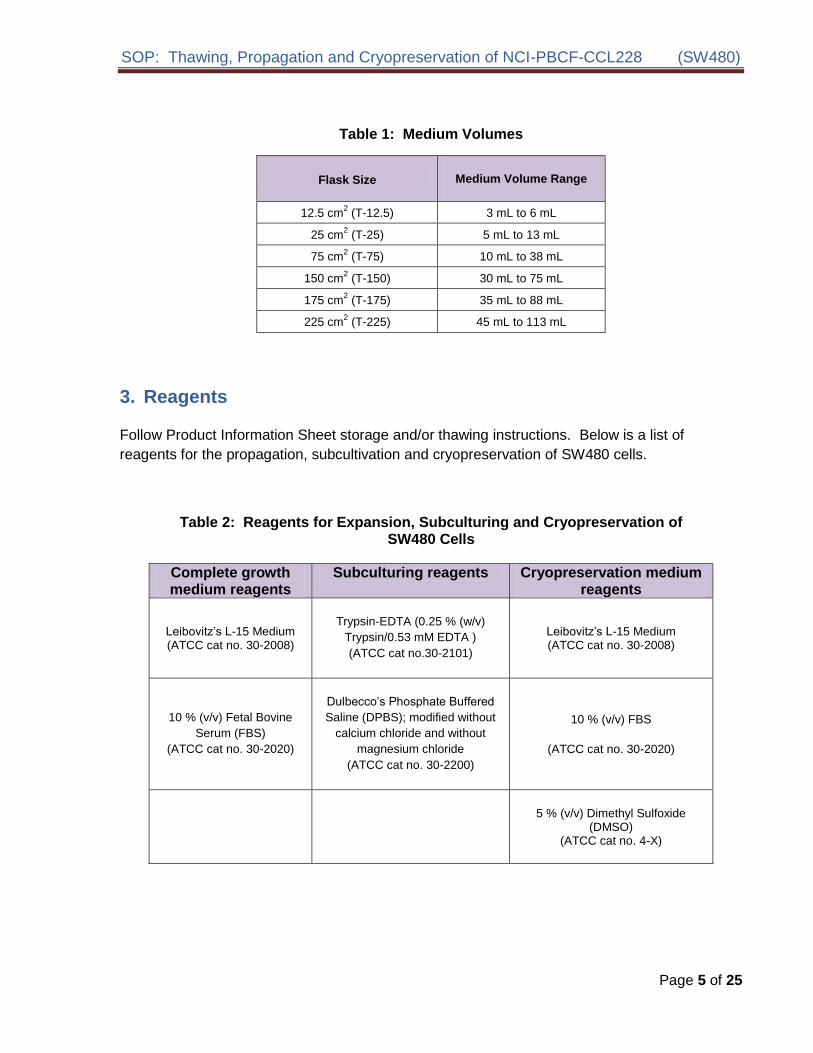
SOP Thawing Propagation and Cryopreservation of NCI-PBCF-CCL228 (SW480)
b Propagating cells
Method
1 Add 9 mL of complete growth medium (L-15 + 10 (vv) FBS) to a 15-mL conical centrifuge
tube
2 Again wipe the outer surface of the vial with sterile gauze wetted with 70 ethanol
3 Using sterile gauze carefully remove the cap from the vial
4 With a 1 mL pipette transfer slowly the completely thawed content of the vial (1 mL cell
suspension) to the 15-mL conical centrifuge tube containing 9 mL complete growth medium
(L-15 + 10 (vv) FBS) Gently resuspend cells by pipetting up and down
5 Centrifuge at 125 xg at room temperature for 8 min to 10 min
6 Carefully aspirate (discard) the medium leaving the pellet undisturbed
7 Using a 10 mL pipette add 10 mL of complete growth medium (L-15 + 10 (vv) FBS)
8 Resuspend pellet by gentle pipetting up and down
9 Using a 1 mL pipette remove 1 mL of cell suspension for cell count and viability Cell counts
are performed using either an automated counter (such as Innovatis Cedex System
Beckman-Coulter ViCell system) or a hemocytometer
10 Record total cell count and viability When an automated system is used attach copies of
the printed results to the record
11 Plate cells in pre-labeled T-25 cm2 flask at about 08 x 105 cellscm2
12 Transfer flask to a 37 degC plusmn 1degC incubator without CO2
NOTE The L-15 medium formulation was devised for use in a free gas exchange with
atmospheric air A CO2 and air mixture is detrimental to cells when using this
medium for cultivation
13 Observe culture daily by eye and under an inverted microscope to ensure culture is free of contamination and culture has not reached confluence Monitor visually the pH of the medium daily If the medium goes from red through orange to yellow change the medium
14 Note In most cases cultures at a high cell density exhaust the medium faster than those at low cell density as is evident from the change in pH A drop in pH is usually accompanied by an increase in cell density which is an indicator to subculture the cells Cells may stop growing when the pH is between pH 7 to pH 6 and loose viability between pH 65 and pH 6
15 If fluid renewal is needed aseptically aspirate the complete growth medium from the flask and discard Add an equivalent volume of fresh complete growth medium to the flask Alternatively perform a fluid addition by adding fresh complete growth medium to the flask
Page 7 of 25
SOP Thawing Propagation and Cryopreservation of NCI-PBCF-CCL228 (SW480)
without removing the existing medium Record fluid change or fluid addition on the Cell Line Expansion Table (see Table 5 in Appendix 6)
16 If subculturing of cells is needed continue to lsquoSubculturing cellsrsquo
Note Subculture when the cells are 80-90 confluent (see photomicrographs in Appendix 1)
c Subculturing cells
Reagents and Material
025 (wv) Trypsin-053 mM EDTA
DPBS
Complete growth medium (L-15 (ATCC cat no 30-2008) + 10 (vv) FBS (ATCC cat
no 30-2020) Plastic pipettes (1 mL 10 mL 25 mL) T-75 cm2 T-225 cm2 polystyrene flasks
Method
1 Aseptically remove medium from the flask
2 Add appropriate volumes of sterile Ca2+- and Mg2+-free DPBS to the side of the flask opposite the cells so as to avoid dislodging the cells (see Table 3)
3 Rinse the cells with DPBS (using a gently rocking motion) and discard
4 Add appropriate volume of 025 (wv) Trypsin-053 mM EDTA solution to the flask (see Table 3)
5 Incubate the flask at 37 oC plusmn 1 oC until the cells round up Observe cells under an
inverted microscope every 5 min When the flask is tilted the attached cells should slide
down the surface This usually occurs after 5 min to 10 min of incubation
Note Do not leave trypsin-EDTA on the cells any longer than necessary as
clumping may result
6 Neutralize the trypsin-EDTAcell suspension by adding an equal volume of complete growth medium (L-15 + 10 (vv) FBS) to each flask Disperse the cells by pipetting gently over the surface of the monolayer Pipette the cell suspension up and down with the tip of the pipette resting on the bottom corner or edge until a single cell suspension is obtained Care should be taken to avoid the creation of foam
7 Using a 1 mL pipette remove 1 mL of cell suspension for total cell count and viability
8 Record total cell count and viability
Page 8 of 25
Type
DPBS Rinse Trypsin-EDTAFlask Flask
Size Buffer
T-flask 125 cm2
(T-125) 1 mL to 3 mL 1 mL to 2 mL
25 cm2
(T-25) 1 mL to 5 mL 1 mL to 3 mL
75 cm2
(T-75) 4 mL to 15 mL 2 mL to 8 mL
150 cm2
(T-150) 8 mL to 30 mL 4 mL to 15 mL
175 cm2
(T-175) 9 mL to 35 mL 5 mL to 20 mL
225 cm2
(T-225) 10 mL to 45 mL 5 mL to 25 mL
SOP Thawing Propagation and Cryopreservation of NCI-PBCF-CCL228 (SW480)
9 Add appropriate volume of fresh complete growth medium (L-15 + 10 (vv) FBS) and
transfer cell suspension (for volume see Table 1) into new pre-labeled flasks at a
seeding density of 4 x 10⁴ viable cellscm2 to 5 x 10⁴ viable cellscm2 or a split ratio of
110 to 112
10 Label all new flasks with the (a) name of cell line (b) passage number (c) date (d) initials of technician
Table 3 - Volume of Rinse Buffer and Trypsin
5 Harvesting of Cells for Cryopreservation
Reagents and Material
025 (wv) Trypsin-053 mM EDTA
DPBS
Complete growth medium (L-15 (ATCC cat no 30-2008) + 10 (vv) FBS (ATCC cat
no 30-2020)
50 mL or 250 mL conical centrifuge tube
Plastic pipettes (1 mL 10 mL 25 mL)
Sterile DMSO
1 mL to 18 mL cryovials
Ice bucket with ice
Method
1 Label cryovials to include information on the name of cell line (b) passage number (c) date
2 Prepare cryopreservation medium by adding DMSO to cold complete growth medium (Lshy
15 + 10 (vv) FBS) at a final concentration of 5 (vv) DMSO Place cryopreservation
medium on ice until ready to use
Page 9 of 25
SOP Thawing Propagation and Cryopreservation of NCI-PBCF-CCL228 (SW480)
3 Aseptically remove medium from the flask
4 Add appropriate volumes of sterile Ca2+- and Mg2+-free DPBS to the side of the flask so as to avoid dislodging the cells (see Table 3)
5 Rinse the cells with DPBS (using a gentle rocking motion) and discard
6 Add appropriate volume of 025 (wv) Trypsin-053 mM EDTA solution to the flask (see Table 3)
7 Incubate the flask at 37 oC plusmn 1 oC until the cells round up Observe cells under an
inverted microscope every 5 min When the flask is tilted the attached cells should slide
down the surface This usually occurs after 5 min to 10 min of incubation
Note Do not leave trypsin-EDTA on the cells any longer than necessary as
clumping may result
8 Neutralize the trypsin-EDTAcell suspension by adding an equal volume of complete growth medium (L-15 + 10 (vv) FBS) to each flask Disperse the cells by pipetting gently over the surface of the monolayer Pipette the cell suspension up and down with the tip of the pipette resting on the bottom corner or edge until a single cell suspension is obtained Care should be taken to avoid the creation of foam
9 Using a 1 mL pipette remove 1 mL of cell suspension for total cell count and viability
10 Record total cell count and viability
11 Spin cells at approximately 125 xg for 5 min to 10 min at room temperature Carefully
aspirate and discard the medium leaving the pellet undisturbed
12 Calculate volume of cryopreservation medium based on the count performed at step 9
and resuspend pellet in cold cryopreservation medium at a viable cell density of 2 x 10⁶
viable cellsmL (acceptable range 2 x 10⁶ viable cellsmL to 3 x 10⁶ viable cellsmL) by
gentle pipetting up and down
13 Dispense 1 mL of cell suspension using a 5 mL or 10 mL pipette into each 1 mL cryovial
14 Place filled cryovials at 2 oC to 8 oC until ready to cryopreserve A minimum equilibration
time of 10 min but no longer than 45 min is necessary to allow DMSO to penetrate the
cells
Note DMSO is toxic to the cells Long exposure in DMSO may affect viability
6 Cryopreservation of Cells
Material
Liquid nitrogen freezer Cryomed Programmable freezer (Forma Scientific cat no 1010) or Mr Frosty (Nalgene cat no 5100)
Page 10 of 25
SOP Thawing Propagation and Cryopreservation of NCI-PBCF-CCL228 (SW480)
Isopropanol
Cryovial rack
a Cryopreservation using a rate-controlled programmable freezerMethod
A slow and reproducible cooling rate is very important to ensure good recovery of cultures A decrease of 1 degC per min to -80 degC followed by rapid freeze at about 15 degC to 30 degC per min drop to -150 degC will usually work for most animal cell cultures The best way to control the cooling process is to use a programmable rate-controlled electronic freezer unit Refer to the manufacturerrsquos handbook for detailed procedure
i Using the Cryomed
Starting the Cryopreservation Process
1 Check that the liquid nitrogen valve that supplies the Cryomed is open
2 Check the gauge to ensure that there is enough liquid nitrogen in the open tank to complete the freeze
3 Install the thermocouple probe so that the tip is immersed midway into the control fluid
Note Be sure that the thermocouple is centered in the vial and the vial is placed centered in the rack The probe should be changed after three uses or if it turns yellow to ensure accurate readings by the controller during the freezing process Old medium may have different freezing characteristics
4 Close and latch Cryomed door
5 Turn on microcomputer computer and monitor
6 Double click the ldquoCryomedrdquo icon The machine may need to be pre-programmed for specific cell type and medium
7 From the top of the screen select MENU RUN FUNCTIONS START RUN
8 Fill out the box which appears on the screen Cell line ID TYPE OF SAMPLE MEDIA NUMBER OF SAMPLES
9 Hit the ESCAPE key and the Cryomed will cool to 4 C
10 Once Cryomed chamber has cooled to 4 C load cryovials onto racks and close the door
11 When the Cryomedrsquos chamber temperature and the sample temperature have reached approximately 4 C press the space bar to initiate the rate controlled cryopreservation process
Page 11 of 25
SOP Thawing Propagation and Cryopreservation of NCI-PBCF-CCL228 (SW480)
Completing the Cryopreservation Process
1 When samples have reached ndash80C an alarm will sound To silence this select ALARM
from the options at the top of the screen
2 Select MENU RUN FUNCTIONSrarr STOP Hit the ESCAPE key to return to the main
menu and select EXIT
3 Immediately transfer vials to liquid nitrogen freezer
4 Shut down the microcomputer and then turn off the monitor
b Cryopreservation using ldquoMr Frostyrdquo
1 One day before freezing cells add 250 mL isopropanol to the bottom of the container and place at 2 oC to 8 oC
2 On the day of the freeze prepare cells for cryopreservation as described above
3 Insert cryovials with the cell suspension in appropriate slots in the container
4 Transfer the container to a -70 degC to -90 degC freezer and store overnight
5 Next day transfer cryovials to the vapor phase of liquid nitrogen freezer
Note Each container has 18 slots which can accommodate 18 cryovials in one freeze
Important information when using the rate-controlled programmable freezer or a manual method (Mr Frosty) for cryopreservation of mammalian cells
Regardless which cooling method is used it is important that the transfer to the final storage location (between -130 degC and -196 degC) be done quickly and efficiently If the transfer cannot be done immediately the vials can be placed on dry ice for a short time This will avoid damage to cultures by inadvertent temporary warming during the transfer process Warming during this transfer process is a major cause of variation in culture viability upon thawing
Always keep the storage temperature below -130 degC for optimum survival Cells may survive storage at higher temperatures but viability will usually decrease over time The ideal storage container is a liquid nitrogen freezer where the cultures are stored in the vapor phase above the liquid nitrogen
Note ATCC does not have experience in the cryopreservation of the SW480 cells by any other method than the Cryomed programmable freezer
Page 12 of 25
SOP Thawing Propagation and Cryopreservation of NCI-PBCF-CCL228 (SW480)
7 Storage
Store cryopreserved cells in the vapor phase of liquid nitrogen freezer (below ndash130 degC) for optimum long-term survival
Note Experiments on long-term storage of animal cell lines at different temperature
levels indicate that a -70 degC storage temperature is not adequate except for very short
period of time A -90 degC storage may be adequate for longer periods depending upon
the cell line preserved The efficiency of recovery however is not as great as when
the cells are stored in vapor phase of the liquid nitrogen freezer
Page 13 of 25
SOP Thawing Propagation and Cryopreservation of NCI-PBCF-CCL228 (SW480)
APPENDIX 1 PHOTOMICROGRAPHS OF NCI-PBCF-CCL228 (SW480) CELLS
Figure 1 Photomicrograph of SW480 cells after one day post-freeze recovery Cells were plated at 80 x 104 viable cellscm2
Figure 2 Photomicrograph of SW480 cells after two days post-freeze recovery Cells were plated at 80 x 104 viable cellscm2
Page 14 of 25
SOP Thawing Propagation and Cryopreservation of NCI-PBCF-CCL228 (SW480)
40X 40X 40X
100X 100X 100X
500 microm500 microm500 microm
200 microm 200 microm 200 microm
Figure 3 Photomicrographs of SW480 cells at various time points after
seeding at a cell density of 5 x 10⁴ viable cellscm2
Page 15 of 25
SOP Thawing Propagation and Cryopreservation of NCI-PBCF-CCL228 (SW480)
APPENDIX 2 GROWTH PROFILE OF NCI-PBCF-CCL228 (SW480) CELLS
000E+00
100E+05
200E+05
300E+05
400E+05
500E+05
600E+05
700E+05
800E+05
900E+05
100E+06
7 8 9
Via
ble
Cell
sc
m2
0 1 2 3 4 5 6
Days in Culture
Figure 4 Growth curve for SW480 cells cells were plated at 5 x 10⁴ viable cellscm2 population doubling time (PDT) is approximately 38 h
Page 16 of 25
SOP Thawing Propagation and Cryopreservation of NCI-PBCF-CCL228 (SW480)
APPENDIX 3 CYTOGENETIC ANALYSIS OF NCI-PBCF-CCL228 SW480 CELLS
NCI-PBCF-CCL228 Karyotype Results
Metaphase Spread Karyotype
Number of metaphase spreads counted 20
Band level 300-400
Number of metaphase spreads karyotyped 10
Chromosome range 50-56
Sex Male
Comments Aneuploid
Karyotype
50~56XX+2der(2)t(218)(q21q111)del(5)(q13q33)+add(7)(q22)+add(7)(q32)
der(10)t (1012)(p15q11)+add(11)(p15)-12der(1214)(p10q10)-14+add(15)(p10)
der(15)t(1215)(q13q15)-18add(19)(p133)+20+add(20)(p13)
+der(20)t(520)(q22p13)x2+21+add(21)(q111)add(21)(q22)+mar1x2
(ISCN nomenclature written based on a diploid karyotype)
Human diploid karyotype (2N) 46XX (female) or 46XY (male)
Page 17 of 25
SOP Thawing Propagation and Cryopreservation of NCI-PBCF-CCL228 (SW480)
Karyotype Summary
In the karyotype image arrows indicate regions of abnormality It should be noted that the karyotype description includes the observed abnormalities from all examined metaphase spreads but due to heterogeneity not all of the karyotyped cells will contain every abnormality
This is a rearranged human cell line of male origin containing 50 to 56 chromosomes per metaphase spread (hyperdiploid) Structural abnormalities include rearrangements to approximately 50 of the 22 different autosomal chromosomes (described below) There is one unidentifiable clonal marker chromosome (markers present in two or more of the examined cells) [+mar1]
The rearrangements include
Addition of unknown material to the short arms (designated by p) of chromosomes 11 15 19 and 20
Addition of unknown material to the long arms (designated by q) of chromosomes 7 and 21 (2 different derivatives of each)
An interstitial deletion of material from the long arm of chromosome 5 between bands q13 and q33
Translocations involving chromosomes 2 and 18 [der(2)t(218)(q21q111)] 10 and 12 [der(10)t (1012)(p15q11) chromosomes 12 and 14 [der(1214)(p10q10)] 12 and 15 [der(15)t(1215)(q13q15)] and chromosomes 5 and 20 [der(20)t(520)(q22p13)]
Numerical changes are based on a diploid karyotype which would contain two copies of
each chromosome (2N) Therefore karyotype designations such as +2 +20 and +21
indicates three copies of structurally normal chromosomes 2 20 and 21 Designations
-18 and -14 indicates one copy of structurally normal chromosomes -18 and -14 Also it
appears that the Y chromosome was lost which is not an uncommon occurrence in cancer
cell lines (ISCN 2009 An International System for Human Cytogenetic Nomenclature
(2009) Editors Lisa G Shaffer Marilyn L Slovak Lynda J Campbell)
Karyotype Procedure
Cell Harvest Cells were allowed to grow to 80-90 confluence Mitotic division was
arrested by treating the cells with KaryoMaxreg colcemid for 20 minutes to 2 hours at 37degC
Cells were harvested using 005 Trypsin-EDTA treated with 0075M KCL hypotonic
solution and then fixed in three changes of a 31 ratio of methanolglacial acetic acid
Slide Preparation Slides were prepared by dropping the cell suspension onto wet glass
slides and allowing them to dry under controlled conditions
G-banding Slides were baked one hour at 90degC trypsinized using 10X trypsin-EDTA
and then stained with Leishmanrsquos stain
Page 18 of 25
SOP Thawing Propagation and Cryopreservation of NCI-PBCF-CCL228 (SW480)
Microscopy Slides were scanned using a 10X objective and metaphase spreads were
analyzed using a 100X plan apochromat objective on an Olympus BX-41 microscope
Imaging and karyotyping were performed using Cytovisionreg software
Analysis Twenty metaphase cells were counted and analyzed and representative
metaphase cells were karyotyped depending on the complexity of the study
Summary of Karyotyping Procedure
G-band karyotyping analysis is performed using GTL banding technique G bands produced with trypsin and Leishman Slides prepared with metaphase spreads are treated with trypsin and stained with Leishmanrsquos This method produces a series of light and dark bands that allow for the positive identification of each chromosome
SW480 cell line karyotyping was carried out by Cell Line Genetics Inc (Madison WI 53719)
Page 19 of 25
SOP Thawing Propagation and Cryopreservation of NCI-PBCF-CCL228 (SW480)
APPENDIX 4 GLOSSARY OF TERMS
Confluent monolayer adherent cell culture in which all cells are in contact with other cells
all around their periphery and no available substrate is left uncovered
Split ratio the divisor of the dilution ration of a cell culture to subculture (eg one flask
divided into four or 100 mL up to 400 mL would be split ratio of 14)
Subculture (or passage) the transfer or transplantation of cells with or without dilution
from one culture vessel to another
Passage No the total number of times the cells in the culture have been subcultured or
passaged (with each subculture the passage number increases by 1)
Population doubling level (PDL) the total number of population doublings of a cell line
since its initiation in vitro (with each subculture the population doubling increases in
relationship to the split ratio at which the cells are plated) See Appendix 7
Population doubling time (doubling time) the time interval calculated during the
logarithmic phase of growth in which cells double in number
Seeding density recommended number of cells per cm2 of substrate when inoculating a
new flask
Epithelial-like adherent cells of a polygonal shape with clear sharp boundaries between
them
Fibroblast-like adherent cells of a spindle or stellate shape
Page 20 of 25
SOP Thawing Propagation and Cryopreservation of NCI-PBCF-CCL228 (SW480)
APPENDIX 5 REFERENCE
1 Culture Of Animal Cells A Manual of Basic Technique by R Ian Freshney 6th edition
published by Wiley-Liss NY 2010
2 Fogh J et al Absence of HeLa cell contamination in 169 cell lines derived from human
tumors J Natl Cancer Inst 58 209-214 1977 PubMed 833871
3 Fogh J et al One hundred and twenty-seven cultured human tumor cell lines producing
tumors in nude mice J Natl Cancer Inst 59 221-226 1977 PubMed 327080
4 Lelbovitz A et al Detection and analysis of a glucose 6-phosphate dehydrogenase
phenotype B cell line contamination J Natl Cancer Inst 63 635-645 1979 PubMed
288927
5 Adachi A et al Productive persistent infection of human colorectal cell lines with human
immunodeficiency virus J Virol 61 209-213 1987 PubMed 3640832
6 Schroy PC et al Detection of p21ras mutations in colorectal adenomas and carcinomas
by enzyme-linked immunosorbent assay Cancer 76 201-209 1995 PubMed 8625092
7 Trainer DL et al Biological characterization and oncogene expression in human
colorectal carcinoma cell lines Int J Cancer 41 287-296 1988 PubMed 3338874
8 Weiss J et al Mutation and expression of the p53 gene in malignant melanoma cell
lines Int J Cancer 54 693-699 1993 PubMed 8514460
9 Nigro JM et al Mutations in the p53 gene occur in diverse human tumour types Nature
342 705-707 1989 PubMed 2531845
10 Barnett SW et al Characterization of human immunodeficiency virus type 1 strains
recovered from the bowel of infected individuals Virology 182 802-809 1991 PubMed
2024498
11 Leibovitz A et al Classification of human colorectal adenocarcinoma cell lines Cancer
Res 36 4562-4569 1976 PubMed 1000501
12 Geiser AG et al Suppression of tumorigenicity in human cell hybrids derived from cell
lines expressing different activated ras oncogenes Cancer Res 49 1572-1577 1989
PubMed 2647289
13 Lahm H et al Secretion of bioactive granulocyte-macrophage colony-stimulating factor
by human colorectal carcinoma cells Cancer Res 54 3700-3702 1994 PubMed
8033086
14 Rodrigues NR et al p53 mutations in colorectal cancer Proc Natl Acad Sci USA 87
7555-7559 1990 PubMed 1699228
Page 21 of 25
SOP Thawing Propagation and Cryopreservation of NCI-PBCF-CCL228 (SW480)
15 Santoro IM Groden J Alternative splicing of the APC gene and its association with terminal differentiation Cancer Res 57 488-494 1997 PubMed 9012479
16 Tsao H et al Novel mutations in the p16CDKN2A binding region of the Cyclinshy
dependent Kinase-4 gene Cancer Res 58 109-113 1998 PubMed 9426066
17 Zhu X et al Cell cycle-dependent modulation of telomerase activity in tumor cells Proc Natl Acad Sci USA 93 6091-6095 1996 PubMed 8650224
18 Witty JP et al Modulation of matrilysin levels in colon carcinoma cell lines affects tumorigenicity in vivo Cancer Res 54 4805-4812 1994 PubMed 8062282
Page 22 of 25
SOP Thawing Propagation and Cryopreservation of NCI-PBCF-CCL228 (SW480)
APPENDIX 6 REAGENT LOT TRACEABILITY AND CELL EXPANSION TABLES
Table 4 Reagent Lot Traceability
Reagent Vendor Catalog Lot Expiration Date
Page 23 of 25
Table 5 Cell Expansion
FROM FLUID CHANGE
Observation under
microscope CELL COUNT TO
By Date Flask qty
size
Pass Confluence
Add Replace
Volume
(in mL)
Viable cellsmL
Total viable cells
Viability
Split Ratio
Flask qty
size
Pass
PDL
Add
Replace
Add
Replace
Add
Replace
Add
Replace
Add
Replace
Add
Replace
Add
Replace
Add
Replace
Add
Replace
SOP Thawing Propagation and Cryopreservation of NCI-PBCF-CCL228 (SW480)
APPENDIX 7 CALCULATION OF POPULATION DOUBLING LEVEL (PDL)
Calculate the PDL of the current passage using the following equation
PDL = X + 3322 (log Y ndash log I)
Where X = initial PDL I = cell inoculum (number of cells plated in the flask) Y = final cell yield (number of cells at the end of the growth period)
APPENDIX 8 SAFETY PRECAUTIONS
Use at least approved Biological Safety Level 2 (BSL-2) facilities and procedures
Wear appropriate Personal Protective Equipment (PPE) such as isolation gown lab coat with sleeve protectors face shield and gloves
Use safety precautions when working with liquid nitrogen nitrogen vapor and cryogenically cooled fixtures
Use liquid nitrogen freezers and liquid nitrogen tanks only in areas with adequate ventilation Liquid nitrogen reduces the concentration of oxygen and can cause suffocation
Wear latex gloves over insulating gloves to prevent liquid nitrogen from soaking in and being held next to the skin Liquid nitrogen is extremely cold and will cause burns and frostbite Metal inventory racks tank components and liquid nitrogen transfer hoses exposed to liquid nitrogen or nitrogen vapor quickly cool to cryogenic temperatures and can cause burns and frostbite
Wear a full face mask when thawing and retrieving vials from liquid nitrogen freezer Danger to the technician derives mainly from the possibility that liquid nitrogen can penetrate the cryovial during storage On warming rapid evaporation of the nitrogen within the confines of such cryovial can cause an aerosol or explosion of the cryovial and contents
Page 25 of 25
SOP Thawing Propagation and Cryopreservation of NCI-PBCF-CCL228 (SW480)
without removing the existing medium Record fluid change or fluid addition on the Cell Line Expansion Table (see Table 5 in Appendix 6)
16 If subculturing of cells is needed continue to lsquoSubculturing cellsrsquo
Note Subculture when the cells are 80-90 confluent (see photomicrographs in Appendix 1)
c Subculturing cells
Reagents and Material
025 (wv) Trypsin-053 mM EDTA
DPBS
Complete growth medium (L-15 (ATCC cat no 30-2008) + 10 (vv) FBS (ATCC cat
no 30-2020) Plastic pipettes (1 mL 10 mL 25 mL) T-75 cm2 T-225 cm2 polystyrene flasks
Method
1 Aseptically remove medium from the flask
2 Add appropriate volumes of sterile Ca2+- and Mg2+-free DPBS to the side of the flask opposite the cells so as to avoid dislodging the cells (see Table 3)
3 Rinse the cells with DPBS (using a gently rocking motion) and discard
4 Add appropriate volume of 025 (wv) Trypsin-053 mM EDTA solution to the flask (see Table 3)
5 Incubate the flask at 37 oC plusmn 1 oC until the cells round up Observe cells under an
inverted microscope every 5 min When the flask is tilted the attached cells should slide
down the surface This usually occurs after 5 min to 10 min of incubation
Note Do not leave trypsin-EDTA on the cells any longer than necessary as
clumping may result
6 Neutralize the trypsin-EDTAcell suspension by adding an equal volume of complete growth medium (L-15 + 10 (vv) FBS) to each flask Disperse the cells by pipetting gently over the surface of the monolayer Pipette the cell suspension up and down with the tip of the pipette resting on the bottom corner or edge until a single cell suspension is obtained Care should be taken to avoid the creation of foam
7 Using a 1 mL pipette remove 1 mL of cell suspension for total cell count and viability
8 Record total cell count and viability
Page 8 of 25
Type
DPBS Rinse Trypsin-EDTAFlask Flask
Size Buffer
T-flask 125 cm2
(T-125) 1 mL to 3 mL 1 mL to 2 mL
25 cm2
(T-25) 1 mL to 5 mL 1 mL to 3 mL
75 cm2
(T-75) 4 mL to 15 mL 2 mL to 8 mL
150 cm2
(T-150) 8 mL to 30 mL 4 mL to 15 mL
175 cm2
(T-175) 9 mL to 35 mL 5 mL to 20 mL
225 cm2
(T-225) 10 mL to 45 mL 5 mL to 25 mL
SOP Thawing Propagation and Cryopreservation of NCI-PBCF-CCL228 (SW480)
9 Add appropriate volume of fresh complete growth medium (L-15 + 10 (vv) FBS) and
transfer cell suspension (for volume see Table 1) into new pre-labeled flasks at a
seeding density of 4 x 10⁴ viable cellscm2 to 5 x 10⁴ viable cellscm2 or a split ratio of
110 to 112
10 Label all new flasks with the (a) name of cell line (b) passage number (c) date (d) initials of technician
Table 3 - Volume of Rinse Buffer and Trypsin
5 Harvesting of Cells for Cryopreservation
Reagents and Material
025 (wv) Trypsin-053 mM EDTA
DPBS
Complete growth medium (L-15 (ATCC cat no 30-2008) + 10 (vv) FBS (ATCC cat
no 30-2020)
50 mL or 250 mL conical centrifuge tube
Plastic pipettes (1 mL 10 mL 25 mL)
Sterile DMSO
1 mL to 18 mL cryovials
Ice bucket with ice
Method
1 Label cryovials to include information on the name of cell line (b) passage number (c) date
2 Prepare cryopreservation medium by adding DMSO to cold complete growth medium (Lshy
15 + 10 (vv) FBS) at a final concentration of 5 (vv) DMSO Place cryopreservation
medium on ice until ready to use
Page 9 of 25
SOP Thawing Propagation and Cryopreservation of NCI-PBCF-CCL228 (SW480)
3 Aseptically remove medium from the flask
4 Add appropriate volumes of sterile Ca2+- and Mg2+-free DPBS to the side of the flask so as to avoid dislodging the cells (see Table 3)
5 Rinse the cells with DPBS (using a gentle rocking motion) and discard
6 Add appropriate volume of 025 (wv) Trypsin-053 mM EDTA solution to the flask (see Table 3)
7 Incubate the flask at 37 oC plusmn 1 oC until the cells round up Observe cells under an
inverted microscope every 5 min When the flask is tilted the attached cells should slide
down the surface This usually occurs after 5 min to 10 min of incubation
Note Do not leave trypsin-EDTA on the cells any longer than necessary as
clumping may result
8 Neutralize the trypsin-EDTAcell suspension by adding an equal volume of complete growth medium (L-15 + 10 (vv) FBS) to each flask Disperse the cells by pipetting gently over the surface of the monolayer Pipette the cell suspension up and down with the tip of the pipette resting on the bottom corner or edge until a single cell suspension is obtained Care should be taken to avoid the creation of foam
9 Using a 1 mL pipette remove 1 mL of cell suspension for total cell count and viability
10 Record total cell count and viability
11 Spin cells at approximately 125 xg for 5 min to 10 min at room temperature Carefully
aspirate and discard the medium leaving the pellet undisturbed
12 Calculate volume of cryopreservation medium based on the count performed at step 9
and resuspend pellet in cold cryopreservation medium at a viable cell density of 2 x 10⁶
viable cellsmL (acceptable range 2 x 10⁶ viable cellsmL to 3 x 10⁶ viable cellsmL) by
gentle pipetting up and down
13 Dispense 1 mL of cell suspension using a 5 mL or 10 mL pipette into each 1 mL cryovial
14 Place filled cryovials at 2 oC to 8 oC until ready to cryopreserve A minimum equilibration
time of 10 min but no longer than 45 min is necessary to allow DMSO to penetrate the
cells
Note DMSO is toxic to the cells Long exposure in DMSO may affect viability
6 Cryopreservation of Cells
Material
Liquid nitrogen freezer Cryomed Programmable freezer (Forma Scientific cat no 1010) or Mr Frosty (Nalgene cat no 5100)
Page 10 of 25
SOP Thawing Propagation and Cryopreservation of NCI-PBCF-CCL228 (SW480)
Isopropanol
Cryovial rack
a Cryopreservation using a rate-controlled programmable freezerMethod
A slow and reproducible cooling rate is very important to ensure good recovery of cultures A decrease of 1 degC per min to -80 degC followed by rapid freeze at about 15 degC to 30 degC per min drop to -150 degC will usually work for most animal cell cultures The best way to control the cooling process is to use a programmable rate-controlled electronic freezer unit Refer to the manufacturerrsquos handbook for detailed procedure
i Using the Cryomed
Starting the Cryopreservation Process
1 Check that the liquid nitrogen valve that supplies the Cryomed is open
2 Check the gauge to ensure that there is enough liquid nitrogen in the open tank to complete the freeze
3 Install the thermocouple probe so that the tip is immersed midway into the control fluid
Note Be sure that the thermocouple is centered in the vial and the vial is placed centered in the rack The probe should be changed after three uses or if it turns yellow to ensure accurate readings by the controller during the freezing process Old medium may have different freezing characteristics
4 Close and latch Cryomed door
5 Turn on microcomputer computer and monitor
6 Double click the ldquoCryomedrdquo icon The machine may need to be pre-programmed for specific cell type and medium
7 From the top of the screen select MENU RUN FUNCTIONS START RUN
8 Fill out the box which appears on the screen Cell line ID TYPE OF SAMPLE MEDIA NUMBER OF SAMPLES
9 Hit the ESCAPE key and the Cryomed will cool to 4 C
10 Once Cryomed chamber has cooled to 4 C load cryovials onto racks and close the door
11 When the Cryomedrsquos chamber temperature and the sample temperature have reached approximately 4 C press the space bar to initiate the rate controlled cryopreservation process
Page 11 of 25
SOP Thawing Propagation and Cryopreservation of NCI-PBCF-CCL228 (SW480)
Completing the Cryopreservation Process
1 When samples have reached ndash80C an alarm will sound To silence this select ALARM
from the options at the top of the screen
2 Select MENU RUN FUNCTIONSrarr STOP Hit the ESCAPE key to return to the main
menu and select EXIT
3 Immediately transfer vials to liquid nitrogen freezer
4 Shut down the microcomputer and then turn off the monitor
b Cryopreservation using ldquoMr Frostyrdquo
1 One day before freezing cells add 250 mL isopropanol to the bottom of the container and place at 2 oC to 8 oC
2 On the day of the freeze prepare cells for cryopreservation as described above
3 Insert cryovials with the cell suspension in appropriate slots in the container
4 Transfer the container to a -70 degC to -90 degC freezer and store overnight
5 Next day transfer cryovials to the vapor phase of liquid nitrogen freezer
Note Each container has 18 slots which can accommodate 18 cryovials in one freeze
Important information when using the rate-controlled programmable freezer or a manual method (Mr Frosty) for cryopreservation of mammalian cells
Regardless which cooling method is used it is important that the transfer to the final storage location (between -130 degC and -196 degC) be done quickly and efficiently If the transfer cannot be done immediately the vials can be placed on dry ice for a short time This will avoid damage to cultures by inadvertent temporary warming during the transfer process Warming during this transfer process is a major cause of variation in culture viability upon thawing
Always keep the storage temperature below -130 degC for optimum survival Cells may survive storage at higher temperatures but viability will usually decrease over time The ideal storage container is a liquid nitrogen freezer where the cultures are stored in the vapor phase above the liquid nitrogen
Note ATCC does not have experience in the cryopreservation of the SW480 cells by any other method than the Cryomed programmable freezer
Page 12 of 25
SOP Thawing Propagation and Cryopreservation of NCI-PBCF-CCL228 (SW480)
7 Storage
Store cryopreserved cells in the vapor phase of liquid nitrogen freezer (below ndash130 degC) for optimum long-term survival
Note Experiments on long-term storage of animal cell lines at different temperature
levels indicate that a -70 degC storage temperature is not adequate except for very short
period of time A -90 degC storage may be adequate for longer periods depending upon
the cell line preserved The efficiency of recovery however is not as great as when
the cells are stored in vapor phase of the liquid nitrogen freezer
Page 13 of 25
SOP Thawing Propagation and Cryopreservation of NCI-PBCF-CCL228 (SW480)
APPENDIX 1 PHOTOMICROGRAPHS OF NCI-PBCF-CCL228 (SW480) CELLS
Figure 1 Photomicrograph of SW480 cells after one day post-freeze recovery Cells were plated at 80 x 104 viable cellscm2
Figure 2 Photomicrograph of SW480 cells after two days post-freeze recovery Cells were plated at 80 x 104 viable cellscm2
Page 14 of 25
SOP Thawing Propagation and Cryopreservation of NCI-PBCF-CCL228 (SW480)
40X 40X 40X
100X 100X 100X
500 microm500 microm500 microm
200 microm 200 microm 200 microm
Figure 3 Photomicrographs of SW480 cells at various time points after
seeding at a cell density of 5 x 10⁴ viable cellscm2
Page 15 of 25
SOP Thawing Propagation and Cryopreservation of NCI-PBCF-CCL228 (SW480)
APPENDIX 2 GROWTH PROFILE OF NCI-PBCF-CCL228 (SW480) CELLS
000E+00
100E+05
200E+05
300E+05
400E+05
500E+05
600E+05
700E+05
800E+05
900E+05
100E+06
7 8 9
Via
ble
Cell
sc
m2
0 1 2 3 4 5 6
Days in Culture
Figure 4 Growth curve for SW480 cells cells were plated at 5 x 10⁴ viable cellscm2 population doubling time (PDT) is approximately 38 h
Page 16 of 25
SOP Thawing Propagation and Cryopreservation of NCI-PBCF-CCL228 (SW480)
APPENDIX 3 CYTOGENETIC ANALYSIS OF NCI-PBCF-CCL228 SW480 CELLS
NCI-PBCF-CCL228 Karyotype Results
Metaphase Spread Karyotype
Number of metaphase spreads counted 20
Band level 300-400
Number of metaphase spreads karyotyped 10
Chromosome range 50-56
Sex Male
Comments Aneuploid
Karyotype
50~56XX+2der(2)t(218)(q21q111)del(5)(q13q33)+add(7)(q22)+add(7)(q32)
der(10)t (1012)(p15q11)+add(11)(p15)-12der(1214)(p10q10)-14+add(15)(p10)
der(15)t(1215)(q13q15)-18add(19)(p133)+20+add(20)(p13)
+der(20)t(520)(q22p13)x2+21+add(21)(q111)add(21)(q22)+mar1x2
(ISCN nomenclature written based on a diploid karyotype)
Human diploid karyotype (2N) 46XX (female) or 46XY (male)
Page 17 of 25
SOP Thawing Propagation and Cryopreservation of NCI-PBCF-CCL228 (SW480)
Karyotype Summary
In the karyotype image arrows indicate regions of abnormality It should be noted that the karyotype description includes the observed abnormalities from all examined metaphase spreads but due to heterogeneity not all of the karyotyped cells will contain every abnormality
This is a rearranged human cell line of male origin containing 50 to 56 chromosomes per metaphase spread (hyperdiploid) Structural abnormalities include rearrangements to approximately 50 of the 22 different autosomal chromosomes (described below) There is one unidentifiable clonal marker chromosome (markers present in two or more of the examined cells) [+mar1]
The rearrangements include
Addition of unknown material to the short arms (designated by p) of chromosomes 11 15 19 and 20
Addition of unknown material to the long arms (designated by q) of chromosomes 7 and 21 (2 different derivatives of each)
An interstitial deletion of material from the long arm of chromosome 5 between bands q13 and q33
Translocations involving chromosomes 2 and 18 [der(2)t(218)(q21q111)] 10 and 12 [der(10)t (1012)(p15q11) chromosomes 12 and 14 [der(1214)(p10q10)] 12 and 15 [der(15)t(1215)(q13q15)] and chromosomes 5 and 20 [der(20)t(520)(q22p13)]
Numerical changes are based on a diploid karyotype which would contain two copies of
each chromosome (2N) Therefore karyotype designations such as +2 +20 and +21
indicates three copies of structurally normal chromosomes 2 20 and 21 Designations
-18 and -14 indicates one copy of structurally normal chromosomes -18 and -14 Also it
appears that the Y chromosome was lost which is not an uncommon occurrence in cancer
cell lines (ISCN 2009 An International System for Human Cytogenetic Nomenclature
(2009) Editors Lisa G Shaffer Marilyn L Slovak Lynda J Campbell)
Karyotype Procedure
Cell Harvest Cells were allowed to grow to 80-90 confluence Mitotic division was
arrested by treating the cells with KaryoMaxreg colcemid for 20 minutes to 2 hours at 37degC
Cells were harvested using 005 Trypsin-EDTA treated with 0075M KCL hypotonic
solution and then fixed in three changes of a 31 ratio of methanolglacial acetic acid
Slide Preparation Slides were prepared by dropping the cell suspension onto wet glass
slides and allowing them to dry under controlled conditions
G-banding Slides were baked one hour at 90degC trypsinized using 10X trypsin-EDTA
and then stained with Leishmanrsquos stain
Page 18 of 25
SOP Thawing Propagation and Cryopreservation of NCI-PBCF-CCL228 (SW480)
Microscopy Slides were scanned using a 10X objective and metaphase spreads were
analyzed using a 100X plan apochromat objective on an Olympus BX-41 microscope
Imaging and karyotyping were performed using Cytovisionreg software
Analysis Twenty metaphase cells were counted and analyzed and representative
metaphase cells were karyotyped depending on the complexity of the study
Summary of Karyotyping Procedure
G-band karyotyping analysis is performed using GTL banding technique G bands produced with trypsin and Leishman Slides prepared with metaphase spreads are treated with trypsin and stained with Leishmanrsquos This method produces a series of light and dark bands that allow for the positive identification of each chromosome
SW480 cell line karyotyping was carried out by Cell Line Genetics Inc (Madison WI 53719)
Page 19 of 25
SOP Thawing Propagation and Cryopreservation of NCI-PBCF-CCL228 (SW480)
APPENDIX 4 GLOSSARY OF TERMS
Confluent monolayer adherent cell culture in which all cells are in contact with other cells
all around their periphery and no available substrate is left uncovered
Split ratio the divisor of the dilution ration of a cell culture to subculture (eg one flask
divided into four or 100 mL up to 400 mL would be split ratio of 14)
Subculture (or passage) the transfer or transplantation of cells with or without dilution
from one culture vessel to another
Passage No the total number of times the cells in the culture have been subcultured or
passaged (with each subculture the passage number increases by 1)
Population doubling level (PDL) the total number of population doublings of a cell line
since its initiation in vitro (with each subculture the population doubling increases in
relationship to the split ratio at which the cells are plated) See Appendix 7
Population doubling time (doubling time) the time interval calculated during the
logarithmic phase of growth in which cells double in number
Seeding density recommended number of cells per cm2 of substrate when inoculating a
new flask
Epithelial-like adherent cells of a polygonal shape with clear sharp boundaries between
them
Fibroblast-like adherent cells of a spindle or stellate shape
Page 20 of 25
SOP Thawing Propagation and Cryopreservation of NCI-PBCF-CCL228 (SW480)
APPENDIX 5 REFERENCE
1 Culture Of Animal Cells A Manual of Basic Technique by R Ian Freshney 6th edition
published by Wiley-Liss NY 2010
2 Fogh J et al Absence of HeLa cell contamination in 169 cell lines derived from human
tumors J Natl Cancer Inst 58 209-214 1977 PubMed 833871
3 Fogh J et al One hundred and twenty-seven cultured human tumor cell lines producing
tumors in nude mice J Natl Cancer Inst 59 221-226 1977 PubMed 327080
4 Lelbovitz A et al Detection and analysis of a glucose 6-phosphate dehydrogenase
phenotype B cell line contamination J Natl Cancer Inst 63 635-645 1979 PubMed
288927
5 Adachi A et al Productive persistent infection of human colorectal cell lines with human
immunodeficiency virus J Virol 61 209-213 1987 PubMed 3640832
6 Schroy PC et al Detection of p21ras mutations in colorectal adenomas and carcinomas
by enzyme-linked immunosorbent assay Cancer 76 201-209 1995 PubMed 8625092
7 Trainer DL et al Biological characterization and oncogene expression in human
colorectal carcinoma cell lines Int J Cancer 41 287-296 1988 PubMed 3338874
8 Weiss J et al Mutation and expression of the p53 gene in malignant melanoma cell
lines Int J Cancer 54 693-699 1993 PubMed 8514460
9 Nigro JM et al Mutations in the p53 gene occur in diverse human tumour types Nature
342 705-707 1989 PubMed 2531845
10 Barnett SW et al Characterization of human immunodeficiency virus type 1 strains
recovered from the bowel of infected individuals Virology 182 802-809 1991 PubMed
2024498
11 Leibovitz A et al Classification of human colorectal adenocarcinoma cell lines Cancer
Res 36 4562-4569 1976 PubMed 1000501
12 Geiser AG et al Suppression of tumorigenicity in human cell hybrids derived from cell
lines expressing different activated ras oncogenes Cancer Res 49 1572-1577 1989
PubMed 2647289
13 Lahm H et al Secretion of bioactive granulocyte-macrophage colony-stimulating factor
by human colorectal carcinoma cells Cancer Res 54 3700-3702 1994 PubMed
8033086
14 Rodrigues NR et al p53 mutations in colorectal cancer Proc Natl Acad Sci USA 87
7555-7559 1990 PubMed 1699228
Page 21 of 25
SOP Thawing Propagation and Cryopreservation of NCI-PBCF-CCL228 (SW480)
15 Santoro IM Groden J Alternative splicing of the APC gene and its association with terminal differentiation Cancer Res 57 488-494 1997 PubMed 9012479
16 Tsao H et al Novel mutations in the p16CDKN2A binding region of the Cyclinshy
dependent Kinase-4 gene Cancer Res 58 109-113 1998 PubMed 9426066
17 Zhu X et al Cell cycle-dependent modulation of telomerase activity in tumor cells Proc Natl Acad Sci USA 93 6091-6095 1996 PubMed 8650224
18 Witty JP et al Modulation of matrilysin levels in colon carcinoma cell lines affects tumorigenicity in vivo Cancer Res 54 4805-4812 1994 PubMed 8062282
Page 22 of 25
SOP Thawing Propagation and Cryopreservation of NCI-PBCF-CCL228 (SW480)
APPENDIX 6 REAGENT LOT TRACEABILITY AND CELL EXPANSION TABLES
Table 4 Reagent Lot Traceability
Reagent Vendor Catalog Lot Expiration Date
Page 23 of 25
Table 5 Cell Expansion
FROM FLUID CHANGE
Observation under
microscope CELL COUNT TO
By Date Flask qty
size
Pass Confluence
Add Replace
Volume
(in mL)
Viable cellsmL
Total viable cells
Viability
Split Ratio
Flask qty
size
Pass
PDL
Add
Replace
Add
Replace
Add
Replace
Add
Replace
Add
Replace
Add
Replace
Add
Replace
Add
Replace
Add
Replace
SOP Thawing Propagation and Cryopreservation of NCI-PBCF-CCL228 (SW480)
APPENDIX 7 CALCULATION OF POPULATION DOUBLING LEVEL (PDL)
Calculate the PDL of the current passage using the following equation
PDL = X + 3322 (log Y ndash log I)
Where X = initial PDL I = cell inoculum (number of cells plated in the flask) Y = final cell yield (number of cells at the end of the growth period)
APPENDIX 8 SAFETY PRECAUTIONS
Use at least approved Biological Safety Level 2 (BSL-2) facilities and procedures
Wear appropriate Personal Protective Equipment (PPE) such as isolation gown lab coat with sleeve protectors face shield and gloves
Use safety precautions when working with liquid nitrogen nitrogen vapor and cryogenically cooled fixtures
Use liquid nitrogen freezers and liquid nitrogen tanks only in areas with adequate ventilation Liquid nitrogen reduces the concentration of oxygen and can cause suffocation
Wear latex gloves over insulating gloves to prevent liquid nitrogen from soaking in and being held next to the skin Liquid nitrogen is extremely cold and will cause burns and frostbite Metal inventory racks tank components and liquid nitrogen transfer hoses exposed to liquid nitrogen or nitrogen vapor quickly cool to cryogenic temperatures and can cause burns and frostbite
Wear a full face mask when thawing and retrieving vials from liquid nitrogen freezer Danger to the technician derives mainly from the possibility that liquid nitrogen can penetrate the cryovial during storage On warming rapid evaporation of the nitrogen within the confines of such cryovial can cause an aerosol or explosion of the cryovial and contents
Page 25 of 25
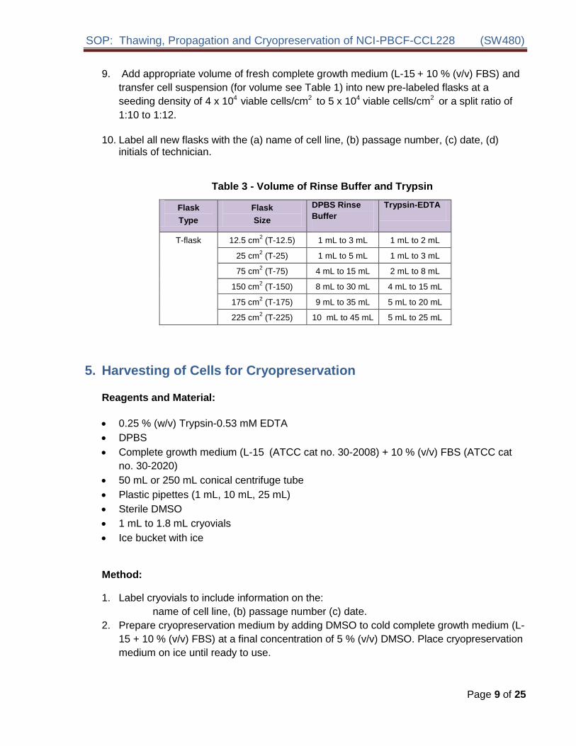
SOP Thawing Propagation and Cryopreservation of NCI-PBCF-CCL228 (SW480)
Isopropanol
Cryovial rack
a Cryopreservation using a rate-controlled programmable freezerMethod
A slow and reproducible cooling rate is very important to ensure good recovery of cultures A decrease of 1 degC per min to -80 degC followed by rapid freeze at about 15 degC to 30 degC per min drop to -150 degC will usually work for most animal cell cultures The best way to control the cooling process is to use a programmable rate-controlled electronic freezer unit Refer to the manufacturerrsquos handbook for detailed procedure
i Using the Cryomed
Starting the Cryopreservation Process
1 Check that the liquid nitrogen valve that supplies the Cryomed is open
2 Check the gauge to ensure that there is enough liquid nitrogen in the open tank to complete the freeze
3 Install the thermocouple probe so that the tip is immersed midway into the control fluid
Note Be sure that the thermocouple is centered in the vial and the vial is placed centered in the rack The probe should be changed after three uses or if it turns yellow to ensure accurate readings by the controller during the freezing process Old medium may have different freezing characteristics
4 Close and latch Cryomed door
5 Turn on microcomputer computer and monitor
6 Double click the ldquoCryomedrdquo icon The machine may need to be pre-programmed for specific cell type and medium
7 From the top of the screen select MENU RUN FUNCTIONS START RUN
8 Fill out the box which appears on the screen Cell line ID TYPE OF SAMPLE MEDIA NUMBER OF SAMPLES
9 Hit the ESCAPE key and the Cryomed will cool to 4 C
10 Once Cryomed chamber has cooled to 4 C load cryovials onto racks and close the door
11 When the Cryomedrsquos chamber temperature and the sample temperature have reached approximately 4 C press the space bar to initiate the rate controlled cryopreservation process
Page 11 of 25
SOP Thawing Propagation and Cryopreservation of NCI-PBCF-CCL228 (SW480)
Completing the Cryopreservation Process
1 When samples have reached ndash80C an alarm will sound To silence this select ALARM
from the options at the top of the screen
2 Select MENU RUN FUNCTIONSrarr STOP Hit the ESCAPE key to return to the main
menu and select EXIT
3 Immediately transfer vials to liquid nitrogen freezer
4 Shut down the microcomputer and then turn off the monitor
b Cryopreservation using ldquoMr Frostyrdquo
1 One day before freezing cells add 250 mL isopropanol to the bottom of the container and place at 2 oC to 8 oC
2 On the day of the freeze prepare cells for cryopreservation as described above
3 Insert cryovials with the cell suspension in appropriate slots in the container
4 Transfer the container to a -70 degC to -90 degC freezer and store overnight
5 Next day transfer cryovials to the vapor phase of liquid nitrogen freezer
Note Each container has 18 slots which can accommodate 18 cryovials in one freeze
Important information when using the rate-controlled programmable freezer or a manual method (Mr Frosty) for cryopreservation of mammalian cells
Regardless which cooling method is used it is important that the transfer to the final storage location (between -130 degC and -196 degC) be done quickly and efficiently If the transfer cannot be done immediately the vials can be placed on dry ice for a short time This will avoid damage to cultures by inadvertent temporary warming during the transfer process Warming during this transfer process is a major cause of variation in culture viability upon thawing
Always keep the storage temperature below -130 degC for optimum survival Cells may survive storage at higher temperatures but viability will usually decrease over time The ideal storage container is a liquid nitrogen freezer where the cultures are stored in the vapor phase above the liquid nitrogen
Note ATCC does not have experience in the cryopreservation of the SW480 cells by any other method than the Cryomed programmable freezer
Page 12 of 25
SOP Thawing Propagation and Cryopreservation of NCI-PBCF-CCL228 (SW480)
7 Storage
Store cryopreserved cells in the vapor phase of liquid nitrogen freezer (below ndash130 degC) for optimum long-term survival
Note Experiments on long-term storage of animal cell lines at different temperature
levels indicate that a -70 degC storage temperature is not adequate except for very short
period of time A -90 degC storage may be adequate for longer periods depending upon
the cell line preserved The efficiency of recovery however is not as great as when
the cells are stored in vapor phase of the liquid nitrogen freezer
Page 13 of 25
SOP Thawing Propagation and Cryopreservation of NCI-PBCF-CCL228 (SW480)
APPENDIX 1 PHOTOMICROGRAPHS OF NCI-PBCF-CCL228 (SW480) CELLS
Figure 1 Photomicrograph of SW480 cells after one day post-freeze recovery Cells were plated at 80 x 104 viable cellscm2
Figure 2 Photomicrograph of SW480 cells after two days post-freeze recovery Cells were plated at 80 x 104 viable cellscm2
Page 14 of 25
SOP Thawing Propagation and Cryopreservation of NCI-PBCF-CCL228 (SW480)
40X 40X 40X
100X 100X 100X
500 microm500 microm500 microm
200 microm 200 microm 200 microm
Figure 3 Photomicrographs of SW480 cells at various time points after
seeding at a cell density of 5 x 10⁴ viable cellscm2
Page 15 of 25
SOP Thawing Propagation and Cryopreservation of NCI-PBCF-CCL228 (SW480)
APPENDIX 2 GROWTH PROFILE OF NCI-PBCF-CCL228 (SW480) CELLS
000E+00
100E+05
200E+05
300E+05
400E+05
500E+05
600E+05
700E+05
800E+05
900E+05
100E+06
7 8 9
Via
ble
Cell
sc
m2
0 1 2 3 4 5 6
Days in Culture
Figure 4 Growth curve for SW480 cells cells were plated at 5 x 10⁴ viable cellscm2 population doubling time (PDT) is approximately 38 h
Page 16 of 25
SOP Thawing Propagation and Cryopreservation of NCI-PBCF-CCL228 (SW480)
APPENDIX 3 CYTOGENETIC ANALYSIS OF NCI-PBCF-CCL228 SW480 CELLS
NCI-PBCF-CCL228 Karyotype Results
Metaphase Spread Karyotype
Number of metaphase spreads counted 20
Band level 300-400
Number of metaphase spreads karyotyped 10
Chromosome range 50-56
Sex Male
Comments Aneuploid
Karyotype
50~56XX+2der(2)t(218)(q21q111)del(5)(q13q33)+add(7)(q22)+add(7)(q32)
der(10)t (1012)(p15q11)+add(11)(p15)-12der(1214)(p10q10)-14+add(15)(p10)
der(15)t(1215)(q13q15)-18add(19)(p133)+20+add(20)(p13)
+der(20)t(520)(q22p13)x2+21+add(21)(q111)add(21)(q22)+mar1x2
(ISCN nomenclature written based on a diploid karyotype)
Human diploid karyotype (2N) 46XX (female) or 46XY (male)
Page 17 of 25
SOP Thawing Propagation and Cryopreservation of NCI-PBCF-CCL228 (SW480)
Karyotype Summary
In the karyotype image arrows indicate regions of abnormality It should be noted that the karyotype description includes the observed abnormalities from all examined metaphase spreads but due to heterogeneity not all of the karyotyped cells will contain every abnormality
This is a rearranged human cell line of male origin containing 50 to 56 chromosomes per metaphase spread (hyperdiploid) Structural abnormalities include rearrangements to approximately 50 of the 22 different autosomal chromosomes (described below) There is one unidentifiable clonal marker chromosome (markers present in two or more of the examined cells) [+mar1]
The rearrangements include
Addition of unknown material to the short arms (designated by p) of chromosomes 11 15 19 and 20
Addition of unknown material to the long arms (designated by q) of chromosomes 7 and 21 (2 different derivatives of each)
An interstitial deletion of material from the long arm of chromosome 5 between bands q13 and q33
Translocations involving chromosomes 2 and 18 [der(2)t(218)(q21q111)] 10 and 12 [der(10)t (1012)(p15q11) chromosomes 12 and 14 [der(1214)(p10q10)] 12 and 15 [der(15)t(1215)(q13q15)] and chromosomes 5 and 20 [der(20)t(520)(q22p13)]
Numerical changes are based on a diploid karyotype which would contain two copies of
each chromosome (2N) Therefore karyotype designations such as +2 +20 and +21
indicates three copies of structurally normal chromosomes 2 20 and 21 Designations
-18 and -14 indicates one copy of structurally normal chromosomes -18 and -14 Also it
appears that the Y chromosome was lost which is not an uncommon occurrence in cancer
cell lines (ISCN 2009 An International System for Human Cytogenetic Nomenclature
(2009) Editors Lisa G Shaffer Marilyn L Slovak Lynda J Campbell)
Karyotype Procedure
Cell Harvest Cells were allowed to grow to 80-90 confluence Mitotic division was
arrested by treating the cells with KaryoMaxreg colcemid for 20 minutes to 2 hours at 37degC
Cells were harvested using 005 Trypsin-EDTA treated with 0075M KCL hypotonic
solution and then fixed in three changes of a 31 ratio of methanolglacial acetic acid
Slide Preparation Slides were prepared by dropping the cell suspension onto wet glass
slides and allowing them to dry under controlled conditions
G-banding Slides were baked one hour at 90degC trypsinized using 10X trypsin-EDTA
and then stained with Leishmanrsquos stain
Page 18 of 25
SOP Thawing Propagation and Cryopreservation of NCI-PBCF-CCL228 (SW480)
Microscopy Slides were scanned using a 10X objective and metaphase spreads were
analyzed using a 100X plan apochromat objective on an Olympus BX-41 microscope
Imaging and karyotyping were performed using Cytovisionreg software
Analysis Twenty metaphase cells were counted and analyzed and representative
metaphase cells were karyotyped depending on the complexity of the study
Summary of Karyotyping Procedure
G-band karyotyping analysis is performed using GTL banding technique G bands produced with trypsin and Leishman Slides prepared with metaphase spreads are treated with trypsin and stained with Leishmanrsquos This method produces a series of light and dark bands that allow for the positive identification of each chromosome
SW480 cell line karyotyping was carried out by Cell Line Genetics Inc (Madison WI 53719)
Page 19 of 25
SOP Thawing Propagation and Cryopreservation of NCI-PBCF-CCL228 (SW480)
APPENDIX 4 GLOSSARY OF TERMS
Confluent monolayer adherent cell culture in which all cells are in contact with other cells
all around their periphery and no available substrate is left uncovered
Split ratio the divisor of the dilution ration of a cell culture to subculture (eg one flask
divided into four or 100 mL up to 400 mL would be split ratio of 14)
Subculture (or passage) the transfer or transplantation of cells with or without dilution
from one culture vessel to another
Passage No the total number of times the cells in the culture have been subcultured or
passaged (with each subculture the passage number increases by 1)
Population doubling level (PDL) the total number of population doublings of a cell line
since its initiation in vitro (with each subculture the population doubling increases in
relationship to the split ratio at which the cells are plated) See Appendix 7
Population doubling time (doubling time) the time interval calculated during the
logarithmic phase of growth in which cells double in number
Seeding density recommended number of cells per cm2 of substrate when inoculating a
new flask
Epithelial-like adherent cells of a polygonal shape with clear sharp boundaries between
them
Fibroblast-like adherent cells of a spindle or stellate shape
Page 20 of 25
SOP Thawing Propagation and Cryopreservation of NCI-PBCF-CCL228 (SW480)
APPENDIX 5 REFERENCE
1 Culture Of Animal Cells A Manual of Basic Technique by R Ian Freshney 6th edition
published by Wiley-Liss NY 2010
2 Fogh J et al Absence of HeLa cell contamination in 169 cell lines derived from human
tumors J Natl Cancer Inst 58 209-214 1977 PubMed 833871
3 Fogh J et al One hundred and twenty-seven cultured human tumor cell lines producing
tumors in nude mice J Natl Cancer Inst 59 221-226 1977 PubMed 327080
4 Lelbovitz A et al Detection and analysis of a glucose 6-phosphate dehydrogenase
phenotype B cell line contamination J Natl Cancer Inst 63 635-645 1979 PubMed
288927
5 Adachi A et al Productive persistent infection of human colorectal cell lines with human
immunodeficiency virus J Virol 61 209-213 1987 PubMed 3640832
6 Schroy PC et al Detection of p21ras mutations in colorectal adenomas and carcinomas
by enzyme-linked immunosorbent assay Cancer 76 201-209 1995 PubMed 8625092
7 Trainer DL et al Biological characterization and oncogene expression in human
colorectal carcinoma cell lines Int J Cancer 41 287-296 1988 PubMed 3338874
8 Weiss J et al Mutation and expression of the p53 gene in malignant melanoma cell
lines Int J Cancer 54 693-699 1993 PubMed 8514460
9 Nigro JM et al Mutations in the p53 gene occur in diverse human tumour types Nature
342 705-707 1989 PubMed 2531845
10 Barnett SW et al Characterization of human immunodeficiency virus type 1 strains
recovered from the bowel of infected individuals Virology 182 802-809 1991 PubMed
2024498
11 Leibovitz A et al Classification of human colorectal adenocarcinoma cell lines Cancer
Res 36 4562-4569 1976 PubMed 1000501
12 Geiser AG et al Suppression of tumorigenicity in human cell hybrids derived from cell
lines expressing different activated ras oncogenes Cancer Res 49 1572-1577 1989
PubMed 2647289
13 Lahm H et al Secretion of bioactive granulocyte-macrophage colony-stimulating factor
by human colorectal carcinoma cells Cancer Res 54 3700-3702 1994 PubMed
8033086
14 Rodrigues NR et al p53 mutations in colorectal cancer Proc Natl Acad Sci USA 87
7555-7559 1990 PubMed 1699228
Page 21 of 25
SOP Thawing Propagation and Cryopreservation of NCI-PBCF-CCL228 (SW480)
15 Santoro IM Groden J Alternative splicing of the APC gene and its association with terminal differentiation Cancer Res 57 488-494 1997 PubMed 9012479
16 Tsao H et al Novel mutations in the p16CDKN2A binding region of the Cyclinshy
dependent Kinase-4 gene Cancer Res 58 109-113 1998 PubMed 9426066
17 Zhu X et al Cell cycle-dependent modulation of telomerase activity in tumor cells Proc Natl Acad Sci USA 93 6091-6095 1996 PubMed 8650224
18 Witty JP et al Modulation of matrilysin levels in colon carcinoma cell lines affects tumorigenicity in vivo Cancer Res 54 4805-4812 1994 PubMed 8062282
Page 22 of 25
SOP Thawing Propagation and Cryopreservation of NCI-PBCF-CCL228 (SW480)
APPENDIX 6 REAGENT LOT TRACEABILITY AND CELL EXPANSION TABLES
Table 4 Reagent Lot Traceability
Reagent Vendor Catalog Lot Expiration Date
Page 23 of 25
Table 5 Cell Expansion
FROM FLUID CHANGE
Observation under
microscope CELL COUNT TO
By Date Flask qty
size
Pass Confluence
Add Replace
Volume
(in mL)
Viable cellsmL
Total viable cells
Viability
Split Ratio
Flask qty
size
Pass
PDL
Add
Replace
Add
Replace
Add
Replace
Add
Replace
Add
Replace
Add
Replace
Add
Replace
Add
Replace
Add
Replace
SOP Thawing Propagation and Cryopreservation of NCI-PBCF-CCL228 (SW480)
APPENDIX 7 CALCULATION OF POPULATION DOUBLING LEVEL (PDL)
Calculate the PDL of the current passage using the following equation
PDL = X + 3322 (log Y ndash log I)
Where X = initial PDL I = cell inoculum (number of cells plated in the flask) Y = final cell yield (number of cells at the end of the growth period)
APPENDIX 8 SAFETY PRECAUTIONS
Use at least approved Biological Safety Level 2 (BSL-2) facilities and procedures
Wear appropriate Personal Protective Equipment (PPE) such as isolation gown lab coat with sleeve protectors face shield and gloves
Use safety precautions when working with liquid nitrogen nitrogen vapor and cryogenically cooled fixtures
Use liquid nitrogen freezers and liquid nitrogen tanks only in areas with adequate ventilation Liquid nitrogen reduces the concentration of oxygen and can cause suffocation
Wear latex gloves over insulating gloves to prevent liquid nitrogen from soaking in and being held next to the skin Liquid nitrogen is extremely cold and will cause burns and frostbite Metal inventory racks tank components and liquid nitrogen transfer hoses exposed to liquid nitrogen or nitrogen vapor quickly cool to cryogenic temperatures and can cause burns and frostbite
Wear a full face mask when thawing and retrieving vials from liquid nitrogen freezer Danger to the technician derives mainly from the possibility that liquid nitrogen can penetrate the cryovial during storage On warming rapid evaporation of the nitrogen within the confines of such cryovial can cause an aerosol or explosion of the cryovial and contents
Page 25 of 25

SOP Thawing Propagation and Cryopreservation of NCI-PBCF-CCL228 (SW480)
Completing the Cryopreservation Process
1 When samples have reached ndash80C an alarm will sound To silence this select ALARM
from the options at the top of the screen
2 Select MENU RUN FUNCTIONSrarr STOP Hit the ESCAPE key to return to the main
menu and select EXIT
3 Immediately transfer vials to liquid nitrogen freezer
4 Shut down the microcomputer and then turn off the monitor
b Cryopreservation using ldquoMr Frostyrdquo
1 One day before freezing cells add 250 mL isopropanol to the bottom of the container and place at 2 oC to 8 oC
2 On the day of the freeze prepare cells for cryopreservation as described above
3 Insert cryovials with the cell suspension in appropriate slots in the container
4 Transfer the container to a -70 degC to -90 degC freezer and store overnight
5 Next day transfer cryovials to the vapor phase of liquid nitrogen freezer
Note Each container has 18 slots which can accommodate 18 cryovials in one freeze
Important information when using the rate-controlled programmable freezer or a manual method (Mr Frosty) for cryopreservation of mammalian cells
Regardless which cooling method is used it is important that the transfer to the final storage location (between -130 degC and -196 degC) be done quickly and efficiently If the transfer cannot be done immediately the vials can be placed on dry ice for a short time This will avoid damage to cultures by inadvertent temporary warming during the transfer process Warming during this transfer process is a major cause of variation in culture viability upon thawing
Always keep the storage temperature below -130 degC for optimum survival Cells may survive storage at higher temperatures but viability will usually decrease over time The ideal storage container is a liquid nitrogen freezer where the cultures are stored in the vapor phase above the liquid nitrogen
Note ATCC does not have experience in the cryopreservation of the SW480 cells by any other method than the Cryomed programmable freezer
Page 12 of 25
SOP Thawing Propagation and Cryopreservation of NCI-PBCF-CCL228 (SW480)
7 Storage
Store cryopreserved cells in the vapor phase of liquid nitrogen freezer (below ndash130 degC) for optimum long-term survival
Note Experiments on long-term storage of animal cell lines at different temperature
levels indicate that a -70 degC storage temperature is not adequate except for very short
period of time A -90 degC storage may be adequate for longer periods depending upon
the cell line preserved The efficiency of recovery however is not as great as when
the cells are stored in vapor phase of the liquid nitrogen freezer
Page 13 of 25
SOP Thawing Propagation and Cryopreservation of NCI-PBCF-CCL228 (SW480)
APPENDIX 1 PHOTOMICROGRAPHS OF NCI-PBCF-CCL228 (SW480) CELLS
Figure 1 Photomicrograph of SW480 cells after one day post-freeze recovery Cells were plated at 80 x 104 viable cellscm2
Figure 2 Photomicrograph of SW480 cells after two days post-freeze recovery Cells were plated at 80 x 104 viable cellscm2
Page 14 of 25
SOP Thawing Propagation and Cryopreservation of NCI-PBCF-CCL228 (SW480)
40X 40X 40X
100X 100X 100X
500 microm500 microm500 microm
200 microm 200 microm 200 microm
Figure 3 Photomicrographs of SW480 cells at various time points after
seeding at a cell density of 5 x 10⁴ viable cellscm2
Page 15 of 25
SOP Thawing Propagation and Cryopreservation of NCI-PBCF-CCL228 (SW480)
APPENDIX 2 GROWTH PROFILE OF NCI-PBCF-CCL228 (SW480) CELLS
000E+00
100E+05
200E+05
300E+05
400E+05
500E+05
600E+05
700E+05
800E+05
900E+05
100E+06
7 8 9
Via
ble
Cell
sc
m2
0 1 2 3 4 5 6
Days in Culture
Figure 4 Growth curve for SW480 cells cells were plated at 5 x 10⁴ viable cellscm2 population doubling time (PDT) is approximately 38 h
Page 16 of 25
SOP Thawing Propagation and Cryopreservation of NCI-PBCF-CCL228 (SW480)
APPENDIX 3 CYTOGENETIC ANALYSIS OF NCI-PBCF-CCL228 SW480 CELLS
NCI-PBCF-CCL228 Karyotype Results
Metaphase Spread Karyotype
Number of metaphase spreads counted 20
Band level 300-400
Number of metaphase spreads karyotyped 10
Chromosome range 50-56
Sex Male
Comments Aneuploid
Karyotype
50~56XX+2der(2)t(218)(q21q111)del(5)(q13q33)+add(7)(q22)+add(7)(q32)
der(10)t (1012)(p15q11)+add(11)(p15)-12der(1214)(p10q10)-14+add(15)(p10)
der(15)t(1215)(q13q15)-18add(19)(p133)+20+add(20)(p13)
+der(20)t(520)(q22p13)x2+21+add(21)(q111)add(21)(q22)+mar1x2
(ISCN nomenclature written based on a diploid karyotype)
Human diploid karyotype (2N) 46XX (female) or 46XY (male)
Page 17 of 25
SOP Thawing Propagation and Cryopreservation of NCI-PBCF-CCL228 (SW480)
Karyotype Summary
In the karyotype image arrows indicate regions of abnormality It should be noted that the karyotype description includes the observed abnormalities from all examined metaphase spreads but due to heterogeneity not all of the karyotyped cells will contain every abnormality
This is a rearranged human cell line of male origin containing 50 to 56 chromosomes per metaphase spread (hyperdiploid) Structural abnormalities include rearrangements to approximately 50 of the 22 different autosomal chromosomes (described below) There is one unidentifiable clonal marker chromosome (markers present in two or more of the examined cells) [+mar1]
The rearrangements include
Addition of unknown material to the short arms (designated by p) of chromosomes 11 15 19 and 20
Addition of unknown material to the long arms (designated by q) of chromosomes 7 and 21 (2 different derivatives of each)
An interstitial deletion of material from the long arm of chromosome 5 between bands q13 and q33
Translocations involving chromosomes 2 and 18 [der(2)t(218)(q21q111)] 10 and 12 [der(10)t (1012)(p15q11) chromosomes 12 and 14 [der(1214)(p10q10)] 12 and 15 [der(15)t(1215)(q13q15)] and chromosomes 5 and 20 [der(20)t(520)(q22p13)]
Numerical changes are based on a diploid karyotype which would contain two copies of
each chromosome (2N) Therefore karyotype designations such as +2 +20 and +21
indicates three copies of structurally normal chromosomes 2 20 and 21 Designations
-18 and -14 indicates one copy of structurally normal chromosomes -18 and -14 Also it
appears that the Y chromosome was lost which is not an uncommon occurrence in cancer
cell lines (ISCN 2009 An International System for Human Cytogenetic Nomenclature
(2009) Editors Lisa G Shaffer Marilyn L Slovak Lynda J Campbell)
Karyotype Procedure
Cell Harvest Cells were allowed to grow to 80-90 confluence Mitotic division was
arrested by treating the cells with KaryoMaxreg colcemid for 20 minutes to 2 hours at 37degC
Cells were harvested using 005 Trypsin-EDTA treated with 0075M KCL hypotonic
solution and then fixed in three changes of a 31 ratio of methanolglacial acetic acid
Slide Preparation Slides were prepared by dropping the cell suspension onto wet glass
slides and allowing them to dry under controlled conditions
G-banding Slides were baked one hour at 90degC trypsinized using 10X trypsin-EDTA
and then stained with Leishmanrsquos stain
Page 18 of 25
SOP Thawing Propagation and Cryopreservation of NCI-PBCF-CCL228 (SW480)
Microscopy Slides were scanned using a 10X objective and metaphase spreads were
analyzed using a 100X plan apochromat objective on an Olympus BX-41 microscope
Imaging and karyotyping were performed using Cytovisionreg software
Analysis Twenty metaphase cells were counted and analyzed and representative
metaphase cells were karyotyped depending on the complexity of the study
Summary of Karyotyping Procedure
G-band karyotyping analysis is performed using GTL banding technique G bands produced with trypsin and Leishman Slides prepared with metaphase spreads are treated with trypsin and stained with Leishmanrsquos This method produces a series of light and dark bands that allow for the positive identification of each chromosome
SW480 cell line karyotyping was carried out by Cell Line Genetics Inc (Madison WI 53719)
Page 19 of 25
SOP Thawing Propagation and Cryopreservation of NCI-PBCF-CCL228 (SW480)
APPENDIX 4 GLOSSARY OF TERMS
Confluent monolayer adherent cell culture in which all cells are in contact with other cells
all around their periphery and no available substrate is left uncovered
Split ratio the divisor of the dilution ration of a cell culture to subculture (eg one flask
divided into four or 100 mL up to 400 mL would be split ratio of 14)
Subculture (or passage) the transfer or transplantation of cells with or without dilution
from one culture vessel to another
Passage No the total number of times the cells in the culture have been subcultured or
passaged (with each subculture the passage number increases by 1)
Population doubling level (PDL) the total number of population doublings of a cell line
since its initiation in vitro (with each subculture the population doubling increases in
relationship to the split ratio at which the cells are plated) See Appendix 7
Population doubling time (doubling time) the time interval calculated during the
logarithmic phase of growth in which cells double in number
Seeding density recommended number of cells per cm2 of substrate when inoculating a
new flask
Epithelial-like adherent cells of a polygonal shape with clear sharp boundaries between
them
Fibroblast-like adherent cells of a spindle or stellate shape
Page 20 of 25
SOP Thawing Propagation and Cryopreservation of NCI-PBCF-CCL228 (SW480)
APPENDIX 5 REFERENCE
1 Culture Of Animal Cells A Manual of Basic Technique by R Ian Freshney 6th edition
published by Wiley-Liss NY 2010
2 Fogh J et al Absence of HeLa cell contamination in 169 cell lines derived from human
tumors J Natl Cancer Inst 58 209-214 1977 PubMed 833871
3 Fogh J et al One hundred and twenty-seven cultured human tumor cell lines producing
tumors in nude mice J Natl Cancer Inst 59 221-226 1977 PubMed 327080
4 Lelbovitz A et al Detection and analysis of a glucose 6-phosphate dehydrogenase
phenotype B cell line contamination J Natl Cancer Inst 63 635-645 1979 PubMed
288927
5 Adachi A et al Productive persistent infection of human colorectal cell lines with human
immunodeficiency virus J Virol 61 209-213 1987 PubMed 3640832
6 Schroy PC et al Detection of p21ras mutations in colorectal adenomas and carcinomas
by enzyme-linked immunosorbent assay Cancer 76 201-209 1995 PubMed 8625092
7 Trainer DL et al Biological characterization and oncogene expression in human
colorectal carcinoma cell lines Int J Cancer 41 287-296 1988 PubMed 3338874
8 Weiss J et al Mutation and expression of the p53 gene in malignant melanoma cell
lines Int J Cancer 54 693-699 1993 PubMed 8514460
9 Nigro JM et al Mutations in the p53 gene occur in diverse human tumour types Nature
342 705-707 1989 PubMed 2531845
10 Barnett SW et al Characterization of human immunodeficiency virus type 1 strains
recovered from the bowel of infected individuals Virology 182 802-809 1991 PubMed
2024498
11 Leibovitz A et al Classification of human colorectal adenocarcinoma cell lines Cancer
Res 36 4562-4569 1976 PubMed 1000501
12 Geiser AG et al Suppression of tumorigenicity in human cell hybrids derived from cell
lines expressing different activated ras oncogenes Cancer Res 49 1572-1577 1989
PubMed 2647289
13 Lahm H et al Secretion of bioactive granulocyte-macrophage colony-stimulating factor
by human colorectal carcinoma cells Cancer Res 54 3700-3702 1994 PubMed
8033086
14 Rodrigues NR et al p53 mutations in colorectal cancer Proc Natl Acad Sci USA 87
7555-7559 1990 PubMed 1699228
Page 21 of 25
SOP Thawing Propagation and Cryopreservation of NCI-PBCF-CCL228 (SW480)
15 Santoro IM Groden J Alternative splicing of the APC gene and its association with terminal differentiation Cancer Res 57 488-494 1997 PubMed 9012479
16 Tsao H et al Novel mutations in the p16CDKN2A binding region of the Cyclinshy
dependent Kinase-4 gene Cancer Res 58 109-113 1998 PubMed 9426066
17 Zhu X et al Cell cycle-dependent modulation of telomerase activity in tumor cells Proc Natl Acad Sci USA 93 6091-6095 1996 PubMed 8650224
18 Witty JP et al Modulation of matrilysin levels in colon carcinoma cell lines affects tumorigenicity in vivo Cancer Res 54 4805-4812 1994 PubMed 8062282
Page 22 of 25
SOP Thawing Propagation and Cryopreservation of NCI-PBCF-CCL228 (SW480)
APPENDIX 6 REAGENT LOT TRACEABILITY AND CELL EXPANSION TABLES
Table 4 Reagent Lot Traceability
Reagent Vendor Catalog Lot Expiration Date
Page 23 of 25
Table 5 Cell Expansion
FROM FLUID CHANGE
Observation under
microscope CELL COUNT TO
By Date Flask qty
size
Pass Confluence
Add Replace
Volume
(in mL)
Viable cellsmL
Total viable cells
Viability
Split Ratio
Flask qty
size
Pass
PDL
Add
Replace
Add
Replace
Add
Replace
Add
Replace
Add
Replace
Add
Replace
Add
Replace
Add
Replace
Add
Replace
SOP Thawing Propagation and Cryopreservation of NCI-PBCF-CCL228 (SW480)
APPENDIX 7 CALCULATION OF POPULATION DOUBLING LEVEL (PDL)
Calculate the PDL of the current passage using the following equation
PDL = X + 3322 (log Y ndash log I)
Where X = initial PDL I = cell inoculum (number of cells plated in the flask) Y = final cell yield (number of cells at the end of the growth period)
APPENDIX 8 SAFETY PRECAUTIONS
Use at least approved Biological Safety Level 2 (BSL-2) facilities and procedures
Wear appropriate Personal Protective Equipment (PPE) such as isolation gown lab coat with sleeve protectors face shield and gloves
Use safety precautions when working with liquid nitrogen nitrogen vapor and cryogenically cooled fixtures
Use liquid nitrogen freezers and liquid nitrogen tanks only in areas with adequate ventilation Liquid nitrogen reduces the concentration of oxygen and can cause suffocation
Wear latex gloves over insulating gloves to prevent liquid nitrogen from soaking in and being held next to the skin Liquid nitrogen is extremely cold and will cause burns and frostbite Metal inventory racks tank components and liquid nitrogen transfer hoses exposed to liquid nitrogen or nitrogen vapor quickly cool to cryogenic temperatures and can cause burns and frostbite
Wear a full face mask when thawing and retrieving vials from liquid nitrogen freezer Danger to the technician derives mainly from the possibility that liquid nitrogen can penetrate the cryovial during storage On warming rapid evaporation of the nitrogen within the confines of such cryovial can cause an aerosol or explosion of the cryovial and contents
Page 25 of 25
SOP Thawing Propagation and Cryopreservation of NCI-PBCF-CCL228 (SW480)
7 Storage
Store cryopreserved cells in the vapor phase of liquid nitrogen freezer (below ndash130 degC) for optimum long-term survival
Note Experiments on long-term storage of animal cell lines at different temperature
levels indicate that a -70 degC storage temperature is not adequate except for very short
period of time A -90 degC storage may be adequate for longer periods depending upon
the cell line preserved The efficiency of recovery however is not as great as when
the cells are stored in vapor phase of the liquid nitrogen freezer
Page 13 of 25
SOP Thawing Propagation and Cryopreservation of NCI-PBCF-CCL228 (SW480)
APPENDIX 1 PHOTOMICROGRAPHS OF NCI-PBCF-CCL228 (SW480) CELLS
Figure 1 Photomicrograph of SW480 cells after one day post-freeze recovery Cells were plated at 80 x 104 viable cellscm2
Figure 2 Photomicrograph of SW480 cells after two days post-freeze recovery Cells were plated at 80 x 104 viable cellscm2
Page 14 of 25
SOP Thawing Propagation and Cryopreservation of NCI-PBCF-CCL228 (SW480)
40X 40X 40X
100X 100X 100X
500 microm500 microm500 microm
200 microm 200 microm 200 microm
Figure 3 Photomicrographs of SW480 cells at various time points after
seeding at a cell density of 5 x 10⁴ viable cellscm2
Page 15 of 25
SOP Thawing Propagation and Cryopreservation of NCI-PBCF-CCL228 (SW480)
APPENDIX 2 GROWTH PROFILE OF NCI-PBCF-CCL228 (SW480) CELLS
000E+00
100E+05
200E+05
300E+05
400E+05
500E+05
600E+05
700E+05
800E+05
900E+05
100E+06
7 8 9
Via
ble
Cell
sc
m2
0 1 2 3 4 5 6
Days in Culture
Figure 4 Growth curve for SW480 cells cells were plated at 5 x 10⁴ viable cellscm2 population doubling time (PDT) is approximately 38 h
Page 16 of 25
SOP Thawing Propagation and Cryopreservation of NCI-PBCF-CCL228 (SW480)
APPENDIX 3 CYTOGENETIC ANALYSIS OF NCI-PBCF-CCL228 SW480 CELLS
NCI-PBCF-CCL228 Karyotype Results
Metaphase Spread Karyotype
Number of metaphase spreads counted 20
Band level 300-400
Number of metaphase spreads karyotyped 10
Chromosome range 50-56
Sex Male
Comments Aneuploid
Karyotype
50~56XX+2der(2)t(218)(q21q111)del(5)(q13q33)+add(7)(q22)+add(7)(q32)
der(10)t (1012)(p15q11)+add(11)(p15)-12der(1214)(p10q10)-14+add(15)(p10)
der(15)t(1215)(q13q15)-18add(19)(p133)+20+add(20)(p13)
+der(20)t(520)(q22p13)x2+21+add(21)(q111)add(21)(q22)+mar1x2
(ISCN nomenclature written based on a diploid karyotype)
Human diploid karyotype (2N) 46XX (female) or 46XY (male)
Page 17 of 25
SOP Thawing Propagation and Cryopreservation of NCI-PBCF-CCL228 (SW480)
Karyotype Summary
In the karyotype image arrows indicate regions of abnormality It should be noted that the karyotype description includes the observed abnormalities from all examined metaphase spreads but due to heterogeneity not all of the karyotyped cells will contain every abnormality
This is a rearranged human cell line of male origin containing 50 to 56 chromosomes per metaphase spread (hyperdiploid) Structural abnormalities include rearrangements to approximately 50 of the 22 different autosomal chromosomes (described below) There is one unidentifiable clonal marker chromosome (markers present in two or more of the examined cells) [+mar1]
The rearrangements include
Addition of unknown material to the short arms (designated by p) of chromosomes 11 15 19 and 20
Addition of unknown material to the long arms (designated by q) of chromosomes 7 and 21 (2 different derivatives of each)
An interstitial deletion of material from the long arm of chromosome 5 between bands q13 and q33
Translocations involving chromosomes 2 and 18 [der(2)t(218)(q21q111)] 10 and 12 [der(10)t (1012)(p15q11) chromosomes 12 and 14 [der(1214)(p10q10)] 12 and 15 [der(15)t(1215)(q13q15)] and chromosomes 5 and 20 [der(20)t(520)(q22p13)]
Numerical changes are based on a diploid karyotype which would contain two copies of
each chromosome (2N) Therefore karyotype designations such as +2 +20 and +21
indicates three copies of structurally normal chromosomes 2 20 and 21 Designations
-18 and -14 indicates one copy of structurally normal chromosomes -18 and -14 Also it
appears that the Y chromosome was lost which is not an uncommon occurrence in cancer
cell lines (ISCN 2009 An International System for Human Cytogenetic Nomenclature
(2009) Editors Lisa G Shaffer Marilyn L Slovak Lynda J Campbell)
Karyotype Procedure
Cell Harvest Cells were allowed to grow to 80-90 confluence Mitotic division was
arrested by treating the cells with KaryoMaxreg colcemid for 20 minutes to 2 hours at 37degC
Cells were harvested using 005 Trypsin-EDTA treated with 0075M KCL hypotonic
solution and then fixed in three changes of a 31 ratio of methanolglacial acetic acid
Slide Preparation Slides were prepared by dropping the cell suspension onto wet glass
slides and allowing them to dry under controlled conditions
G-banding Slides were baked one hour at 90degC trypsinized using 10X trypsin-EDTA
and then stained with Leishmanrsquos stain
Page 18 of 25
SOP Thawing Propagation and Cryopreservation of NCI-PBCF-CCL228 (SW480)
Microscopy Slides were scanned using a 10X objective and metaphase spreads were
analyzed using a 100X plan apochromat objective on an Olympus BX-41 microscope
Imaging and karyotyping were performed using Cytovisionreg software
Analysis Twenty metaphase cells were counted and analyzed and representative
metaphase cells were karyotyped depending on the complexity of the study
Summary of Karyotyping Procedure
G-band karyotyping analysis is performed using GTL banding technique G bands produced with trypsin and Leishman Slides prepared with metaphase spreads are treated with trypsin and stained with Leishmanrsquos This method produces a series of light and dark bands that allow for the positive identification of each chromosome
SW480 cell line karyotyping was carried out by Cell Line Genetics Inc (Madison WI 53719)
Page 19 of 25
SOP Thawing Propagation and Cryopreservation of NCI-PBCF-CCL228 (SW480)
APPENDIX 4 GLOSSARY OF TERMS
Confluent monolayer adherent cell culture in which all cells are in contact with other cells
all around their periphery and no available substrate is left uncovered
Split ratio the divisor of the dilution ration of a cell culture to subculture (eg one flask
divided into four or 100 mL up to 400 mL would be split ratio of 14)
Subculture (or passage) the transfer or transplantation of cells with or without dilution
from one culture vessel to another
Passage No the total number of times the cells in the culture have been subcultured or
passaged (with each subculture the passage number increases by 1)
Population doubling level (PDL) the total number of population doublings of a cell line
since its initiation in vitro (with each subculture the population doubling increases in
relationship to the split ratio at which the cells are plated) See Appendix 7
Population doubling time (doubling time) the time interval calculated during the
logarithmic phase of growth in which cells double in number
Seeding density recommended number of cells per cm2 of substrate when inoculating a
new flask
Epithelial-like adherent cells of a polygonal shape with clear sharp boundaries between
them
Fibroblast-like adherent cells of a spindle or stellate shape
Page 20 of 25
SOP Thawing Propagation and Cryopreservation of NCI-PBCF-CCL228 (SW480)
APPENDIX 5 REFERENCE
1 Culture Of Animal Cells A Manual of Basic Technique by R Ian Freshney 6th edition
published by Wiley-Liss NY 2010
2 Fogh J et al Absence of HeLa cell contamination in 169 cell lines derived from human
tumors J Natl Cancer Inst 58 209-214 1977 PubMed 833871
3 Fogh J et al One hundred and twenty-seven cultured human tumor cell lines producing
tumors in nude mice J Natl Cancer Inst 59 221-226 1977 PubMed 327080
4 Lelbovitz A et al Detection and analysis of a glucose 6-phosphate dehydrogenase
phenotype B cell line contamination J Natl Cancer Inst 63 635-645 1979 PubMed
288927
5 Adachi A et al Productive persistent infection of human colorectal cell lines with human
immunodeficiency virus J Virol 61 209-213 1987 PubMed 3640832
6 Schroy PC et al Detection of p21ras mutations in colorectal adenomas and carcinomas
by enzyme-linked immunosorbent assay Cancer 76 201-209 1995 PubMed 8625092
7 Trainer DL et al Biological characterization and oncogene expression in human
colorectal carcinoma cell lines Int J Cancer 41 287-296 1988 PubMed 3338874
8 Weiss J et al Mutation and expression of the p53 gene in malignant melanoma cell
lines Int J Cancer 54 693-699 1993 PubMed 8514460
9 Nigro JM et al Mutations in the p53 gene occur in diverse human tumour types Nature
342 705-707 1989 PubMed 2531845
10 Barnett SW et al Characterization of human immunodeficiency virus type 1 strains
recovered from the bowel of infected individuals Virology 182 802-809 1991 PubMed
2024498
11 Leibovitz A et al Classification of human colorectal adenocarcinoma cell lines Cancer
Res 36 4562-4569 1976 PubMed 1000501
12 Geiser AG et al Suppression of tumorigenicity in human cell hybrids derived from cell
lines expressing different activated ras oncogenes Cancer Res 49 1572-1577 1989
PubMed 2647289
13 Lahm H et al Secretion of bioactive granulocyte-macrophage colony-stimulating factor
by human colorectal carcinoma cells Cancer Res 54 3700-3702 1994 PubMed
8033086
14 Rodrigues NR et al p53 mutations in colorectal cancer Proc Natl Acad Sci USA 87
7555-7559 1990 PubMed 1699228
Page 21 of 25
SOP Thawing Propagation and Cryopreservation of NCI-PBCF-CCL228 (SW480)
15 Santoro IM Groden J Alternative splicing of the APC gene and its association with terminal differentiation Cancer Res 57 488-494 1997 PubMed 9012479
16 Tsao H et al Novel mutations in the p16CDKN2A binding region of the Cyclinshy
dependent Kinase-4 gene Cancer Res 58 109-113 1998 PubMed 9426066
17 Zhu X et al Cell cycle-dependent modulation of telomerase activity in tumor cells Proc Natl Acad Sci USA 93 6091-6095 1996 PubMed 8650224
18 Witty JP et al Modulation of matrilysin levels in colon carcinoma cell lines affects tumorigenicity in vivo Cancer Res 54 4805-4812 1994 PubMed 8062282
Page 22 of 25
SOP Thawing Propagation and Cryopreservation of NCI-PBCF-CCL228 (SW480)
APPENDIX 6 REAGENT LOT TRACEABILITY AND CELL EXPANSION TABLES
Table 4 Reagent Lot Traceability
Reagent Vendor Catalog Lot Expiration Date
Page 23 of 25
Table 5 Cell Expansion
FROM FLUID CHANGE
Observation under
microscope CELL COUNT TO
By Date Flask qty
size
Pass Confluence
Add Replace
Volume
(in mL)
Viable cellsmL
Total viable cells
Viability
Split Ratio
Flask qty
size
Pass
PDL
Add
Replace
Add
Replace
Add
Replace
Add
Replace
Add
Replace
Add
Replace
Add
Replace
Add
Replace
Add
Replace
SOP Thawing Propagation and Cryopreservation of NCI-PBCF-CCL228 (SW480)
APPENDIX 7 CALCULATION OF POPULATION DOUBLING LEVEL (PDL)
Calculate the PDL of the current passage using the following equation
PDL = X + 3322 (log Y ndash log I)
Where X = initial PDL I = cell inoculum (number of cells plated in the flask) Y = final cell yield (number of cells at the end of the growth period)
APPENDIX 8 SAFETY PRECAUTIONS
Use at least approved Biological Safety Level 2 (BSL-2) facilities and procedures
Wear appropriate Personal Protective Equipment (PPE) such as isolation gown lab coat with sleeve protectors face shield and gloves
Use safety precautions when working with liquid nitrogen nitrogen vapor and cryogenically cooled fixtures
Use liquid nitrogen freezers and liquid nitrogen tanks only in areas with adequate ventilation Liquid nitrogen reduces the concentration of oxygen and can cause suffocation
Wear latex gloves over insulating gloves to prevent liquid nitrogen from soaking in and being held next to the skin Liquid nitrogen is extremely cold and will cause burns and frostbite Metal inventory racks tank components and liquid nitrogen transfer hoses exposed to liquid nitrogen or nitrogen vapor quickly cool to cryogenic temperatures and can cause burns and frostbite
Wear a full face mask when thawing and retrieving vials from liquid nitrogen freezer Danger to the technician derives mainly from the possibility that liquid nitrogen can penetrate the cryovial during storage On warming rapid evaporation of the nitrogen within the confines of such cryovial can cause an aerosol or explosion of the cryovial and contents
Page 25 of 25
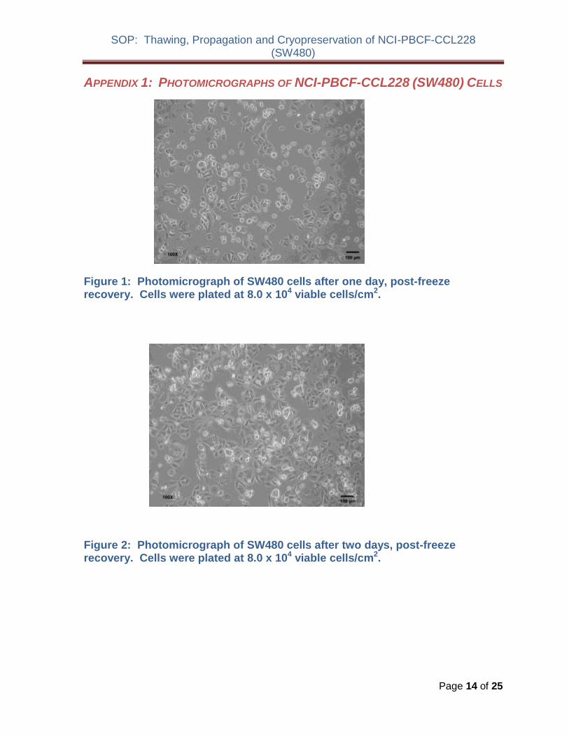
SOP Thawing Propagation and Cryopreservation of NCI-PBCF-CCL228 (SW480)
APPENDIX 1 PHOTOMICROGRAPHS OF NCI-PBCF-CCL228 (SW480) CELLS
Figure 1 Photomicrograph of SW480 cells after one day post-freeze recovery Cells were plated at 80 x 104 viable cellscm2
Figure 2 Photomicrograph of SW480 cells after two days post-freeze recovery Cells were plated at 80 x 104 viable cellscm2
Page 14 of 25
SOP Thawing Propagation and Cryopreservation of NCI-PBCF-CCL228 (SW480)
40X 40X 40X
100X 100X 100X
500 microm500 microm500 microm
200 microm 200 microm 200 microm
Figure 3 Photomicrographs of SW480 cells at various time points after
seeding at a cell density of 5 x 10⁴ viable cellscm2
Page 15 of 25
SOP Thawing Propagation and Cryopreservation of NCI-PBCF-CCL228 (SW480)
APPENDIX 2 GROWTH PROFILE OF NCI-PBCF-CCL228 (SW480) CELLS
000E+00
100E+05
200E+05
300E+05
400E+05
500E+05
600E+05
700E+05
800E+05
900E+05
100E+06
7 8 9
Via
ble
Cell
sc
m2
0 1 2 3 4 5 6
Days in Culture
Figure 4 Growth curve for SW480 cells cells were plated at 5 x 10⁴ viable cellscm2 population doubling time (PDT) is approximately 38 h
Page 16 of 25
SOP Thawing Propagation and Cryopreservation of NCI-PBCF-CCL228 (SW480)
APPENDIX 3 CYTOGENETIC ANALYSIS OF NCI-PBCF-CCL228 SW480 CELLS
NCI-PBCF-CCL228 Karyotype Results
Metaphase Spread Karyotype
Number of metaphase spreads counted 20
Band level 300-400
Number of metaphase spreads karyotyped 10
Chromosome range 50-56
Sex Male
Comments Aneuploid
Karyotype
50~56XX+2der(2)t(218)(q21q111)del(5)(q13q33)+add(7)(q22)+add(7)(q32)
der(10)t (1012)(p15q11)+add(11)(p15)-12der(1214)(p10q10)-14+add(15)(p10)
der(15)t(1215)(q13q15)-18add(19)(p133)+20+add(20)(p13)
+der(20)t(520)(q22p13)x2+21+add(21)(q111)add(21)(q22)+mar1x2
(ISCN nomenclature written based on a diploid karyotype)
Human diploid karyotype (2N) 46XX (female) or 46XY (male)
Page 17 of 25
SOP Thawing Propagation and Cryopreservation of NCI-PBCF-CCL228 (SW480)
Karyotype Summary
In the karyotype image arrows indicate regions of abnormality It should be noted that the karyotype description includes the observed abnormalities from all examined metaphase spreads but due to heterogeneity not all of the karyotyped cells will contain every abnormality
This is a rearranged human cell line of male origin containing 50 to 56 chromosomes per metaphase spread (hyperdiploid) Structural abnormalities include rearrangements to approximately 50 of the 22 different autosomal chromosomes (described below) There is one unidentifiable clonal marker chromosome (markers present in two or more of the examined cells) [+mar1]
The rearrangements include
Addition of unknown material to the short arms (designated by p) of chromosomes 11 15 19 and 20
Addition of unknown material to the long arms (designated by q) of chromosomes 7 and 21 (2 different derivatives of each)
An interstitial deletion of material from the long arm of chromosome 5 between bands q13 and q33
Translocations involving chromosomes 2 and 18 [der(2)t(218)(q21q111)] 10 and 12 [der(10)t (1012)(p15q11) chromosomes 12 and 14 [der(1214)(p10q10)] 12 and 15 [der(15)t(1215)(q13q15)] and chromosomes 5 and 20 [der(20)t(520)(q22p13)]
Numerical changes are based on a diploid karyotype which would contain two copies of
each chromosome (2N) Therefore karyotype designations such as +2 +20 and +21
indicates three copies of structurally normal chromosomes 2 20 and 21 Designations
-18 and -14 indicates one copy of structurally normal chromosomes -18 and -14 Also it
appears that the Y chromosome was lost which is not an uncommon occurrence in cancer
cell lines (ISCN 2009 An International System for Human Cytogenetic Nomenclature
(2009) Editors Lisa G Shaffer Marilyn L Slovak Lynda J Campbell)
Karyotype Procedure
Cell Harvest Cells were allowed to grow to 80-90 confluence Mitotic division was
arrested by treating the cells with KaryoMaxreg colcemid for 20 minutes to 2 hours at 37degC
Cells were harvested using 005 Trypsin-EDTA treated with 0075M KCL hypotonic
solution and then fixed in three changes of a 31 ratio of methanolglacial acetic acid
Slide Preparation Slides were prepared by dropping the cell suspension onto wet glass
slides and allowing them to dry under controlled conditions
G-banding Slides were baked one hour at 90degC trypsinized using 10X trypsin-EDTA
and then stained with Leishmanrsquos stain
Page 18 of 25
SOP Thawing Propagation and Cryopreservation of NCI-PBCF-CCL228 (SW480)
Microscopy Slides were scanned using a 10X objective and metaphase spreads were
analyzed using a 100X plan apochromat objective on an Olympus BX-41 microscope
Imaging and karyotyping were performed using Cytovisionreg software
Analysis Twenty metaphase cells were counted and analyzed and representative
metaphase cells were karyotyped depending on the complexity of the study
Summary of Karyotyping Procedure
G-band karyotyping analysis is performed using GTL banding technique G bands produced with trypsin and Leishman Slides prepared with metaphase spreads are treated with trypsin and stained with Leishmanrsquos This method produces a series of light and dark bands that allow for the positive identification of each chromosome
SW480 cell line karyotyping was carried out by Cell Line Genetics Inc (Madison WI 53719)
Page 19 of 25
SOP Thawing Propagation and Cryopreservation of NCI-PBCF-CCL228 (SW480)
APPENDIX 4 GLOSSARY OF TERMS
Confluent monolayer adherent cell culture in which all cells are in contact with other cells
all around their periphery and no available substrate is left uncovered
Split ratio the divisor of the dilution ration of a cell culture to subculture (eg one flask
divided into four or 100 mL up to 400 mL would be split ratio of 14)
Subculture (or passage) the transfer or transplantation of cells with or without dilution
from one culture vessel to another
Passage No the total number of times the cells in the culture have been subcultured or
passaged (with each subculture the passage number increases by 1)
Population doubling level (PDL) the total number of population doublings of a cell line
since its initiation in vitro (with each subculture the population doubling increases in
relationship to the split ratio at which the cells are plated) See Appendix 7
Population doubling time (doubling time) the time interval calculated during the
logarithmic phase of growth in which cells double in number
Seeding density recommended number of cells per cm2 of substrate when inoculating a
new flask
Epithelial-like adherent cells of a polygonal shape with clear sharp boundaries between
them
Fibroblast-like adherent cells of a spindle or stellate shape
Page 20 of 25
SOP Thawing Propagation and Cryopreservation of NCI-PBCF-CCL228 (SW480)
APPENDIX 5 REFERENCE
1 Culture Of Animal Cells A Manual of Basic Technique by R Ian Freshney 6th edition
published by Wiley-Liss NY 2010
2 Fogh J et al Absence of HeLa cell contamination in 169 cell lines derived from human
tumors J Natl Cancer Inst 58 209-214 1977 PubMed 833871
3 Fogh J et al One hundred and twenty-seven cultured human tumor cell lines producing
tumors in nude mice J Natl Cancer Inst 59 221-226 1977 PubMed 327080
4 Lelbovitz A et al Detection and analysis of a glucose 6-phosphate dehydrogenase
phenotype B cell line contamination J Natl Cancer Inst 63 635-645 1979 PubMed
288927
5 Adachi A et al Productive persistent infection of human colorectal cell lines with human
immunodeficiency virus J Virol 61 209-213 1987 PubMed 3640832
6 Schroy PC et al Detection of p21ras mutations in colorectal adenomas and carcinomas
by enzyme-linked immunosorbent assay Cancer 76 201-209 1995 PubMed 8625092
7 Trainer DL et al Biological characterization and oncogene expression in human
colorectal carcinoma cell lines Int J Cancer 41 287-296 1988 PubMed 3338874
8 Weiss J et al Mutation and expression of the p53 gene in malignant melanoma cell
lines Int J Cancer 54 693-699 1993 PubMed 8514460
9 Nigro JM et al Mutations in the p53 gene occur in diverse human tumour types Nature
342 705-707 1989 PubMed 2531845
10 Barnett SW et al Characterization of human immunodeficiency virus type 1 strains
recovered from the bowel of infected individuals Virology 182 802-809 1991 PubMed
2024498
11 Leibovitz A et al Classification of human colorectal adenocarcinoma cell lines Cancer
Res 36 4562-4569 1976 PubMed 1000501
12 Geiser AG et al Suppression of tumorigenicity in human cell hybrids derived from cell
lines expressing different activated ras oncogenes Cancer Res 49 1572-1577 1989
PubMed 2647289
13 Lahm H et al Secretion of bioactive granulocyte-macrophage colony-stimulating factor
by human colorectal carcinoma cells Cancer Res 54 3700-3702 1994 PubMed
8033086
14 Rodrigues NR et al p53 mutations in colorectal cancer Proc Natl Acad Sci USA 87
7555-7559 1990 PubMed 1699228
Page 21 of 25
SOP Thawing Propagation and Cryopreservation of NCI-PBCF-CCL228 (SW480)
15 Santoro IM Groden J Alternative splicing of the APC gene and its association with terminal differentiation Cancer Res 57 488-494 1997 PubMed 9012479
16 Tsao H et al Novel mutations in the p16CDKN2A binding region of the Cyclinshy
dependent Kinase-4 gene Cancer Res 58 109-113 1998 PubMed 9426066
17 Zhu X et al Cell cycle-dependent modulation of telomerase activity in tumor cells Proc Natl Acad Sci USA 93 6091-6095 1996 PubMed 8650224
18 Witty JP et al Modulation of matrilysin levels in colon carcinoma cell lines affects tumorigenicity in vivo Cancer Res 54 4805-4812 1994 PubMed 8062282
Page 22 of 25
SOP Thawing Propagation and Cryopreservation of NCI-PBCF-CCL228 (SW480)
APPENDIX 6 REAGENT LOT TRACEABILITY AND CELL EXPANSION TABLES
Table 4 Reagent Lot Traceability
Reagent Vendor Catalog Lot Expiration Date
Page 23 of 25
Table 5 Cell Expansion
FROM FLUID CHANGE
Observation under
microscope CELL COUNT TO
By Date Flask qty
size
Pass Confluence
Add Replace
Volume
(in mL)
Viable cellsmL
Total viable cells
Viability
Split Ratio
Flask qty
size
Pass
PDL
Add
Replace
Add
Replace
Add
Replace
Add
Replace
Add
Replace
Add
Replace
Add
Replace
Add
Replace
Add
Replace
SOP Thawing Propagation and Cryopreservation of NCI-PBCF-CCL228 (SW480)
APPENDIX 7 CALCULATION OF POPULATION DOUBLING LEVEL (PDL)
Calculate the PDL of the current passage using the following equation
PDL = X + 3322 (log Y ndash log I)
Where X = initial PDL I = cell inoculum (number of cells plated in the flask) Y = final cell yield (number of cells at the end of the growth period)
APPENDIX 8 SAFETY PRECAUTIONS
Use at least approved Biological Safety Level 2 (BSL-2) facilities and procedures
Wear appropriate Personal Protective Equipment (PPE) such as isolation gown lab coat with sleeve protectors face shield and gloves
Use safety precautions when working with liquid nitrogen nitrogen vapor and cryogenically cooled fixtures
Use liquid nitrogen freezers and liquid nitrogen tanks only in areas with adequate ventilation Liquid nitrogen reduces the concentration of oxygen and can cause suffocation
Wear latex gloves over insulating gloves to prevent liquid nitrogen from soaking in and being held next to the skin Liquid nitrogen is extremely cold and will cause burns and frostbite Metal inventory racks tank components and liquid nitrogen transfer hoses exposed to liquid nitrogen or nitrogen vapor quickly cool to cryogenic temperatures and can cause burns and frostbite
Wear a full face mask when thawing and retrieving vials from liquid nitrogen freezer Danger to the technician derives mainly from the possibility that liquid nitrogen can penetrate the cryovial during storage On warming rapid evaporation of the nitrogen within the confines of such cryovial can cause an aerosol or explosion of the cryovial and contents
Page 25 of 25
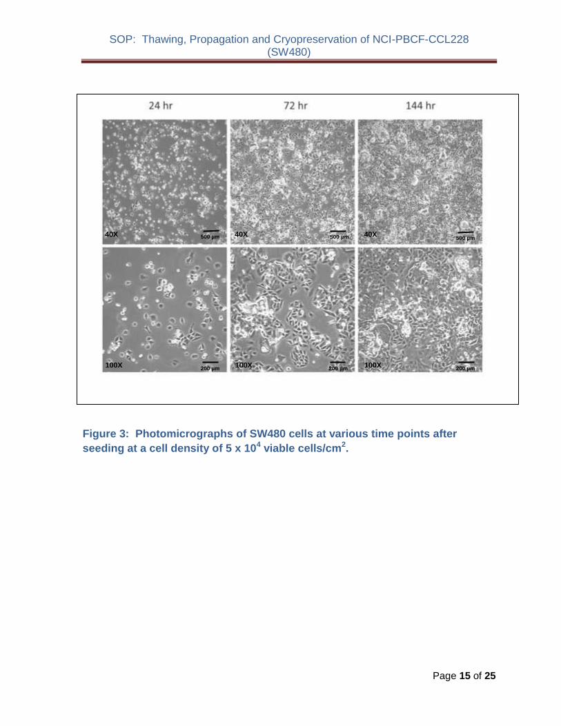
SOP Thawing Propagation and Cryopreservation of NCI-PBCF-CCL228 (SW480)
APPENDIX 2 GROWTH PROFILE OF NCI-PBCF-CCL228 (SW480) CELLS
000E+00
100E+05
200E+05
300E+05
400E+05
500E+05
600E+05
700E+05
800E+05
900E+05
100E+06
7 8 9
Via
ble
Cell
sc
m2
0 1 2 3 4 5 6
Days in Culture
Figure 4 Growth curve for SW480 cells cells were plated at 5 x 10⁴ viable cellscm2 population doubling time (PDT) is approximately 38 h
Page 16 of 25
SOP Thawing Propagation and Cryopreservation of NCI-PBCF-CCL228 (SW480)
APPENDIX 3 CYTOGENETIC ANALYSIS OF NCI-PBCF-CCL228 SW480 CELLS
NCI-PBCF-CCL228 Karyotype Results
Metaphase Spread Karyotype
Number of metaphase spreads counted 20
Band level 300-400
Number of metaphase spreads karyotyped 10
Chromosome range 50-56
Sex Male
Comments Aneuploid
Karyotype
50~56XX+2der(2)t(218)(q21q111)del(5)(q13q33)+add(7)(q22)+add(7)(q32)
der(10)t (1012)(p15q11)+add(11)(p15)-12der(1214)(p10q10)-14+add(15)(p10)
der(15)t(1215)(q13q15)-18add(19)(p133)+20+add(20)(p13)
+der(20)t(520)(q22p13)x2+21+add(21)(q111)add(21)(q22)+mar1x2
(ISCN nomenclature written based on a diploid karyotype)
Human diploid karyotype (2N) 46XX (female) or 46XY (male)
Page 17 of 25
SOP Thawing Propagation and Cryopreservation of NCI-PBCF-CCL228 (SW480)
Karyotype Summary
In the karyotype image arrows indicate regions of abnormality It should be noted that the karyotype description includes the observed abnormalities from all examined metaphase spreads but due to heterogeneity not all of the karyotyped cells will contain every abnormality
This is a rearranged human cell line of male origin containing 50 to 56 chromosomes per metaphase spread (hyperdiploid) Structural abnormalities include rearrangements to approximately 50 of the 22 different autosomal chromosomes (described below) There is one unidentifiable clonal marker chromosome (markers present in two or more of the examined cells) [+mar1]
The rearrangements include
Addition of unknown material to the short arms (designated by p) of chromosomes 11 15 19 and 20
Addition of unknown material to the long arms (designated by q) of chromosomes 7 and 21 (2 different derivatives of each)
An interstitial deletion of material from the long arm of chromosome 5 between bands q13 and q33
Translocations involving chromosomes 2 and 18 [der(2)t(218)(q21q111)] 10 and 12 [der(10)t (1012)(p15q11) chromosomes 12 and 14 [der(1214)(p10q10)] 12 and 15 [der(15)t(1215)(q13q15)] and chromosomes 5 and 20 [der(20)t(520)(q22p13)]
Numerical changes are based on a diploid karyotype which would contain two copies of
each chromosome (2N) Therefore karyotype designations such as +2 +20 and +21
indicates three copies of structurally normal chromosomes 2 20 and 21 Designations
-18 and -14 indicates one copy of structurally normal chromosomes -18 and -14 Also it
appears that the Y chromosome was lost which is not an uncommon occurrence in cancer
cell lines (ISCN 2009 An International System for Human Cytogenetic Nomenclature
(2009) Editors Lisa G Shaffer Marilyn L Slovak Lynda J Campbell)
Karyotype Procedure
Cell Harvest Cells were allowed to grow to 80-90 confluence Mitotic division was
arrested by treating the cells with KaryoMaxreg colcemid for 20 minutes to 2 hours at 37degC
Cells were harvested using 005 Trypsin-EDTA treated with 0075M KCL hypotonic
solution and then fixed in three changes of a 31 ratio of methanolglacial acetic acid
Slide Preparation Slides were prepared by dropping the cell suspension onto wet glass
slides and allowing them to dry under controlled conditions
G-banding Slides were baked one hour at 90degC trypsinized using 10X trypsin-EDTA
and then stained with Leishmanrsquos stain
Page 18 of 25
SOP Thawing Propagation and Cryopreservation of NCI-PBCF-CCL228 (SW480)
Microscopy Slides were scanned using a 10X objective and metaphase spreads were
analyzed using a 100X plan apochromat objective on an Olympus BX-41 microscope
Imaging and karyotyping were performed using Cytovisionreg software
Analysis Twenty metaphase cells were counted and analyzed and representative
metaphase cells were karyotyped depending on the complexity of the study
Summary of Karyotyping Procedure
G-band karyotyping analysis is performed using GTL banding technique G bands produced with trypsin and Leishman Slides prepared with metaphase spreads are treated with trypsin and stained with Leishmanrsquos This method produces a series of light and dark bands that allow for the positive identification of each chromosome
SW480 cell line karyotyping was carried out by Cell Line Genetics Inc (Madison WI 53719)
Page 19 of 25
SOP Thawing Propagation and Cryopreservation of NCI-PBCF-CCL228 (SW480)
APPENDIX 4 GLOSSARY OF TERMS
Confluent monolayer adherent cell culture in which all cells are in contact with other cells
all around their periphery and no available substrate is left uncovered
Split ratio the divisor of the dilution ration of a cell culture to subculture (eg one flask
divided into four or 100 mL up to 400 mL would be split ratio of 14)
Subculture (or passage) the transfer or transplantation of cells with or without dilution
from one culture vessel to another
Passage No the total number of times the cells in the culture have been subcultured or
passaged (with each subculture the passage number increases by 1)
Population doubling level (PDL) the total number of population doublings of a cell line
since its initiation in vitro (with each subculture the population doubling increases in
relationship to the split ratio at which the cells are plated) See Appendix 7
Population doubling time (doubling time) the time interval calculated during the
logarithmic phase of growth in which cells double in number
Seeding density recommended number of cells per cm2 of substrate when inoculating a
new flask
Epithelial-like adherent cells of a polygonal shape with clear sharp boundaries between
them
Fibroblast-like adherent cells of a spindle or stellate shape
Page 20 of 25
SOP Thawing Propagation and Cryopreservation of NCI-PBCF-CCL228 (SW480)
APPENDIX 5 REFERENCE
1 Culture Of Animal Cells A Manual of Basic Technique by R Ian Freshney 6th edition
published by Wiley-Liss NY 2010
2 Fogh J et al Absence of HeLa cell contamination in 169 cell lines derived from human
tumors J Natl Cancer Inst 58 209-214 1977 PubMed 833871
3 Fogh J et al One hundred and twenty-seven cultured human tumor cell lines producing
tumors in nude mice J Natl Cancer Inst 59 221-226 1977 PubMed 327080
4 Lelbovitz A et al Detection and analysis of a glucose 6-phosphate dehydrogenase
phenotype B cell line contamination J Natl Cancer Inst 63 635-645 1979 PubMed
288927
5 Adachi A et al Productive persistent infection of human colorectal cell lines with human
immunodeficiency virus J Virol 61 209-213 1987 PubMed 3640832
6 Schroy PC et al Detection of p21ras mutations in colorectal adenomas and carcinomas
by enzyme-linked immunosorbent assay Cancer 76 201-209 1995 PubMed 8625092
7 Trainer DL et al Biological characterization and oncogene expression in human
colorectal carcinoma cell lines Int J Cancer 41 287-296 1988 PubMed 3338874
8 Weiss J et al Mutation and expression of the p53 gene in malignant melanoma cell
lines Int J Cancer 54 693-699 1993 PubMed 8514460
9 Nigro JM et al Mutations in the p53 gene occur in diverse human tumour types Nature
342 705-707 1989 PubMed 2531845
10 Barnett SW et al Characterization of human immunodeficiency virus type 1 strains
recovered from the bowel of infected individuals Virology 182 802-809 1991 PubMed
2024498
11 Leibovitz A et al Classification of human colorectal adenocarcinoma cell lines Cancer
Res 36 4562-4569 1976 PubMed 1000501
12 Geiser AG et al Suppression of tumorigenicity in human cell hybrids derived from cell
lines expressing different activated ras oncogenes Cancer Res 49 1572-1577 1989
PubMed 2647289
13 Lahm H et al Secretion of bioactive granulocyte-macrophage colony-stimulating factor
by human colorectal carcinoma cells Cancer Res 54 3700-3702 1994 PubMed
8033086
14 Rodrigues NR et al p53 mutations in colorectal cancer Proc Natl Acad Sci USA 87
7555-7559 1990 PubMed 1699228
Page 21 of 25
SOP Thawing Propagation and Cryopreservation of NCI-PBCF-CCL228 (SW480)
15 Santoro IM Groden J Alternative splicing of the APC gene and its association with terminal differentiation Cancer Res 57 488-494 1997 PubMed 9012479
16 Tsao H et al Novel mutations in the p16CDKN2A binding region of the Cyclinshy
dependent Kinase-4 gene Cancer Res 58 109-113 1998 PubMed 9426066
17 Zhu X et al Cell cycle-dependent modulation of telomerase activity in tumor cells Proc Natl Acad Sci USA 93 6091-6095 1996 PubMed 8650224
18 Witty JP et al Modulation of matrilysin levels in colon carcinoma cell lines affects tumorigenicity in vivo Cancer Res 54 4805-4812 1994 PubMed 8062282
Page 22 of 25
SOP Thawing Propagation and Cryopreservation of NCI-PBCF-CCL228 (SW480)
APPENDIX 6 REAGENT LOT TRACEABILITY AND CELL EXPANSION TABLES
Table 4 Reagent Lot Traceability
Reagent Vendor Catalog Lot Expiration Date
Page 23 of 25
Table 5 Cell Expansion
FROM FLUID CHANGE
Observation under
microscope CELL COUNT TO
By Date Flask qty
size
Pass Confluence
Add Replace
Volume
(in mL)
Viable cellsmL
Total viable cells
Viability
Split Ratio
Flask qty
size
Pass
PDL
Add
Replace
Add
Replace
Add
Replace
Add
Replace
Add
Replace
Add
Replace
Add
Replace
Add
Replace
Add
Replace
SOP Thawing Propagation and Cryopreservation of NCI-PBCF-CCL228 (SW480)
APPENDIX 7 CALCULATION OF POPULATION DOUBLING LEVEL (PDL)
Calculate the PDL of the current passage using the following equation
PDL = X + 3322 (log Y ndash log I)
Where X = initial PDL I = cell inoculum (number of cells plated in the flask) Y = final cell yield (number of cells at the end of the growth period)
APPENDIX 8 SAFETY PRECAUTIONS
Use at least approved Biological Safety Level 2 (BSL-2) facilities and procedures
Wear appropriate Personal Protective Equipment (PPE) such as isolation gown lab coat with sleeve protectors face shield and gloves
Use safety precautions when working with liquid nitrogen nitrogen vapor and cryogenically cooled fixtures
Use liquid nitrogen freezers and liquid nitrogen tanks only in areas with adequate ventilation Liquid nitrogen reduces the concentration of oxygen and can cause suffocation
Wear latex gloves over insulating gloves to prevent liquid nitrogen from soaking in and being held next to the skin Liquid nitrogen is extremely cold and will cause burns and frostbite Metal inventory racks tank components and liquid nitrogen transfer hoses exposed to liquid nitrogen or nitrogen vapor quickly cool to cryogenic temperatures and can cause burns and frostbite
Wear a full face mask when thawing and retrieving vials from liquid nitrogen freezer Danger to the technician derives mainly from the possibility that liquid nitrogen can penetrate the cryovial during storage On warming rapid evaporation of the nitrogen within the confines of such cryovial can cause an aerosol or explosion of the cryovial and contents
Page 25 of 25
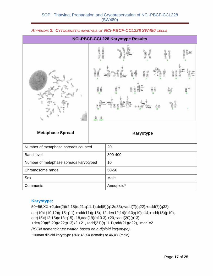
SOP Thawing Propagation and Cryopreservation of NCI-PBCF-CCL228 (SW480)
APPENDIX 4 GLOSSARY OF TERMS
Confluent monolayer adherent cell culture in which all cells are in contact with other cells
all around their periphery and no available substrate is left uncovered
Split ratio the divisor of the dilution ration of a cell culture to subculture (eg one flask
divided into four or 100 mL up to 400 mL would be split ratio of 14)
Subculture (or passage) the transfer or transplantation of cells with or without dilution
from one culture vessel to another
Passage No the total number of times the cells in the culture have been subcultured or
passaged (with each subculture the passage number increases by 1)
Population doubling level (PDL) the total number of population doublings of a cell line
since its initiation in vitro (with each subculture the population doubling increases in
relationship to the split ratio at which the cells are plated) See Appendix 7
Population doubling time (doubling time) the time interval calculated during the
logarithmic phase of growth in which cells double in number
Seeding density recommended number of cells per cm2 of substrate when inoculating a
new flask
Epithelial-like adherent cells of a polygonal shape with clear sharp boundaries between
them
Fibroblast-like adherent cells of a spindle or stellate shape
Page 20 of 25
SOP Thawing Propagation and Cryopreservation of NCI-PBCF-CCL228 (SW480)
APPENDIX 5 REFERENCE
1 Culture Of Animal Cells A Manual of Basic Technique by R Ian Freshney 6th edition
published by Wiley-Liss NY 2010
2 Fogh J et al Absence of HeLa cell contamination in 169 cell lines derived from human
tumors J Natl Cancer Inst 58 209-214 1977 PubMed 833871
3 Fogh J et al One hundred and twenty-seven cultured human tumor cell lines producing
tumors in nude mice J Natl Cancer Inst 59 221-226 1977 PubMed 327080
4 Lelbovitz A et al Detection and analysis of a glucose 6-phosphate dehydrogenase
phenotype B cell line contamination J Natl Cancer Inst 63 635-645 1979 PubMed
288927
5 Adachi A et al Productive persistent infection of human colorectal cell lines with human
immunodeficiency virus J Virol 61 209-213 1987 PubMed 3640832
6 Schroy PC et al Detection of p21ras mutations in colorectal adenomas and carcinomas
by enzyme-linked immunosorbent assay Cancer 76 201-209 1995 PubMed 8625092
7 Trainer DL et al Biological characterization and oncogene expression in human
colorectal carcinoma cell lines Int J Cancer 41 287-296 1988 PubMed 3338874
8 Weiss J et al Mutation and expression of the p53 gene in malignant melanoma cell
lines Int J Cancer 54 693-699 1993 PubMed 8514460
9 Nigro JM et al Mutations in the p53 gene occur in diverse human tumour types Nature
342 705-707 1989 PubMed 2531845
10 Barnett SW et al Characterization of human immunodeficiency virus type 1 strains
recovered from the bowel of infected individuals Virology 182 802-809 1991 PubMed
2024498
11 Leibovitz A et al Classification of human colorectal adenocarcinoma cell lines Cancer
Res 36 4562-4569 1976 PubMed 1000501
12 Geiser AG et al Suppression of tumorigenicity in human cell hybrids derived from cell
lines expressing different activated ras oncogenes Cancer Res 49 1572-1577 1989
PubMed 2647289
13 Lahm H et al Secretion of bioactive granulocyte-macrophage colony-stimulating factor
by human colorectal carcinoma cells Cancer Res 54 3700-3702 1994 PubMed
8033086
14 Rodrigues NR et al p53 mutations in colorectal cancer Proc Natl Acad Sci USA 87
7555-7559 1990 PubMed 1699228
Page 21 of 25
SOP Thawing Propagation and Cryopreservation of NCI-PBCF-CCL228 (SW480)
15 Santoro IM Groden J Alternative splicing of the APC gene and its association with terminal differentiation Cancer Res 57 488-494 1997 PubMed 9012479
16 Tsao H et al Novel mutations in the p16CDKN2A binding region of the Cyclinshy
dependent Kinase-4 gene Cancer Res 58 109-113 1998 PubMed 9426066
17 Zhu X et al Cell cycle-dependent modulation of telomerase activity in tumor cells Proc Natl Acad Sci USA 93 6091-6095 1996 PubMed 8650224
18 Witty JP et al Modulation of matrilysin levels in colon carcinoma cell lines affects tumorigenicity in vivo Cancer Res 54 4805-4812 1994 PubMed 8062282
Page 22 of 25
SOP Thawing Propagation and Cryopreservation of NCI-PBCF-CCL228 (SW480)
APPENDIX 6 REAGENT LOT TRACEABILITY AND CELL EXPANSION TABLES
Table 4 Reagent Lot Traceability
Reagent Vendor Catalog Lot Expiration Date
Page 23 of 25
Table 5 Cell Expansion
FROM FLUID CHANGE
Observation under
microscope CELL COUNT TO
By Date Flask qty
size
Pass Confluence
Add Replace
Volume
(in mL)
Viable cellsmL
Total viable cells
Viability
Split Ratio
Flask qty
size
Pass
PDL
Add
Replace
Add
Replace
Add
Replace
Add
Replace
Add
Replace
Add
Replace
Add
Replace
Add
Replace
Add
Replace
SOP Thawing Propagation and Cryopreservation of NCI-PBCF-CCL228 (SW480)
APPENDIX 7 CALCULATION OF POPULATION DOUBLING LEVEL (PDL)
Calculate the PDL of the current passage using the following equation
PDL = X + 3322 (log Y ndash log I)
Where X = initial PDL I = cell inoculum (number of cells plated in the flask) Y = final cell yield (number of cells at the end of the growth period)
APPENDIX 8 SAFETY PRECAUTIONS
Use at least approved Biological Safety Level 2 (BSL-2) facilities and procedures
Wear appropriate Personal Protective Equipment (PPE) such as isolation gown lab coat with sleeve protectors face shield and gloves
Use safety precautions when working with liquid nitrogen nitrogen vapor and cryogenically cooled fixtures
Use liquid nitrogen freezers and liquid nitrogen tanks only in areas with adequate ventilation Liquid nitrogen reduces the concentration of oxygen and can cause suffocation
Wear latex gloves over insulating gloves to prevent liquid nitrogen from soaking in and being held next to the skin Liquid nitrogen is extremely cold and will cause burns and frostbite Metal inventory racks tank components and liquid nitrogen transfer hoses exposed to liquid nitrogen or nitrogen vapor quickly cool to cryogenic temperatures and can cause burns and frostbite
Wear a full face mask when thawing and retrieving vials from liquid nitrogen freezer Danger to the technician derives mainly from the possibility that liquid nitrogen can penetrate the cryovial during storage On warming rapid evaporation of the nitrogen within the confines of such cryovial can cause an aerosol or explosion of the cryovial and contents
Page 25 of 25
SOP Thawing Propagation and Cryopreservation of NCI-PBCF-CCL228 (SW480)
APPENDIX 5 REFERENCE
1 Culture Of Animal Cells A Manual of Basic Technique by R Ian Freshney 6th edition
published by Wiley-Liss NY 2010
2 Fogh J et al Absence of HeLa cell contamination in 169 cell lines derived from human
tumors J Natl Cancer Inst 58 209-214 1977 PubMed 833871
3 Fogh J et al One hundred and twenty-seven cultured human tumor cell lines producing
tumors in nude mice J Natl Cancer Inst 59 221-226 1977 PubMed 327080
4 Lelbovitz A et al Detection and analysis of a glucose 6-phosphate dehydrogenase
phenotype B cell line contamination J Natl Cancer Inst 63 635-645 1979 PubMed
288927
5 Adachi A et al Productive persistent infection of human colorectal cell lines with human
immunodeficiency virus J Virol 61 209-213 1987 PubMed 3640832
6 Schroy PC et al Detection of p21ras mutations in colorectal adenomas and carcinomas
by enzyme-linked immunosorbent assay Cancer 76 201-209 1995 PubMed 8625092
7 Trainer DL et al Biological characterization and oncogene expression in human
colorectal carcinoma cell lines Int J Cancer 41 287-296 1988 PubMed 3338874
8 Weiss J et al Mutation and expression of the p53 gene in malignant melanoma cell
lines Int J Cancer 54 693-699 1993 PubMed 8514460
9 Nigro JM et al Mutations in the p53 gene occur in diverse human tumour types Nature
342 705-707 1989 PubMed 2531845
10 Barnett SW et al Characterization of human immunodeficiency virus type 1 strains
recovered from the bowel of infected individuals Virology 182 802-809 1991 PubMed
2024498
11 Leibovitz A et al Classification of human colorectal adenocarcinoma cell lines Cancer
Res 36 4562-4569 1976 PubMed 1000501
12 Geiser AG et al Suppression of tumorigenicity in human cell hybrids derived from cell
lines expressing different activated ras oncogenes Cancer Res 49 1572-1577 1989
PubMed 2647289
13 Lahm H et al Secretion of bioactive granulocyte-macrophage colony-stimulating factor
by human colorectal carcinoma cells Cancer Res 54 3700-3702 1994 PubMed
8033086
14 Rodrigues NR et al p53 mutations in colorectal cancer Proc Natl Acad Sci USA 87
7555-7559 1990 PubMed 1699228
Page 21 of 25
SOP Thawing Propagation and Cryopreservation of NCI-PBCF-CCL228 (SW480)
15 Santoro IM Groden J Alternative splicing of the APC gene and its association with terminal differentiation Cancer Res 57 488-494 1997 PubMed 9012479
16 Tsao H et al Novel mutations in the p16CDKN2A binding region of the Cyclinshy
dependent Kinase-4 gene Cancer Res 58 109-113 1998 PubMed 9426066
17 Zhu X et al Cell cycle-dependent modulation of telomerase activity in tumor cells Proc Natl Acad Sci USA 93 6091-6095 1996 PubMed 8650224
18 Witty JP et al Modulation of matrilysin levels in colon carcinoma cell lines affects tumorigenicity in vivo Cancer Res 54 4805-4812 1994 PubMed 8062282
Page 22 of 25
SOP Thawing Propagation and Cryopreservation of NCI-PBCF-CCL228 (SW480)
APPENDIX 6 REAGENT LOT TRACEABILITY AND CELL EXPANSION TABLES
Table 4 Reagent Lot Traceability
Reagent Vendor Catalog Lot Expiration Date
Page 23 of 25
Table 5 Cell Expansion
FROM FLUID CHANGE
Observation under
microscope CELL COUNT TO
By Date Flask qty
size
Pass Confluence
Add Replace
Volume
(in mL)
Viable cellsmL
Total viable cells
Viability
Split Ratio
Flask qty
size
Pass
PDL
Add
Replace
Add
Replace
Add
Replace
Add
Replace
Add
Replace
Add
Replace
Add
Replace
Add
Replace
Add
Replace
SOP Thawing Propagation and Cryopreservation of NCI-PBCF-CCL228 (SW480)
APPENDIX 7 CALCULATION OF POPULATION DOUBLING LEVEL (PDL)
Calculate the PDL of the current passage using the following equation
PDL = X + 3322 (log Y ndash log I)
Where X = initial PDL I = cell inoculum (number of cells plated in the flask) Y = final cell yield (number of cells at the end of the growth period)
APPENDIX 8 SAFETY PRECAUTIONS
Use at least approved Biological Safety Level 2 (BSL-2) facilities and procedures
Wear appropriate Personal Protective Equipment (PPE) such as isolation gown lab coat with sleeve protectors face shield and gloves
Use safety precautions when working with liquid nitrogen nitrogen vapor and cryogenically cooled fixtures
Use liquid nitrogen freezers and liquid nitrogen tanks only in areas with adequate ventilation Liquid nitrogen reduces the concentration of oxygen and can cause suffocation
Wear latex gloves over insulating gloves to prevent liquid nitrogen from soaking in and being held next to the skin Liquid nitrogen is extremely cold and will cause burns and frostbite Metal inventory racks tank components and liquid nitrogen transfer hoses exposed to liquid nitrogen or nitrogen vapor quickly cool to cryogenic temperatures and can cause burns and frostbite
Wear a full face mask when thawing and retrieving vials from liquid nitrogen freezer Danger to the technician derives mainly from the possibility that liquid nitrogen can penetrate the cryovial during storage On warming rapid evaporation of the nitrogen within the confines of such cryovial can cause an aerosol or explosion of the cryovial and contents
Page 25 of 25

SOP Thawing Propagation and Cryopreservation of NCI-PBCF-CCL228 (SW480)
15 Santoro IM Groden J Alternative splicing of the APC gene and its association with terminal differentiation Cancer Res 57 488-494 1997 PubMed 9012479
16 Tsao H et al Novel mutations in the p16CDKN2A binding region of the Cyclinshy
dependent Kinase-4 gene Cancer Res 58 109-113 1998 PubMed 9426066
17 Zhu X et al Cell cycle-dependent modulation of telomerase activity in tumor cells Proc Natl Acad Sci USA 93 6091-6095 1996 PubMed 8650224
18 Witty JP et al Modulation of matrilysin levels in colon carcinoma cell lines affects tumorigenicity in vivo Cancer Res 54 4805-4812 1994 PubMed 8062282
Page 22 of 25
SOP Thawing Propagation and Cryopreservation of NCI-PBCF-CCL228 (SW480)
APPENDIX 6 REAGENT LOT TRACEABILITY AND CELL EXPANSION TABLES
Table 4 Reagent Lot Traceability
Reagent Vendor Catalog Lot Expiration Date
Page 23 of 25
Table 5 Cell Expansion
FROM FLUID CHANGE
Observation under
microscope CELL COUNT TO
By Date Flask qty
size
Pass Confluence
Add Replace
Volume
(in mL)
Viable cellsmL
Total viable cells
Viability
Split Ratio
Flask qty
size
Pass
PDL
Add
Replace
Add
Replace
Add
Replace
Add
Replace
Add
Replace
Add
Replace
Add
Replace
Add
Replace
Add
Replace
SOP Thawing Propagation and Cryopreservation of NCI-PBCF-CCL228 (SW480)
APPENDIX 7 CALCULATION OF POPULATION DOUBLING LEVEL (PDL)
Calculate the PDL of the current passage using the following equation
PDL = X + 3322 (log Y ndash log I)
Where X = initial PDL I = cell inoculum (number of cells plated in the flask) Y = final cell yield (number of cells at the end of the growth period)
APPENDIX 8 SAFETY PRECAUTIONS
Use at least approved Biological Safety Level 2 (BSL-2) facilities and procedures
Wear appropriate Personal Protective Equipment (PPE) such as isolation gown lab coat with sleeve protectors face shield and gloves
Use safety precautions when working with liquid nitrogen nitrogen vapor and cryogenically cooled fixtures
Use liquid nitrogen freezers and liquid nitrogen tanks only in areas with adequate ventilation Liquid nitrogen reduces the concentration of oxygen and can cause suffocation
Wear latex gloves over insulating gloves to prevent liquid nitrogen from soaking in and being held next to the skin Liquid nitrogen is extremely cold and will cause burns and frostbite Metal inventory racks tank components and liquid nitrogen transfer hoses exposed to liquid nitrogen or nitrogen vapor quickly cool to cryogenic temperatures and can cause burns and frostbite
Wear a full face mask when thawing and retrieving vials from liquid nitrogen freezer Danger to the technician derives mainly from the possibility that liquid nitrogen can penetrate the cryovial during storage On warming rapid evaporation of the nitrogen within the confines of such cryovial can cause an aerosol or explosion of the cryovial and contents
Page 25 of 25
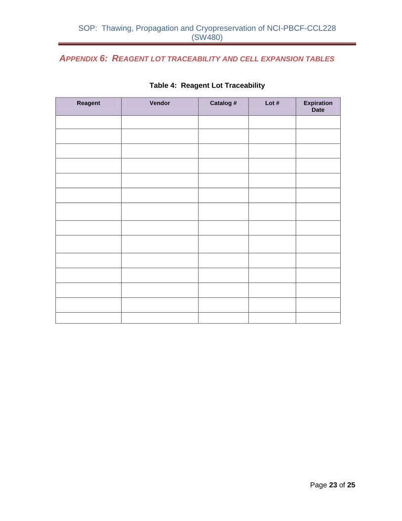
SOP Thawing Propagation and Cryopreservation of NCI-PBCF-CCL228 (SW480)
APPENDIX 6 REAGENT LOT TRACEABILITY AND CELL EXPANSION TABLES
Table 4 Reagent Lot Traceability
Reagent Vendor Catalog Lot Expiration Date
Page 23 of 25
Table 5 Cell Expansion
FROM FLUID CHANGE
Observation under
microscope CELL COUNT TO
By Date Flask qty
size
Pass Confluence
Add Replace
Volume
(in mL)
Viable cellsmL
Total viable cells
Viability
Split Ratio
Flask qty
size
Pass
PDL
Add
Replace
Add
Replace
Add
Replace
Add
Replace
Add
Replace
Add
Replace
Add
Replace
Add
Replace
Add
Replace
SOP Thawing Propagation and Cryopreservation of NCI-PBCF-CCL228 (SW480)
APPENDIX 7 CALCULATION OF POPULATION DOUBLING LEVEL (PDL)
Calculate the PDL of the current passage using the following equation
PDL = X + 3322 (log Y ndash log I)
Where X = initial PDL I = cell inoculum (number of cells plated in the flask) Y = final cell yield (number of cells at the end of the growth period)
APPENDIX 8 SAFETY PRECAUTIONS
Use at least approved Biological Safety Level 2 (BSL-2) facilities and procedures
Wear appropriate Personal Protective Equipment (PPE) such as isolation gown lab coat with sleeve protectors face shield and gloves
Use safety precautions when working with liquid nitrogen nitrogen vapor and cryogenically cooled fixtures
Use liquid nitrogen freezers and liquid nitrogen tanks only in areas with adequate ventilation Liquid nitrogen reduces the concentration of oxygen and can cause suffocation
Wear latex gloves over insulating gloves to prevent liquid nitrogen from soaking in and being held next to the skin Liquid nitrogen is extremely cold and will cause burns and frostbite Metal inventory racks tank components and liquid nitrogen transfer hoses exposed to liquid nitrogen or nitrogen vapor quickly cool to cryogenic temperatures and can cause burns and frostbite
Wear a full face mask when thawing and retrieving vials from liquid nitrogen freezer Danger to the technician derives mainly from the possibility that liquid nitrogen can penetrate the cryovial during storage On warming rapid evaporation of the nitrogen within the confines of such cryovial can cause an aerosol or explosion of the cryovial and contents
Page 25 of 25
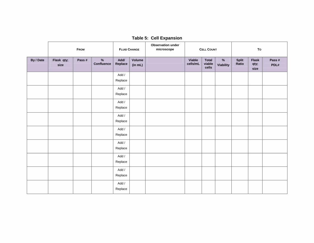