neonatal hypoxic ischemic brain injury management in the first hours dongli song, md, phd santa...
Post on 21-Dec-2015
213 views
TRANSCRIPT
Neonatal Hypoxic Ischemic Brain Injury
Management in the First Hours
Dongli Song, MD, PhD
Santa Clara Valley Medical Center
Neonatology

History
• Baby girl F, born to a 35 year old G5P4 mother with good antenatal care. This pregnancy complicated by GDM, diet control.
• Blood group O positive; Hep B neg: HIV neg: RPR neg: Rubella immune: GBS negative
• Mom admitted at 37+2 weeks with active vaginal bleeding. US showed placenta abruption.

Pediatric team get called to the DR stat
• Infant was delivered via stat c/s. • At delivery, she was floppy with no respiratory effort and
no heart rate.
• Bag and mask ventilation started immediately, HR > 100 bpm at 3 min, and some respiration effort noted at 5 min. She was intubated at 7 min for poor sustained respiration. Color improved but remained floppy at 10
min. Apgar score 0@1 min; 4@ 5 min; 5@ 10 min.
What should you ask OB/L&D staff in the DR?

Get cord blood gas
Answer: ask OB/L&D staff to send cord blood gas.
Cord arterial gas: pH 6.8, PCO2 103, Bicarb 15 and BD19.7
• Cord blood gas provides critical information regarding the severity and/or duration of hypoxic ischemic insults prior to delivery.
• Cord arterial gas (from UA) is a part of the criteria for hypothermia treatment.
• If cord blood gas is not available, get infant ABG within first hour of life.
Physical examination
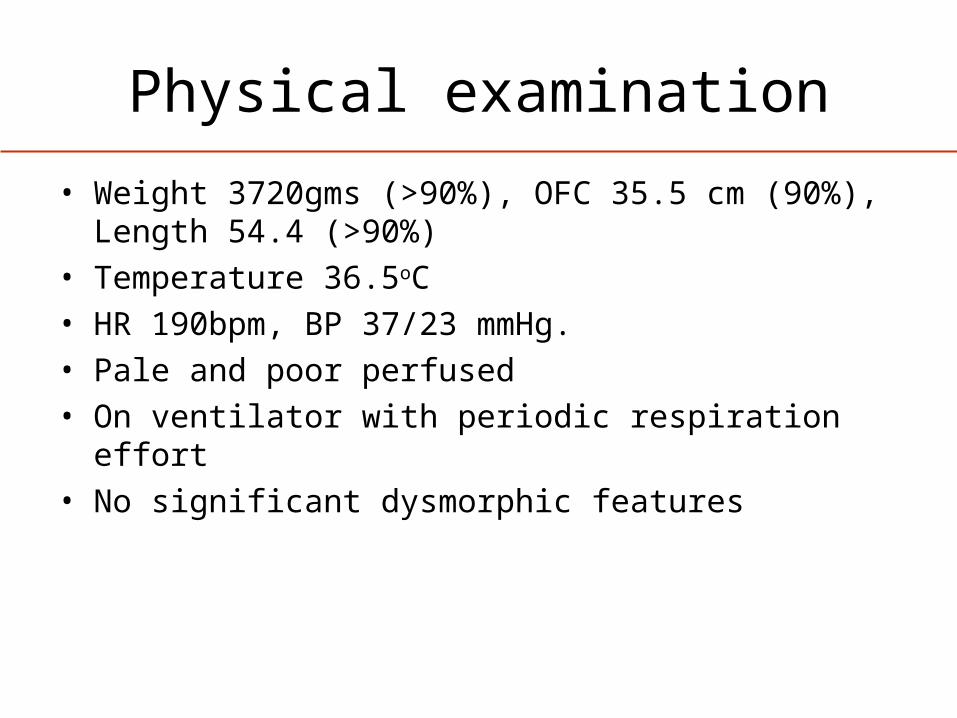
• Weight 3720gms (>90%), OFC 35.5 cm (90%), Length 54.4 (>90%)
• Temperature 36.5oC • HR 190bpm, BP 37/23 mmHg. • Pale and poor perfused• On ventilator with periodic respiration effort• No significant dysmorphic features
Neurological examination
• Does this infant display encephalopathy?
• How could the neurological examination have been done/documented to show this?

Neurological examination
A systemic detailed neuro exam were performed and documented:
• Level of Consciousness: poor eye opening to stimulation, no sustained alertness
• Movements and Tone: minimal spontaneous activity, hypotonia
• Brainstem/Autonomic Functions: pupils constricted but reactive, no suck, no gag
• Reflexes: incomplete Moro, no DTR

NICHD Exam Criteria for HypothermiaModerate
EncephalopathySevere
Encephalopathy
1. Consciousness Lethargic Stupor/coma
2. Activity Decreased No Activity
3. Posture Distal Flexion Decerebrate
4. Tone Hypotonia (focal or general)
Flaccid
5. Primitive ReflexesSuckMoro
WeakIncomplete
AbsentAbsent
6. AutonomicPupilsHeart RateRespirations
ConstrictedBradycardia
Periodic
Fixed; UnequalVariable HR
Apnea
Lab tests
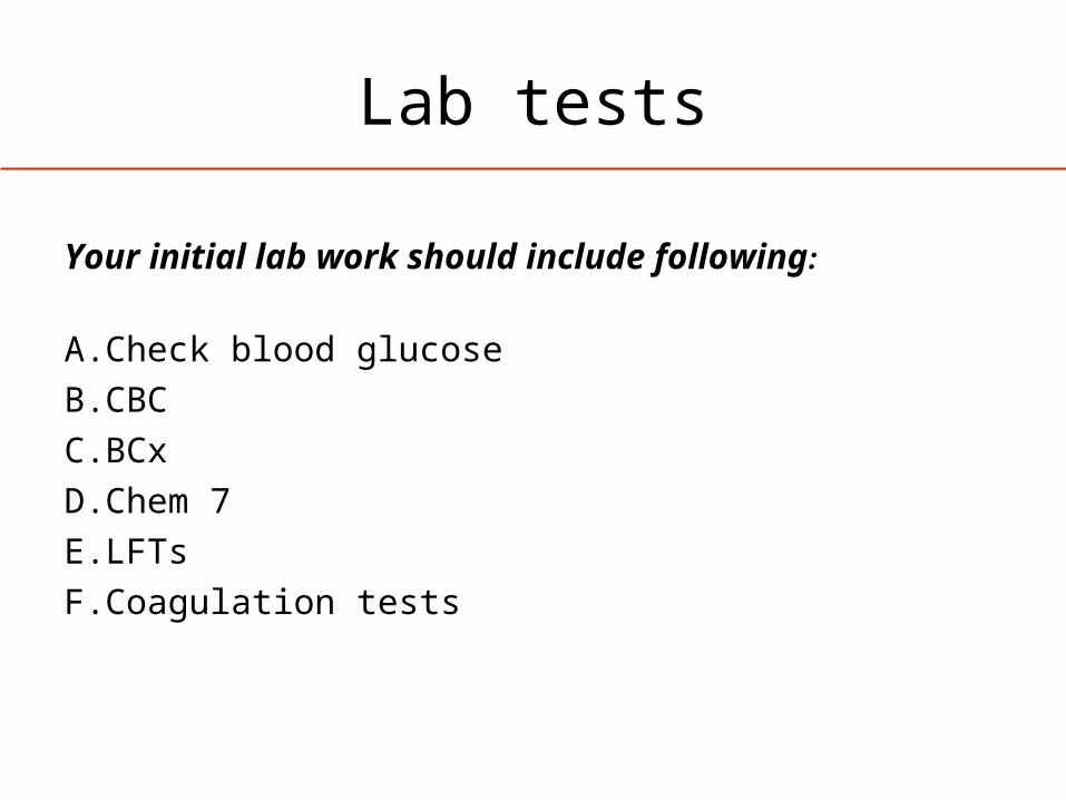
Your initial lab work should include following:
A. Check blood glucose
B. CBC
C.BCx
D.Chem 7
E. LFTs
F. Coagulation tests

Lab tests
Answer: All above.
• Correcting hypoglycemia is critical for brain protection.• Mom had placenta abruption, HCT and platelet count will
help to determine if blood product transfusion is indicated.
• Increase in creatinine indicates kidney injury, and elevation of LFTs and coagulopathy indicates liver damage.

Lab results
• Cord arterial gas: pH 6.8, PCO2 103, Bicarb 15 and BD 19.7.
• Blood glucose 15• CBC: WBC 17.7k, HCT 30%, platelet count
141K• Creatinine 1.3• AST 945, ALT 220 • PT/PTT/INR significant prolonged

Antepartum Risk Factors
• Socioeconomic Status
• Family History • Infertility treatment• Maternal thyroid
disease
• Severe pre-eclampsia• Bleeding in pregnancy• Viral illness• Abnormal placenta• IUGR• Postmaturity

Intrapartum Risk Factors
• Operative vaginal delivery or emergency C-section
• Maternal fever• Occipito-posterior presentation• Acute intrapartum events: cord prolapse,
abruptio placentae…

Heterogeneous cause
Badawi N. et al. BMJ.1998
Hypothermia treatment
One hour later, fluid boluses were given, hypoglycemia was corrected and FFP transfusion was started. Infant started to have spontaneous respiration effort and movements and her tone improved.
Your next treatment plan include:
A. Start hypothermia treatment ASAP
B. Obtain brain imagine to confirm hypoxic-ischemic brain injury before start hypothermia treatment
C. Continue monitoring. Hypothermia will not be indicated if infant’s condition significantly improved at 6hr of life.

Hypothermia treatment
Answer: A and B
Diagnosis of Neonatal Encephalopathy is Clinical
• Careful history and neurological exam• Laboratory studies to exclude “mimics” of hypoxia-
ischemia– Metabolic abnormalities
• including inborn errors of metabolism– Infection– Acute bilirubin encephalopathy– Stroke

Diagnosis – Neuro imaging
HUS - may detect basal ganglia and thalamic injury, not sensitive to cortical injury. Most useful in detecting and following intracranial bleeding.
CT - can detect diffuse cortical neuronal injury, most useful to r/o intracranial hemorrhage that requiring immediate surgical intervention. Concerns for radiation.
MRI - is the study choice of assessing HI brain injury. It provides specific information regarding the injury pattern, severity and evolution.
Neuro imaging is not an absolute requirement for initiating hypothermia treatment for HIE.

Copyright ©Radiological Society of North America, 2006
Chao, C. P. et al. Radiographics 2006;26:S159-S172
Cortical InjuryCortical Injury
Basal Ganglia Injury
Parent’s questions
You talked to infant’s father and explained to him that the his baby is critically ill and may have suffered serious brain injury.
He asked:• What causes her brain injury?• Is my baby going to die? • If she survived, will she be normal?• What can you do to save my baby?
Significance
• Incidence of HIE: 1-2/1000 live births
*California: 4.5/1000 live births• HIE is a major cause of infant mortality and
morbidity with significant long term neurological deficits:
• 15 - 20% die in infancy and 20 -25% survived with some neurological abnormalities including cerebral palsy, cortical visual impairment, seizures, developmental delay and mental retardation.
Hypothermia treatment
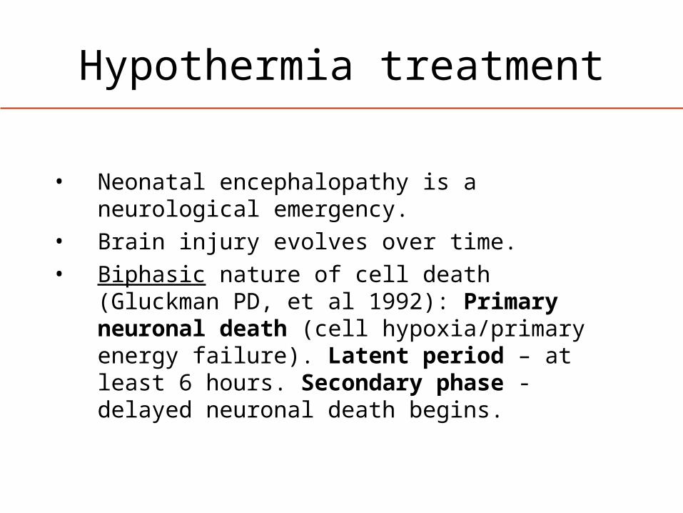
• Neonatal encephalopathy is a neurological emergency.
• Brain injury evolves over time.• Biphasic nature of cell death (Gluckman PD, et al
1992): Primary neuronal death (cell hypoxia/primary energy failure). Latent period – at least 6 hours. Secondary phase - delayed neuronal death begins.

Mechanisms of ischemic brain injury
Delayed neuronal death
Hypoxia-ischemia
Primaryneuronal death
Cytotoxic mechanisms
1 hour
6 hours Days
Modified from Gunn and Thoresen, 2006
HypothermiaHypothermia

Mechanisms of ischemic brain injury
Ferriero D. NEJM 2004
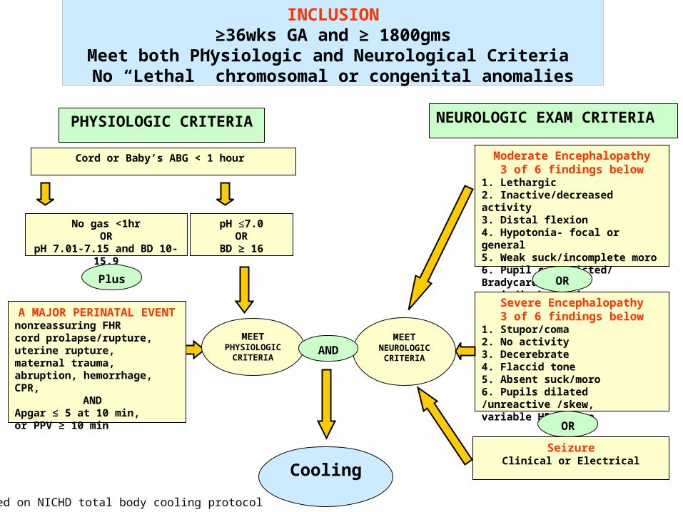
INCLUSION≥36wks GA and ≥ 1800gms
Meet both Physiologic and Neurological Criteria No “Lethal” chromosomal or congenital anomalies
PHYSIOLOGIC CRITERIA NEUROLOGIC EXAM CRITERIA
Cord or Baby’s ABG < 1 hour
No gas <1hrOR
pH 7.01-7.15 and BD 10-15.9
Moderate Encephalopathy3 of 6 findings below
1. Lethargic2. Inactive/decreased activity3. Distal flexion4. Hypotonia- focal or general5. Weak suck/incomplete moro6. Pupil constricted/ Bradycardia /periodic breathing
pH ≤7.0OR
BD ≥ 16
SeizureClinical or Electrical
OR
Severe Encephalopathy3 of 6 findings below
1. Stupor/coma2. No activity3. Decerebrate4. Flaccid tone5. Absent suck/moro6. Pupils dilated /unreactive /skew, variable HR, apnea
OR
MEETPHYSIOLOGIC CRITERIA
MEETNEUROLOGIC
CRITERIAAND
Plus
Cooling
A MAJOR PERINATAL EVENTnonreassuring FHR cord prolapse/rupture,uterine rupture, maternal trauma, abruption, hemorrhage, CPR, ANDApgar ≤ 5 at 10 min, or PPV ≥ 10 min
Based on NICHD total body cooling protocol
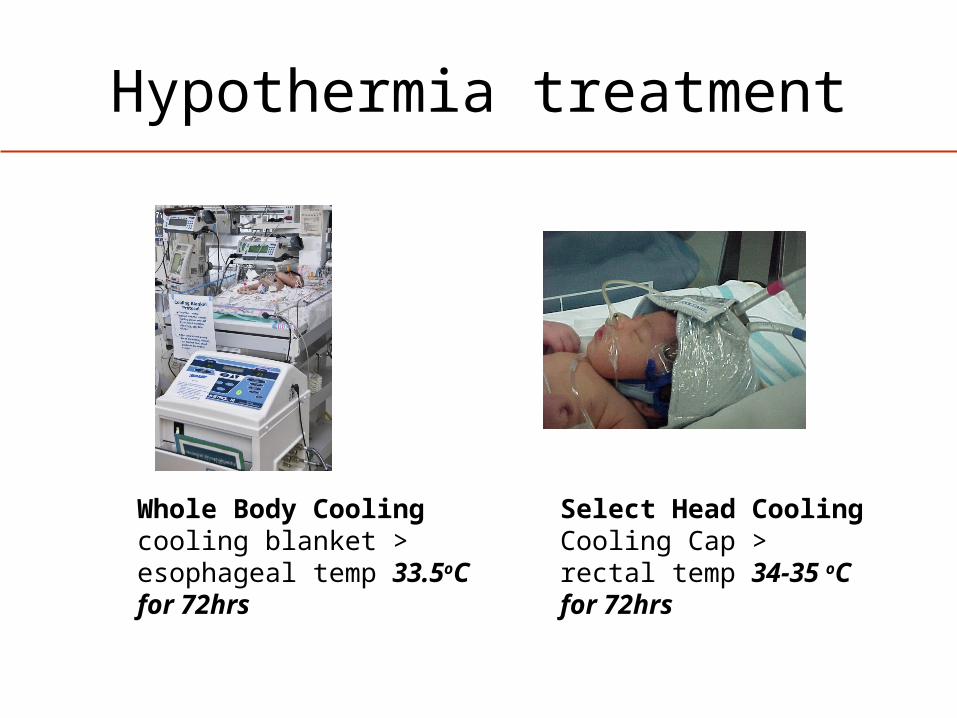
Hypothermia treatment
Whole Body Coolingcooling blanket > esophageal temp 33.5oC for 72hrs
Select Head Cooling Cooling Cap >rectal temp 34-35 oC for 72hrs
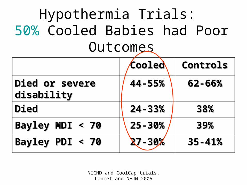
NICHD and CoolCap trials, Lancet and NEJM 2005
Hypothermia Trials: 50% Cooled Babies had Poor
OutcomesCooledCooled ControlsControls
Died or severe Died or severe disabilitydisability
44-55%44-55% 62-66%62-66%
DiedDied 24-33%24-33% 38%38%
Bayley MDI < 70Bayley MDI < 70 25-30%25-30% 39%39%
Bayley PDI < 70Bayley PDI < 70 27-30%27-30% 35-41%35-41%
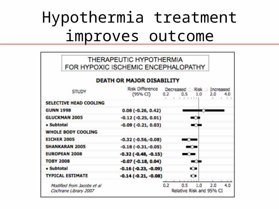
Hypothermia treatment improves outcome
Hypothermia treatment Potential adverse effects
-Hypotension-Cardiac arrhythmia (mainly sinus bradycardia ) -Persistent acidosis-Increased oxygen consumption-Increased blood viscosity-Reduction in platelet count-Pulmonary hemorrhage-Sepsis-Necrotizing enterocolitis
-no severe side effects have been reported so far
Best patient care depends on
• Close communication with family
• Multidisciplinary care
• Neurology– neurological examination (structured /routine), diagnosis, prognosis, follow up
• Radiology – timing and interpretation
• Physical and occupational therapy – evaluation, pre-discharge examination

“The world
belongs to the
enthusiast who
keeps cool”
Thank you!