neonatal inclusion conjunctivitis in australia - bjo.bmj.com · brit. j. ophthal. (i969) 53, 670...
TRANSCRIPT
Brit. J. Ophthal. (I969) 53, 670
Neonatal inclusion conjunctivitis inAustralia
D. HANSMAN
Tlhe Women's Hospital, Sydney, Australia
Trachoma has been known to occur in Australia since the latter part of the 9gth century.Laboratory confirmation was first made by Rodger and Priestley (19I5), who studied casesof trachoma in Western Queensland and found characteristic inclusions within conjunc-tival epithelial cells. This work was reported 8 years after the original description of cellinclusions in trachoma by Halberstaedter and von Prowazek (I 907). Although previouslycommon, the infection has now largely disappeared from the white population, but a largereservoir of infection continues to exist among the Aborigines, as shown by investigationsconducted in Western Australia (Mann, 955 i960), the Northern Territory (Flynn,1957), and South Australia (Moore, Howarth, Wilson, Derrington, and Surman, I965;Hardy, Surman, and Howarth, I967).The trachoma agent was first isolated by T'ang, Chang, Huang, and Wang (1957) in
Peking. These workers used embryonated eggs, inoculated by the yolk sac route and thenincubated at 350C. This work was confirmed by Collier and Sowa (I959) in London.Subsequently, isolations were made from patients with trachoma in Australia by Perretand Mann (I960) in Perth, and by Howarth and his colleagues in Adelaide (Howarth,I966). These workers found a high incidence of infection among Aboriginal children insome districts of Western Australia and South Australia.
Inclusion conjunctivitis is due to a micro-organism which by laboratory methods isindistinguishable from that which causes trachoma, and identical inclusions are producedwithin conjunctival epithelium. The causative agents of these infections have beengrouped together as TRIC agent. Inclusion conjunctivitis occurs in newborn infants andthe infection is usually derived from the cervix of the mother during birth. Sometimeschildren and adults are infected, usually from contaminated water in swimming pools.
Neonatal inclusion conjunctivitis has not been reported from Australia, despite attemptsto demonstrate this infection. However, recent evidence obtained at this hospital suggeststhat inclusion conjunctivitis does occur. During a study of conjunctivitis in newbornbabies, cases of severe purulent ophthalmia were seen, from which cultures for bacteriayielded negative results, but further investigations revealed the presence of typical inclu-sions within conjunctival epithelial cells (Hansman, 1969). This article describes furthercases; in most of these infants it has been possible to follow the course of the infection.
Material and methods
Specimens ofconjunctival exudate were collected by heat-sterilized cotton-wool swabs and inoculatedon plates of blood agar and heated blood agar. In most cases specimens were plated immediately
Received for publication March 24, I969Address for reprints: The Women's Hospital, Crown Street, Sydney, N.S.W. 2010, Australia
on 28 March 2019 by guest. P
rotected by copyright.http://bjo.bm
j.com/
Br J O
phthalmol: first published as 10.1136/bjo.53.10.670 on 1 O
ctober 1969. Dow
nloaded from

Neonatal inclusion conjunctivitis in Australia 671
after collection. Cultures were incubated in a jar with added "Carbogen" for at least 2 days. Thecriterion for the diagnosis of bacterial conjunctivitis was significant growth of a potential pathogen.
Conjunctival smears were collected with a stainless steel ophthalmic spud and films prepared onglass slides which had been previously cleaned in a mixture of potassium dichromate and sulphuricacid. After air-drying, the smears were fixed with methanol for 3 minutes and then stained over-night with dilute Giemsa. After staining, the smears were washed in running tap water, dried in anincubator, and mounted before examination. As evidence of inclusion conjunctivitis, conjunctivalsmears were examined for the presence of basophilic, "initial body" inclusions, and Halberstaedter-Prowazek (HP) inclusions.When possible, cervical smears were collected from the mother of the affected infant and these
were fixed and stained by the method described.
Results
From February to May, I968, a period of 4 months, nine cases of severe purulent con-junctivitis were observed, in which bacterial pathogens could not be implicated. Thefindings are summarized in the Table. The conjunctivitis was unilateral in four cases andaffected both eyes in five. In three infants both eyes were involved simultaneously, but intwo (Cases 3 and 7) the conjunctivitis was at first unilateral with intervals of one and 8days, respectively, before the contralateral eye was affected.
Table Summary of nine cases
Case Age at onset Distribution Treatment before Giemsa-stained conjunctivalno.(days) ~~~~~collection of Y
conjunctival smears smear
I 6 Bilateral "Neosporin" HP inclusion*PenicillinChloramphenicol
2 6 Bilateral "Neosporin" Inclusions not seen3 4 Bilateral None HP inclusions4 5 Bilateral "Neosporin" Inclusions not seen
Chloramphenicol5 7 Unilateral "Neosporin" HP inclusions6 2 Unilateral "Neosporin" Inclusions not seen7 6 Bilateral "Neosporin" HP inclusion8 8 (see text) Unilateral "Neosporin" Basophilic inclusions9 6 Unilateral "Neosporin" Basophilic and HP inclusions
Chloramphenicol
*HP = Halberstaedter-Prowazek
Conjunctival smears showed numerous neutrophils with many mononuclear cells whichresembled monocytes and lymphocytes; degenerate leucocytes were often found in largenumbers. HP inclusions were detected in smears from four cases, basophilic inclusions inone, and both types of inclusion in specimens from another infant. In three cases, despitea careful search, inclusions were not seen, so that the cause of the conjunctivitis in thesebabies must remain in doubt. The age at onset in the six inclusion-positive cases wasfrom 4 to 8 days.
Cervical smears were collected from five of the mothers and examined for inclusions, allwith negative results. Examination of conjunctival smears from the infants of thesemothers had revealed inclusions in two cases.
on 28 March 2019 by guest. P
rotected by copyright.http://bjo.bm
j.com/
Br J O
phthalmol: first published as 10.1136/bjo.53.10.670 on 1 O
ctober 1969. Dow
nloaded from
672 to. Hansman
Representative cases
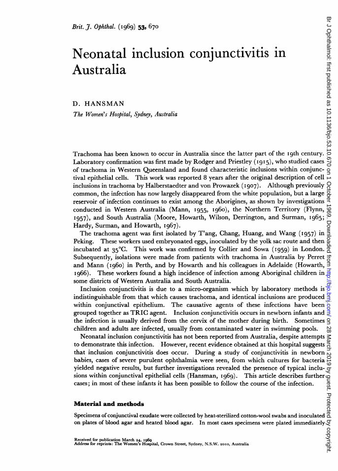
Case IThis infant developed conjunctivitis at the age of 6 days, and the condition was at first treated with"Neosporin" drops. Pathogenic bacteria were not isolated. On the next day the baby was statedto show bilateral severe conjunctivitis with oedema and erythema of the lids. Treatment waschanged to penicillin. After another 2 days, penicillin was stopped and chloramphenicol adminis-tered. At the age of Io days, after treatment with chloramphenicol for one day, there was slightexudate, lid oedema, and chemosis. Conjunctival smears, collected at this stage, showed numerousneutrophils and some mononuclear cells which resembled lymphocytes and monocytes; an epithelialcell containing a typical HP inclusion was detected (Fig. I). The conjunctivitis slowly improved.At the age of 13 days, moderate chemosis and slight purulent exudate were found. The infant wasdischarged from hospital at the age of i8 days. The infant's mother was an unmarried girl of 18years.
4~~~~~~~~~~~~~~~~~~~~~~~~~~~
FIG. I Case I Conjunctival smear stained with Giemsa: epithelial cell containing Halber-staedter-Prowazek inclusion. x 960
Case 7
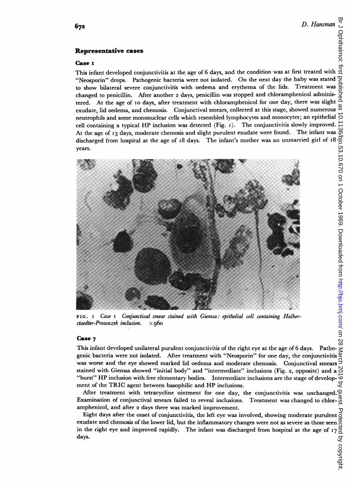
This infant developed unilateral purulent conjunctivitis of the right eye at the age of 6 days. Patho-genic bacteria were not isolated. After treatment with "Neosporin" for one day, the conjunctivitiswas worse and the eye showed marked lid oedema and moderate chemosis. Conjunctival smearsstained with Giemsa showed "initial body" and "intermediate" inclusions (Fig. 2, opposite) and a"burst" HP inclusion with free elementary bodies. Intermediate inclusions are the stage of develop-ment of the TRIC agent between basophilic and HP inclusions.
After treatment with tetracycline ointment for one day, the conjunctivitis was unchanged.Examination of conjunctival smears failed to reveal inclusions. Treatment was changed to chlor-amphenicol, and after 2 days there was marked improvement.
Eight days after the onset of conjunctivitis, the left eye was involved, showing moderate purulentexudate and chemosis of the lower lid, but the inflammatory changes were not as severe as those seenin the right eye and improved rapidly. The infant was discharged from hospital at the age of I7days.
T% "-- --
on 28 March 2019 by guest. P
rotected by copyright.http://bjo.bm
j.com/
Br J O
phthalmol: first published as 10.1136/bjo.53.10.670 on 1 O
ctober 1969. Dow
nloaded from

Neonatal inclusion conjunctivitis in Australia
The infant's mother was an unmarried girl of 17 years; cervical smears collected I3 days afterdelivery were examined for inclusions with a negative result.
I '1''
FIG. 2 Case 7 Conjunctival smear stained with Giemsa: group of epithelial cells, some con-taining initial body and intermediate inclusions, also a "burst" Halberstaedter-Prowazek inclusionwithfree elementary bodies. x 960
Case 8This infant developed mild conjunctivitis on the day of birth; bacteriological cultures yielded nogrowth. The conjunctivitis was treated with "Neosporin" for 8 days, and the infant then showedunilateral purulent conjunctivitis with moderate lid oedema and marked chemosis. Cultures wereagain negative and conjunctival smears stained with Giemsa showed basophilic inclusions. Treat-ment was changed to chloramphenicol drops which produced a marked improvement after one day,although chemosis of the lower lid persisted for at least a week. The infant was discharged fromhospital when aged 17 days.The infant's mother was an unmarried woman aged 25 years. It was considered that the mild
initial conjunctivitis was probably not due to TRIC agent infection, which usually has an incubationperiod of 6 to 8 days. However, Sowa, Sowa, and Collier (I968) reported the case of a neonatewho developed TRIC agent infection at the age of one day.
Response to treatment
During this investigation, all cases of conjunctivitis were initially treated with "Neosporin"drops (which contain a mixture of three antibiotics: neomycin, polymyxin B, and grami-cidin). If the conjunctivitis failed to improve, treatment was changed to chloramphenicolor penicillin drops. In none of the nine cases did the conjunctivitis respond to "Neo-sporin" and in five (including three of the inclusion-positive cases) the degree of inflamma-tion increased during treatment. Six of the infants were subsequently treated withchloramphenicol and one with penicillin; one was treated with penicillin and subsequentlywith chloramphenicol; and another with tetracycline and then chloramphenicol. Allresponded, but improvement was not usually evident until at least 2 days' treatment hadbeen given.
673
on 28 March 2019 by guest. P
rotected by copyright.http://bjo.bm
j.com/
Br J O
phthalmol: first published as 10.1136/bjo.53.10.670 on 1 O
ctober 1969. Dow
nloaded from

D. Hansman
After the acute phase of inflammation had subsided, mild conjunctivitis, manifest byslight exudate from the eye and thickening of the conjunctiva of the lower lid, oftenpersisted. Some cases showed transverse ridging or corrugation of the lower lid con-junctiva, an appearance similar to that described by Allen (i944). In two infantsapparent healing had occurred 7 days after onset of the conjunctivitis, but the othersshowed slower resolution of infection. The mean period in hospital after conjunctivitisbegan was 26 days, and all except one had recovered sufficiently to be discharged fromhospital after 5 weeks. Most of the babies (6/9) were adopted and this tended to increasethe duration of their stay in hospital.
In most cases it was not possible to collect conjunctival smears before treatment wasbegun. Inclusions were found despite previous treatment with "Neosporin" (Cases 5, 7and 8), "Neosporin" and chloramphenicol (Case 9), and "Neosporin", penicillin, andchloramphenicol (Case i).
Discussion
In all cases the babies were born to parents of European descent. Inclusion conjunctivitishas not been observed in Aboriginal babies, only small numbers of whom are born at thehospital where the investigation was carried out. It would appear to be of someimportance to establish whether inclusion conjunctivitis is restricted to Europeans inAustralia. Inclusion conjunctivitis is said to be unknown among American Indians,while trachoma is common among Indians living upon reservations in the south-west ofthe United States.
All but one of the infants in this study were born to young unmarried women, whosemean age was I9 years (range i6 to 25). Because of this it was difficult to arrange for thecollection of cervical smears after delivery, and we were unable to re-examine the infantsafter discharge from hospital. It has usually been accepted (Thygeson, 1934; Thygesonand Stone, 1942) that complete and permanent healing occurs in neonatal inclusionconjunctivitis. However, recent evidence (Freedman and ten others, I967; Watson andGairdner, I 968) suggests that in some cases corneal involvement, with formation ofpannus,and conjunctival scarring develop. Follow-up of these infants is therefore desirable.
Cervical smears from five of the mothers were examined but inclusions were not found.Inclusions may be difficult to find in such material and even when both microscopy andegg inoculation techniques are used negative results are common in women who havegiven birth to infants with proven infection (Hanna, Zichosch, Dawson, Thygeson, andJawetz, I962; Sowa and others, I968).
Clinically, it is not possible to distinguish inclusion conjunctivitis from severe bacterialconjunctivitis. Although the laboratory diagnosis may be made presumptively onnegative bacteriological results, the demonstration of inclusion bodies is essential for acertain diagnosis.
Summary
Until I969, neonatal inclusion conjunctivitis had not been described from Australia.Nine cases of purulent conjunctivitis in newborn babies, from whom bacterial pathogenswere not isolated, are reported. In six of the nine cases, basophilic or Halberstaedter-Prowazek inclusions were found within conjunctival epithelial cells. All of the babieswere born to parents of European descent.
674
on 28 March 2019 by guest. P
rotected by copyright.http://bjo.bm
j.com/
Br J O
phthalmol: first published as 10.1136/bjo.53.10.670 on 1 O
ctober 1969. Dow
nloaded from

Neonatal inclusion conjunctivitis in Australia 675
It is a pleasure to thank Prof. Ida Mann, Perth, and Dr. Douglas D. Smith, associate professor of bacteriology,the University of New South Wales, for their assistance and advice during this investigation. Thanks arealso due to Dr. G. Burfitt-Williams, honorary ophthalmologist to the Women's Hospital, for his ready co-operation. For the photomicrographs, I am indebted to Dr. James Halley, associate professor of pathology,the University of New South Wales.
References
ALLEN, J. H. (i944) Amer. J. Ophthal., 27, 833COLLIER, L. H., and SOWA, J. (I959) Lancet, I, 993
FLYNN, F. (I957) Med. J. Aust., 2, 269FREEDMAN, A., AL-HUSSAINI, M. K., DUNLOP, E. M. C., EMARAH, M. H. M., GARLAND, J. A., HARPER, I. A.,
JONES, B. R., RACE, J. W., DU TOIT, M. S., TREHARNE, J. D., and WRIGHT, D. J. M. (I 966) Trans.ophthal. Soc. U.K., 86, 313
HALBERSTAEDTER, L., and VON PROWAZEK, S. (1907) Arb. kais. Gesundh. Amt. Berl., 26, 44HANNA, L., ZICHOSCH, J., DAWSON, C., THYGESON, P., and JAWETZ, E. (I962) Amer. J. Ophthal., 53,
774HANSMAN, D. (I969) Med. j. Aust., , I 51
HARDY, D., SURMAN, P. G., and HOWARTH, W. H. (I967) Brit. J. Ophthal., 51, 54
HOWARTH, W. H. (I966) Med. J. Aust., 2, 337
MANN, I. (1955) Trans. ophthal. Soc. Aust., 15, 9- (I960) Brit. J. Ophthal., 44, 321
MOORE, M. C., HOWARTH, W. H., WILSON, K. J., DERRINGTON, A. W., and SURMAN, P. G. (I965) Med. J.Aust., 2, 441
PERRET, D., and MANN, I. (I960) Brit. J. Ophthal., 44, 503RODGER, D., and PRIESTLEY, H. (I9I5) Med. J. Aust., 2, 25
SOWA, S., SOWA, j., and COLLIER, L. H. (I968) Lancet, 2, 243
T ANG, F. F., CHANG, H. L., HUANG, Y. T., and WANG, K. C. (I957) Chin. med. J., 75, 429
THYGESON, P. (1934) Amer. J. Ophthal., 17, IOI9and STONE, W. (I 942) Arch. Ophthal. (Chicago), 27, 9 I
WATSON, P. G. and GAIRDNER, D. (I968) Brit. med. J., 3, 527
on 28 March 2019 by guest. P
rotected by copyright.http://bjo.bm
j.com/
Br J O
phthalmol: first published as 10.1136/bjo.53.10.670 on 1 O
ctober 1969. Dow
nloaded from