nephropathy in zucker diabetic fat rat is associated...
TRANSCRIPT
Nephropathy in Zucker Diabetic Fat Rat Is Associated withOxidative and Nitrosative Stress: Prevention by ChronicTherapy with a Peroxynitrite Scavenger Ebselen
PRAVEEN N. CHANDER,* OLGA GEALEKMAN,† SERGEY V. BRODSKY,†
SABAN ELITOK,† AKIHIRO TOJO,§ MARK CRABTREE,¶ STEVEN S. GROSS, ¶ andMICHAEL S. GOLIGORSKY†‡
Departments of *Pathology and †Medicine, and ‡Renal Research Institute, New York Medical College,Valhalla, New York; §Department of Medicine, Tokyo University, Tokyo, Japan; and ¶Department ofPharmacology, Weill Medical College, Cornell University, New York, New York
Abstract. Zucker diabetic fat (ZDF) rats with the metabolicsyndrome and hyperlipidemia develop focal and segmentalsclerosis. The role of oxidative and nitrosative stress in thenephropathy in ZDF was studied. Renal histology, function,and immunohistologic and biochemical parameters of oxida-tive and nitrosative stress were evaluated at 8 and 22 wk of agein ZDF and Zucker lean (ZL) rats and after chronic treatmentwith ebselen, an antioxidant and peroxinitrite scavenger. At 8wk, ZDF rats showed hyperglycemia, no proteinuria or ne-phropathy, but higher levels of dihydrobiopterin and 3-nitro-tyrosine (3-NT)–modified proteins compared with age-matched ZL rats. At 22 wk, ZDF rats developed focal andsegmental sclerosis, proteinuria, decreased creatinine clear-ance, and renal tissue levels of glutathione and tetrahydrobiop-terin with further elevation in dihydrobiopterin and 3-NT–modified proteins, in contrast to age-matched ZL rats. Renalimmunohistologic expression of lipid peroxidation products
and 3-NT–modified proteins also increased in 22-wk-old ZDFbut not in ZL rats. Chronic ebselen treatment of ZDF ratsrestored renal tissue levels of glutathione and tetrahydrobiop-terin; prevented significant accumulation of dihydrobiopterin,lipid peroxidation products, and 3-NT–modified proteins; andameliorated focal and segmental sclerosis, proteinuria, and fallin creatinine clearance without affecting mean BP, bodyweight, and blood glucose, compared with the untreated ZDFrats. Chronic ebselen therapy also ameliorated vasculopathywith lipid deposits and tubulointerstitial scarring, inflamma-tion, and upregulated �-smooth muscle actin expression. Thesefindings suggest that ZDF rats develop a progressive nephrop-athy with glomerular, vascular, and tubulointerstitial pathol-ogy. Oxidative and nitrosative stress predates the nephropathy,which is improved by peroxinitrite scavenger ebselen, and thusconsidered its cause and not consequence.
The growing population of patients with the metabolic syn-drome and with obesity-associated type 2 diabetes has resultedin a dramatic increase in the number of patients who haveESRD and require dialytic life support (1,2). This challenge isbeing met by interdisciplinary efforts to elucidate the charac-teristics and mechanisms of nephropathy and to develop animalmodels and therapeutic tools. The Zucker diabetic fat (ZDF) ratrepresents a well-characterized model of the metabolic syn-drome. Developed more than three decades ago (3), the patho-physiologic mechanism of this model has recently been attrib-uted to the missense mutation of leptin receptor (4). Theevolution of the metabolic syndrome in the ZDF rat is associ-ated with the development and progression of nephropathy,
together with obesity, insulin resistance, hyperglycemia, andhyperlipidemia (5–10). Focal and segmental sclerosis (FSGS)develops at the age of 18 wk, although severe hyperlipidemia,hyperglycemia, and obesity predate it (8).
Previous studies suggested the role of oxidative stress andendothelial dysfunction in the pathophysiology of systemicvasculopathy in ZDF rats (11–13). Indeed, increased genera-tion of superoxide radicals has been demonstrated directly inthe islets of Langerhans from young prediabetic ZDF rats (14).Whether these mechanisms are operant in the kidney andwhether they play a pathophysiologic role in the progression ofnephropathy in the ZDF rat remains a compelling but anunexplored possibility.
We attempted to address this possibility by morphologic,biochemical, and functional testing of ZDF rats during theevolution of nephropathy. In addition, animals receivedchronic therapy with a seleno-organic compound, ebselen, abona fide peroxynitrite scavenger and an antioxidant (15,16).In our previous studies, ebselen ameliorated oxidative andnitrosative stress associated with acute renal ischemia (17) andprevented macrovascular disease in ZDF rats (13), and it wasfound to be well tolerated by rats at the doses used. To examine
Received March 31, 2004; Accepted May 28, 2004.Correspondence to Dr. Praveen N. Chander, Department of Pathology, NewYork Medical College, Valhalla, NY 10595. Phone: 914-594-4172; Fax:914-594-4161; E-mail: [email protected]
1046-6673/1509-2391Journal of the American Society of NephrologyCopyright © 2004 by the American Society of Nephrology
DOI: 10.1097/01.ASN.0000135971.88164.2C
J Am Soc Nephrol 15: 2391–2403, 2004
the efficacy of ebselen in preventing nephropathy in ZDF rats,we initiated chronic therapy at 8 wk, the age when nephropathyis undetectable, and continued until the age of 22 wk, whennephropathy advances to chronic renal insufficiency.
Materials and MethodsMaterials
The following antibodies were used: anti–�-smooth muscle actin(�-SMA; DakoCytomation, Carpinteria, CA), anti–CD-68 (SerotecLtd, Oxford, UK), 4-hydroxy-2-nonenal (HNE; gift of Dr. M. Tanaka,Nagoya Univ., Japan), and anti–3-nitrotyrosine (3-NT; Upstate Bio-technology, Lake Placid, NY). Ebselen was purchased from AlexisBiochemicals (San Diego, CA) or provided by Daiichi PharmaceuticalCo. (Tokyo, Japan).
Experimental AnimalsStudies were carried out in ZDF and lean control (ZL) rats (Charles
River Laboratories, Wilmington, MA) aged 8 and 22 wk. At least fiveanimals were used in each group. The animals were housed in animalquarters kept at 20 to 22°C with a 12-h light/dark cycle and wereallowed free access to rat diet and water throughout the study. ZDFrats were randomly divided into two groups. The first group receiveddaily ebselen administered by gavage in two doses, 5 mg/kg body wteach, dissolved in 5% CM-Cellulose (Sigma, St. Louis, MO) startingat the age of 8 wk and continuing until the rats were killed at 22 wk.The control (vehicle) groups of ZDF and ZL rats were treated with 5%CM-Cellulose.
Before the rats were killed, they were anesthetized by intraperito-neal injection of Ketamin-Xylazin (60 and 7.7 mg/kg body wt, re-spectively). A mid-laparotomy was performed, the abdominal aortawas cannulated with a P-50 catheter, and mean BP (MBP) wasmeasured using a pressure monitor BP-1 (WPI). Subsequently, bloodwas collected, and the right kidney was removed, cross-sectioned,fixed, and processed for paraffin embedding or cryopreservation andcryosectioning. Glucose concentration in the blood was measuredusing the modified Trinder color reaction according to the manufac-turer’s protocol (Raichem, San Diego, CA). The animal study protocolwas approved by the institutional Animal Care and Use Committee.
Histologic and Immunohistologic EvaluationMidcoronal cross-sections of kidneys from all animals were paraf-
fin embedded, cut at 2- to 3-� thickness, and stained with hematoxylinand eosin, periodic acid-Schiff, and Trichrome stains. Glomeruli wereevaluated for size and mesangial expansion. On Trichrome stain, 100consecutive glomeruli from one end of the section from each animalwere counted for the presence of lesions of FSGS. Foci of tubuloin-terstitial scars and of interstitial inflammation were also counted in thesame area that contained these 100 glomeruli in each section. Tubu-lointerstitial scarring index was evaluated by counting the total num-ber of atrophic or atrophying tubular profiles in these foci of scarringper 100 glomeruli. Each of the 100 glomeruli counted were scoredfrom 0 to 3� on the basis of the glomerular tuft surface area oblit-erated with sclerosis (0 � none, 1 � �25%, 2 � up to 50%, and 3 ��50%), and glomerulosclerosis index was calculated by adding all ofthe scores and dividing by 100. A 2- to 4-� cryosection from eachkidney was evaluated with Oil-Red-O staining. Tubular staining wasevaluated from 0 to 3� on the basis of the degree and distribution ofthe positive droplets.
For immunohistology, 2- to 3-�-thick paraffin sections werestained by using specific antibodies to �-SMA (1 �g/ml), CD-68 (50
�g/ml), HNE (25 �g/ml), and 3-NT (5 �g/ml). Immunoperoxidasestaining was performed by using LSAB� system (DakoCytomation)according to the manufacturer’s protocol. AEC peroxidase substratekit (Vector Laboratories, Burlingame, CA) was used to visualize thestaining. For �-SMA, the number of glomeruli with positive mesan-gial staining was counted among 100 consecutive glomeruli. Bow-man’s capsules and tubular profiles surrounded with positive stainingwere also counted as indicators of interstitial myofibroblastic activa-tion per 100 glomeruli. Immunostaining for HNE and 3-NT wasgraded semiquantitatively from 0 to 3� in glomeruli; cortical, outer,and inner medullary tubules; and blood vessels. For further evaluationof the 3-NT immunoexpression in glomeruli, 10 of the most intenselystained glomeruli from sections of each animal were captured bydigital imaging and submitted for image analysis by using AdobePhotoshop 7.0 software. The image complexity was reduced using“Curves” and “Replace Color” tools until all but specific stainingremained. The specific staining was then replaced by a primary color.Single color area was measured with the Histogram tool. All numbersare given as a percentage of specific staining in glomerular tuft area.
Pterin analysis by CoulArray ElectrochemicalDetection
Tissues (kidney) were rinsed of blood using ice cold PBS solution(pH 7.2 to 7.4) and then homogenized in 10 ml/g wet wt tissue in abuffer that consisted of 50 mM Tris, 150 mM NaCl, 0.1 mM EDTA,and 20 mM CHAPS (pH 7.4). Ice-cold acid precipitation buffer (0.1M phosphoric acid, 0.23 M TCA) was added to a 100-�l portion of thesample (3:1 vol/vol) and then centrifuged for 1 min at 12,000 � g at4°C. Two aliquots of supernatant (120 �l) were removed into HPLCvials for the analysis of total biopterin, tetrahydrobiopterin (BH4),dihydrobiopterin (q-BH2), and 7,8-BH2 (18).
To the first vial, 1 �l of sodium bisulfite (final concentration 3mM) was added immediately followed by 1 �l of dithioerythritol(DTE; 6 mM final). To the second, DTE (6 mM final) only was added.Samples were then injected onto an isocratic HPLC system withmultichannel electrochemical CoulArray (ESA Inc., Chelmsford,MA) detection with a 100-mm C-18 column (Microsorb-MV; Varian,Palo Alto, CA) running mobile phase, comprising 50 mM sodiumacetate, 5 mM citric acid, 48 �M EDTA, and 0.3 mM DTE (pH 5.2).The flow rate was set at 0.75 ml/min, and the temperature was set at30°C. The optimum potential for detection of BH4 was determined tobe 125 mV. Two other sequential electrodes were set at �350 mV and700 mV to reduce the BH4 and oxidize all of the pterin within thesample, respectively, allowing for confirmation of BH4 presence anddetection of all pterins, irrespective of their redox state. Quantitationof q-BH2 was done by subtracting value sample 1 from sample 2.7,8-BH2 is quantified by fluorescence detection linked to the fourthchannel of the electrochemical detector. Quantitation of BH4 and7,8-BH2 was done by comparison with external standards after nor-malizing for sample protein content.
Glutathione Analysis in the Renal TissueA microtiter plate enzymatic recycling assay was adapted (19,20),
in which cellular glutathione (GSH) is oxidized by 5,5'-dithiobis-(2-nitrobenzoic acid) (DTNB) and reduced by NADPH in the presence ofglutathione reductase. A buffer that contained sodium phosphate (125mM) and EDTA (1 mM) and a reaction solution (2.8 ml of 1 mMDTNB, 3.75 ml of 1 mM NADPH, 5.85 ml of buffer, and 20 U ofGSH reductase) were freshly prepared. GSH standards and kidneyhomogenate (50 �l) were loaded onto a microtiter plate. Immediately,100 �l of the reaction mixture was added and the rate of 2-nitro-5-
2392 Journal of the American Society of Nephrology J Am Soc Nephrol 15: 2391–2403, 2004
thiobenzoic acid formation was quantified at 405 nm over a 2-minperiod.
Protein-Incorporated 3-NT Assay of Renal TissueAll chemicals, unless otherwise stated, were purchased from Sigma
Chemical Co. The water used for the HPLC mobile phase and samplepreparation was from a MilliQ water purification system (Millipore,MA) and �18 M� resistance.
Kidneys were rinsed of blood using ice-cold PBS solution (pH 7.2to 7.4), minced with scissors, and then homogenized in 10 ml/g wet wttissue in a buffer that consisted of 50 mM Tris, 150 mM NaCl, 0.1mM EDTA, and 20 mM CHAPS (pH 7.4). Ice-cold acid precipitationbuffer (0.1 M phosphoric acid and 0.23 M TCA) was added to a100-�l portion of the sample (3:1 vol/vol), allowed to set for 5 min atroom temperature, and then centrifuged for 15 min at 12,000 � g at4°C. The supernatant was removed and placed in HPLC vials for theinjection of the sample.
A second 100-�l aliquot of the tissue homogenate was subjected toproteolytic digestion before the extent of protein incorporated 3-NTwas quantified. Proteinase K (1 U/10 mg protein) was added to thetissue homogenate and incubated for 8 h at 55°C. Samples wereallowed to come to room temperature before the addition of theprecipitation buffer and further preparation as above.
An isocratic HPLC system with multichannel electrochemical Coul-Array detection was used to effectively resolve 3-NT from back-ground species with a 100-mm C-18 column (Microsorb-MV) runningthe mobile phase, 90 mM sodium acetate, 35 mM citric acid, 130 �MEDTA, and 460 �M sodium octane sulfonate (pH 4.35) (21,22). Theflow rate was set at 0.75 ml/min, and the temperature was set at 30°C.The optimum potential for detection of 3-NT was found to be 800 mV.For maximum selectivity of the system for 3-NT, two other electrodeswere set at either side of the optimum potential for 3-NT, 700mV and900 mV, respectively. Further confirmation of the elution of 3-NTwas established by addition of 10 mM sodium hydrosulfite to nitro-tyrosine. This treatment chemically reduces 3-nitro- to 3-amino-ty-rosine, silencing the electrochemical signal.
Statistical AnalysesThe data were expressed as mean � SEM. The means of two
populations were compared by a t test. For multiple comparisons,one-way ANOVA was used, followed by Tukey posttest. Differenceswere considered significant at P � 0.05.
ResultsGeneral Characterization of the Model
At 8 wk of age, ZDF rats were obese (332 � 7 versus 214� 9 g in age-matched ZL rats; P � 0.05); developed mildhyperglycemia (198 � 11 versus 144 � 16 mg/dl in ZL rats;P � 0.05); but showed no hypertension (98 � 12 versus 105 �4 mmHg in ZL rats), decline in creatinine clearance (Ccr; 2.6� 0.2 versus 2.8 � 0.2 ml/min in ZL rats), or proteinuria (32.9� 1.2 versus 16.2 � 0.6 mg/dl in ZL rats). By the age of 22wk, the glucose level in ZDF rats averaged 400 � 33 mg/dl(181 � 17 mg/dl in age-matched ZL rats; P � 0.01), Ccrdecreased to 1.9 � 0.4 ml/min (3.2 � 0.2 ml/min in ZL rats;P � 0.05), and proteinuria increased to 197.7 � 15.7 mg/d(29.2 � 2.7 in ZL rats; P � 0.001). MBP remained withinnormal range in both ZDF and ZL animals (108 � 5 versus 103� 3 mmHg). Ebselen treatment affected neither BP (101 � 6
mmHg) nor the level of hyperglycemia (393 � 18 mg/dl) orbody weight (410 � 28 g) in ZDF rats (Figure 1).
Morphologic Characterization of the ModelConsonant with the lack of proteinuria and normal renal
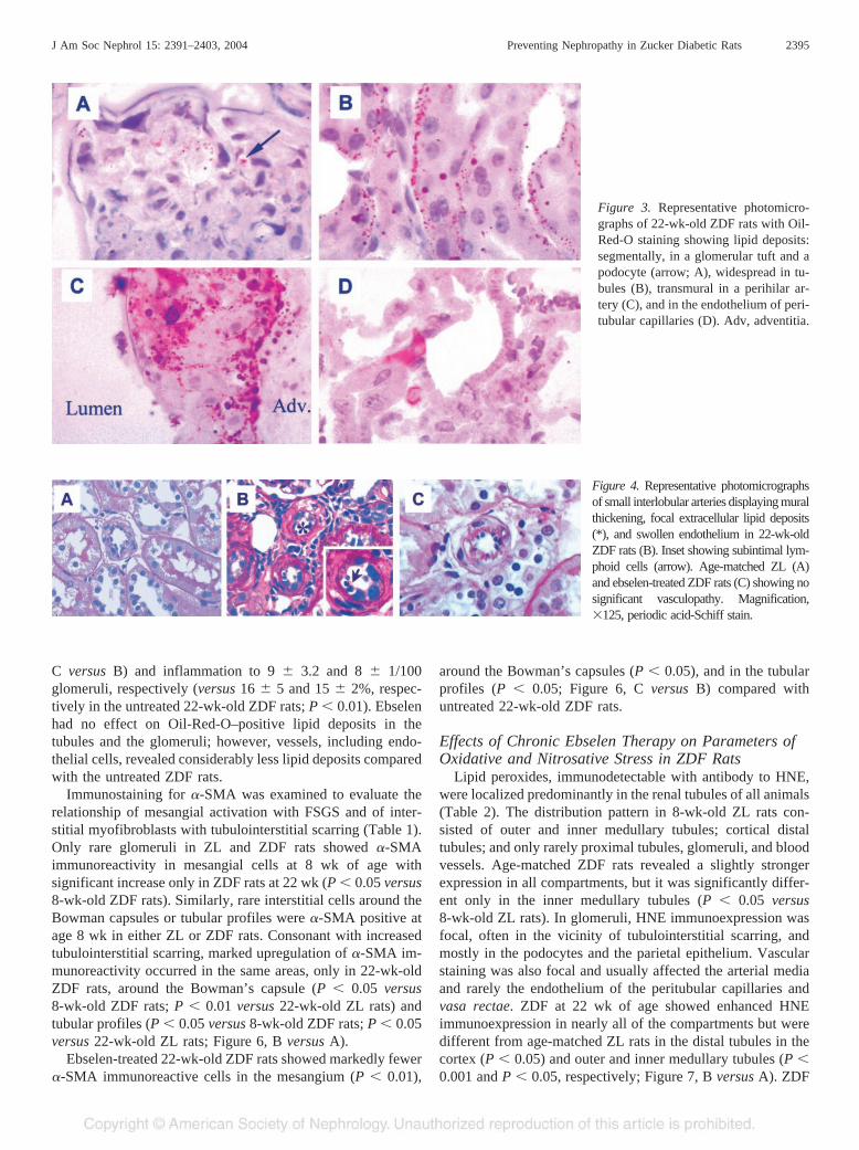
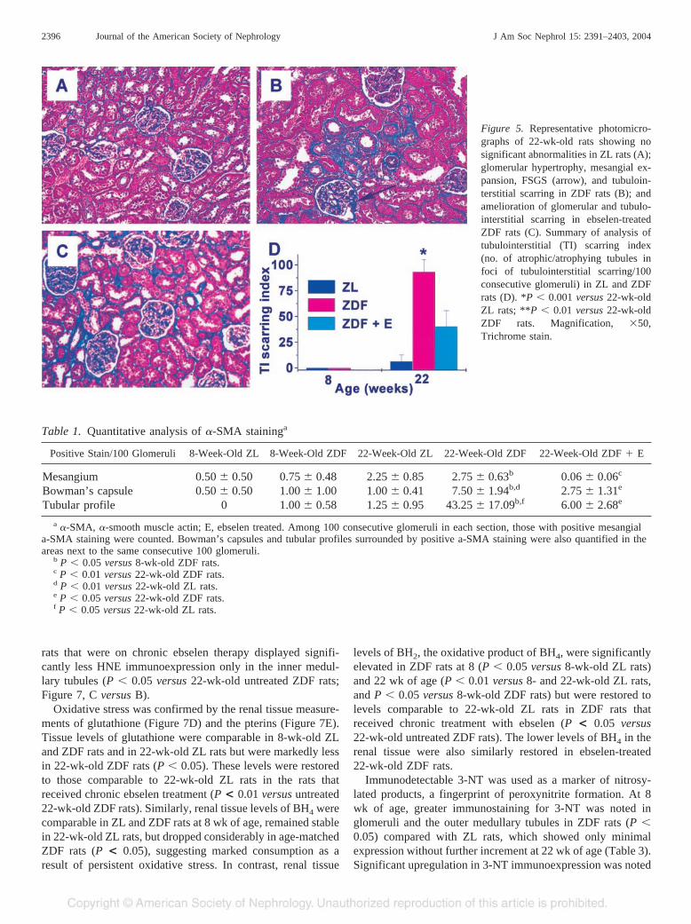
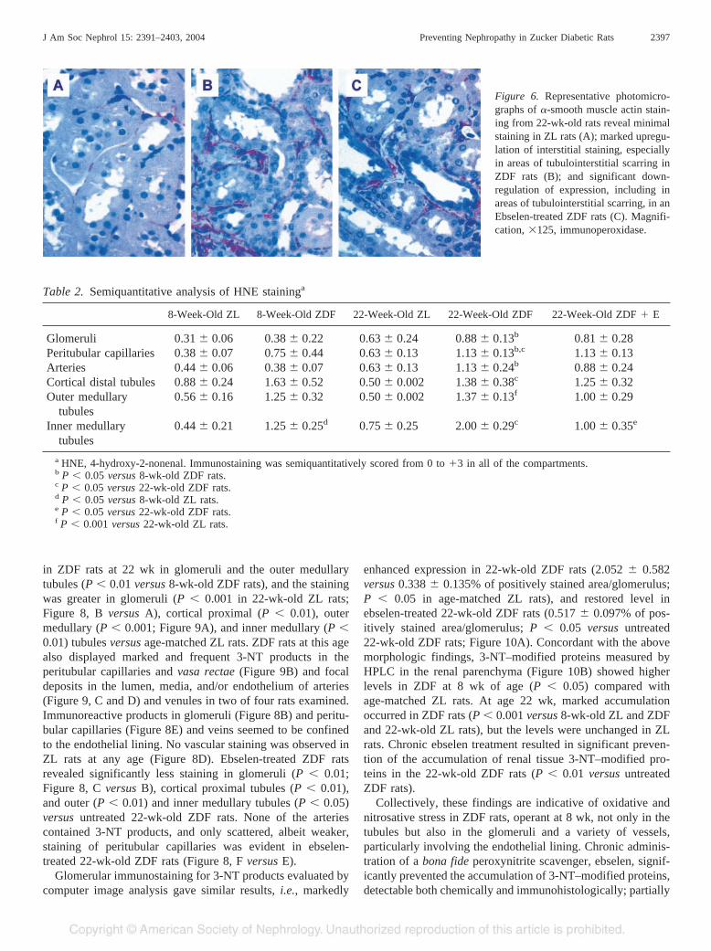
function, 8-wk-old ZDF animals showed neither glomerulo-sclerosis nor evidence of tubulointerstitial scarring or inflam-mation. Compared with 8-wk-old ZL rats, glomeruli in ZDFrats seemed slightly hypertrophied with further increment at 22wk of age along with mild to moderate mesangial expansion(Figure 2, B versus A and D versus C in age-matched ZL rats).FSGS was noted in 9.5 � 1.8% glomeruli (versus 1.2 � 2.7 in22-wk-old ZL rats; P � 0.001) with focally swollen andvacuolated podocytes that contained protein resorption dropletsoverlying such areas and frequent segmental adhesions to theBowman’s capsules (Figure 2D). Glomerulosclerosis indexwas scored at 0.34 � 0.04 in 22-wk-old ZDF (versus 0.02 �0.02 in 22-wk-old ZL rats; P � 0.001). Oil-Red-O stainingrevealed lipid deposits in the tubular epithelium, focal in 8-wk-old and widespread in 22 wk-old ZDF rats (Figure 3B). At thisage, lipid deposits also accumulated focally in podocytes andglomerular capillary tufts (Figure 3A) with or without overtFSGS and in the endothelial cells of peritubular capillaries,vasa rectae (Figure 3D), and arteries of all sizes includingperihilar and interlobar arteries. Lipid deposits were extra- andintracellular in the medial layer of these larger (Figure 3C) anda few smaller arteries (Figure 4B) with thickened vessel walls,focally swollen endothelium, and occasionally subintimal leu-kocytes (Figure 4, B versus A) compared with age-matched ZLrats despite comparable MBP, suggesting a lipid-induced vas-culopathy. Trichrome staining revealed patchy areas of tubu-lointerstitial scars in 22-wk-old ZDF rats (16 � 5 versus 1.2 �2.7/100 glomeruli in 22-wk-old ZL rats; P � 0.001; Figure 5,B versus A) and aggregates of mononuclear leukocytes. Sim-ilar aggregates were seen in other areas without overt scars (15� 2 versus 1.8 � 2 22-wk-old ZL rats; P � 0.001). From 35to 50% of the leukocytes stained positive for monocyte/mac-rophage marker CD-68. A significantly greater number ofatrophic/atrophying tubular profiles, representing the total tu-bulointerstitial scarring index, were quantified in 22-wk-oldZDF rats (93.25 � 0.12 versus 6.40 � 6.40/100 glomeruli in22-wk-old ZL rats; P � 0.001). ZDF rats at 8 wk of age andZL rats at 8 and 22 wk age showed no significant scars orinflammation.
Effects of Chronic Ebselen Treatment on RenalFunction, Morphology, Tubulointerstitial Scarring, and�-SMA Immunoexpression
Ebselen-treated 22-wk-old ZDF rats showed partial amelio-ration in proteinuria (123.5 � 11.2 versus 197.7 � 15.7 mg/24h; P � 0.001 versus untreated 22-wk-old ZDF rats), plasmacreatinine and Ccr (0.8 � 0.1 versus 1.6 � 0.3 mg/dl and 1.9� 0.4 versus 2.5 � 0.6 ml/min, respectively; P � 0.05 versusuntreated 22-wk-old ZDF rats) despite the absence of anynotable changes in the body weight, MBP, or blood glucose. Inparallel with the improved functional parameters, morphologicindices of renal damage were also downgraded compared with
J Am Soc Nephrol 15: 2391–2403, 2004 Preventing Nephropathy in Zucker Diabetic Rats 2393
the untreated ZDF rats. These included glomeruli with FSGS(5.3 � 1 versus 9.5 � 1.8/100 glomeruli in the untreated22-wk-old ZDF rats; P � 0.001; Figure 2, D versus F), glo-merulosclerosis index (0.15 � 0.04 versus 0.34 � 0.04 in the
untreated 22-wk-old ZDF rats; P � 0.001; Figure 2E), tubu-lointerstitial scarring index (40.25 � 15.34 versus 93.25 �12.11/100 glomeruli in the untreated 22-wk-old ZDF rats; P �0.001; Figure 5D), and foci of tubulointerstitial scars (Figure 5,
Figure 1. Effect of age and ebselen on body weight (BW; A), blood glucose (BG; B), mean BP (MBP; C), creatinine clearance (Ccr; D), andurinary protein excretion (UPE; E).
Figure 2. Representative glomerular photomicrographs display no significant abnormalities in 8- (A) and 22-wk-old (C) ZL rats. Glomerularhypertrophy and mesangial expansion are evident in 8-wk-old ZDF rats (B) with development of focal segmental glomerulosclerosis (FSGS)by 22 wk of age, associated with Bowman’s capsular adhesions, segmental lipid deposits in capillary tufts, and large protein reabsorptiondroplets in the podocytes (D). Chronic ebselen treatment was associated with lower prevalence of FSGS and severity of glomerulosclerosis (GS)index (F). Summary of semiquantitative analysis of GS index (E). *P � 0.01 versus 22-wk-old ZL rats; **P � 0.001 versus 22-wk-old ZDFrats. ZDF � E, ebselen treated. Magnification, �100, periodic acid-Schiff stain.
2394 Journal of the American Society of Nephrology J Am Soc Nephrol 15: 2391–2403, 2004
C versus B) and inflammation to 9 � 3.2 and 8 � 1/100glomeruli, respectively (versus 16 � 5 and 15 � 2%, respec-tively in the untreated 22-wk-old ZDF rats; P � 0.01). Ebselenhad no effect on Oil-Red-O–positive lipid deposits in thetubules and the glomeruli; however, vessels, including endo-thelial cells, revealed considerably less lipid deposits comparedwith the untreated ZDF rats.
Immunostaining for �-SMA was examined to evaluate therelationship of mesangial activation with FSGS and of inter-stitial myofibroblasts with tubulointerstitial scarring (Table 1).Only rare glomeruli in ZL and ZDF rats showed �-SMAimmunoreactivity in mesangial cells at 8 wk of age withsignificant increase only in ZDF rats at 22 wk (P � 0.05 versus8-wk-old ZDF rats). Similarly, rare interstitial cells around theBowman capsules or tubular profiles were �-SMA positive atage 8 wk in either ZL or ZDF rats. Consonant with increasedtubulointerstitial scarring, marked upregulation of �-SMA im-munoreactivity occurred in the same areas, only in 22-wk-oldZDF rats, around the Bowman’s capsule (P � 0.05 versus8-wk-old ZDF rats; P � 0.01 versus 22-wk-old ZL rats) andtubular profiles (P � 0.05 versus 8-wk-old ZDF rats; P � 0.05versus 22-wk-old ZL rats; Figure 6, B versus A).
Ebselen-treated 22-wk-old ZDF rats showed markedly fewer�-SMA immunoreactive cells in the mesangium (P � 0.01),
around the Bowman’s capsules (P � 0.05), and in the tubularprofiles (P � 0.05; Figure 6, C versus B) compared withuntreated 22-wk-old ZDF rats.
Effects of Chronic Ebselen Therapy on Parameters ofOxidative and Nitrosative Stress in ZDF Rats
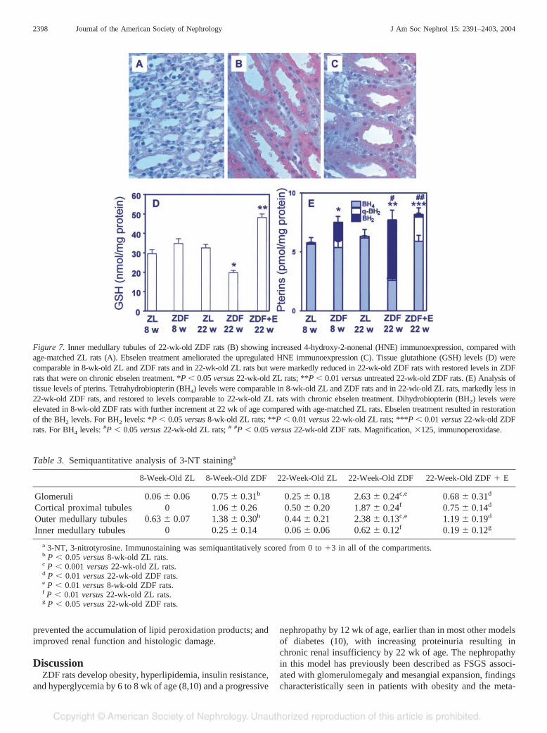
Lipid peroxides, immunodetectable with antibody to HNE,were localized predominantly in the renal tubules of all animals(Table 2). The distribution pattern in 8-wk-old ZL rats con-sisted of outer and inner medullary tubules; cortical distaltubules; and only rarely proximal tubules, glomeruli, and bloodvessels. Age-matched ZDF rats revealed a slightly strongerexpression in all compartments, but it was significantly differ-ent only in the inner medullary tubules (P � 0.05 versus8-wk-old ZL rats). In glomeruli, HNE immunoexpression wasfocal, often in the vicinity of tubulointerstitial scarring, andmostly in the podocytes and the parietal epithelium. Vascularstaining was also focal and usually affected the arterial mediaand rarely the endothelium of the peritubular capillaries andvasa rectae. ZDF at 22 wk of age showed enhanced HNEimmunoexpression in nearly all of the compartments but weredifferent from age-matched ZL rats in the distal tubules in thecortex (P � 0.05) and outer and inner medullary tubules (P �0.001 and P � 0.05, respectively; Figure 7, B versus A). ZDF
Figure 3. Representative photomicro-graphs of 22-wk-old ZDF rats with Oil-Red-O staining showing lipid deposits:segmentally, in a glomerular tuft and apodocyte (arrow; A), widespread in tu-bules (B), transmural in a perihilar ar-tery (C), and in the endothelium of peri-tubular capillaries (D). Adv, adventitia.
Figure 4. Representative photomicrographsof small interlobular arteries displaying muralthickening, focal extracellular lipid deposits(*), and swollen endothelium in 22-wk-oldZDF rats (B). Inset showing subintimal lym-phoid cells (arrow). Age-matched ZL (A)and ebselen-treated ZDF rats (C) showing nosignificant vasculopathy. Magnification,�125, periodic acid-Schiff stain.
J Am Soc Nephrol 15: 2391–2403, 2004 Preventing Nephropathy in Zucker Diabetic Rats 2395
rats that were on chronic ebselen therapy displayed signifi-cantly less HNE immunoexpression only in the inner medul-lary tubules (P � 0.05 versus 22-wk-old untreated ZDF rats;Figure 7, C versus B).
Oxidative stress was confirmed by the renal tissue measure-ments of glutathione (Figure 7D) and the pterins (Figure 7E).Tissue levels of glutathione were comparable in 8-wk-old ZLand ZDF rats and in 22-wk-old ZL rats but were markedly lessin 22-wk-old ZDF rats (P � 0.05). These levels were restoredto those comparable to 22-wk-old ZL rats in the rats thatreceived chronic ebselen treatment (P < 0.01 versus untreated22-wk-old ZDF rats). Similarly, renal tissue levels of BH4 werecomparable in ZL and ZDF rats at 8 wk of age, remained stablein 22-wk-old ZL rats, but dropped considerably in age-matchedZDF rats (P < 0.05), suggesting marked consumption as aresult of persistent oxidative stress. In contrast, renal tissue
levels of BH2, the oxidative product of BH4, were significantlyelevated in ZDF rats at 8 (P � 0.05 versus 8-wk-old ZL rats)and 22 wk of age (P � 0.01 versus 8- and 22-wk-old ZL rats,and P � 0.05 versus 8-wk-old ZDF rats) but were restored tolevels comparable to 22-wk-old ZL rats in ZDF rats thatreceived chronic treatment with ebselen (P < 0.05 versus22-wk-old untreated ZDF rats). The lower levels of BH4 in therenal tissue were also similarly restored in ebselen-treated22-wk-old ZDF rats.
Immunodetectable 3-NT was used as a marker of nitrosy-lated products, a fingerprint of peroxynitrite formation. At 8wk of age, greater immunostaining for 3-NT was noted inglomeruli and the outer medullary tubules in ZDF rats (P �0.05) compared with ZL rats, which showed only minimalexpression without further increment at 22 wk of age (Table 3).Significant upregulation in 3-NT immunoexpression was noted
Figure 5. Representative photomicro-graphs of 22-wk-old rats showing nosignificant abnormalities in ZL rats (A);glomerular hypertrophy, mesangial ex-pansion, FSGS (arrow), and tubuloin-terstitial scarring in ZDF rats (B); andamelioration of glomerular and tubulo-interstitial scarring in ebselen-treatedZDF rats (C). Summary of analysis oftubulointerstitial (TI) scarring index(no. of atrophic/atrophying tubules infoci of tubulointerstitial scarring/100consecutive glomeruli) in ZL and ZDFrats (D). *P � 0.001 versus 22-wk-oldZL rats; **P � 0.01 versus 22-wk-oldZDF rats. Magnification, �50,Trichrome stain.
Table 1. Quantitative analysis of �-SMA staininga
Positive Stain/100 Glomeruli 8-Week-Old ZL 8-Week-Old ZDF 22-Week-Old ZL 22-Week-Old ZDF 22-Week-Old ZDF � E
Mesangium 0.50 � 0.50 0.75 � 0.48 2.25 � 0.85 2.75 � 0.63b 0.06 � 0.06c
Bowman’s capsule 0.50 � 0.50 1.00 � 1.00 1.00 � 0.41 7.50 � 1.94b,d 2.75 � 1.31e
Tubular profile 0 1.00 � 0.58 1.25 � 0.95 43.25 � 17.09b,f 6.00 � 2.68e
a �-SMA, �-smooth muscle actin; E, ebselen treated. Among 100 consecutive glomeruli in each section, those with positive mesangiala-SMA staining were counted. Bowman’s capsules and tubular profiles surrounded by positive a-SMA staining were also quantified in theareas next to the same consecutive 100 glomeruli.
b P � 0.05 versus 8-wk-old ZDF rats.c P � 0.01 versus 22-wk-old ZDF rats.d P � 0.01 versus 22-wk-old ZL rats.e P � 0.05 versus 22-wk-old ZDF rats.f P � 0.05 versus 22-wk-old ZL rats.
2396 Journal of the American Society of Nephrology J Am Soc Nephrol 15: 2391–2403, 2004
in ZDF rats at 22 wk in glomeruli and the outer medullarytubules (P � 0.01 versus 8-wk-old ZDF rats), and the stainingwas greater in glomeruli (P � 0.001 in 22-wk-old ZL rats;Figure 8, B versus A), cortical proximal (P � 0.01), outermedullary (P � 0.001; Figure 9A), and inner medullary (P �0.01) tubules versus age-matched ZL rats. ZDF rats at this agealso displayed marked and frequent 3-NT products in theperitubular capillaries and vasa rectae (Figure 9B) and focaldeposits in the lumen, media, and/or endothelium of arteries(Figure 9, C and D) and venules in two of four rats examined.Immunoreactive products in glomeruli (Figure 8B) and peritu-bular capillaries (Figure 8E) and veins seemed to be confinedto the endothelial lining. No vascular staining was observed inZL rats at any age (Figure 8D). Ebselen-treated ZDF ratsrevealed significantly less staining in glomeruli (P � 0.01;Figure 8, C versus B), cortical proximal tubules (P � 0.01),and outer (P � 0.01) and inner medullary tubules (P � 0.05)versus untreated 22-wk-old ZDF rats. None of the arteriescontained 3-NT products, and only scattered, albeit weaker,staining of peritubular capillaries was evident in ebselen-treated 22-wk-old ZDF rats (Figure 8, F versus E).
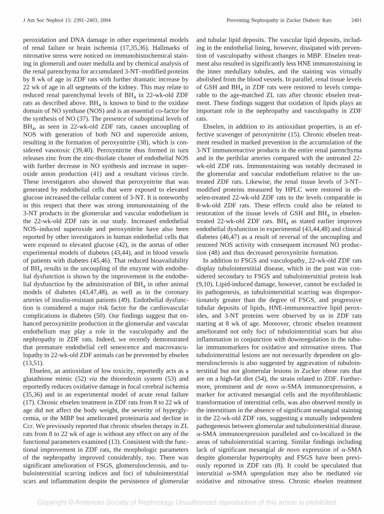
Glomerular immunostaining for 3-NT products evaluated bycomputer image analysis gave similar results, i.e., markedly
enhanced expression in 22-wk-old ZDF rats (2.052 � 0.582versus 0.338 � 0.135% of positively stained area/glomerulus;P � 0.05 in age-matched ZL rats), and restored level inebselen-treated 22-wk-old ZDF rats (0.517 � 0.097% of pos-itively stained area/glomerulus; P � 0.05 versus untreated22-wk-old ZDF rats; Figure 10A). Concordant with the abovemorphologic findings, 3-NT–modified proteins measured byHPLC in the renal parenchyma (Figure 10B) showed higherlevels in ZDF at 8 wk of age (P � 0.05) compared withage-matched ZL rats. At age 22 wk, marked accumulationoccurred in ZDF rats (P � 0.001 versus 8-wk-old ZL and ZDFand 22-wk-old ZL rats), but the levels were unchanged in ZLrats. Chronic ebselen treatment resulted in significant preven-tion of the accumulation of renal tissue 3-NT–modified pro-teins in the 22-wk-old ZDF rats (P � 0.01 versus untreatedZDF rats).
Collectively, these findings are indicative of oxidative andnitrosative stress in ZDF rats, operant at 8 wk, not only in thetubules but also in the glomeruli and a variety of vessels,particularly involving the endothelial lining. Chronic adminis-tration of a bona fide peroxynitrite scavenger, ebselen, signif-icantly prevented the accumulation of 3-NT–modified proteins,detectable both chemically and immunohistologically; partially
Figure 6. Representative photomicro-graphs of �-smooth muscle actin stain-ing from 22-wk-old rats reveal minimalstaining in ZL rats (A); marked upregu-lation of interstitial staining, especiallyin areas of tubulointerstitial scarring inZDF rats (B); and significant down-regulation of expression, including inareas of tubulointerstitial scarring, in anEbselen-treated ZDF rats (C). Magnifi-cation, �125, immunoperoxidase.
Table 2. Semiquantitative analysis of HNE staininga
8-Week-Old ZL 8-Week-Old ZDF 22-Week-Old ZL 22-Week-Old ZDF 22-Week-Old ZDF � E
Glomeruli 0.31 � 0.06 0.38 � 0.22 0.63 � 0.24 0.88 � 0.13b 0.81 � 0.28Peritubular capillaries 0.38 � 0.07 0.75 � 0.44 0.63 � 0.13 1.13 � 0.13b,c 1.13 � 0.13Arteries 0.44 � 0.06 0.38 � 0.07 0.63 � 0.13 1.13 � 0.24b 0.88 � 0.24Cortical distal tubules 0.88 � 0.24 1.63 � 0.52 0.50 � 0.002 1.38 � 0.38c 1.25 � 0.32Outer medullary
tubules0.56 � 0.16 1.25 � 0.32 0.50 � 0.002 1.37 � 0.13f 1.00 � 0.29
Inner medullarytubules
0.44 � 0.21 1.25 � 0.25d 0.75 � 0.25 2.00 � 0.29c 1.00 � 0.35e
a HNE, 4-hydroxy-2-nonenal. Immunostaining was semiquantitatively scored from 0 to �3 in all of the compartments.b P � 0.05 versus 8-wk-old ZDF rats.c P � 0.05 versus 22-wk-old ZDF rats.d P � 0.05 versus 8-wk-old ZL rats.e P � 0.05 versus 22-wk-old ZDF rats.f P � 0.001 versus 22-wk-old ZL rats.
J Am Soc Nephrol 15: 2391–2403, 2004 Preventing Nephropathy in Zucker Diabetic Rats 2397
prevented the accumulation of lipid peroxidation products; andimproved renal function and histologic damage.
DiscussionZDF rats develop obesity, hyperlipidemia, insulin resistance,
and hyperglycemia by 6 to 8 wk of age (8,10) and a progressive
nephropathy by 12 wk of age, earlier than in most other modelsof diabetes (10), with increasing proteinuria resulting inchronic renal insufficiency by 22 wk of age. The nephropathyin this model has previously been described as FSGS associ-ated with glomerulomegaly and mesangial expansion, findingscharacteristically seen in patients with obesity and the meta-
Figure 7. Inner medullary tubules of 22-wk-old ZDF rats (B) showing increased 4-hydroxy-2-nonenal (HNE) immunoexpression, compared withage-matched ZL rats (A). Ebselen treatment ameliorated the upregulated HNE immunoexpression (C). Tissue glutathione (GSH) levels (D) werecomparable in 8-wk-old ZL and ZDF rats and in 22-wk-old ZL rats but were markedly reduced in 22-wk-old ZDF rats with restored levels in ZDFrats that were on chronic ebselen treatment. *P � 0.05 versus 22-wk-old ZL rats; **P � 0.01 versus untreated 22-wk-old ZDF rats. (E) Analysis oftissue levels of pterins. Tetrahydrobiopterin (BH4) levels were comparable in 8-wk-old ZL and ZDF rats and in 22-wk-old ZL rats, markedly less in22-wk-old ZDF rats, and restored to levels comparable to 22-wk-old ZL rats with chronic ebselen treatment. Dihydrobiopterin (BH2) levels wereelevated in 8-wk-old ZDF rats with further increment at 22 wk of age compared with age-matched ZL rats. Ebselen treatment resulted in restorationof the BH2 levels. For BH2 levels: *P � 0.05 versus 8-wk-old ZL rats; **P � 0.01 versus 22-wk-old ZL rats; ***P � 0.01 versus 22-wk-old ZDFrats. For BH4 levels: #P � 0.05 versus 22-wk-old ZL rats; # #P � 0.05 versus 22-wk-old ZDF rats. Magnification, �125, immunoperoxidase.
Table 3. Semiquantitative analysis of 3-NT staininga
8-Week-Old ZL 8-Week-Old ZDF 22-Week-Old ZL 22-Week-Old ZDF 22-Week-Old ZDF � E
Glomeruli 0.06 � 0.06 0.75 � 0.31b 0.25 � 0.18 2.63 � 0.24c,e 0.68 � 0.31d
Cortical proximal tubules 0 1.06 � 0.26 0.50 � 0.20 1.87 � 0.24f 0.75 � 0.14d
Outer medullary tubules 0.63 � 0.07 1.38 � 0.30b 0.44 � 0.21 2.38 � 0.13c,e 1.19 � 0.19d
Inner medullary tubules 0 0.25 � 0.14 0.06 � 0.06 0.62 � 0.12f 0.19 � 0.12g
a 3-NT, 3-nitrotyrosine. Immunostaining was semiquantitatively scored from 0 to �3 in all of the compartments.b P � 0.05 versus 8-wk-old ZL rats.c P � 0.001 versus 22-wk-old ZL rats.d P � 0.01 versus 22-wk-old ZDF rats.e P � 0.01 versus 8-wk-old ZDF rats.f P � 0.01 versus 22-wk-old ZL rats.g P � 0.05 versus 22-wk-old ZDF rats.
2398 Journal of the American Society of Nephrology J Am Soc Nephrol 15: 2391–2403, 2004
bolic syndrome (23,24) associated with type 2 diabetic milieu.Except for an isolated report describing the tubulointerstitialchanges in Zucker obese rats (7), most investigators havementioned these changes only in passing and as secondarypathology (9,10). Renal vascular pathology has not been de-scribed discretely.
FSGS in ZDF rats has been variably ascribed to glomerularhyperperfusion (9) or early podocyte injury induced by hyper-
lipidemia and cholesterol-induced chemotactic monocyte/mac-rophage influx into the glomeruli by some (8) and diabeticmilieu but not hypertension or obesity by other investigators(10). The role of oxidative and nitrosative stress in the patho-genesis of glomerulopathy and nephropathy in these animals,although suggested (8), has not been evaluated. The data pre-sented herein provide morphologic and functional character-ization of progressive nephropathy, including the extraglo-
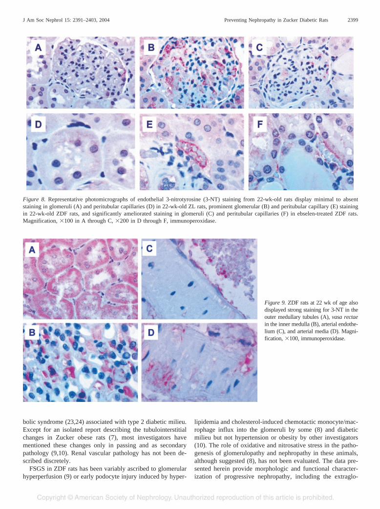
Figure 8. Representative photomicrographs of endothelial 3-nitrotyrosine (3-NT) staining from 22-wk-old rats display minimal to absentstaining in glomeruli (A) and peritubular capillaries (D) in 22-wk-old ZL rats, prominent glomerular (B) and peritubular capillary (E) stainingin 22-wk-old ZDF rats, and significantly ameliorated staining in glomeruli (C) and peritubular capillaries (F) in ebselen-treated ZDF rats.Magnification, �100 in A through C, �200 in D through F, immunoperoxidase.
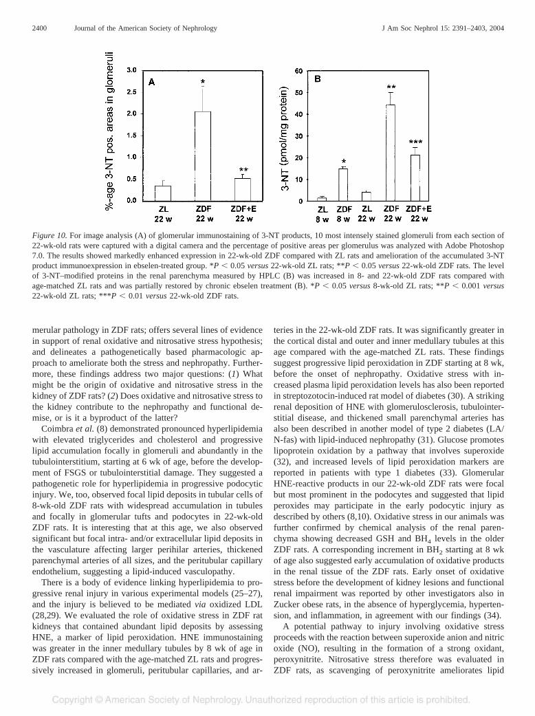
Figure 9. ZDF rats at 22 wk of age alsodisplayed strong staining for 3-NT in theouter medullary tubules (A), vasa rectaein the inner medulla (B), arterial endothe-lium (C), and arterial media (D). Magni-fication, �100, immunoperoxidase.
J Am Soc Nephrol 15: 2391–2403, 2004 Preventing Nephropathy in Zucker Diabetic Rats 2399
merular pathology in ZDF rats; offers several lines of evidencein support of renal oxidative and nitrosative stress hypothesis;and delineates a pathogenetically based pharmacologic ap-proach to ameliorate both the stress and nephropathy. Further-more, these findings address two major questions: (1) Whatmight be the origin of oxidative and nitrosative stress in thekidney of ZDF rats? (2) Does oxidative and nitrosative stress tothe kidney contribute to the nephropathy and functional de-mise, or is it a byproduct of the latter?
Coimbra et al. (8) demonstrated pronounced hyperlipidemiawith elevated triglycerides and cholesterol and progressivelipid accumulation focally in glomeruli and abundantly in thetubulointerstitium, starting at 6 wk of age, before the develop-ment of FSGS or tubulointerstitial damage. They suggested apathogenetic role for hyperlipidemia in progressive podocyticinjury. We, too, observed focal lipid deposits in tubular cells of8-wk-old ZDF rats with widespread accumulation in tubulesand focally in glomerular tufts and podocytes in 22-wk-oldZDF rats. It is interesting that at this age, we also observedsignificant but focal intra- and/or extracellular lipid deposits inthe vasculature affecting larger perihilar arteries, thickenedparenchymal arteries of all sizes, and the peritubular capillaryendothelium, suggesting a lipid-induced vasculopathy.
There is a body of evidence linking hyperlipidemia to pro-gressive renal injury in various experimental models (25–27),and the injury is believed to be mediated via oxidized LDL(28,29). We evaluated the role of oxidative stress in ZDF ratkidneys that contained abundant lipid deposits by assessingHNE, a marker of lipid peroxidation. HNE immunostainingwas greater in the inner medullary tubules by 8 wk of age inZDF rats compared with the age-matched ZL rats and progres-sively increased in glomeruli, peritubular capillaries, and ar-
teries in the 22-wk-old ZDF rats. It was significantly greater inthe cortical distal and outer and inner medullary tubules at thisage compared with the age-matched ZL rats. These findingssuggest progressive lipid peroxidation in ZDF starting at 8 wk,before the onset of nephropathy. Oxidative stress with in-creased plasma lipid peroxidation levels has also been reportedin streptozotocin-induced rat model of diabetes (30). A strikingrenal deposition of HNE with glomerulosclerosis, tubulointer-stitial disease, and thickened small parenchymal arteries hasalso been described in another model of type 2 diabetes (LA/N-fas) with lipid-induced nephropathy (31). Glucose promoteslipoprotein oxidation by a pathway that involves superoxide(32), and increased levels of lipid peroxidation markers arereported in patients with type 1 diabetes (33). GlomerularHNE-reactive products in our 22-wk-old ZDF rats were focalbut most prominent in the podocytes and suggested that lipidperoxides may participate in the early podocytic injury asdescribed by others (8,10). Oxidative stress in our animals wasfurther confirmed by chemical analysis of the renal paren-chyma showing decreased GSH and BH4 levels in the olderZDF rats. A corresponding increment in BH2 starting at 8 wkof age also suggested early accumulation of oxidative productsin the renal tissue of the ZDF rats. Early onset of oxidativestress before the development of kidney lesions and functionalrenal impairment was reported by other investigators also inZucker obese rats, in the absence of hyperglycemia, hyperten-sion, and inflammation, in agreement with our findings (34).
A potential pathway to injury involving oxidative stressproceeds with the reaction between superoxide anion and nitricoxide (NO), resulting in the formation of a strong oxidant,peroxynitrite. Nitrosative stress therefore was evaluated inZDF rats, as scavenging of peroxynitrite ameliorates lipid
Figure 10. For image analysis (A) of glomerular immunostaining of 3-NT products, 10 most intensely stained glomeruli from each section of22-wk-old rats were captured with a digital camera and the percentage of positive areas per glomerulus was analyzed with Adobe Photoshop7.0. The results showed markedly enhanced expression in 22-wk-old ZDF compared with ZL rats and amelioration of the accumulated 3-NTproduct immunoexpression in ebselen-treated group. *P � 0.05 versus 22-wk-old ZL rats; **P � 0.05 versus 22-wk-old ZDF rats. The levelof 3-NT–modified proteins in the renal parenchyma measured by HPLC (B) was increased in 8- and 22-wk-old ZDF rats compared withage-matched ZL rats and was partially restored by chronic ebselen treatment (B). *P � 0.05 versus 8-wk-old ZL rats; **P � 0.001 versus22-wk-old ZL rats; ***P � 0.01 versus 22-wk-old ZDF rats.
2400 Journal of the American Society of Nephrology J Am Soc Nephrol 15: 2391–2403, 2004
peroxidation and DNA damage in other experimental modelsof renal failure or brain ischemia (17,35,36). Hallmarks ofnitrosative stress were noticed on immunohistochemical stain-ing in glomeruli and outer medulla and by chemical analysis ofthe renal parenchyma for accumulated 3-NT–modified proteinsby 8 wk of age in ZDF rats with further dramatic increase by22 wk of age in all segments of the kidney. This may relate toreduced renal parenchymal levels of BH4 in 22-wk-old ZDFrats as described above. BH4 is known to bind to the oxidasedomain of NO synthase (NOS) and is an essential co-factor forthe synthesis of NO (37). The presence of suboptimal levels ofBH4, as seen in 22-wk-old ZDF rats, causes uncoupling ofNOS with generation of both NO and superoxide anions,resulting in the formation of peroxynitrite (38), which is con-sidered vasotoxic (39,40). Peroxynitrite thus formed in turnreleases zinc from the zinc-thiolate cluster of endothelial NOSwith further decrease in NO synthesis and increase in super-oxide anion production (41) and a resultant vicious circle.These investigators also showed that peroxynitrite that wasgenerated by endothelial cells that were exposed to elevatedglucose increased the cellular content of 3-NT. It is noteworthyin this respect that there was strong immunostaining of the3-NT products in the glomerular and vascular endothelium inthe 22-wk-old ZDF rats in our study. Increased endothelialNOS–induced superoxide and peroxynitrite have also beenreported by other investigators in human endothelial cells thatwere exposed to elevated glucose (42), in the aortas of otherexperimental models of diabetes (43,44), and in blood vesselsof patients with diabetes (45,46). That reduced bioavailabilityof BH4 results in the uncoupling of the enzyme with endothe-lial dysfunction is shown by the improvement in the endothe-lial dysfunction by the administration of BH4 in other animalmodels of diabetes (43,47,48), as well as in the coronaryarteries of insulin-resistant patients (49). Endothelial dysfunc-tion is considered a major risk factor for the cardiovascularcomplications in diabetes (50). Our findings suggest that en-hanced peroxynitrite production in the glomerular and vascularendothelium may play a role in the vasculopathy and thenephropathy in ZDF rats. Indeed, we recently demonstratedthat premature endothelial cell senescence and macrovascu-lopathy in 22-wk-old ZDF animals can be prevented by ebselen(13,51).
Ebselen, an antioxidant of low toxicity, reportedly acts as aglutathione mimic (52) via the thioredoxin system (53) andreportedly reduces oxidative damage in focal cerebral ischemia(35,36) and in an experimental model of acute renal failure(17). Chronic ebselen treatment in ZDF rats from 8 to 22 wk ofage did not affect the body weight, the severity of hypergly-cemia, or the MBP but ameliorated proteinuria and decline inCcr. We previously reported that chronic ebselen therapy in ZLrats from 8 to 22 wk of age is without any effect on any of thefunctional parameters examined (13). Consistent with the func-tional improvement in ZDF rats, the morphologic parametersof the nephropathy improved considerably, too. There wassignificant amelioration of FSGS, glomerulosclerosis, and tu-bulointerstitial scarring indices and foci of tubulointerstitialscars and inflammation despite the persistence of glomerular
and tubular lipid deposits. The vascular lipid deposits, includ-ing in the endothelial lining, however, dissipated with preven-tion of vasculopathy without changes in MBP. Ebselen treat-ment also resulted in significantly less HNE immunostaining inthe inner medullary tubules, and the staining was virtuallyabolished from the blood vessels. In parallel, renal tissue levelsof GSH and BH4 in ZDF rats were restored to levels compa-rable to the age-matched ZL rats after chronic ebselen treat-ment. These findings suggest that oxidation of lipids plays animportant role in the nephropathy and vasculopathy in ZDFrats.
Ebselen, in addition to its antioxidant properties, is an ef-fective scavenger of peroxynitrite (15). Chronic ebselen treat-ment resulted in marked prevention in the accumulation of the3-NT immunoreactive products in the entire renal parenchymaand in the perihilar arteries compared with the untreated 22-wk-old ZDF rats. Immunostaining was notably decreased inthe glomerular and vascular endothelium relative to the un-treated ZDF rats. Likewise, the renal tissue levels of 3-NT–modified proteins measured by HPLC were restored in eb-selen-treated 22-wk-old ZDF rats to the levels comparable in8-wk-old ZDF rats. These effects could also be related torestoration of the tissue levels of GSH and BH4 in ebselen-treated 22-wk-old ZDF rats. BH4 as stated earlier improvesendothelial dysfunction in experimental (43,44,48) and clinicaldiabetes (46,47) as a result of reversal of the uncoupling andrestored NOS activity with consequent increased NO produc-tion (48) and thus decreased peroxynitrite formation.
In addition to FSGS and vasculopathy, 22-wk-old ZDF ratsdisplay tubulointerstitial disease, which in the past was con-sidered secondary to FSGS and tubulointerstitial protein leak(9,10). Lipid-induced damage, however, cannot be excluded inits pathogenesis, as tubulointerstitial scarring was dispropor-tionately greater than the degree of FSGS, and progressivetubular deposits of lipids, HNE-immunoreactive lipid perox-ides, and 3-NT proteins were observed by us in ZDF ratsstarting at 8 wk of age. Moreover, chronic ebselen treatmentameliorated not only foci of tubulointerstitial scars but alsoinflammation in conjunction with downregulation in the tubu-lar immunomarkers for oxidative and nitrosative stress. Thattubulointerstitial lesions are not necessarily dependent on glo-merulosclerosis is also suggested by aggravation of tubuloin-terstitial but not glomerular lesions in Zucker obese rats thatare on a high-fat diet (54), the strain related to ZDF. Further-more, prominent and de novo �-SMA immunoexpression, amarker for activated mesangial cells and the myofibroblastictransformation of interstitial cells, was also observed mostly inthe interstitium in the absence of significant mesangial stainingin the 22-wk-old ZDF rats, suggesting a mutually independentpathogenesis between glomerular and tubulointerstitial disease.�-SMA immunoexpression paralleled and co-localized in theareas of tubulointerstitial scarring. Similar findings includinglack of significant mesangial de novo expression of �-SMAdespite glomerular hypertrophy and FSGS have been previ-ously reported in ZDF rats (8). It could be speculated thatinterstitial �-SMA upregulation may also be mediated viaoxidative and nitrosative stress. Chronic ebselen treatment
J Am Soc Nephrol 15: 2391–2403, 2004 Preventing Nephropathy in Zucker Diabetic Rats 2401
indeed not only ameliorated tubulointerstitial scarring and in-flammation but also attenuated �-SMA immunoexpressioneven in areas of tubulointerstitial scars, suggesting a correla-tion with oxidative/nitrosative stress.
In conclusion, we have confirmed that ZDF rats with themetabolic syndrome develop nephropathy with FSGS, protein-uria, and renal failure and in addition demonstrated the occur-rence of lipid-induced vasculopathy and tubulointerstitial dis-ease in this rat model. Lipid peroxidation and 3-NT–modifiedproducts accumulate in the kidneys starting at 8 wk of age,before the onset of the nephropathy, with further accentuationin 22-wk-old ZDF rats, suggesting these to be (1) the origin ofthe oxidative and nitrosative stress and (2) the cause and notthe consequence of the nephropathy in ZDF. Ebselen, a scav-enger of peroxynitrite and an antioxidant, resulted in improve-ment of renal function (proteinuria and Ccr), morphology(FSGS, tubulointerstitial scars and inflammation, and vascu-lopathy), and amelioration in chemical and immunomarkers ofnitrosative and oxidative stress. The beneficial effects of eb-selen, without any effect on or any additional therapy forhyperlipidemia, lipidosis, obesity with the associated meta-bolic syndrome, hyperglycemia, or MBP, strongly argue thatoxidative and nitrosative stress are the primary culprits in thepathogenesis of the nephropathy, including the tubulointersti-tial pathology and the microvascular complications in the ZDFrats.
AcknowledgmentsThese studies were supported in part by National Institutes of
Health Grants DK 45462, 45695, and 54602 (M.S.G.); AmericanHeart Association grants (S.B.); and the Westchester Artificial KidneyFoundation.
References1. Ritz E, Stefanski A: Diabetic nephropathy in type II diabetes.
Am J Kidney Dis 27: 167–194, 19962. Hall J, Kuo J, daSilva A, DePaula R, Liu J, Tallam L: Obesity-
associated hypertension and kidney disease. Curr Opin NephrolHypertens 12: 195–200, 2003
3. Zucker LM: Hereditary obesity in the rat associated with hyper-lipemia. Ann N Y Acad Sci 131: 447–458, 1965
4. Phillips MS, Liu Q, Hammond H, Dugan V, Hey P, Caskey C,Hess JF: Leptin receptor missense mutation in the fatty Zuckerrat. Nat Genet 13: 18–19, 1996
5. Kasiske BL, O’Donnell M, Keane WF: The Zucker rat model ofobesity, insulin resistance, hyperlipidemia and renal injury. Hy-pertension 19: I110–I115, 1992
6. Kasiske BL, Cleary M, O’Donnell M, Keane WF: Effects ofgenetic obesity on renal structure and function in the Zucker rat.J Lab Clin Med 106: 598–604, 1985
7. Magil AB: TI lesions in young Zucker rats. Am J Kidney Dis 25:478–485, 1995
8. Coimbra T, Janssen U, Grone H, Ostendorf T, Kunter U, SchmidtH, Brabant G, Floege J: Early events leading to renal injury inobese Zucker (fatty) rats with type II diabetes. Kidney Int 57:167–182, 2000
9. Gassler N, Elger M, Kranzlin B, Kriz W, Gretz N: Podocyteinjury underlies the progression of focal segmental glomerulo-sclerosis in the fa/fa Zucker rat. Kidney Int 60: 106–116, 2001
10. Hoshi S, Shu Y, Yoshida F, Inagaki T, Sonoda J, Watanabe T,Nomoto K, Nagata M: Podocyte injury promotes progressivenephropathy in Zucker diabetic fatty rats. Lab Invest 82: 25–35,2002
11. Laight D, Desai K, Anggard E, Carrier MJ: Endothelial dysfunc-tion accompanies a pro-oxidant, pro-diabetic challenge in theinsulin resistant, obese Zucker rat in vivo. Eur J Pharmacol 402:95–99, 2000
12. Pannirselvam M, Verma S, Anderson T, Triggle CR: Cellularbasis of endothelial dysfunction in small mesenteric arteries fromspontaneously diabetic (db/db-/-) mice: Decreased tetrahydro-biopterin bioavailability. Br J Pharmacol 136: 255–263, 2002
13. Brodsky SV, Gealekman O, Chen J, Zhang F, Togashi N,Crabtree M, Gross SS, Nasjletti A, Goligorsky MS: Preventionand reversal of premature endothelial cell senescence and vas-culopathy in obesity-induced diabetes by ebselen. Circ Res 94:377–384, 2004
14. Bindokas V, Kuznetsov A, Sreenan S, Polonsky K, Roe M,Philipson LH: Visualizing superoxide production in normal anddiabetic islets of Langerhans. J Biol Chem 278: 9796–9801,2003
15. Masumoto H, Kissner R, Koppenol W, Sies H: Kinetic study ofthe reaction of ebselen with peroxynitrite. FEBS Lett 398: 179–182, 1996
16. Yamaguchi T, Sano K, Takakura K, Saito I, Shinohara Y, AsanoT, Yasuhara H: Ebselen in acute ischemic stroke; a placebo-controlled, double-blind clinical trial. Stroke 29: 12–17, 1998
17. Noiri E, Nakao A, Uchida K, Tsukahara H, Ohno M, Fujita T,Brodsky S, Goligorsky MS: Scavenging of peroxynitrite amelio-rates lipid peroxidation and DNA damage in experimental isch-emic acute renal failure. Am J Physiol Renal Physiol 281: F948–F957, 2001
18. Heales S, Hyland K: Determination of quinonoid dihydrobiop-terin by high-performance liquid chromatography and electro-chemical detection. J Chromatogr 494: 77–85, 1989
19. Baker MA, Cerniglia GJ, Zaman A: Microtiter plate assay for themeasurement of glutathione and glutathione disulfide in largenumbers of biological samples. Anal Biochem 190: 360–365,1990
20. Griffith OW: Determination of glutathione and glutathione di-sulfide using glutathione reductase and 2-vinylpyridine. AnalBiochem 106: 207–212, 1980
21. Crabtree M, Hao G, Gross SS: Detection of cysteine S-nitrosy-lation and tyrosine 3-nitration in kidney proteins. Methods MolMed 86: 373–384, 2003
22. Maruyama W, Hashizume Y, Matsubara K, Naoi M: Identifica-tion of 3-nitro-L-tyrosine, a product of nitric oxide and superox-ide, as an indicator of oxidative stress in the human brain.J Chromatogr B Biomed Appl 676: 153–158, 1996
23. Verani RR: Obesity-associated focal segmental glomeruloscle-rosis: Pathological features of the lesion and relationship withcardiomegaly and hyperlipidemia. Am J Kidney Dis 20: 629–634, 1992
24. Kambham N, Markowitz GS, Valeri AM, Lin J, D’Agati VD:Obesity-related glomerulopathy: An emerging epidemic. KidneyInt 59: 1498–1509, 2001
25. Kasiske BL, O’Donnell MP, Cowardin W, Keane WF: Lipidsand the kidney. Hypertension 15: 443–450, 1990
26. Eddy AA: Interstitial inflammation and fibrosis in rats withdiet-induced hypercholesterolemia. Kidney Int 50: 1139–1149,1996
2402 Journal of the American Society of Nephrology J Am Soc Nephrol 15: 2391–2403, 2004
27. Keane WF, Mulcahy WS, Kasiske BL, Kim Y, O’Donnell MP:Hyperlipidemia and progressive renal disease. Kidney Int Suppl31: S41–S48, 1991
28. Ding G, van Goor H, Ricardo SD, Orlowski JM, Diamond JR:Oxidized LDL stimulates the expression of TGF-beta and fi-bronectin in human glomerular epithelial cells. Kidney Int 51:147–154, 1997
29. Diamond JR: The role of reactive oxygen species in animalmodels of glomerular disease. Am J Kidney Dis 19: 292–300,1992
30. Onozato ML, Tojo A, Goto A, Fujita T, Wilcox CS: Oxidativestress and nitric oxide synthase in rat diabetic nephropathy:effects of ACEI and ARB. Kidney Int 61: 186–194, 2002
31. Dominguez JH, Tang N, Xu W, Evan AP, Siakotos AN, AgarwalR, Walsh J, Deeg M, Pratt JH, March KL, Monnier VM, WeissMF, Baynes JW, Peterson R: Studies of renal injury III: Lipid-induced nephropathy in type II diabetes. Kidney Int 57: 92–104,2000
32. Kawamura M, Heinecke JW, Chait A: Pathophysiological con-centrations of glucose promote oxidative modification of lowdensity lipoprotein by a superoxide-dependent pathway. J ClinInvest 94: 771–778, 1994
33. Ruiz C, Alegria A, Barbera R, Farre R, Lagarda MJ: Lipidperoxidation and antioxidant enzyme activities in patients withtype 1 diabetes mellitus. Scand J Clin Lab Invest 59: 99–105,1999
34. Poirier B, Lannaud-Bournoville M, Conti M, Bazin R, Michel O,Bariety J, Chevalier J, Myara I: Oxidative stress occurs in ab-sence of hyperglycaemia and inflammation in the onset of kidneylesions in normotensive obese rats. Nephrol Dial Transplant 15:467–476, 2000
35. Imai H, Graham DI, Masayasu H, Macrae IM: Antioxidantebselen reduces oxidative damage in focal cerebral ischemia.Free Radic Biol Med 34: 56–63, 2003
36. Imai H, Masayasu H, Dewar D, Graham DI, Macrae IM: Ebselenprotects both gray and white matter in a rodent model of focalcerebral ischemia. Stroke 32: 2149–2154, 2001
37. Mayer B, Werner ER: In search of a function for tetrahydrobiop-terin in the biosynthesis of nitric oxide. Naunyn SchmiedebergsArch Pharmacol 351: 453–463, 1995
38. Wever RM, van Dam T, van Rijn HJ, de Groot F, Rabelink TJ:Tetrahydrobiopterin regulates superoxide and nitric oxide gener-ation by recombinant endothelial nitric oxide synthase. BiochemBiophys Res Commun 237: 340–344, 1997
39. Gryglewski RJ, Palmer RM, Moncada S: Superoxide anion isinvolved in the breakdown of endothelium-derived vascular re-laxing factor. Nature 320: 454–456, 1986
40. Milstien S, Katusic Z: Oxidation of tetrahydrobiopterin by per-oxynitrite: Implications for vascular endothelial function. Bio-chem Biophys Res Commun 263: 681–684, 1999
41. Zou MH, Shi C, Cohen RA: Oxidation of the zinc-thiolatecomplex and uncoupling of endothelial nitric oxide synthase byperoxynitrite. J Clin Invest 109: 817–826, 2002
42. Cosentino F, Hishikawa K, Katusic ZS, Luscher TF: High glu-cose increases nitric oxide synthase expression and superoxideanion generation in human aortic endothelial cells. Circulation96: 25–28, 1997
43. Shinozaki K, Kashiwagi A, Nishio Y, Okamura T, Yoshida Y,Masada M, Toda N, Kikkawa R: Abnormal biopterin metabolismis a major cause of impaired endothelium-dependent relaxationthrough nitric oxide/O2- imbalance in insulin-resistant rat aorta.Diabetes 48: 2437–2445, 1999
44. Shinozaki K, Nishio Y, Okamura T, Yoshida Y, Maegawa H,Kojima H, Masada M, Toda N, Kikkawa R, Kashiwagi A: Oraladministration of tetrahydrobiopterin prevents endothelial dys-function and vascular oxidative stress in the aortas of insulin-resistant rats. Circ Res 87: 566–573, 2000
45. Kossenjans W, Eis A, Sahay R, Brockman D, Myatt L: Role ofperoxynitrite in altered fetal-placental vascular reactivity in dia-betes or preeclampsia. Am J Physiol Heart Circ Physiol 278:H1311–H1319, 2000
46. Heitzer T, Krohn K, Albers S, Meinertz T: Tetrahydrobiopterinimproves endothelium-dependent vasodilation by increasing ni-tric oxide activity in patients with type II diabetes mellitus.Diabetologia 43: 1435–1438, 2000
47. Pieper GM: Acute amelioration of diabetic endothelial dysfunc-tion with a derivative of the nitric oxide synthase cofactor,tetrahydrobiopterin. J Cardiovasc Pharmacol 29: 8–15, 1997
48. Pannirselvam M, Verma S, Anderson TJ, Triggle CR: Cellularbasis of endothelial dysfunction in small mesenteric arteries fromspontaneously diabetic (db/db -/-) mice: Role of decreased tet-rahydrobiopterin bioavailability. Br J Pharmacol 136: 255–263,2002
49. Shinozaki K, Hirayama A, Nishio Y, Yoshida Y, Ohtani T,Okamura T, Masada M, Kikkawa R, Kodama K, Kashiwagi A:Coronary endothelial dysfunction in the insulin-resistant state islinked to abnormal pteridine metabolism and vascular oxidativestress. J Am Coll Cardiol 38: 1821–1828, 2001
50. De Vriese AS, Verbeuren TJ, Van de Voorde J, Lameire NH,Vanhoutte PM: Endothelial dysfunction in diabetes. Br J Phar-macol 130: 963–974, 2000
51. Chen J, Brodsky SV, Goligorsky DM, Hampel DJ, Li H, GrossSS, Goligorsky MS: Glycated collagen I induces premature se-nescence-like phenotypic changes in endothelial cells. Circ Res90: 1290–1298, 2002
52. Muller A, Cadenas E, Graf P, Sies H: A novel biologically activeseleno-organic compound–I. Glutathione peroxidase-like activityin vitro and antioxidant capacity of PZ 51 (Ebselen). BiochemPharmacol 33: 3235–3239, 1984
53. Zhao R, Holmgren A: A novel antioxidant mechanism of ebseleninvolving ebselen diselenide, a substrate of mammalian thiore-doxin and thioredoxin reductase. J Biol Chem 277: 39456–39462, 2002
54. Matsuda S, Arai T, Iwata K, Oka M, Nagase M: A high-fat dietaggravates TI, but not glomerular lesions in obese Zucker rats.Kidney Int Suppl 71: S150–S152, 1999
J Am Soc Nephrol 15: 2391–2403, 2004 Preventing Nephropathy in Zucker Diabetic Rats 2403