nerve activates contraction -...
TRANSCRIPT
Chapter 6
The Muscular System
Introduction to Muscular System
Tissue Types
Naming of Skeletal Muscles
Day 1
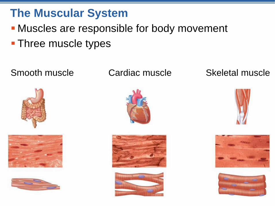
The Muscular System
Muscles are responsible for body movement
Three muscle types
Smooth muscle Cardiac muscle Skeletal muscle
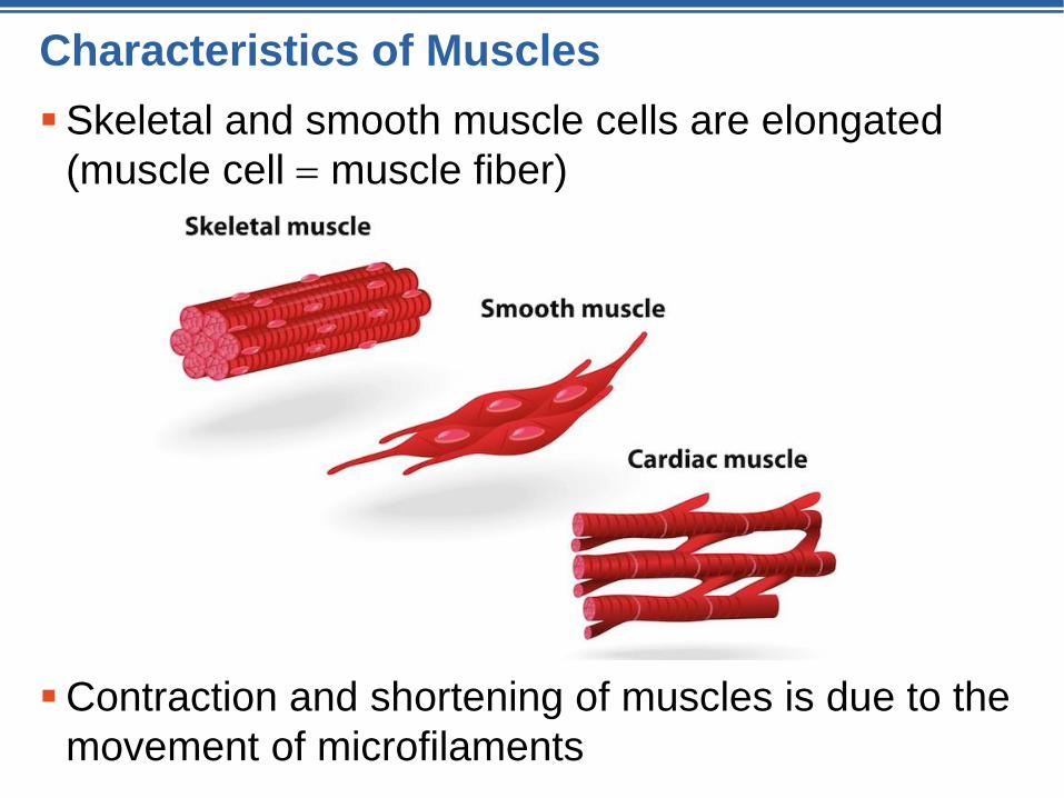
Characteristics of Muscles
Skeletal and smooth muscle cells are elongated
(muscle cell muscle fiber)
Contraction and shortening of muscles is due to the
movement of microfilaments
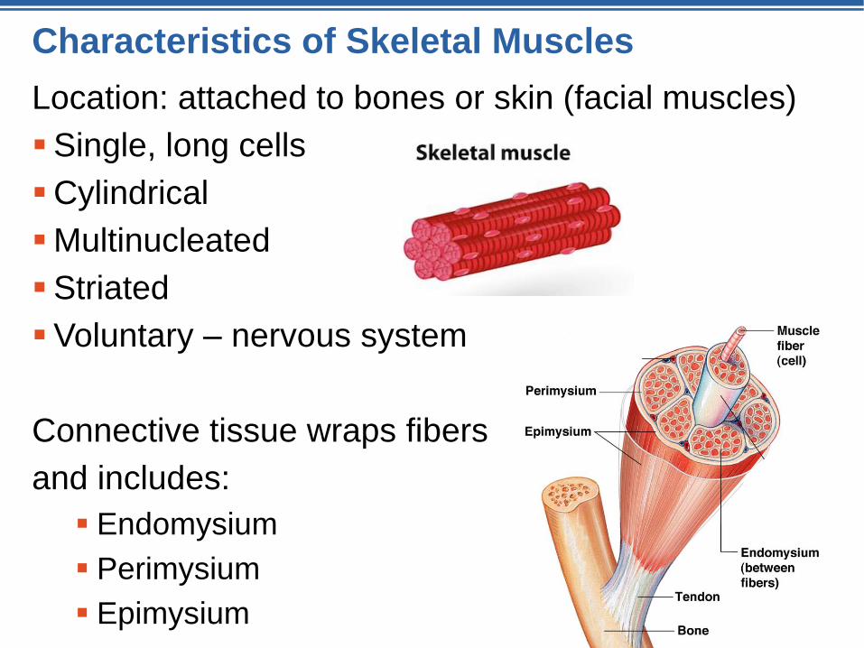
Characteristics of Skeletal Muscles
Location: attached to bones or skin (facial muscles)
Single, long cells
Cylindrical
Multinucleated
Striated
Voluntary – nervous system
Connective tissue wraps fibers
and includes:
Endomysium
Perimysium
Epimysium
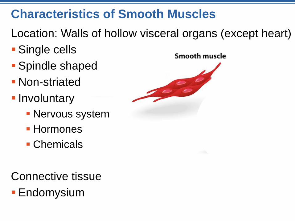
Characteristics of Smooth Muscles
Location: Walls of hollow visceral organs (except heart)
Single cells
Spindle shaped
Non-striated
Involuntary
Nervous system
Hormones
Chemicals
Connective tissue
Endomysium
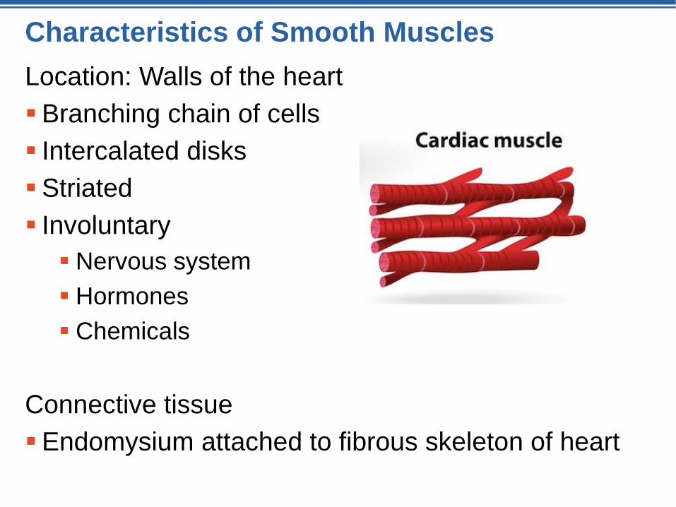
Characteristics of Smooth Muscles
Location: Walls of the heart
Branching chain of cells
Intercalated disks
Striated
Involuntary
Nervous system
Hormones
Chemicals
Connective tissue
Endomysium attached to fibrous skeleton of heart
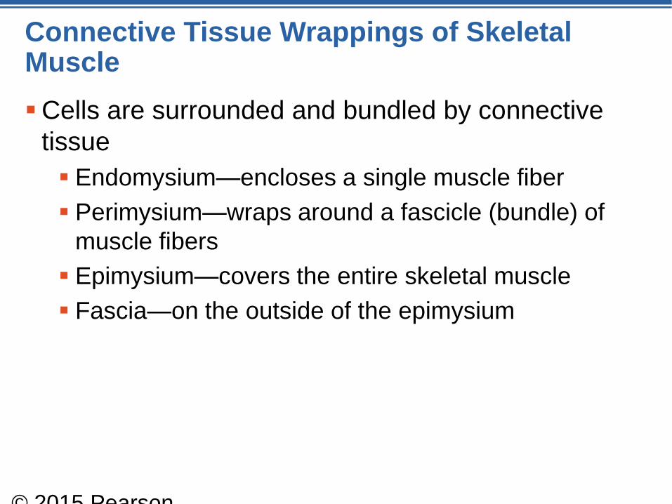
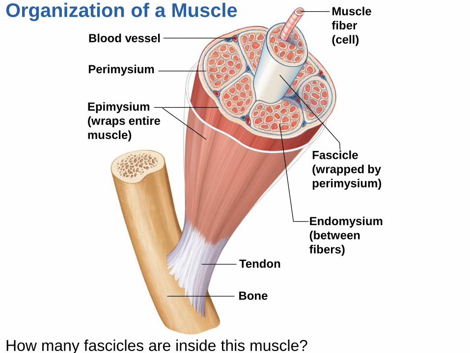
Connective Tissue Wrappings of Skeletal Muscle
Cells are surrounded and bundled by connective
tissue
Endomysium—encloses a single muscle fiber
Perimysium—wraps around a fascicle (bundle) of
muscle fibers
Epimysium—covers the entire skeletal muscle
Fascia—on the outside of the epimysium
© 2015 Pearson
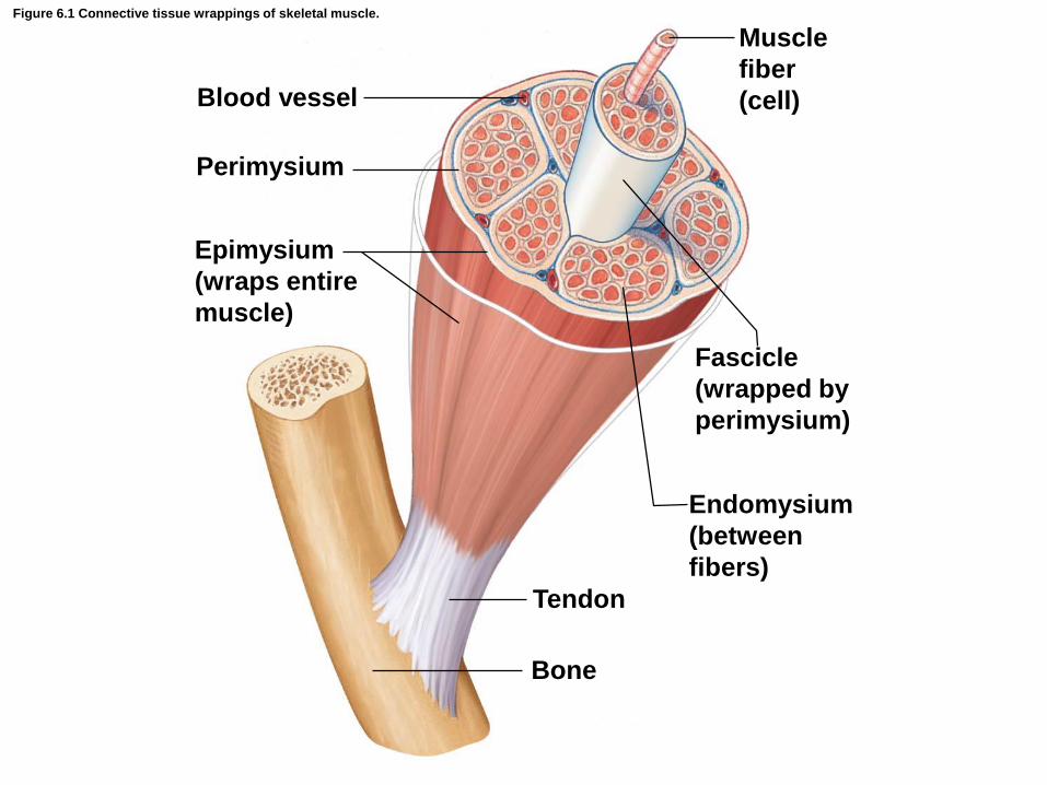
Figure 6.1 Connective tissue wrappings of skeletal muscle.
Blood vessel
Perimysium
Muscle
fiber
(cell)
Fascicle
(wrapped by
perimysium)
Endomysium
(between
fibers)
Tendon
Bone
Epimysium
(wraps entire
muscle)
Skeletal Muscle Attachments
Epimysium blends into a connective tissue
attachment
Tendons—cordlike structures
Mostly collagen fibers
Often cross a joint because of their toughness and
small size
Aponeuroses—sheetlike structures
Attach muscles indirectly to bones, cartilages, or
connective tissue coverings
© 2015 Pearson
Skeletal Muscle Attachments
Sites of muscle attachment
Bones
Cartilages
Connective tissue coverings
© 2015 Pearson
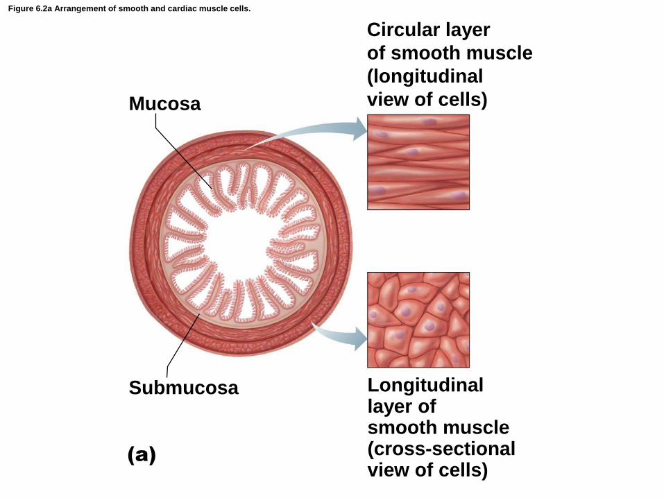
Figure 6.2a Arrangement of smooth and cardiac muscle cells.
Mucosa
Circular layer
of smooth muscle
(longitudinal
view of cells)
Submucosa
(a)
Longitudinallayer ofsmooth muscle(cross-sectionalview of cells)
© 2015 Pearson Education, Inc.
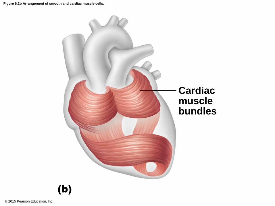
Figure 6.2b Arrangement of smooth and cardiac muscle cells.
Cardiacmusclebundles
(b)
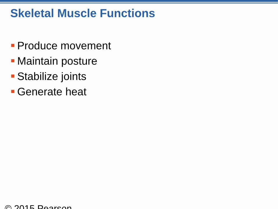
Skeletal Muscle Functions
Produce movement
Maintain posture
Stabilize joints
Generate heat
© 2015 Pearson
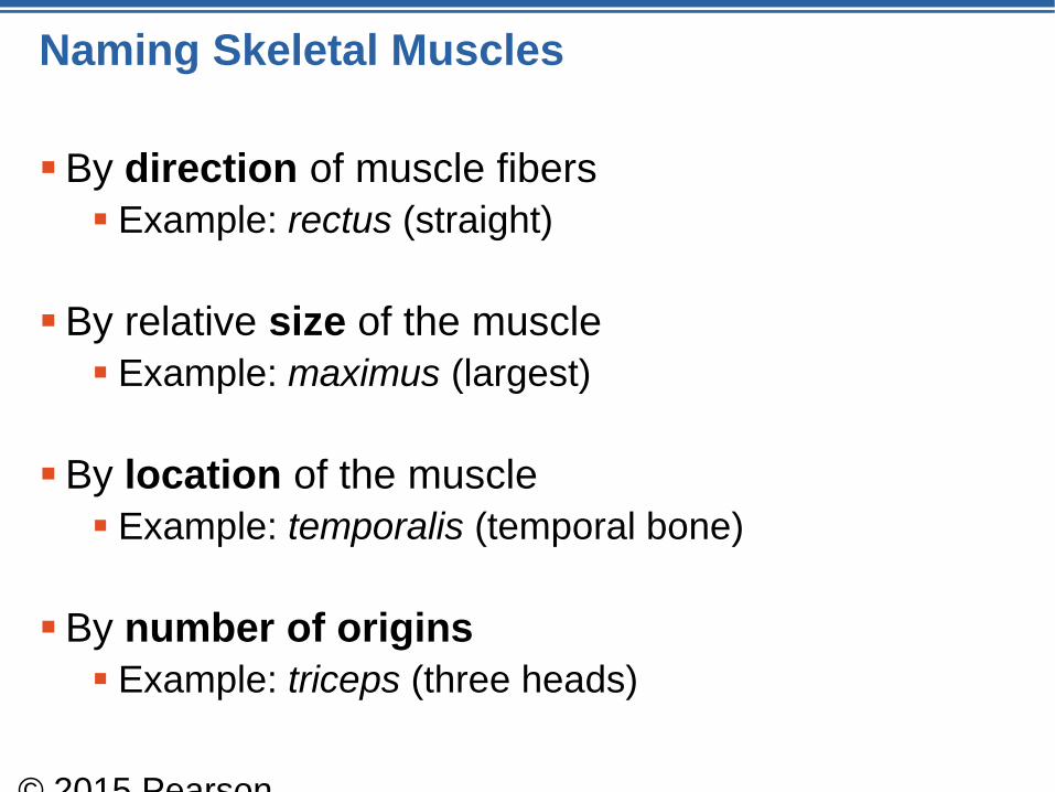
Naming Skeletal Muscles
By direction of muscle fibers
Example: rectus (straight)
By relative size of the muscle
Example: maximus (largest)
By location of the muscle
Example: temporalis (temporal bone)
By number of origins
Example: triceps (three heads)
© 2015 Pearson
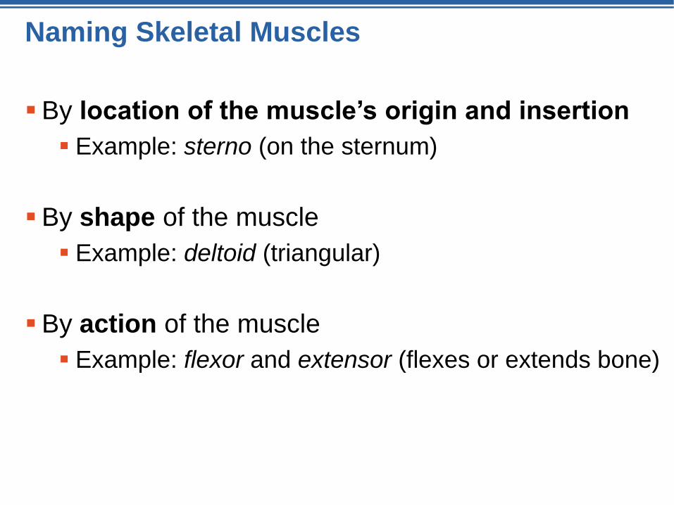
Naming Skeletal Muscles
By location of the muscle’s origin and insertion
Example: sterno (on the sternum)
By shape of the muscle
Example: deltoid (triangular)
By action of the muscle
Example: flexor and extensor (flexes or extends bone)
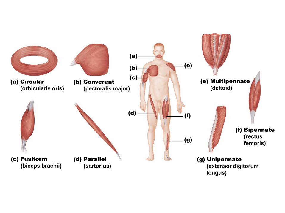
(a)
(b)
(c)
(e)
(f)
(g)
(a) Circular
(orbicularis oris)
(b) Converent
(pectoralis major)
(c) Fusiform
(biceps brachii)
(d) Parallel
(sartorius)
(e) Multipennate
(deltoid)
(g) Unipennate
(extensor digitorum
longus)
(f) Bipennate
(rectus
femoris)
(d)
Cranial
aponeurosis
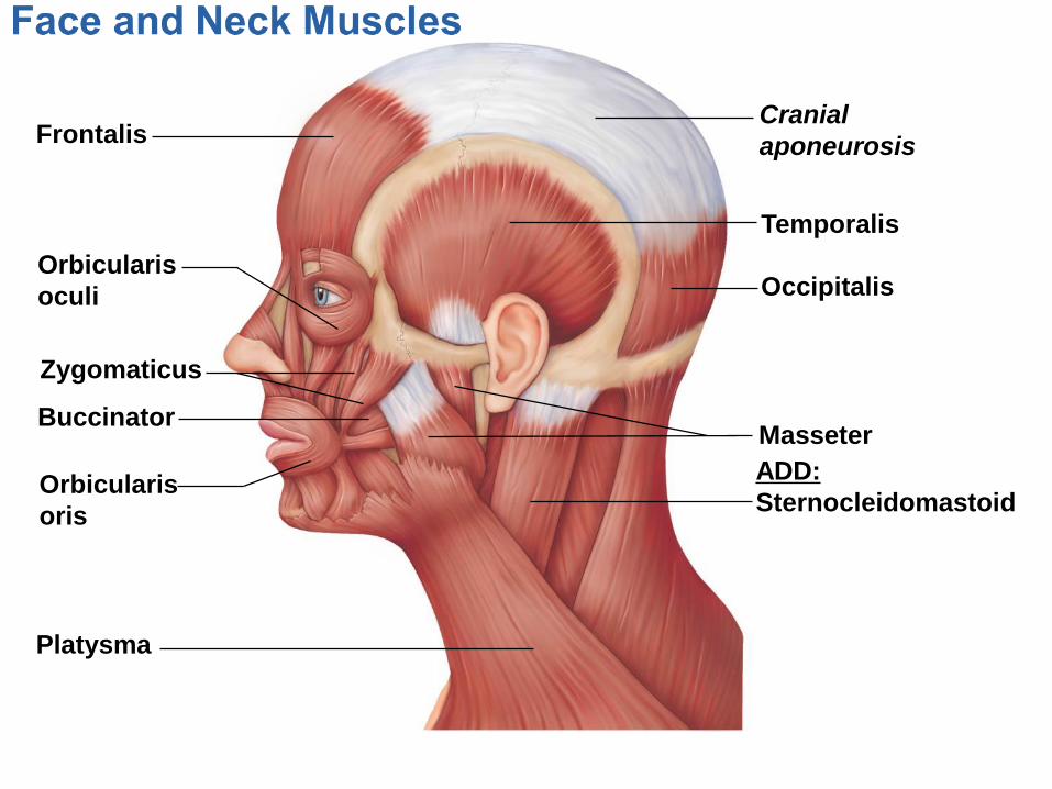
Frontalis
Orbicularis
oculi
Zygomaticus
Buccinator
Orbicularis
oris
Platysma
Masseter
ADD:
Sternocleidomastoid
Occipitalis
Temporalis
Cranial
aponeurosis
Microscopic Skeletal Anatomy
Sliding Filament Theory
Day 2
Blood vessel
Perimysium
Muscle
fiber
(cell)
Fascicle
(wrapped by
perimysium)
Endomysium
(between
fibers)
Tendon
Bone
Epimysium
(wraps entire
muscle)
How many fascicles are inside this muscle?
Organization of a Muscle
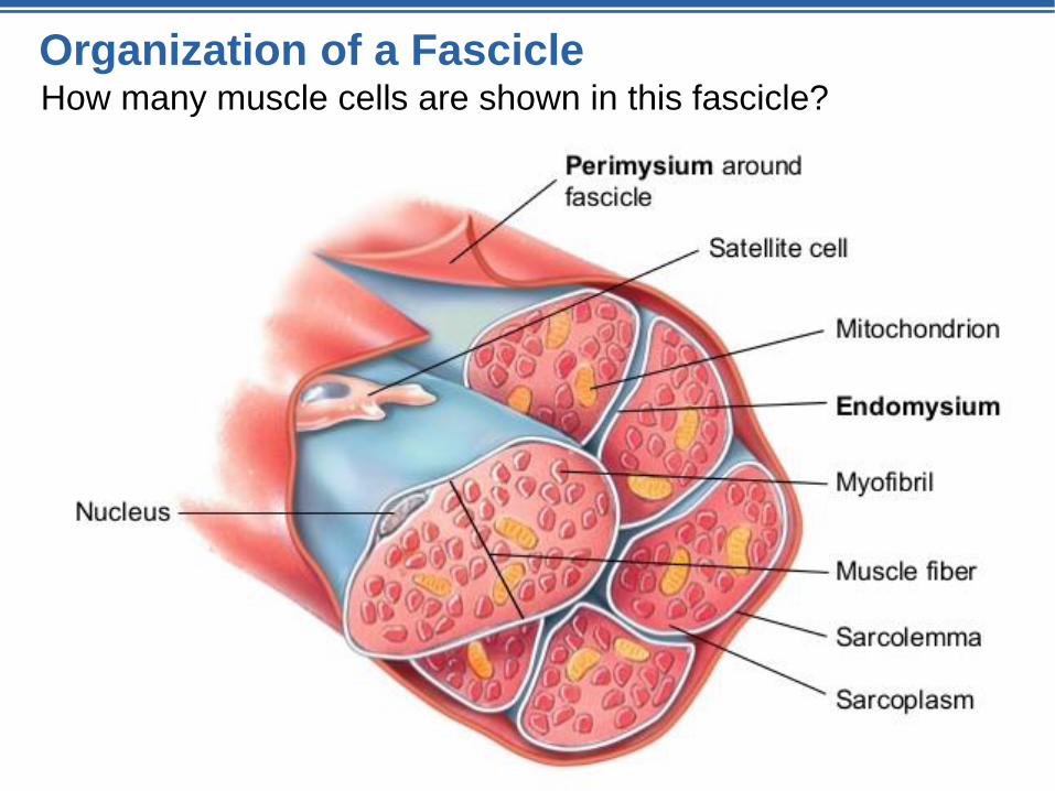
Organization of a FascicleHow many muscle cells are shown in this fascicle?
Inside the Muscle Cell
(skeletal muscle fiber)
Myofibrils
• Long, striated
organelle
Mitochondria
Sarcolemma
Cell plasma
membrane
Sarcolemma
Myofibril
NucleusDark
(A) band
Light
(I) band
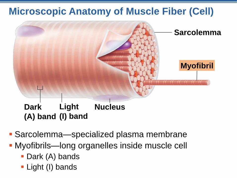
Microscopic Anatomy of Muscle Fiber (Cell)
Sarcolemma—specialized plasma membrane
Myofibrils—long organelles inside muscle cell
Dark (A) bands
Light (I) bands
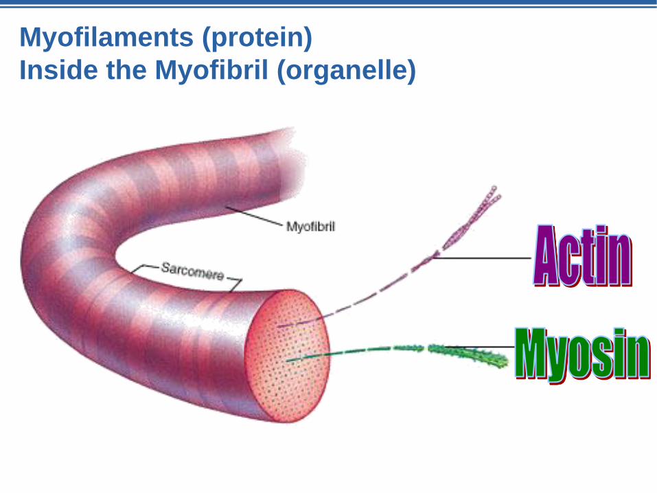
Myofilaments (protein)
Inside the Myofibril (organelle)
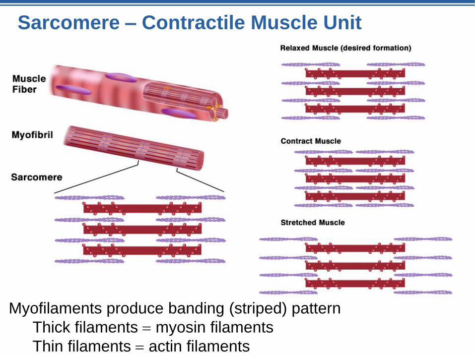
Sarcomere – Contractile Muscle Unit
Myofilaments produce banding (striped) pattern
Thick filaments myosin filaments
Thin filaments actin filaments
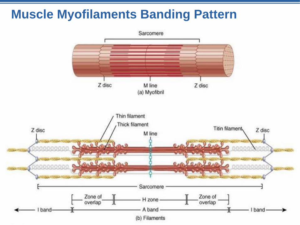
Muscle Myofilaments Banding Pattern
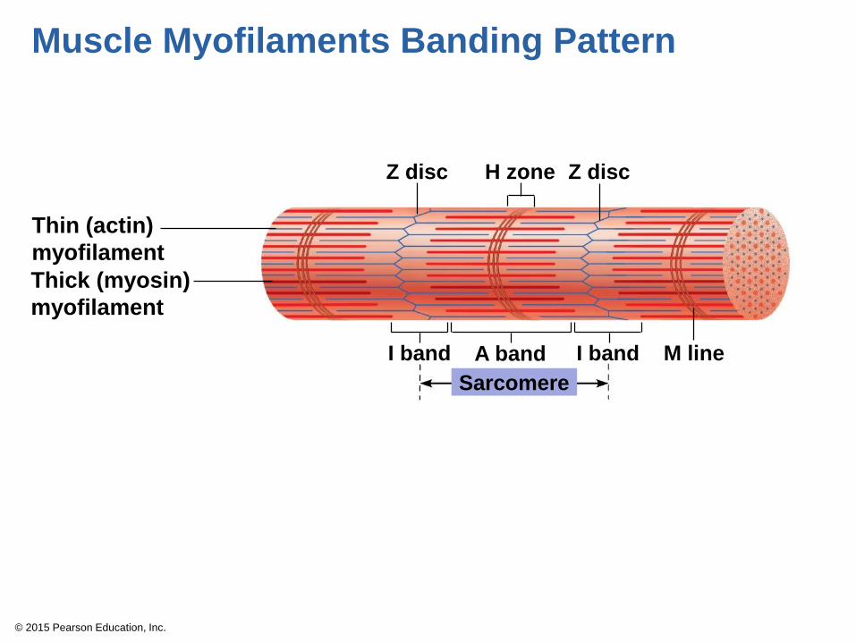
© 2015 Pearson Education, Inc.
Z discZ disc H zone
Thin (actin)
myofilament
Thick (myosin)
myofilament
I band A band M lineI band
Sarcomere
Muscle Myofilaments Banding Pattern
© 2015 Pearson Education, Inc.
Figure 6.3c Anatomy of a skeletal muscle fiber (cell).
Thin (actin)
myofilament
Thick (myosin)
myofilament
Sarcomere (segment of a myofibril)
Z discM line
Z disc
Sarcomere
(c)
© 2015 Pearson Education, Inc.
Myosin Actin
Z
I
Z
I
H
A
(a) Relaxed sarcomere
A
Z
I
Z
I
(b) Fully contracted sarcomere
Muscle Contraction: The Sliding Filament Theory
A&P Flix™: The Cross Bridge Cycle
https://www.youtube.com/watch?v=7O_ZHyPeIIA
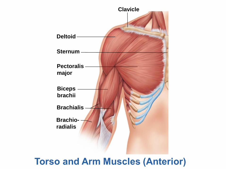
Clavicle
Deltoid
Sternum
Pectoralis
major
Biceps
brachii
Brachialis
Brachio-
radialis
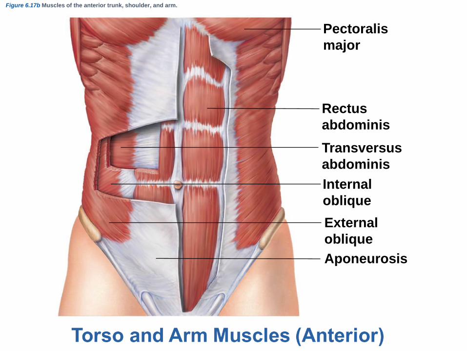
Figure 6.17b Muscles of the anterior trunk, shoulder, and arm.
Pectoralis
major
Rectus
abdominis
Transversus
abdominis
Internal
oblique
External
oblique
Aponeurosis
Skeletal Muscle Activity
and the Neuromuscular Junction
Day 3
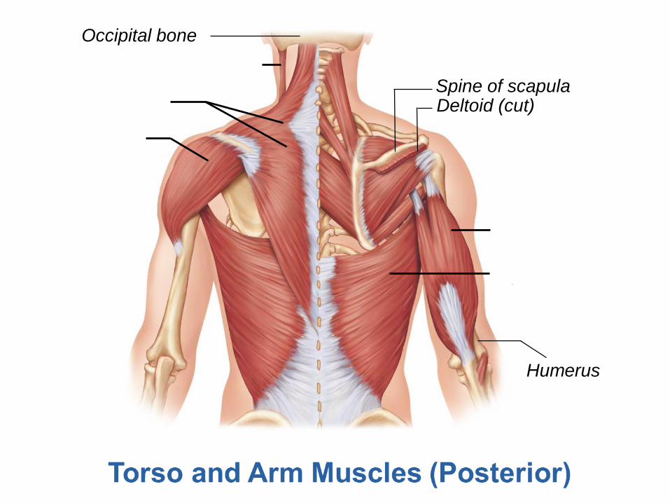
Occipital bone
Spine of scapulaDeltoid (cut)
Humerus

Occipital bone
Spine of scapulaDeltoid (cut)
Triceps brachii
Latissimus dorsi
Humerus
Deltoid
Trapezius
Sternocleidomastoid
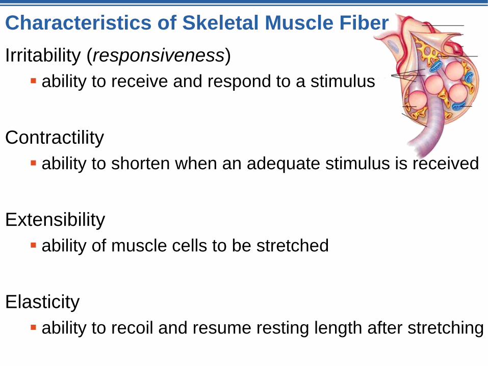
Characteristics of Skeletal Muscle Fiber
Irritability (responsiveness)
ability to receive and respond to a stimulus
Contractility
ability to shorten when an adequate stimulus is received
Extensibility
ability of muscle cells to be stretched
Elasticity
ability to recoil and resume resting length after stretching

Contraction of Skeletal Muscle
Sliding Filament Theory
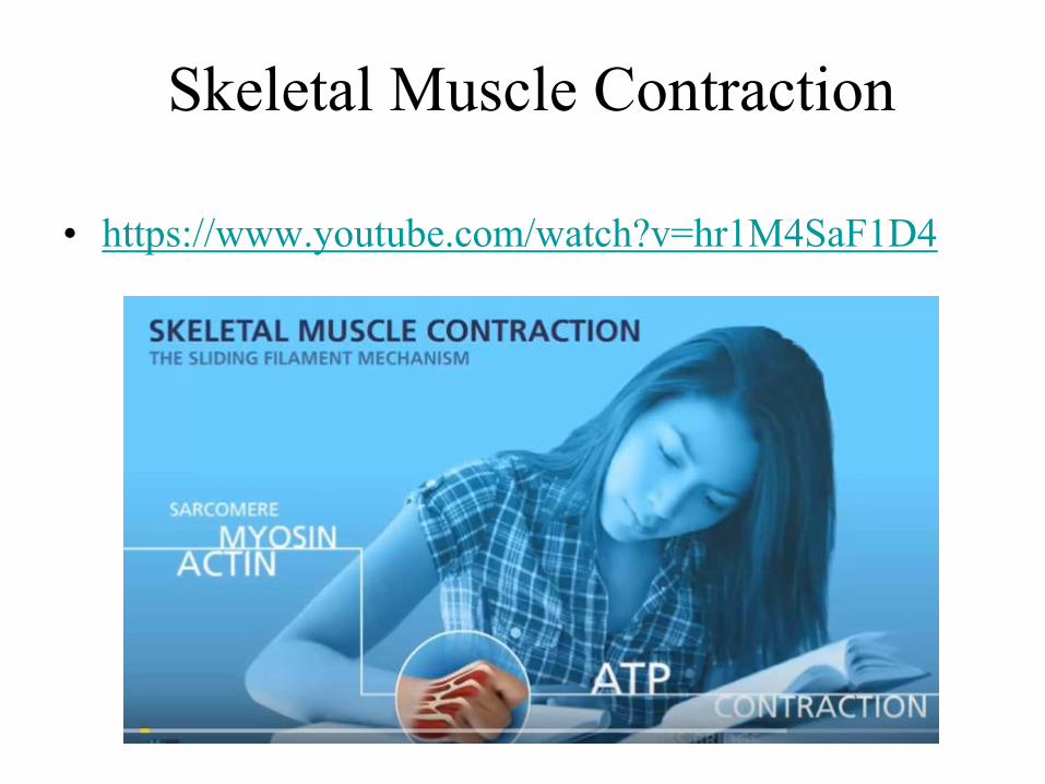
Skeletal Muscle Contraction
• https://www.youtube.com/watch?v=hr1M4SaF1D4
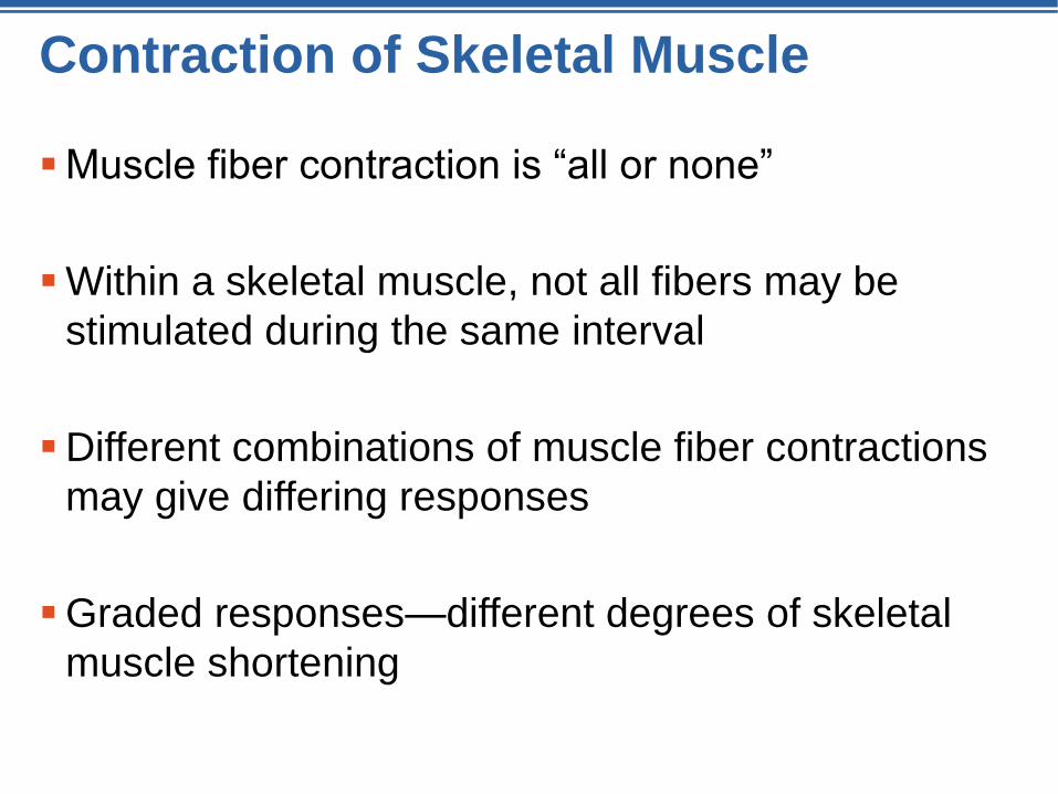
Contraction of Skeletal Muscle
Muscle fiber contraction is “all or none”
Within a skeletal muscle, not all fibers may be
stimulated during the same interval
Different combinations of muscle fiber contractions
may give differing responses
Graded responses—different degrees of skeletal
muscle shortening

Graded Responses Can Be Produced By…
Changing the frequency of muscle stimulation
Changing the number of cells stimulated at one time
Types of Graded Responses
Twitch• Single, brief contraction
• Not a normal function
(Stimuli)
Ten
sio
n (
g)
(Stimuli)
Te
ns
ion
(g
)
Summing of contractions• One contraction immediately
followed by another
• Stimulations are frequent, the
muscle does not completely
return to a resting state
• Effects are “summed” (added)
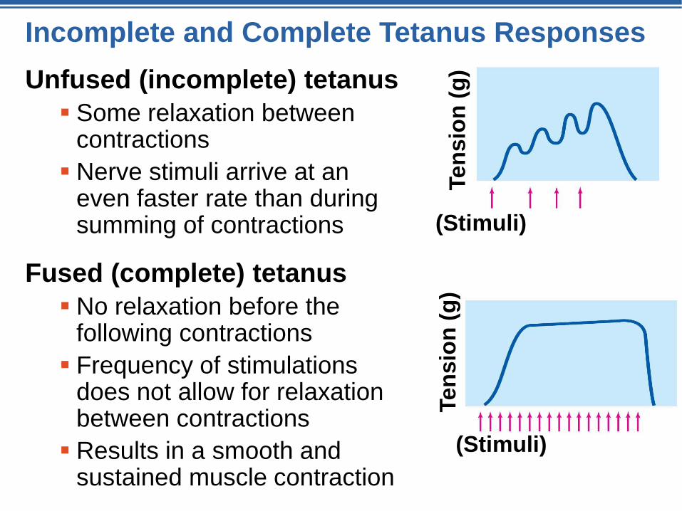
Incomplete and Complete Tetanus Responses
Unfused (incomplete) tetanus
Some relaxation between contractions
Nerve stimuli arrive at an even faster rate than during summing of contractions
Fused (complete) tetanus
No relaxation before the following contractions
Frequency of stimulations does not allow for relaxation between contractions
Results in a smooth and sustained muscle contraction
(Stimuli)
Ten
sio
n (
g)
(Stimuli)Te
nsio
n (
g)
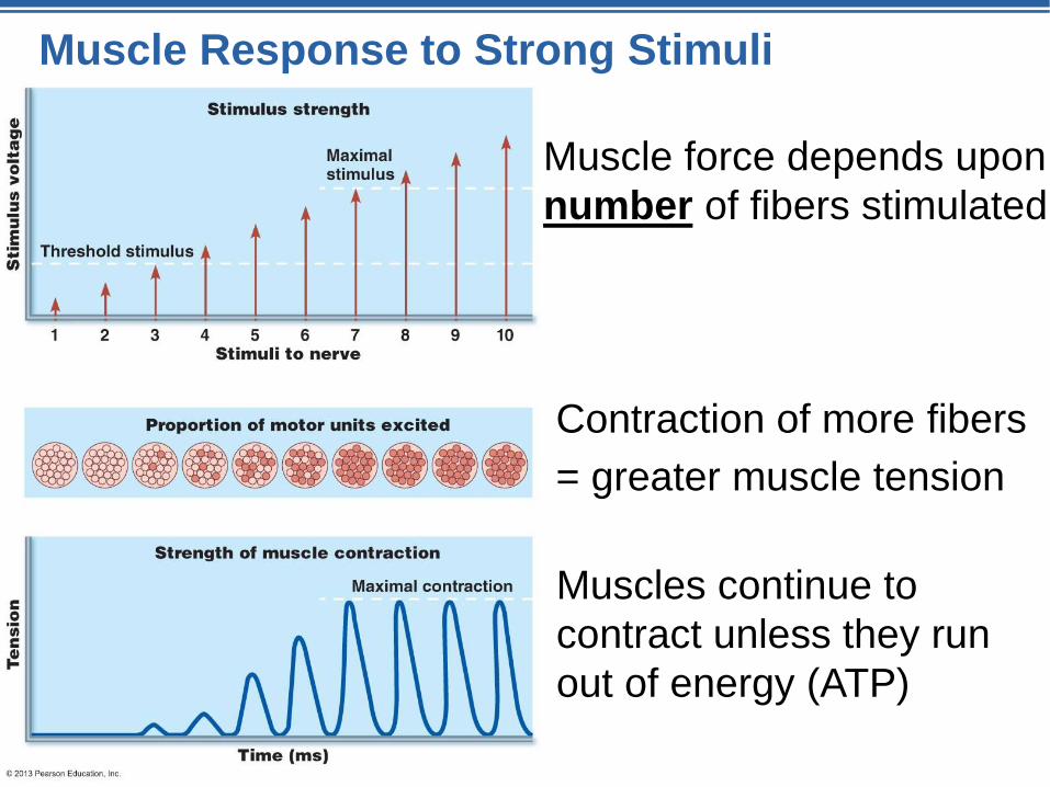
Muscle Response to Strong Stimuli
Muscle force depends upon
number of fibers stimulated
Contraction of more fibers
= greater muscle tension
Muscles continue to
contract unless they run
out of energy (ATP)
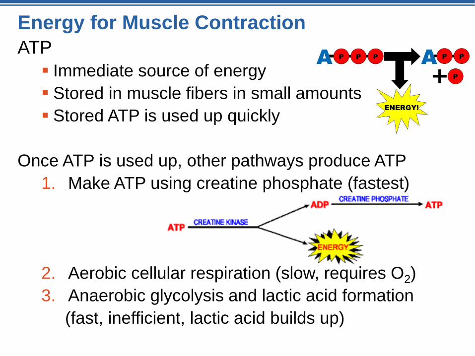
Energy for Muscle Contraction
ATP
Immediate source of energy
Stored in muscle fibers in small amounts
Stored ATP is used up quickly
Once ATP is used up, other pathways produce ATP
1. Make ATP using creatine phosphate (fastest)
2. Aerobic cellular respiration (slow, requires O2)
3. Anaerobic glycolysis and lactic acid formation
(fast, inefficient, lactic acid builds up)
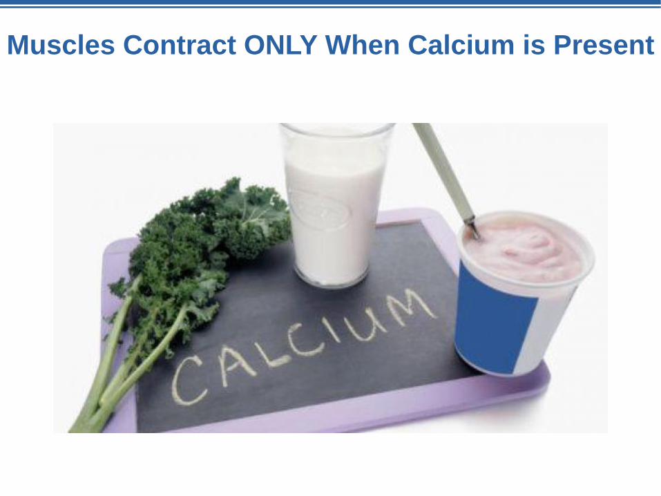
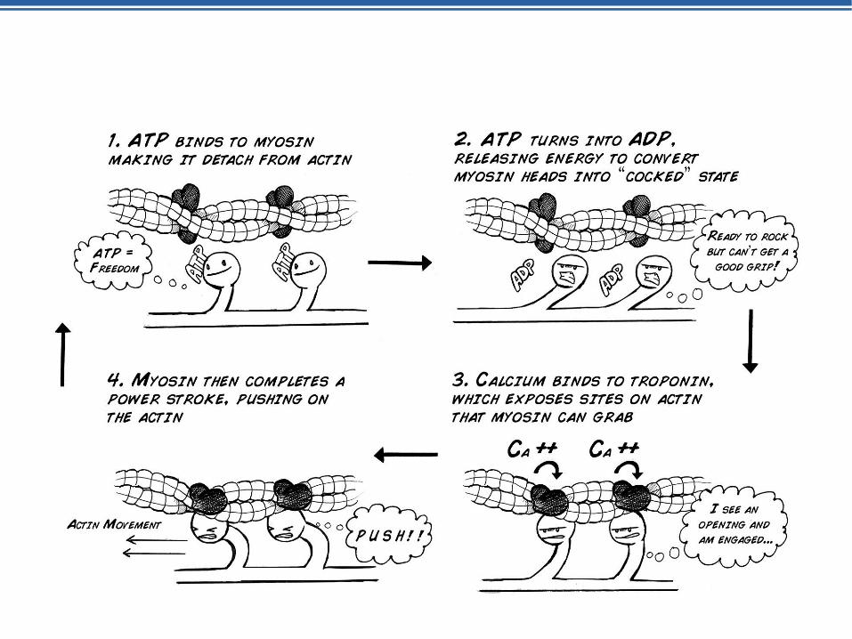
Muscles Contract ONLY When Calcium is Present
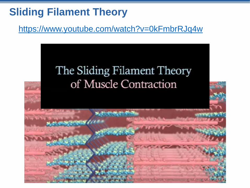
Sliding Filament Theory
https://www.youtube.com/watch?v=0kFmbrRJq4w
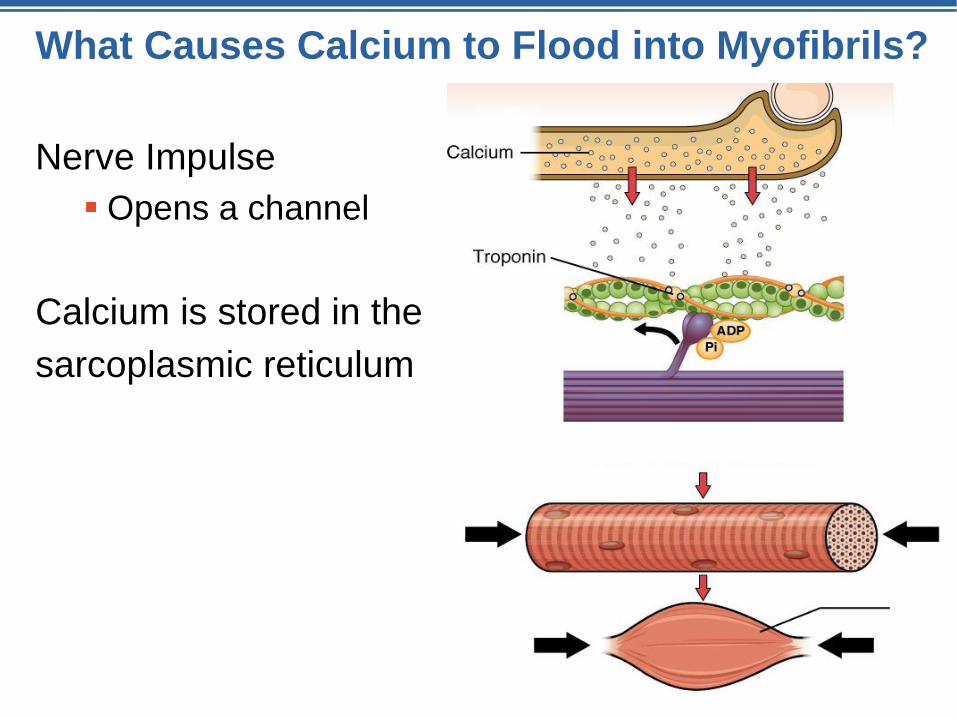
What Causes Calcium to Flood into Myofibrils?
Nerve Impulse
Opens a channel
Calcium is stored in the
sarcoplasmic reticulum
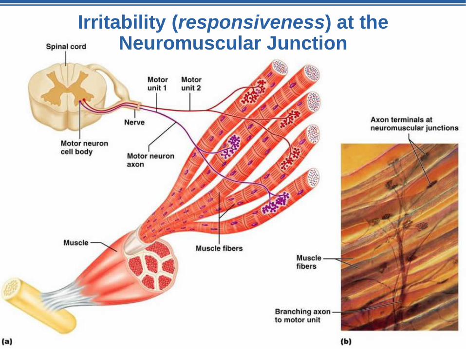
Irritability (responsiveness) at the Neuromuscular Junction
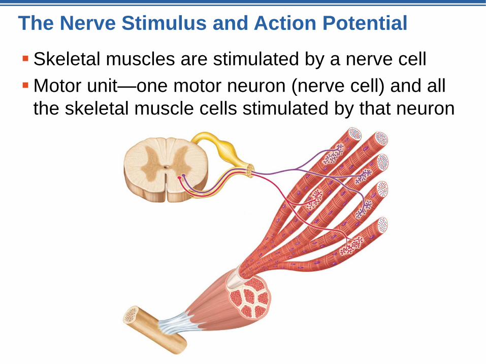
The Nerve Stimulus and Action Potential
Skeletal muscles are stimulated by a nerve cell
Motor unit—one motor neuron (nerve cell) and all
the skeletal muscle cells stimulated by that neuron
The Nerve Stimulus and Action Potential
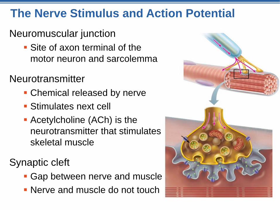
Neuromuscular junction
Site of axon terminal of the
motor neuron and sarcolemma
Neurotransmitter
Chemical released by nerve
Stimulates next cell
Acetylcholine (ACh) is the
neurotransmitter that stimulates
skeletal muscle
Synaptic cleft
Gap between nerve and muscle
Nerve and muscle do not touch
Contraction of Skeletal Muscle
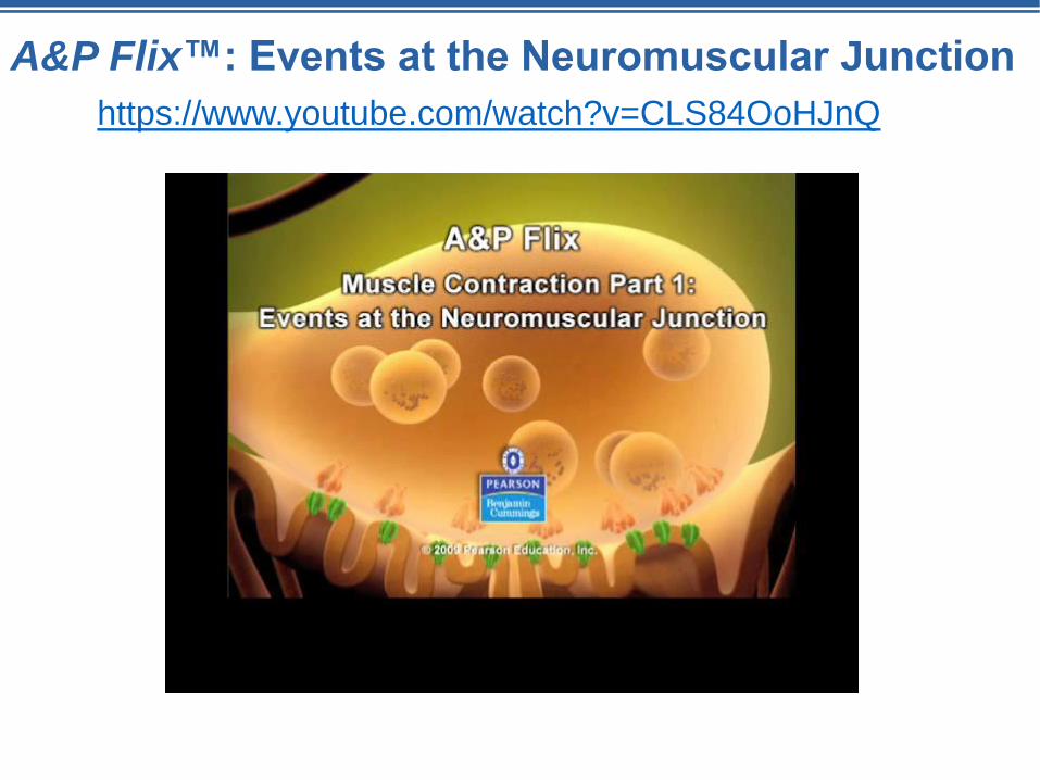
A&P Flix™: Events at the Neuromuscular Junction
https://www.youtube.com/watch?v=CLS84OoHJnQ
The Neuromuscular Junction
Day 4
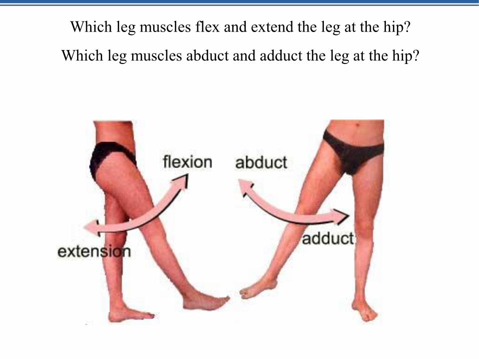
Which leg muscles flex and extend the leg at the hip?
Which leg muscles abduct and adduct the leg at the hip?
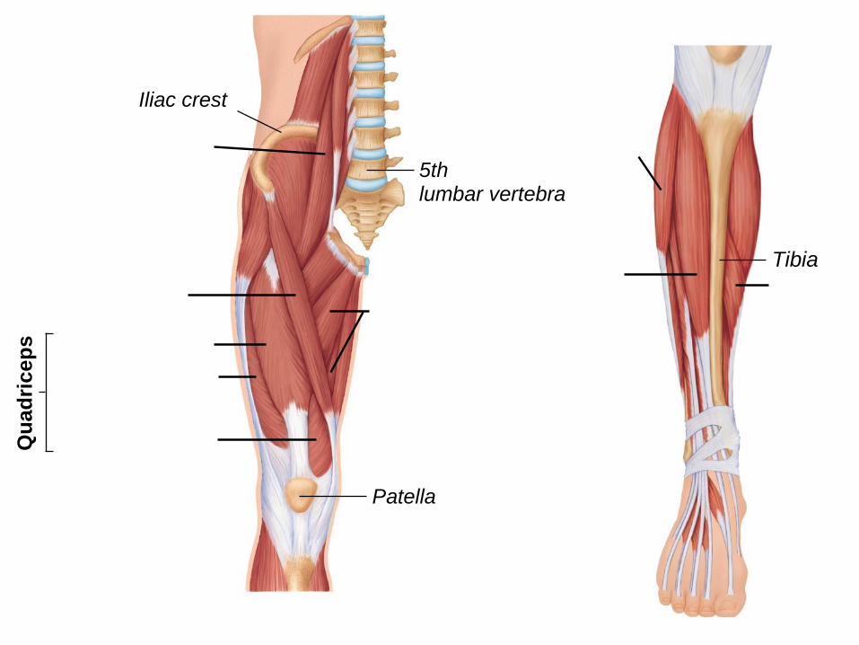
Tibia
Iliac crest
Qu
ad
ric
ep
s
5thlumbar vertebra
Patella
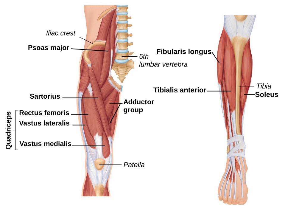
Tibia
Iliac crest
Qu
ad
ric
ep
s
5thlumbar vertebra
Patella
Sartorius
Rectus femoris
Vastus lateralis
Vastus medialis
Adductorgroup
Psoas majorFibularis longus
Tibialis anteriorSoleus
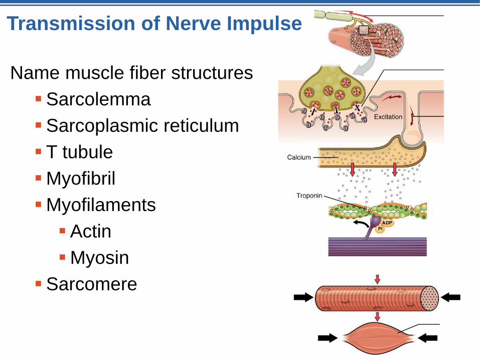
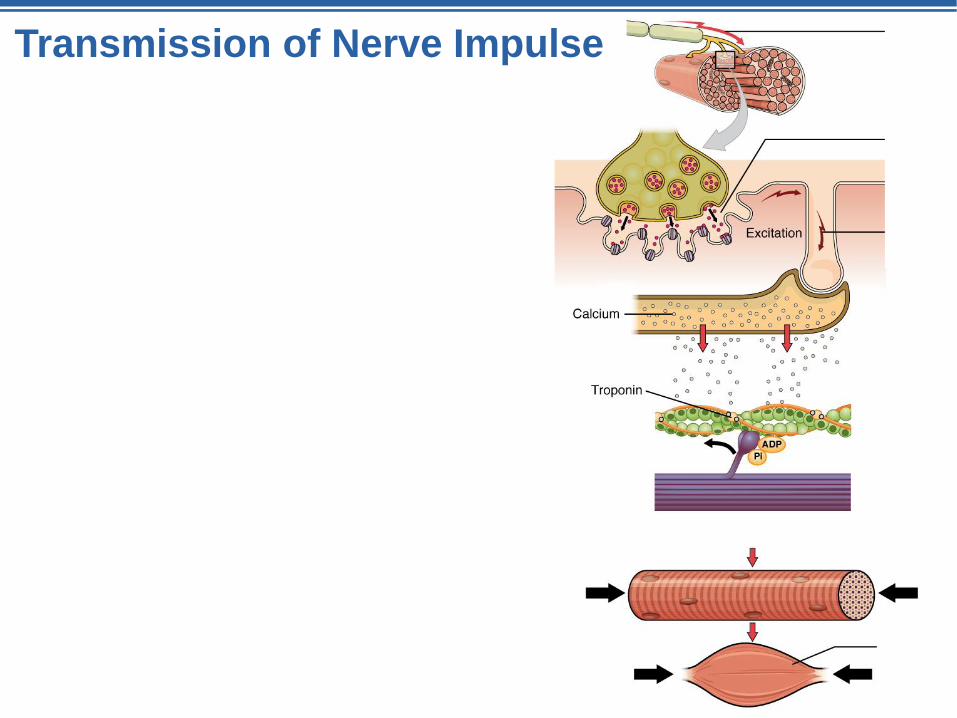
Transmission of Nerve Impulse
Name muscle fiber structures
Sarcolemma
Sarcoplasmic reticulum
T tubule
Myofibril
Myofilaments
Actin
Myosin
Sarcomere
Neuromuscular Junction and the
Story of Muscle Contraction
Day 5
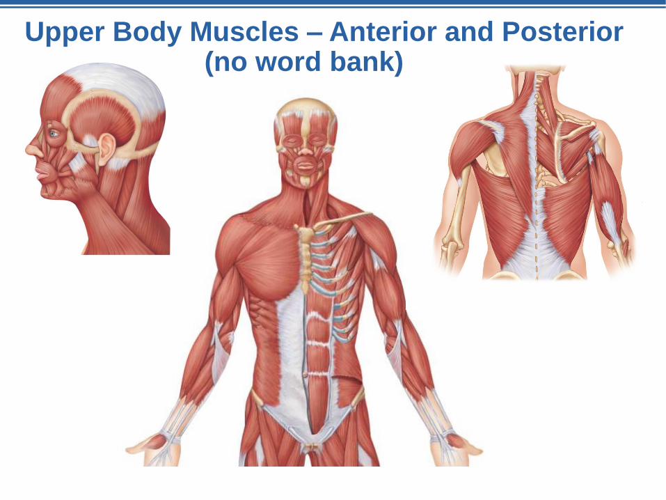
Upper Body Muscles – Anterior and Posterior(no word bank)
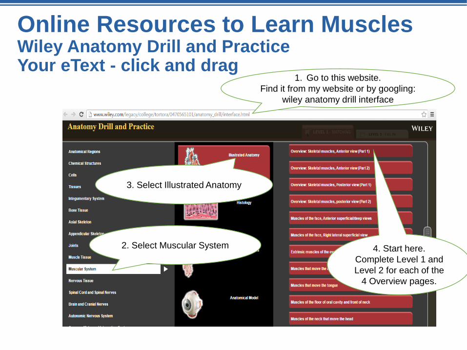
Online Resources to Learn MusclesWiley Anatomy Drill and PracticeYour eText - click and drag
1. Go to this website.
Find it from my website or by googling:
wiley anatomy drill interface
2. Select Muscular System
3. Select Illustrated Anatomy
4. Start here.
Complete Level 1 and
Level 2 for each of the
4 Overview pages.

Transmission of Nerve Impulse
Name muscle fiber structures
Sarcolemma
Sarcoplasmic reticulum
T tubule
Myofibril
Myofilaments
Actin
Myosin
Sarcomere

A&P Flix™: Events at the Neuromuscular Junction
https://www.youtube.com/watch?v=CLS84OoHJnQ

Events of the Neuromuscular Junction part 2
https://www.youtube.com/watch?v=IOkn1ldFO60

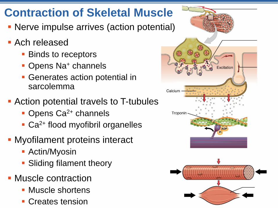
Contraction of Skeletal Muscle Nerve impulse arrives (action potential)
Ach released
Binds to receptors
Opens Na+ channels
Generates action potential in sarcolemma
Action potential travels to T-tubules
Opens Ca2+ channels
Ca2+ flood myofibril organelles
Myofilament proteins interact
Actin/Myosin
Sliding filament theory
Muscle contraction
Muscle shortens
Creates tension
Transmission of Nerve Impulse
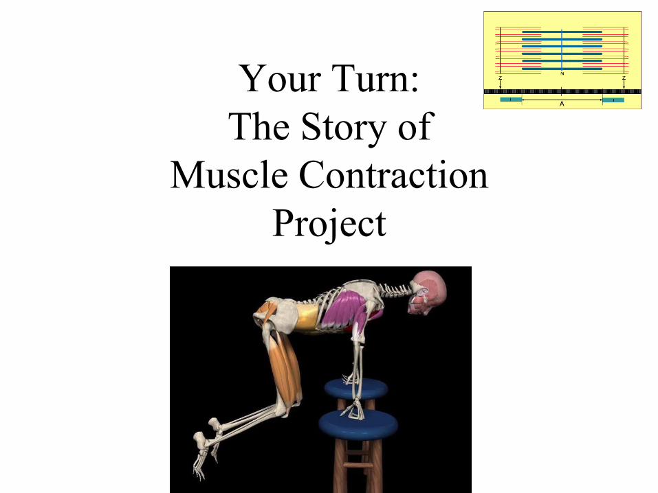
Your Turn:
The Story of
Muscle Contraction
Project
Contraction of Skeletal Muscle Nerve impulse arrives (action potential)
Ach released
Binds to receptors
Opens Na+ channels
Generates action potential in sarcolemma
Action potential travels to T-tubules
Opens Ca2+ channels
Ca2+ flood myofibril organelles
Myofilament proteins interact
Actin/Myosin
Sliding filament theory
Muscle contraction
Muscle shortens
Creates tension
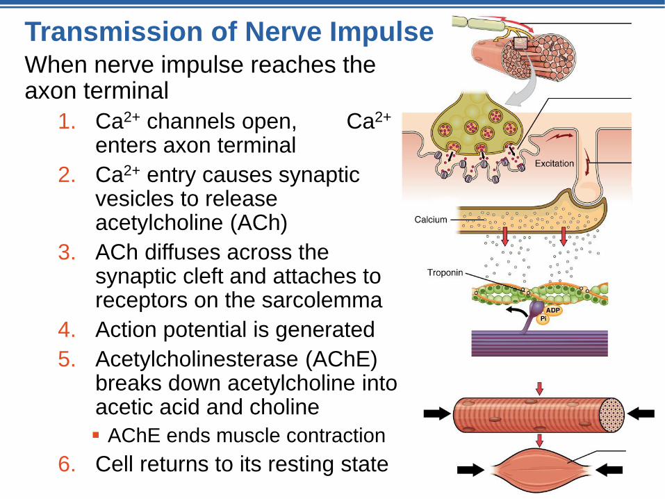
Transmission of Nerve ImpulseWhen nerve impulse reaches the axon terminal
1. Ca2+ channels open, Ca2+
enters axon terminal
2. Ca2+ entry causes synaptic vesicles to release acetylcholine (ACh)
3. ACh diffuses across the synaptic cleft and attaches to receptors on the sarcolemma
4. Action potential is generated
5. Acetylcholinesterase (AChE) breaks down acetylcholine into acetic acid and choline
AChE ends muscle contraction
6. Cell returns to its resting state


START HERE
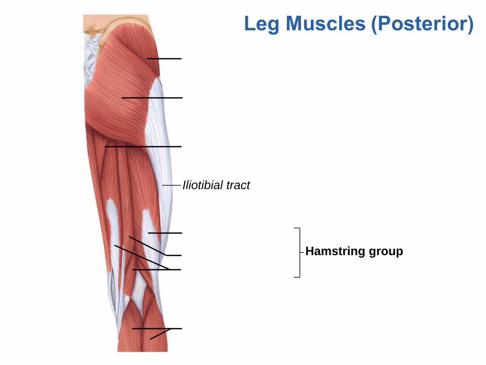
Iliotibial tract
Hamstring group
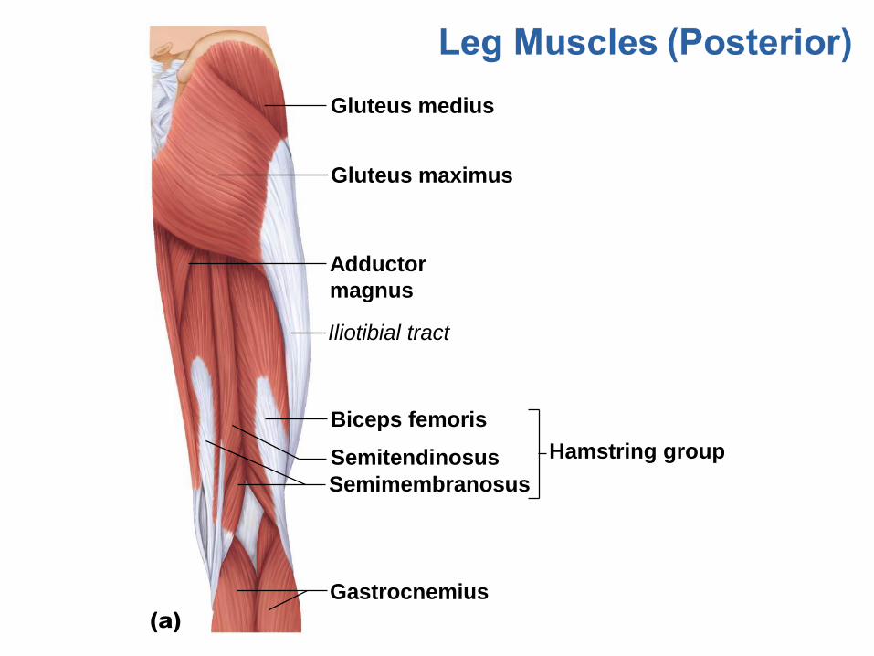
Gluteus medius
Gluteus maximus
Adductor
magnus
Iliotibial tract
Biceps femoris
Semitendinosus
Semimembranosus
Hamstring group
Gastrocnemius
(a)
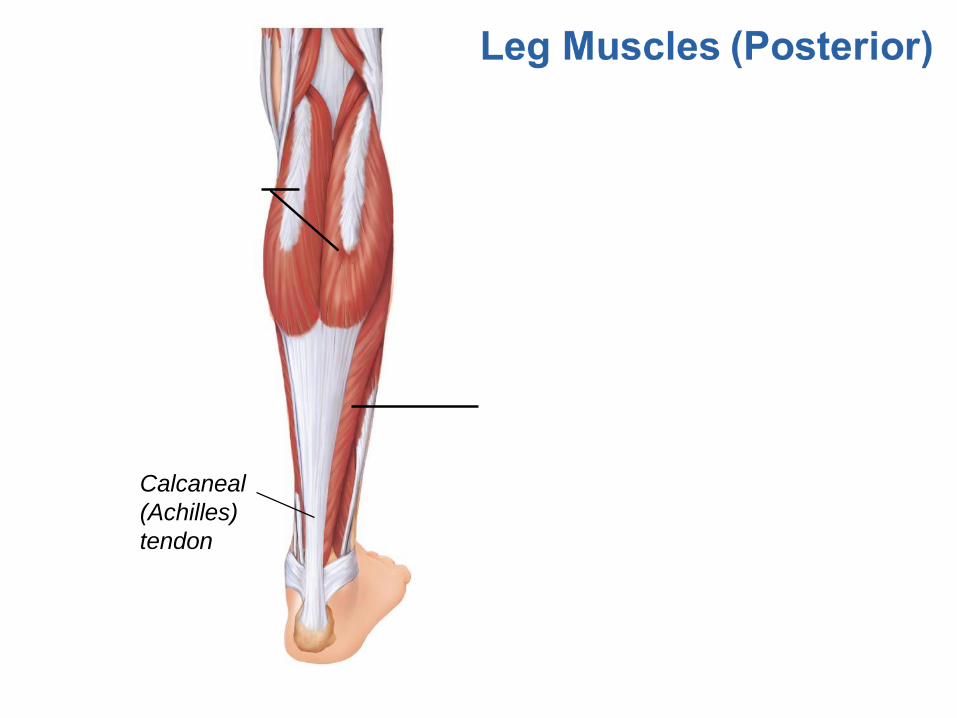
Calcaneal
(Achilles)
tendon
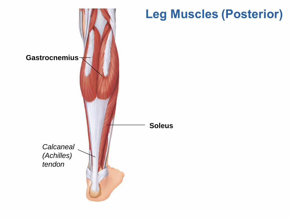
Gastrocnemius
Soleus
Calcaneal
(Achilles)
tendon
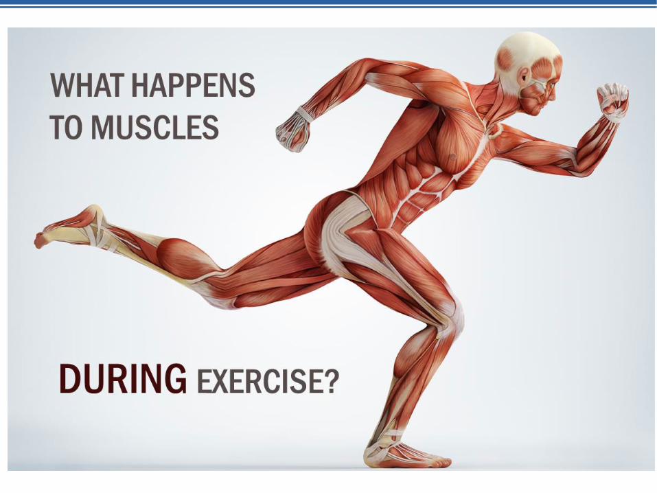
Muscle Movement
Muscle Fatigue
Day 6

Muscle Fatigue and Oxygen Deficit
If muscle activity is strenuous and prolonged,
muscle fatigue occurs because:
Ionic imbalances occur
Lactic acid accumulates in the muscle
Energy (ATP) supply decreases
After exercise, the oxygen deficit is repaid by rapid,
deep breathing

Your Turn:
The Story of
Muscle Contraction
Project
Muscle Movement & Exercise
Developmental Aspects
Day 7

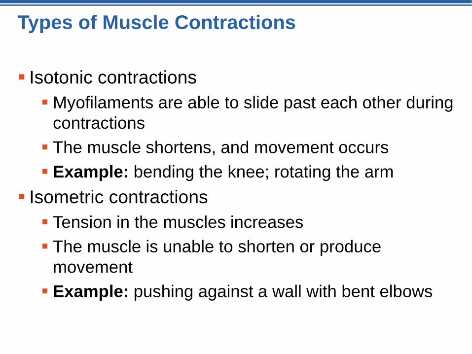
Types of Muscle Contractions
Isotonic contractions
Myofilaments are able to slide past each other during
contractions
The muscle shortens, and movement occurs
Example: bending the knee; rotating the arm
Isometric contractions
Tension in the muscles increases
The muscle is unable to shorten or produce
movement
Example: pushing against a wall with bent elbows
Muscle Tone
Muscle tone keeps muscles healthy and ready to
react
Result of a staggered series of nerve impulses
delivered to different cells within the muscle
If the nerve supply is destroyed, the muscle loses
tone, becomes paralyzed, and atrophies
© 2015 Pearson
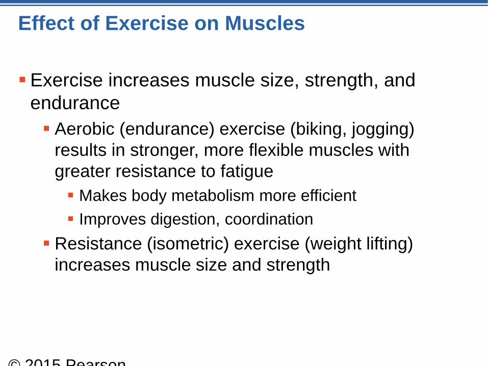
Effect of Exercise on Muscles
Exercise increases muscle size, strength, and
endurance
Aerobic (endurance) exercise (biking, jogging)
results in stronger, more flexible muscles with
greater resistance to fatigue
Makes body metabolism more efficient
Improves digestion, coordination
Resistance (isometric) exercise (weight lifting)
increases muscle size and strength
© 2015 Pearson
© 2015 Pearson Education, Inc.
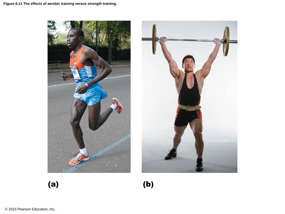
Figure 6.11 The effects of aerobic training versus strength training.
(a) (b)

© 2015 Pearson Education, Inc.
Table 6.2 The Five Golden Rules of Skeletal Muscle Activity.
Leg Muscles (Posterior)
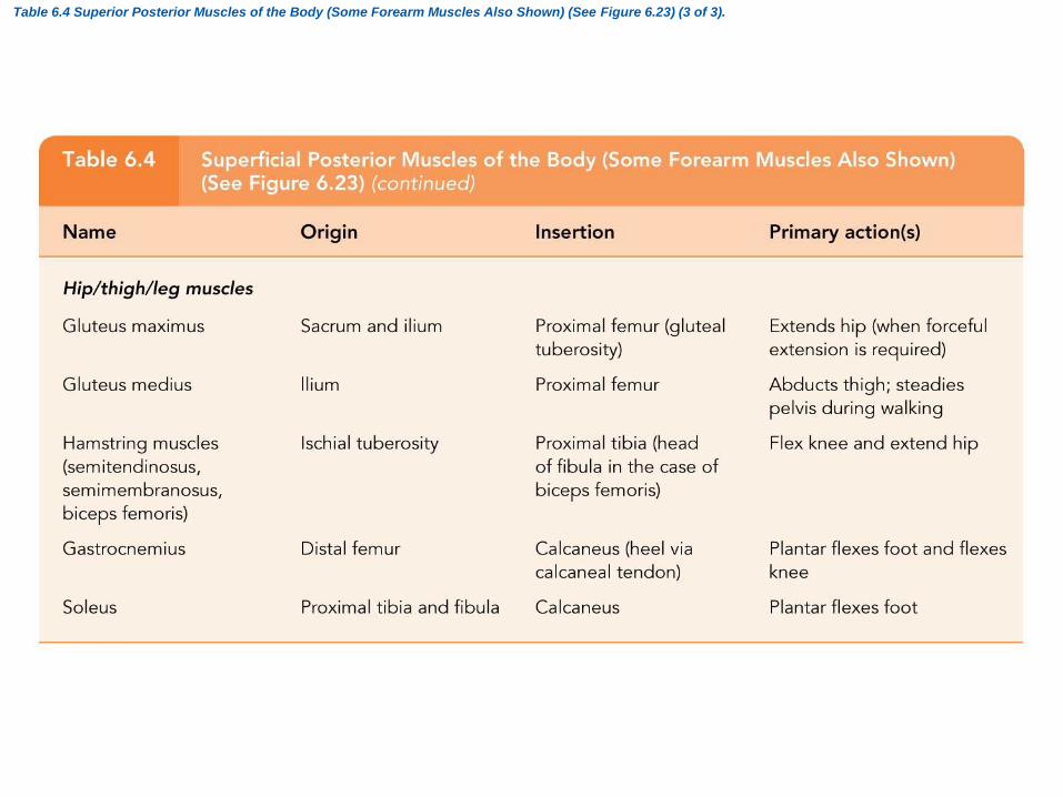
Table 6.4 Superior Posterior Muscles of the Body (Some Forearm Muscles Also Shown) (See Figure 6.23) (3 of 3).

Leg Muscles (no word bank)
Review
Anatomy – Skeletal Muscles
Physiology – Muscle Contraction,
Neuromuscular Junction,
Movement
Day 8