nerve activates contraction -...
TRANSCRIPT

ELAINE N. MARIEB
EIGHTH EDITION
Copyright © 2006 Pearson Education, Inc., publishing as Benjamin Cummings
PowerPoint® Lecture Slide Presentation by Jerry L. Cook, Sam Houston University
ESSENTIALS
OF HUMAN
ANATOMY
& PHYSIOLOGY
Special Senses
Copyright © 2006 Pearson Education, Inc., publishing as Benjamin Cummings
The Senses General senses of touch
Temperature
Pressure
Pain
Special senses
Smell
Taste
Sight
Hearing
Equilibrium
Copyright © 2006 Pearson Education, Inc., publishing as Benjamin Cummings
The Eye and Vision
70 percent of all sensory receptors are in the
eyes
2.5 cm (1 inch) diameter &
5 cm (2 inches) apart
Each eye has over a million nerve fibers
Protection for the eye
Most of the eye is enclosed in a bony orbit
A cushion of fat surrounds most of the eye
Copyright © 2006 Pearson Education, Inc., publishing as Benjamin Cummings
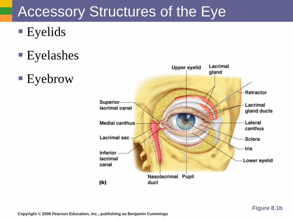
Accessory Structures of the Eye
Eyelids
Eyelashes
Eyebrow
Figure 8.1b
Copyright © 2006 Pearson Education, Inc., publishing as Benjamin Cummings
Accessory Structures of the Eye
Conjunctiva
Membrane that lines the eyelids
Connects to the surface of the eye
Secretes mucus to lubricate the eye
Copyright © 2006 Pearson Education, Inc., publishing as Benjamin Cummings
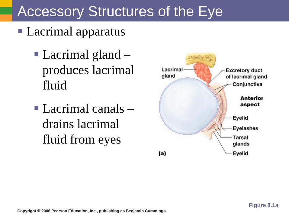
Accessory Structures of the Eye
Lacrimal apparatus
Lacrimal gland –
produces lacrimal
fluid
Lacrimal canals –
drains lacrimal
fluid from eyes
Figure 8.1a
Copyright © 2006 Pearson Education, Inc., publishing as Benjamin Cummings
Function of the Lacrimal Apparatus
Properties of lacrimal fluid
Dilute salt solution (tears)
Contains antibodies and lysozyme
Protects, moistens, and lubricates the eye
Empties into the nasal cavity
Copyright © 2006 Pearson Education, Inc., publishing as Benjamin Cummings
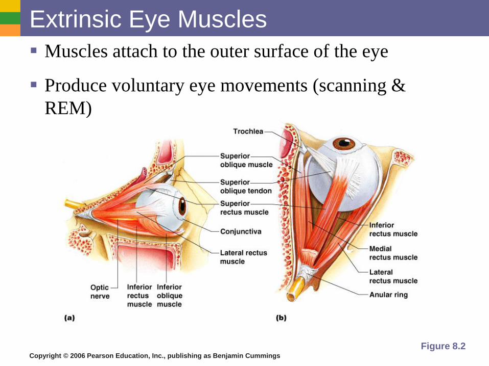
Extrinsic Eye Muscles
Muscles attach to the outer surface of the eye
Produce voluntary eye movements (scanning &
REM)
Figure 8.2
Copyright © 2006 Pearson Education, Inc., publishing as Benjamin Cummings
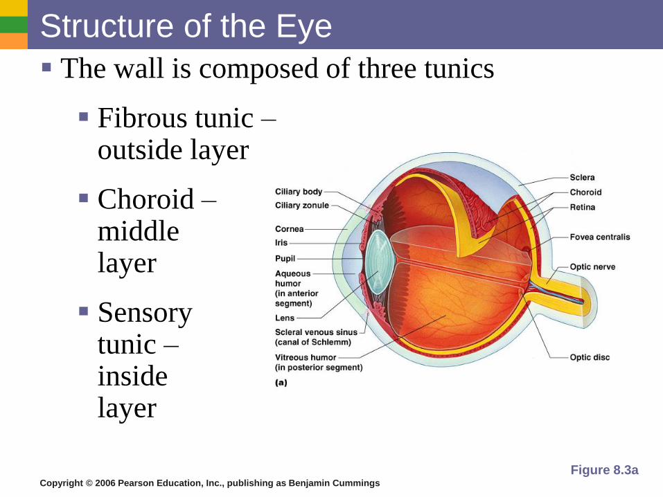
Structure of the Eye
The wall is composed of three tunics
Fibrous tunic – outside layer
Choroid – middle layer
Sensory tunic – inside layer
Figure 8.3a
Copyright © 2006 Pearson Education, Inc., publishing as Benjamin Cummings
The Fibrous Tunic
Sclera
White connective tissue layer – maintains shape
Seen anteriorly as the “white of the eye”
Cornea
Transparent, central anterior portion
Avascular but contains pain receptors
Allows for light to pass through
Repairs itself easily
The only human tissue that can be transplanted
without fear of rejection
Copyright © 2006 Pearson Education, Inc., publishing as Benjamin Cummings
Choroid Layer
Blood-rich nutritive tunic
Non-reflective pigment prevents light from
scattering
Modified interiorly into two structures
Cilliary body – smooth muscle
Controls shape of lens
Iris - pigmented layer that gives eye color
Pupil – rounded muscular opening in the iris
Copyright © 2006 Pearson Education, Inc., publishing as Benjamin Cummings
Sensory Tunic (Retina)
Contains receptor cells (photoreceptors)
Rods
Cones – bright light, high acuity, color
Signals leave the retina toward the brain
through the optic nerve
Copyright © 2006 Pearson Education, Inc., publishing as Benjamin Cummings
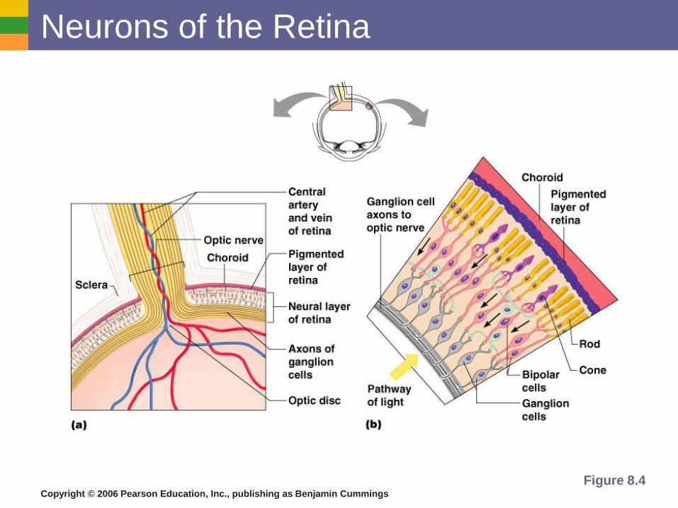
Neurons of the Retina
Figure 8.4
Copyright © 2006 Pearson Education, Inc., publishing as Benjamin Cummings
Neurons of the Retina and Vision
Rods
Most are found towards the edges of the
retina
Allow dim light vision and peripheral
vision
Perception is all in gray tones
Creates fuzzy images
Copyright © 2006 Pearson Education, Inc., publishing as Benjamin Cummings
Neurons of the Retina and Vision
Cones
Used in bright light
Allow for detailed color vision
Densest in the center of the retina
Fovea centralis – area of the retina with
only cones
No photoreceptor cells are at the optic disk,
or blind spot (nerve gathering place)
Copyright © 2006 Pearson Education, Inc., publishing as Benjamin Cummings
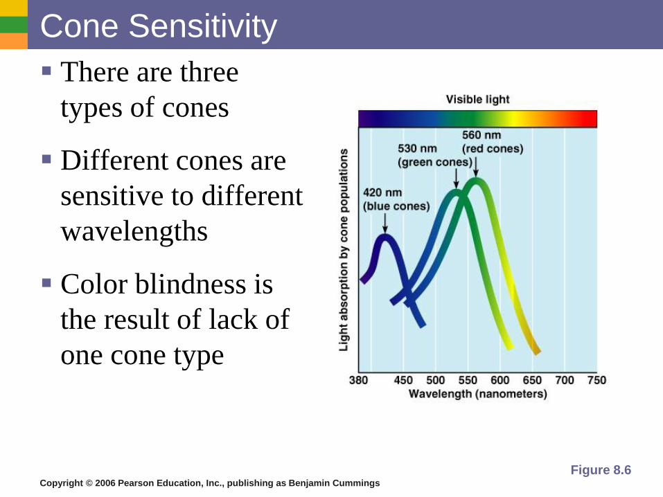
Cone Sensitivity
There are three
types of cones
Different cones are
sensitive to different
wavelengths
Color blindness is
the result of lack of
one cone type
Figure 8.6
Copyright © 2006 Pearson Education, Inc., publishing as Benjamin Cummings
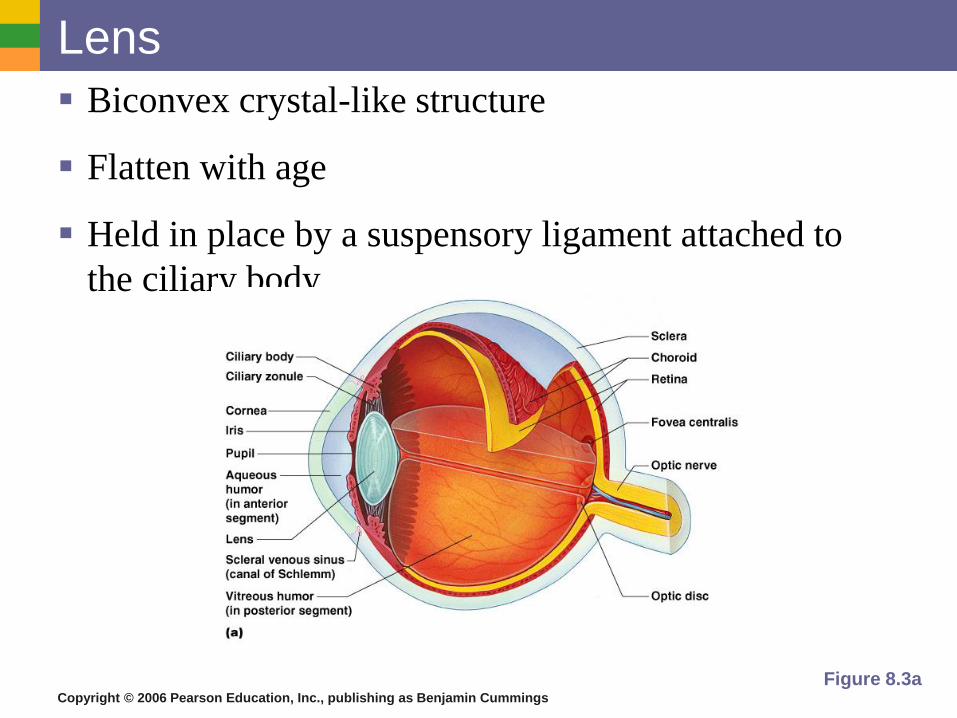
Lens
Biconvex crystal-like structure
Flatten with age
Held in place by a suspensory ligament attached to
the ciliary body
Figure 8.3a
Copyright © 2006 Pearson Education, Inc., publishing as Benjamin Cummings
Internal Eye Chamber Fluids
Aqueous humor
Watery fluid found in chamber between the lens and cornea
Similar to blood plasma
Helps maintain intraocular pressure -shape
Provides nutrients for the lens and cornea
Reabsorbed into venous blood through the canal of Schlemm
Refracts light
Copyright © 2006 Pearson Education, Inc., publishing as Benjamin Cummings
Internal Eye Chamber Fluids
Vitreous humor
Gel-like substance behind the lens
Keeps the eye from collapsing –shape
Lasts a lifetime and is not replaced
Refracts light
Copyright © 2006 Pearson Education, Inc., publishing as Benjamin Cummings
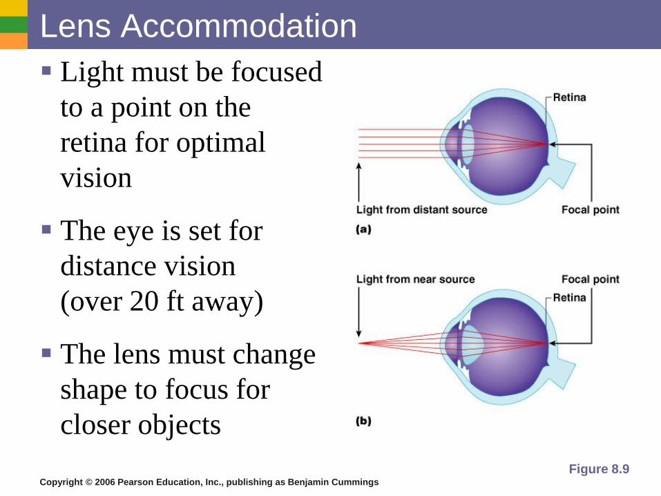
Lens Accommodation
Light must be focused
to a point on the
retina for optimal
vision
The eye is set for
distance vision
(over 20 ft away)
The lens must change
shape to focus for
closer objects
Figure 8.9
Copyright © 2006 Pearson Education, Inc., publishing as Benjamin Cummings
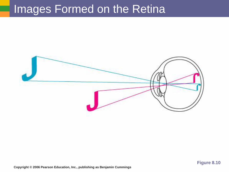
Images Formed on the Retina
Figure 8.10
Copyright © 2006 Pearson Education, Inc., publishing as Benjamin Cummings
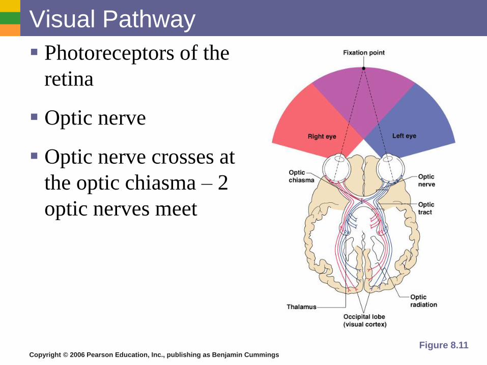
Visual Pathway
Photoreceptors of the
retina
Optic nerve
Optic nerve crosses at
the optic chiasma – 2
optic nerves meet
Figure 8.11
Copyright © 2006 Pearson Education, Inc., publishing as Benjamin Cummings
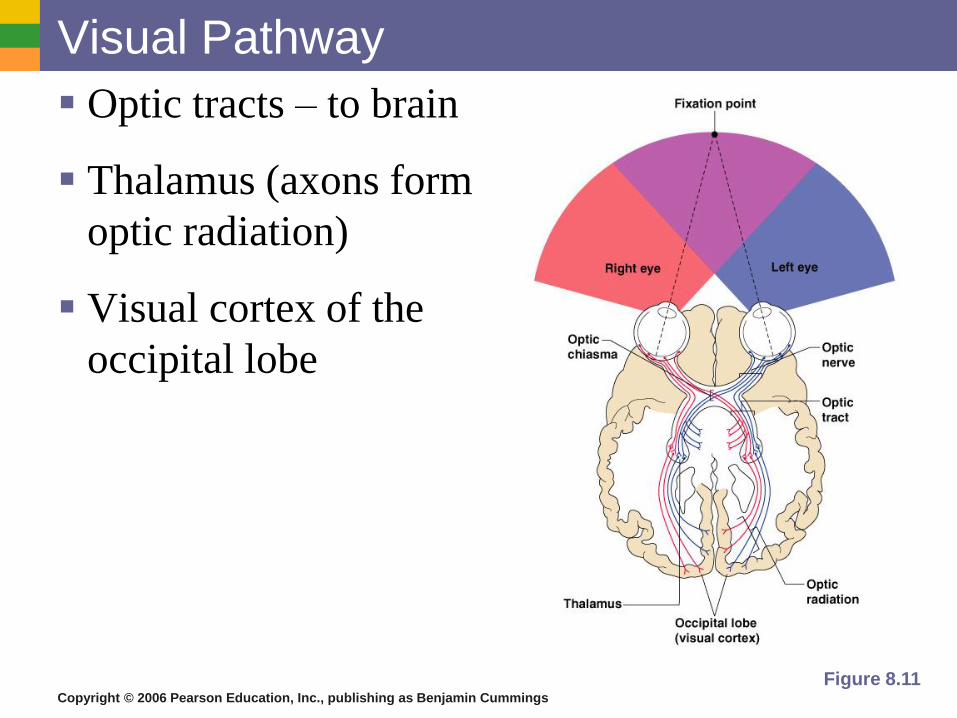
Visual Pathway
Optic tracts – to brain
Thalamus (axons form
optic radiation)
Visual cortex of the
occipital lobe
Figure 8.11
Copyright © 2006 Pearson Education, Inc., publishing as Benjamin Cummings
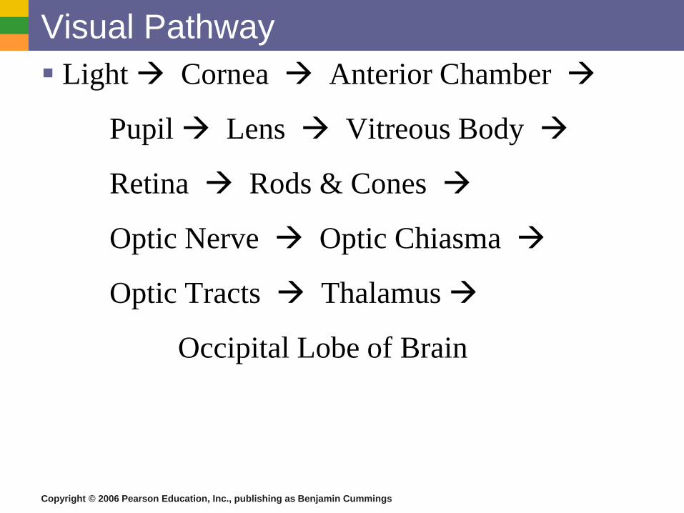
Visual Pathway
Light Cornea Anterior Chamber
Pupil Lens Vitreous Body
Retina Rods & Cones
Optic Nerve Optic Chiasma
Optic Tracts Thalamus
Occipital Lobe of Brain
Copyright © 2006 Pearson Education, Inc., publishing as Benjamin Cummings
Eye Reflexes
Internal muscles are controlled by the autonomic nervous system
Bright light causes pupils to constrict through action of ciliary bodies
Viewing close objects causes accommodation
External muscles control eye movement to follow objects
Viewing close objects causes convergence (eyes moving medially)
Copyright © 2006 Pearson Education, Inc., publishing as Benjamin Cummings
Eye Disorders Stye – infection of sebaceous gland
Conjunctivitis – “pink eye”
Glaucoma – overproduction of aqueous humor or obstruction of canal
Tonometer measures intraocular P
Miotic drugs – constrict pupil
Cataracts – lens become cloudy
Macular Degeneration – occurs with age
Dry – gradual thinning of retina
Wet – leaks under retina form blisters
Copyright © 2006 Pearson Education, Inc., publishing as Benjamin Cummings
Eye Disorders
Detached retina – occurs with age or a
forceful blow “curtain drawn”
Night blindness
Color blindness – red & green cones are most
commonly affected
Copyright © 2006 Pearson Education, Inc., publishing as Benjamin Cummings
Vision Defects
Presbyopia – lens loses elasticity
Hyperopia – aka hypermetropia “far-sighted”
– eyeball short (convex lens)
Myopia – “near-sighted”
– eyeball elongated (concave lens)
Amblyopia – reduction, dimness of vision
Astigmatism – irregular curvature of cornea or lens
Diplopia – blurred vision
Strabismus – crossed eyes
ELAINE N. MARIEB
EIGHTH EDITION
Copyright © 2006 Pearson Education, Inc., publishing as Benjamin Cummings
PowerPoint® Lecture Slide Presentation by Jerry L. Cook, Sam Houston University
ESSENTIALS
OF HUMAN
ANATOMY
& PHYSIOLOGY
Special Senses
Copyright © 2006 Pearson Education, Inc., publishing as Benjamin Cummings
The Ear
Houses two senses
Hearing
Equilibrium (balance)
Receptors are mechanoreceptors
Different organs house receptors for each
sense
Copyright © 2006 Pearson Education, Inc., publishing as Benjamin Cummings
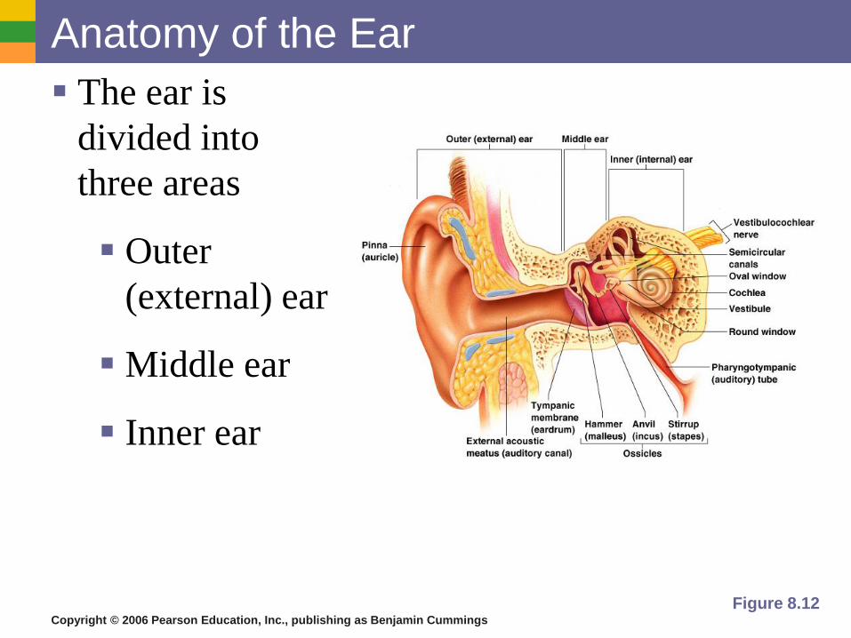
Anatomy of the Ear
The ear is
divided into
three areas
Outer
(external) ear
Middle ear
Inner ear
Figure 8.12
Copyright © 2006 Pearson Education, Inc., publishing as Benjamin Cummings
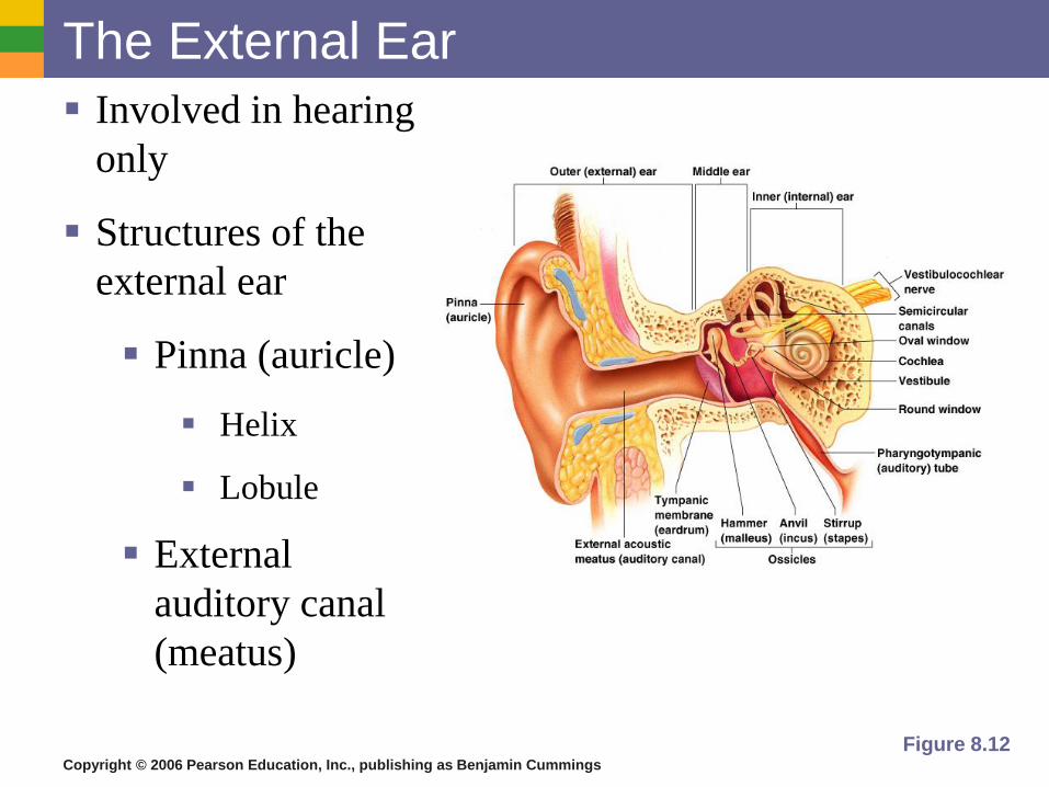
The External Ear
Involved in hearing
only
Structures of the
external ear
Pinna (auricle)
Helix
Lobule
External
auditory canal
(meatus)
Figure 8.12
Copyright © 2006 Pearson Education, Inc., publishing as Benjamin Cummings
The External Auditory Canal (Meatus)
Narrow chamber in the temporal bone
Lined with skin
Ceruminous (wax) glands are present
Cerumen – ear wax
Ends at the tympanic membrane
Copyright © 2006 Pearson Education, Inc., publishing as Benjamin Cummings
The Middle Ear or Tympanic Cavity
Air-filled cavity within the temporal bone
Only involved in the sense of hearing
Copyright © 2006 Pearson Education, Inc., publishing as Benjamin Cummings
The Middle Ear or Tympanic Cavity
Two tubes are associated with the ear
The opening from the auditory canal is
covered by the tympanic membrane
The auditory tube (eustachian tube)
connecting the middle ear with the throat
Allows for equalizing pressure during
yawning or swallowing
This tube is otherwise collapsed
Copyright © 2006 Pearson Education, Inc., publishing as Benjamin Cummings
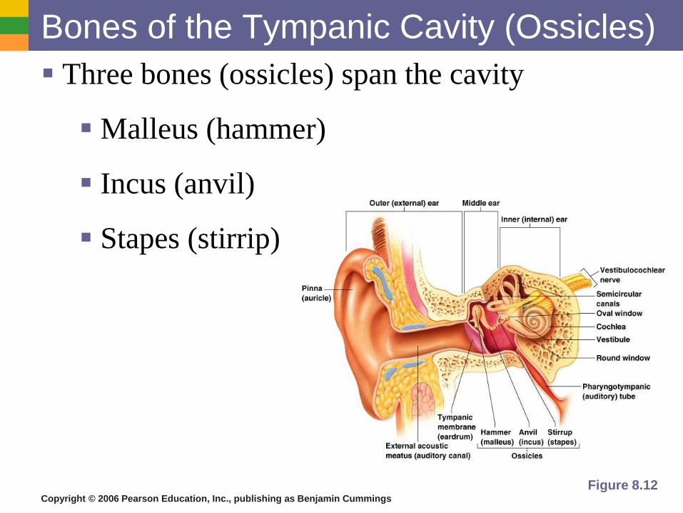
Bones of the Tympanic Cavity (Ossicles)
Three bones (ossicles) span the cavity
Malleus (hammer)
Incus (anvil)
Stapes (stirrip)
Figure 8.12
Copyright © 2006 Pearson Education, Inc., publishing as Benjamin Cummings
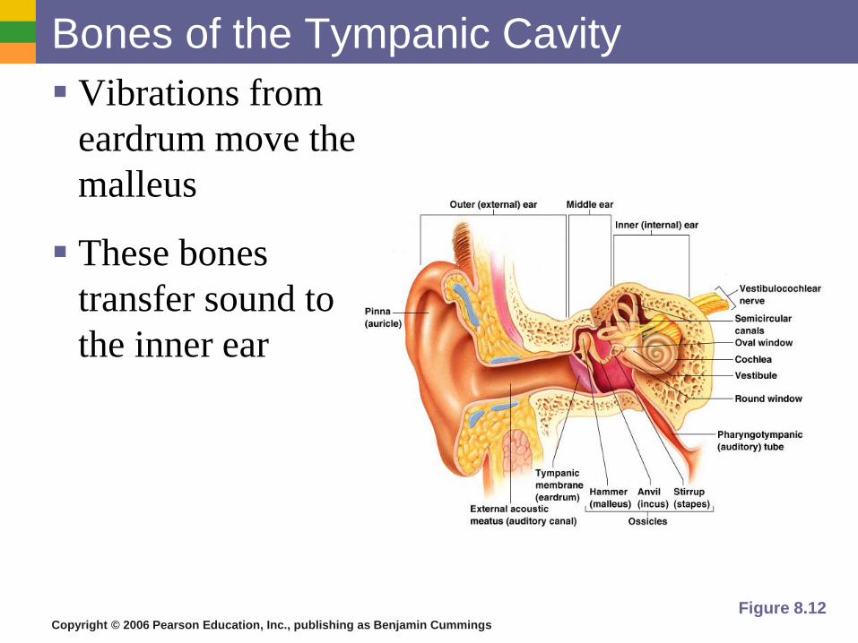
Bones of the Tympanic Cavity
Vibrations from
eardrum move the
malleus
These bones
transfer sound to
the inner ear
Figure 8.12
Copyright © 2006 Pearson Education, Inc., publishing as Benjamin Cummings
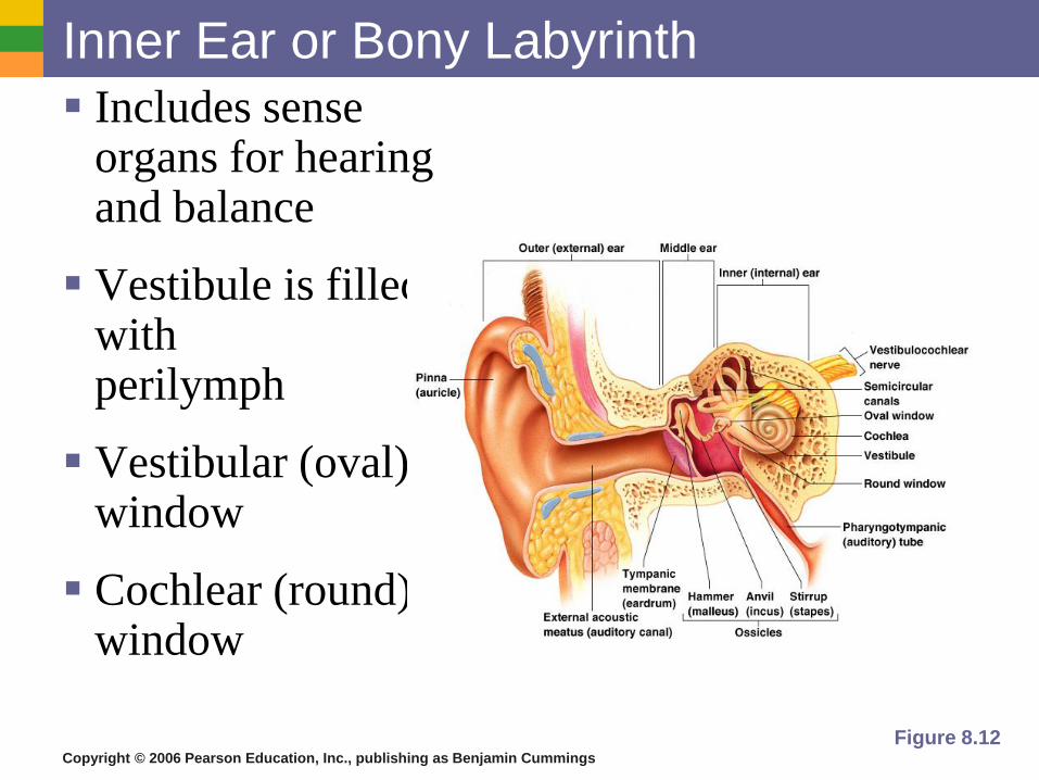
Inner Ear or Bony Labyrinth
Includes sense organs for hearing and balance
Vestibule is filled with perilymph
Vestibular (oval) window
Cochlear (round) window
Figure 8.12
Copyright © 2006 Pearson Education, Inc., publishing as Benjamin Cummings
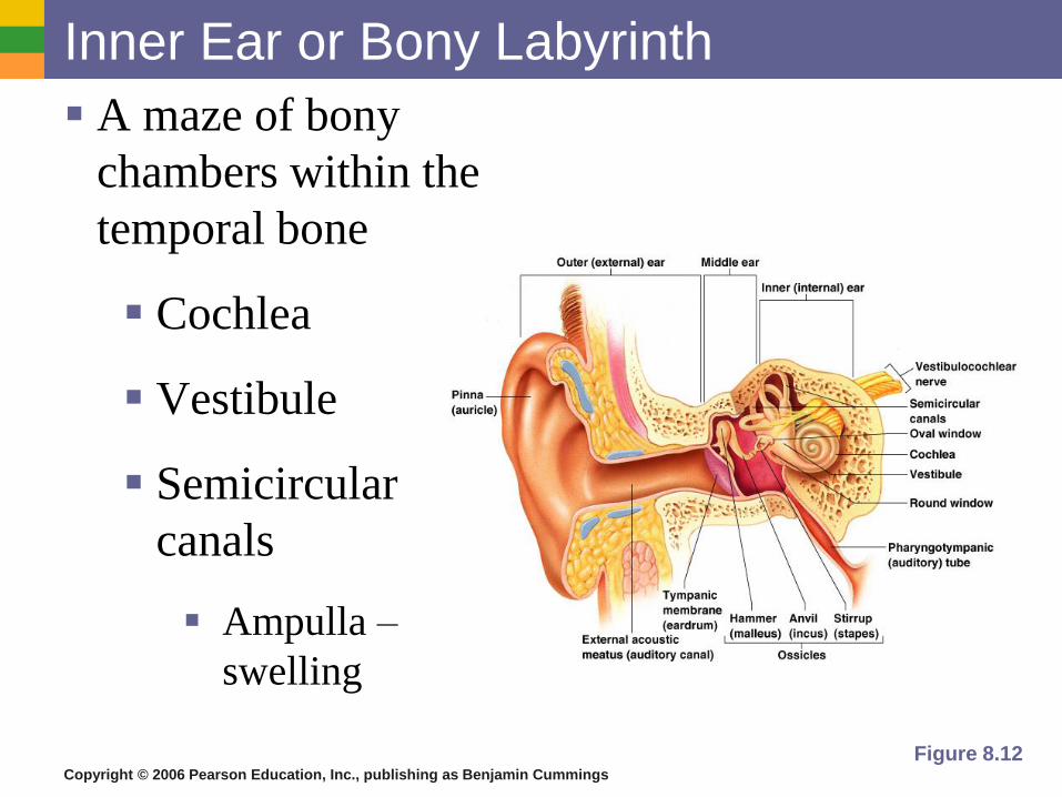
Inner Ear or Bony Labyrinth
A maze of bony
chambers within the
temporal bone
Cochlea
Vestibule
Semicircular
canals
Ampulla –
swelling
Figure 8.12
Copyright © 2006 Pearson Education, Inc., publishing as Benjamin Cummings
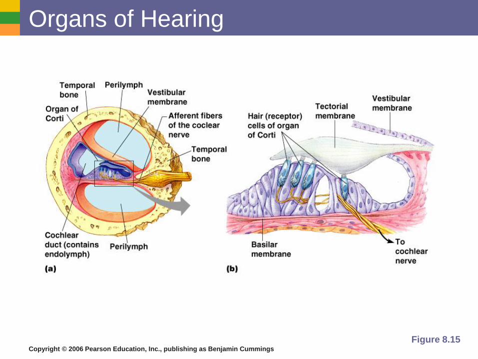
Organs of Hearing
Organ of Corti
Located within the cochlea
Receptors = hair cells on the basilar membrane
Gel-like membrane is capable of bending hair cells
Cochlear nerve attached to hair cells transmits nerve impulses to auditory cortex on temporal lobe
Copyright © 2006 Pearson Education, Inc., publishing as Benjamin Cummings
Organs of Hearing
Figure 8.15
Copyright © 2006 Pearson Education, Inc., publishing as Benjamin Cummings
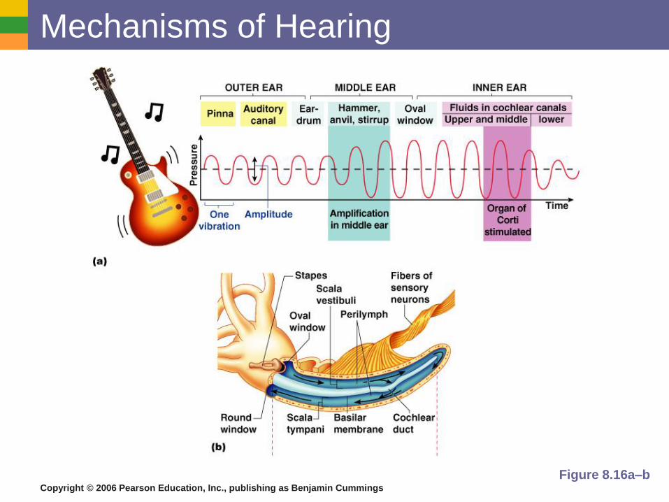
Mechanisms of Hearing
Vibrations from sound waves move
membrane
Hair cells are bent by the membrane
An action potential starts in the cochlear
nerve
Continued stimulation can lead to adaptation
Decibels – measurement of sound
ie. damage = 90 dB for 8 hrs/100 dB for 2
hrs
Copyright © 2006 Pearson Education, Inc., publishing as Benjamin Cummings
Mechanisms of Hearing
Figure 8.16a–b
Copyright © 2006 Pearson Education, Inc., publishing as Benjamin Cummings
Ear Disorders
Deafness – any hearing loss
Otitis Media – middle ear infection
Myringotomy – opening in tympanic membrane
Otosclerosis – genetic disorder
Tinnitus – ringing of ear without stimulus
Presbycusis – deafness due to aging
Meniere’s Disease – condition affecting semicircular
canals
Vertigo - dizziness
Copyright © 2006 Pearson Education, Inc., publishing as Benjamin Cummings
Types of Hearing Loss
Conductive – sound to ear is blocked
Sensorineural – damage to inner ear
Copyright © 2006 Pearson Education, Inc., publishing as Benjamin Cummings
Pathway of Hearing
sound waves pinna meatus
tympanic membrane ossicles
cochlear receptors cochlear nerve
thalamus temporal lobe of brain
Copyright © 2006 Pearson Education, Inc., publishing as Benjamin Cummings
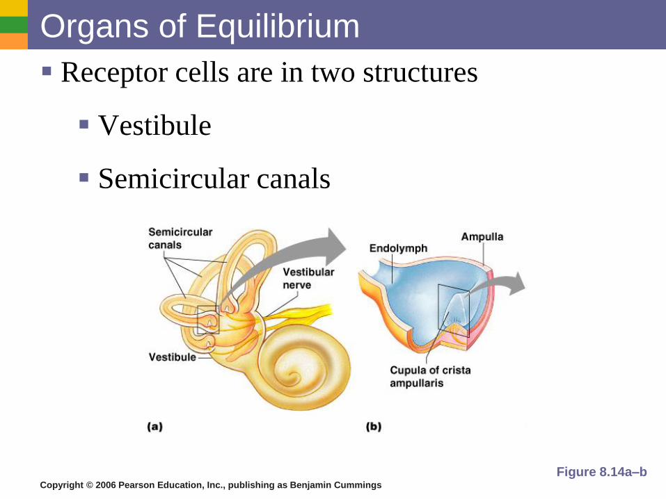
Organs of Equilibrium
Receptor cells are in two structures
Vestibule
Semicircular canals
Figure 8.14a–b
Copyright © 2006 Pearson Education, Inc., publishing as Benjamin Cummings
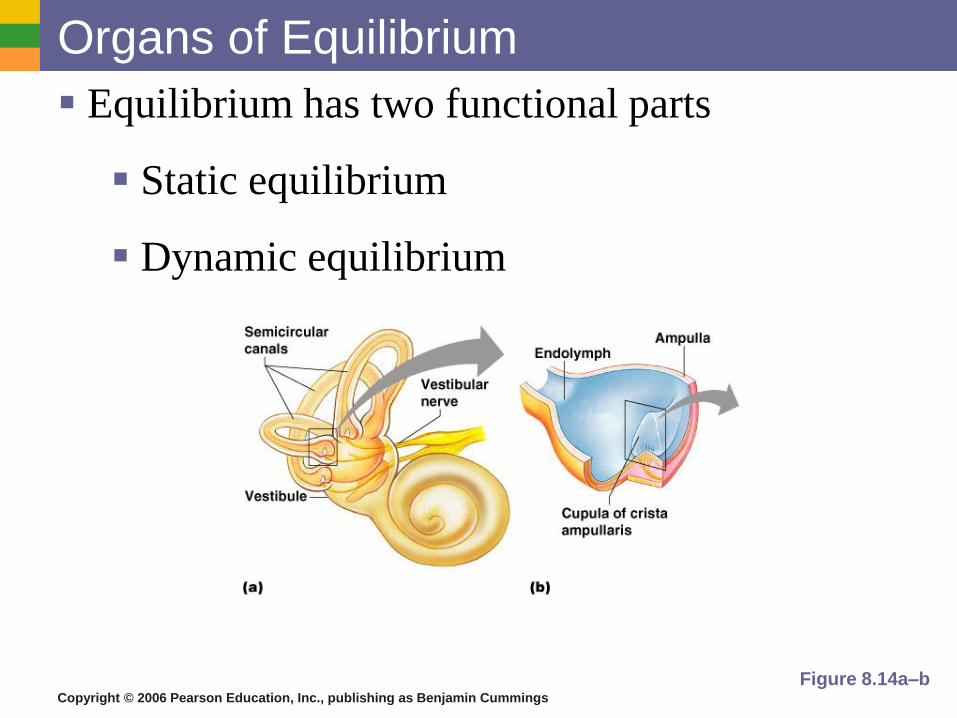
Organs of Equilibrium
Equilibrium has two functional parts
Static equilibrium
Dynamic equilibrium
Figure 8.14a–b
Copyright © 2006 Pearson Education, Inc., publishing as Benjamin Cummings
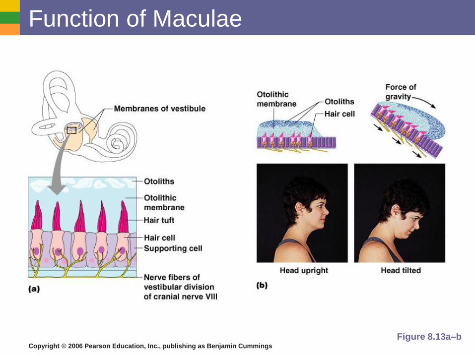
Static Equilibrium
Maculae – receptors in the vestibule
Report on the position of the head
Send information via the vestibular nerve
Anatomy of the maculae
Hair cells are embedded in the membrane
Tiny stones float in a gel around the hair cells
Movements cause stones to bend the hair cells
Copyright © 2006 Pearson Education, Inc., publishing as Benjamin Cummings
Function of Maculae
Figure 8.13a–b
Copyright © 2006 Pearson Education, Inc., publishing as Benjamin Cummings
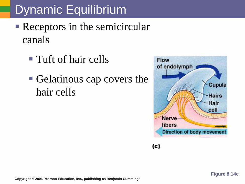
Dynamic Equilibrium
Receptors in the semicircular
canals
Tuft of hair cells
Gelatinous cap covers the
hair cells
Figure 8.14c
Copyright © 2006 Pearson Education, Inc., publishing as Benjamin Cummings
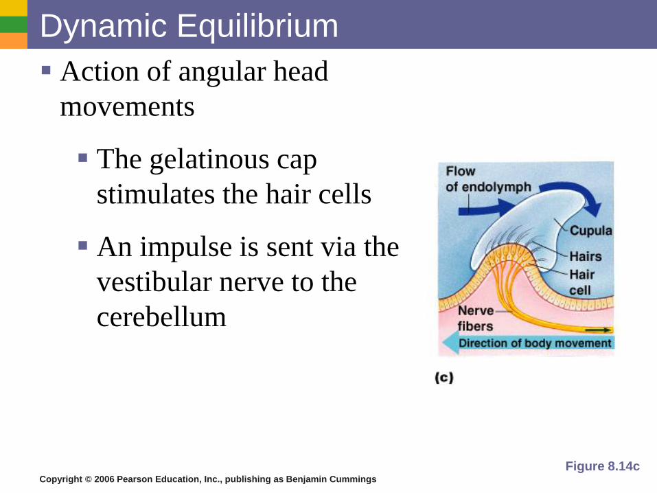
Dynamic Equilibrium
Action of angular head
movements
The gelatinous cap
stimulates the hair cells
An impulse is sent via the
vestibular nerve to the
cerebellum
Figure 8.14c
Copyright © 2006 Pearson Education, Inc., publishing as Benjamin Cummings
Pathway of Equilibrium
Head movement
equilibrium receptors in semicircular
canals & vestibules
vestibular nerve cerebellum of brain

ELAINE N. MARIEB
EIGHTH EDITION
Copyright © 2006 Pearson Education, Inc., publishing as Benjamin Cummings
PowerPoint® Lecture Slide Presentation by Jerry L. Cook, Sam Houston University
ESSENTIALS
OF HUMAN
ANATOMY
& PHYSIOLOGY
Special Senses
Copyright © 2006 Pearson Education, Inc., publishing as Benjamin Cummings
Chemical Senses – Taste and Smell
Both senses use chemoreceptors
Stimulated by chemicals in solution
Taste has four types of receptors
Smell can differentiate a large range of
chemicals
Both senses complement each other and
respond to many of the same stimuli
Copyright © 2006 Pearson Education, Inc., publishing as Benjamin Cummings
Olfaction – The Sense of Smell
Olfactory receptors are in the roof of the nasal
cavity in olfactory ephithelia
Olfactory hairs (neurons) with long cilia
Chemicals must be dissolved in mucus for
detection
Impulses are transmitted via olfactory
receptor cells, olfactory bulb and olfactory
tract
Interpretation of smells is made in the cortex
Copyright © 2006 Pearson Education, Inc., publishing as Benjamin Cummings
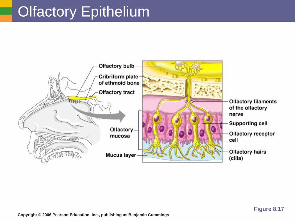
Olfactory Epithelium
Figure 8.17
Copyright © 2006 Pearson Education, Inc., publishing as Benjamin Cummings
Disorders of Nose
Rhinitis – inflammation of nose lining
Nasal polyps – growths in nasal cavity
Deviated nasal septum – bend in cartilage of
septum
Anosmia – “without smells”
Olfactory auras
Copyright © 2006 Pearson Education, Inc., publishing as Benjamin Cummings
Pathway of Smell
volatile molecules inhaled
olfactory epithelia olfactory hairs
olfactory receptor cells
olfactory bulb olfactory tract
limbic system thalmus
frontal cortex of brain
Copyright © 2006 Pearson Education, Inc., publishing as Benjamin Cummings
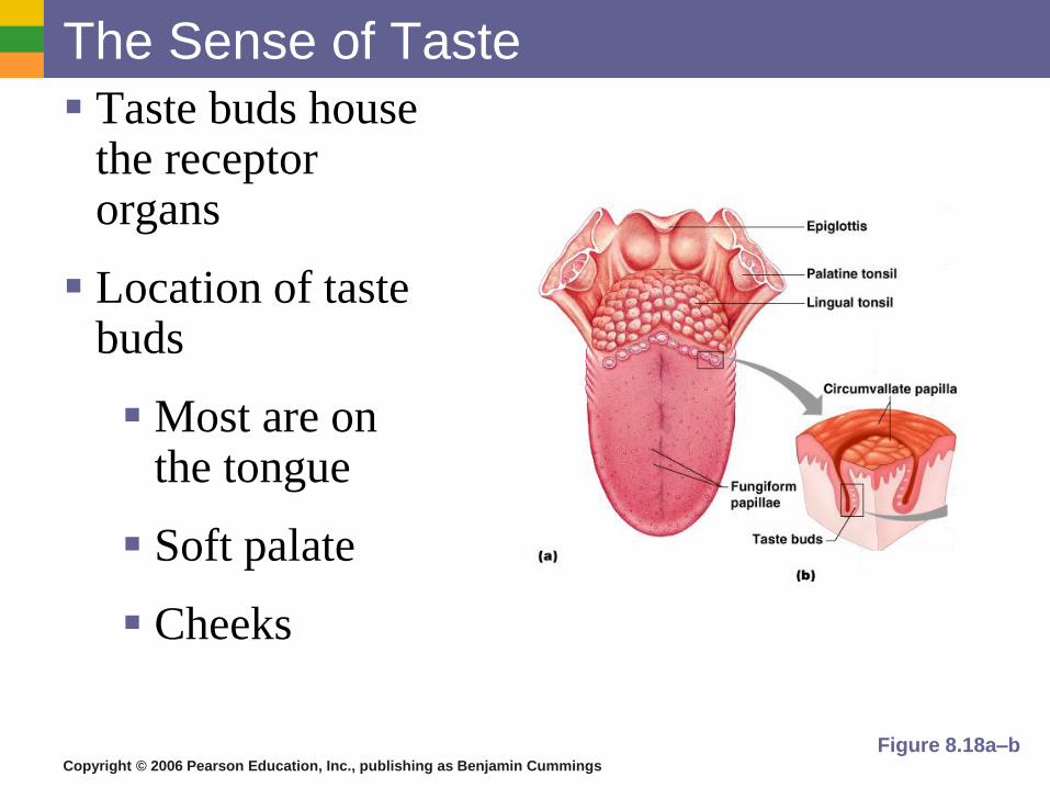
The Sense of Taste
Taste buds house the receptor organs
Location of taste buds
Most are on the tongue
Soft palate
Cheeks
Figure 8.18a–b
Copyright © 2006 Pearson Education, Inc., publishing as Benjamin Cummings
The Tongue and Taste
The tongue is covered with projections called
papillae
Taste buds are found on the sides of papillae
Copyright © 2006 Pearson Education, Inc., publishing as Benjamin Cummings
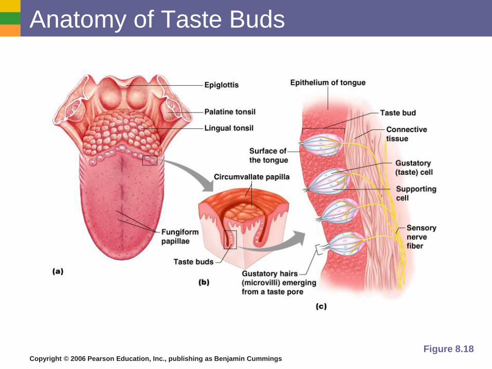
Structure of Taste Buds
Gustatory cells are the receptors
Have gustatory hairs (long microvilli)
Hairs are stimulated by chemicals
dissolved in saliva
Copyright © 2006 Pearson Education, Inc., publishing as Benjamin Cummings
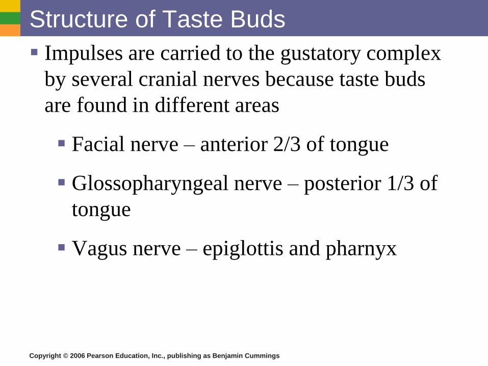
Structure of Taste Buds
Impulses are carried to the gustatory complex
by several cranial nerves because taste buds
are found in different areas
Facial nerve – anterior 2/3 of tongue
Glossopharyngeal nerve – posterior 1/3 of
tongue
Vagus nerve – epiglottis and pharnyx
Copyright © 2006 Pearson Education, Inc., publishing as Benjamin Cummings
Anatomy of Taste Buds
Figure 8.18
Copyright © 2006 Pearson Education, Inc., publishing as Benjamin Cummings
Taste Sensations Sweet receptors
Sugars
Saccharine
Some amino acids
Sour receptors
Acids
Bitter receptors (protective)
Alkaloids
Salty receptors
Metal ions
Copyright © 2006 Pearson Education, Inc., publishing as Benjamin Cummings
Pathway of Taste
chemical in solution taste buds
gustatory hairs
facial/glossopharyngeal/vagus nerves
medulla thalamus
parietal lobe of brain
Copyright © 2006 Pearson Education, Inc., publishing as Benjamin Cummings
Developmental Aspects of the Special
Senses
Formed early in embryonic development
Eyes are outgrowths of the brain
All special senses are functional at birth