nerve activates contraction - ponder independent … · blood cell formation- aka hematopoiesis,...
TRANSCRIPT
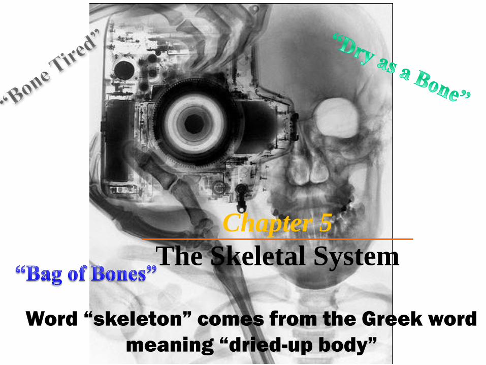
Chapter 5
The Skeletal System
Word “skeleton” comes from the Greek word
meaning “dried-up body”
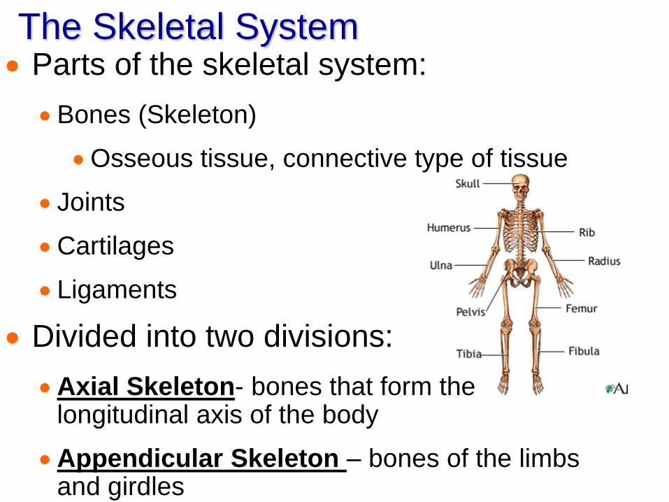
The Skeletal System Parts of the skeletal system:
Bones (Skeleton)
Osseous tissue, connective type of tissue
Joints
Cartilages
Ligaments
Divided into two divisions:
Axial Skeleton- bones that form the longitudinal axis of the body
Appendicular Skeleton – bones of the limbs and girdles
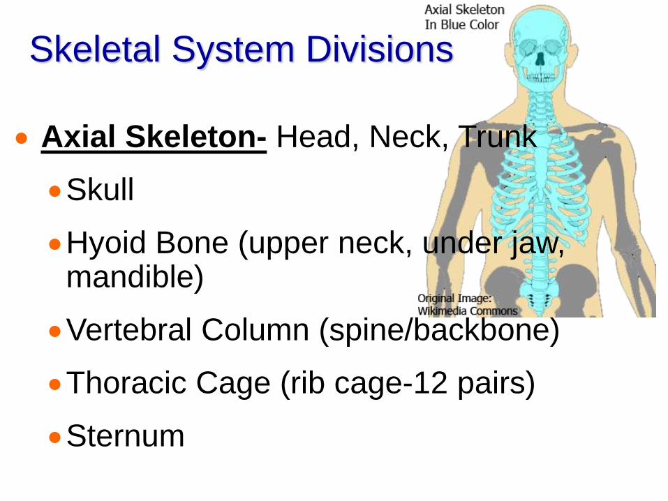
Skeletal System Divisions
Axial Skeleton- Head, Neck, Trunk
Skull
Hyoid Bone (upper neck, under jaw, mandible)
Vertebral Column (spine/backbone)
Thoracic Cage (rib cage-12 pairs)
Sternum

Skeletal System Divisions
Appendicular Skeleton- limbs, limb connectors
Pectoral Girdle (scapula & clavicle)
Upper Limbs (arms)
Pelvic Girdle (coxal bones)
Lower Limbs (legs)
Functions of Bones Support of the Body – are like “steel
girders” and “reinforced concrete”
Protection of Soft Organs- ex. Skull/brain
Movement – bones attached to skeletal
muscles by tendons use bones as levers for
moving (muscles “pull” on bones)
Storage of Minerals and Fats- storehouse,
esp. Ca3PO4, Mg, Na, K, & CO3
Blood Cell Formation- aka hematopoiesis,
occurs in red bone marrow
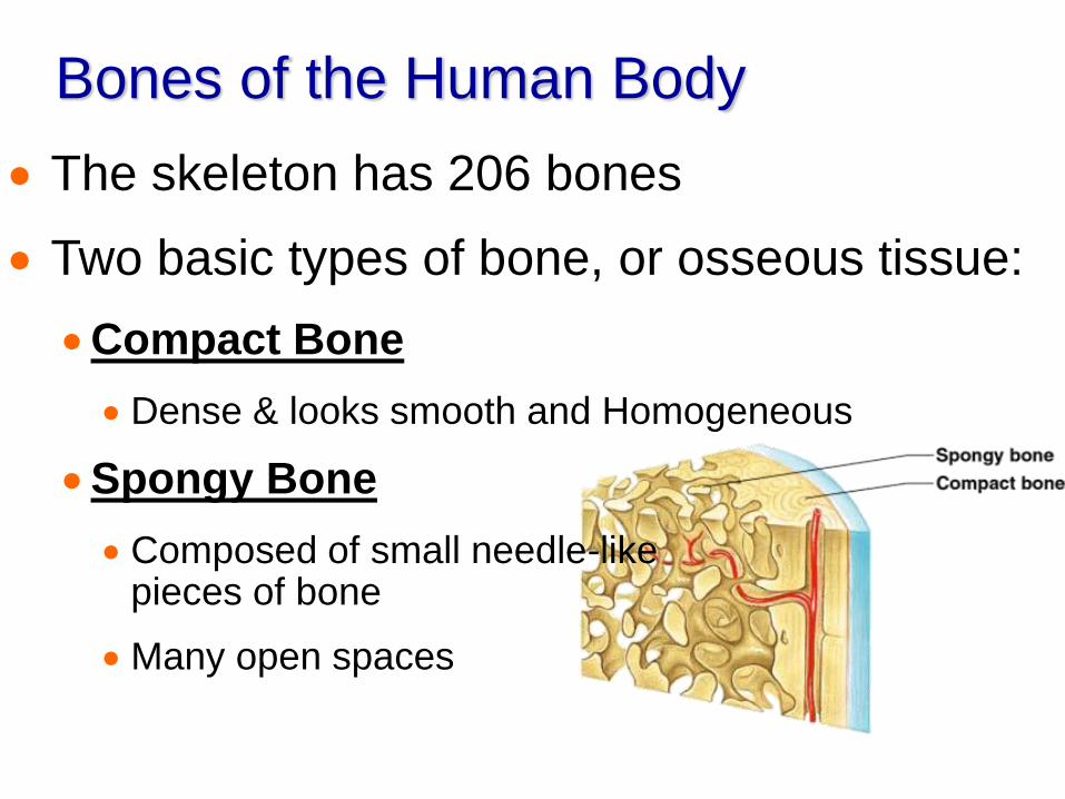
Bones of the Human Body
The skeleton has 206 bones
Two basic types of bone, or osseous tissue:
Compact Bone
Dense & looks smooth and Homogeneous
Spongy Bone
Composed of small needle-like pieces of bone
Many open spaces

Classification of Bones on Basis of Shape
Bones come in many sizes and shapes
Unique shape of each bone fulfills a particular need, they are classified by shape (long, short, flat, & irregular
Pisiform Bone- in wrist the size & shape of a pea
Femur Bone- thigh bone, is nearly two feet long & has large, ball-shaped head
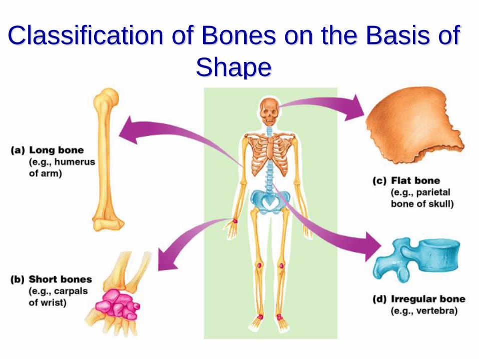
Classification of Bones on the Basis of
Shape
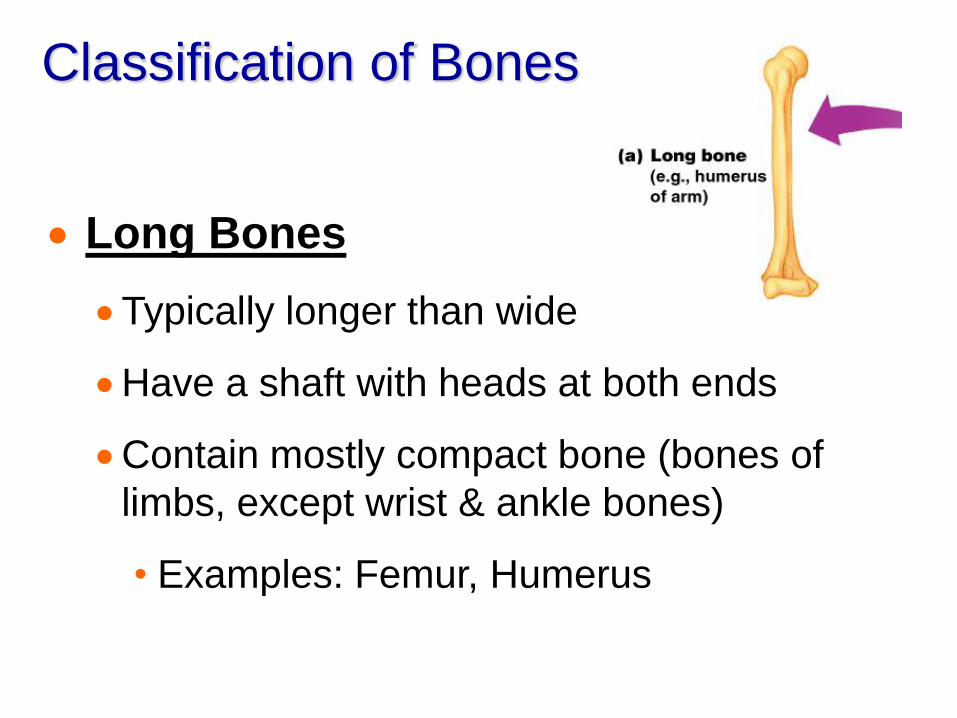
Classification of Bones
Long Bones
Typically longer than wide
Have a shaft with heads at both ends
Contain mostly compact bone (bones of
limbs, except wrist & ankle bones)
• Examples: Femur, Humerus

Classification of Bones
Short Bones
Generally cube-shape
Contain mostly spongy bone
Sesamoid bones- form tendons, are a special
short bone
Examples: Carpals, Tarsals, Petella(kneecap)
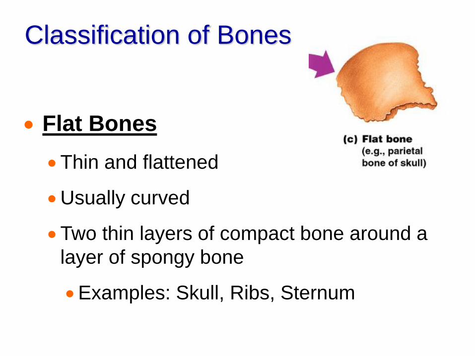
Classification of Bones
Flat Bones
Thin and flattened
Usually curved
Two thin layers of compact bone around a
layer of spongy bone
Examples: Skull, Ribs, Sternum
Classification of Bones
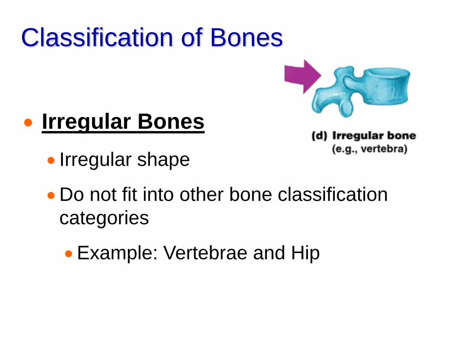
Irregular Bones
Irregular shape
Do not fit into other bone classification
categories
Example: Vertebrae and Hip
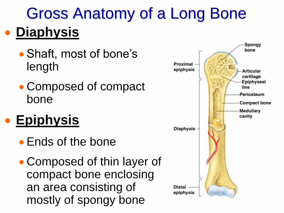
Gross Anatomy of a Long Bone Diaphysis
Shaft, most of bone’s length
Composed of compact bone
Epiphysis
Ends of the bone
Composed of thin layer of compact bone enclosing an area consisting of mostly of spongy bone
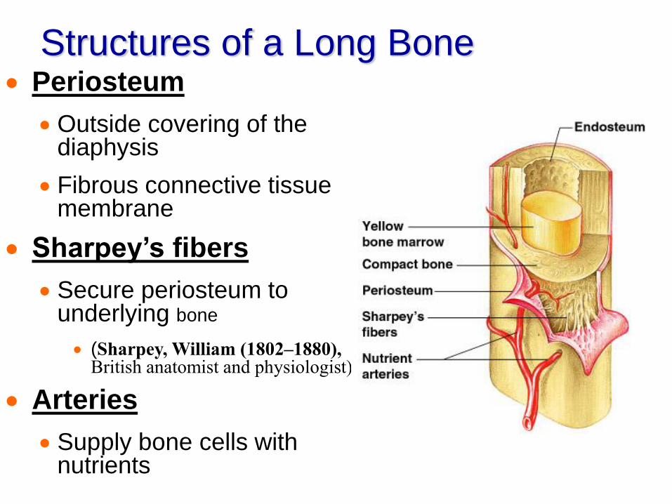
Structures of a Long Bone Periosteum
Outside covering of the diaphysis
Fibrous connective tissue membrane
Sharpey’s fibers
Secure periosteum to underlying bone
(Sharpey, William (1802–1880), British anatomist and physiologist)
Arteries
Supply bone cells with nutrients
Structures of a Long Bone
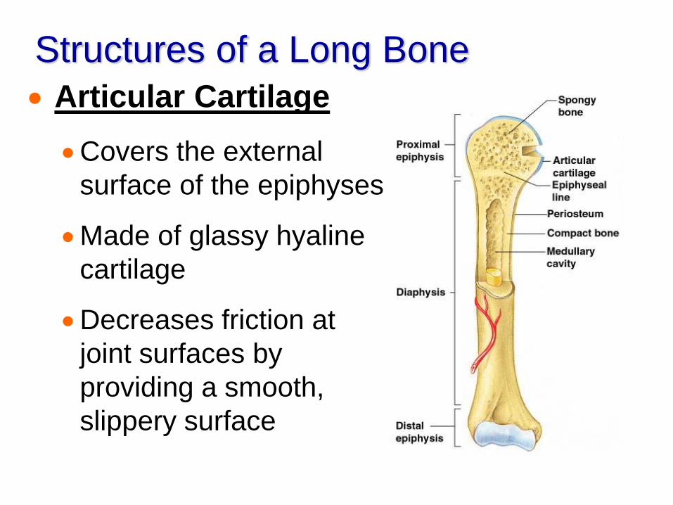
Articular Cartilage
Covers the external
surface of the epiphyses
Made of glassy hyaline
cartilage
Decreases friction at
joint surfaces by
providing a smooth,
slippery surface
Structures of a Long Bone
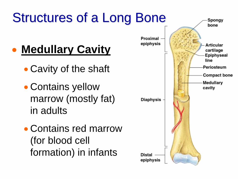
Medullary Cavity
Cavity of the shaft
Contains yellow
marrow (mostly fat)
in adults
Contains red marrow
(for blood cell
formation) in infants
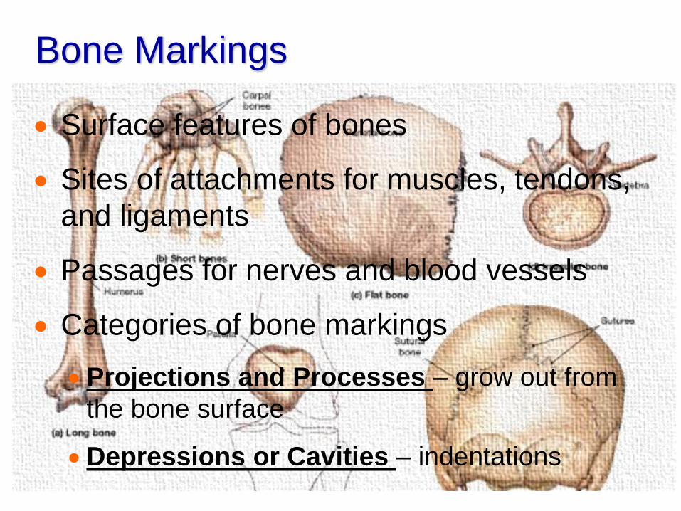
Bone Markings
Surface features of bones
Sites of attachments for muscles, tendons,
and ligaments
Passages for nerves and blood vessels
Categories of bone markings
Projections and Processes – grow out from
the bone surface
Depressions or Cavities – indentations

Microscopic Anatomy of Bone
Osteon (Haversian System)
A unit of bone
Central (Haversian) canal
Opening in the center of an osteon
Carries blood vessels and nerves
Perforating (Volkman’s) canal
Canal perpendicular to the central canal
Carries blood vessels and nerves
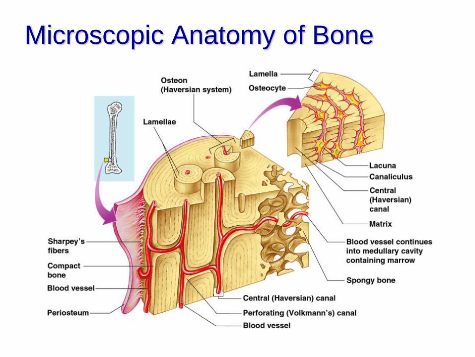
Microscopic Anatomy of Bone
Microscopic Anatomy of Bone
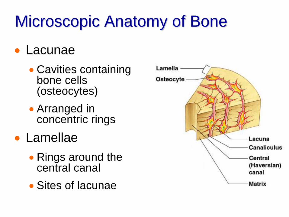
Lacunae
Cavities containing bone cells (osteocytes)
Arranged in concentric rings
Lamellae
Rings around the central canal
Sites of lacunae
Microscopic Anatomy of Bone
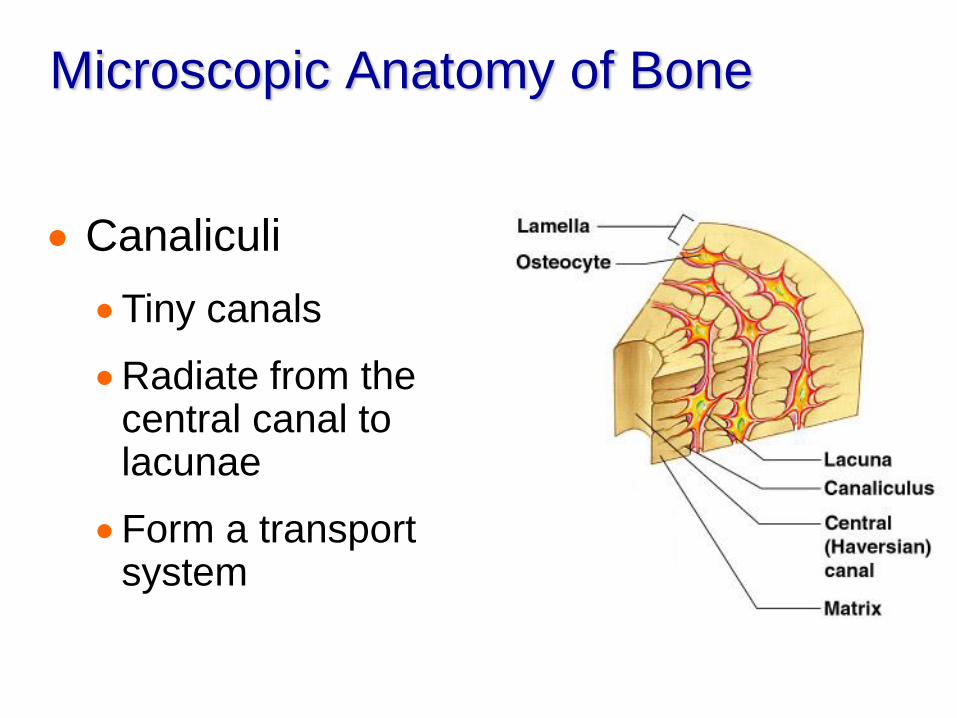
Canaliculi
Tiny canals
Radiate from the central canal to lacunae
Form a transport system
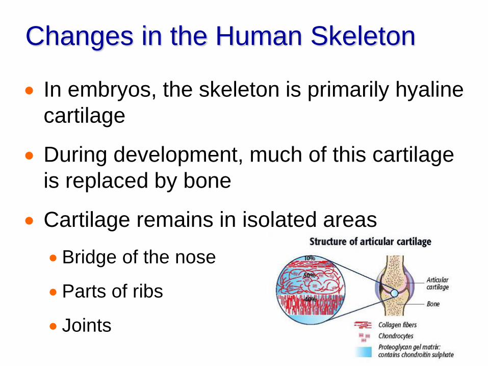
Changes in the Human Skeleton
In embryos, the skeleton is primarily hyaline
cartilage
During development, much of this cartilage
is replaced by bone
Cartilage remains in isolated areas
Bridge of the nose
Parts of ribs
Joints
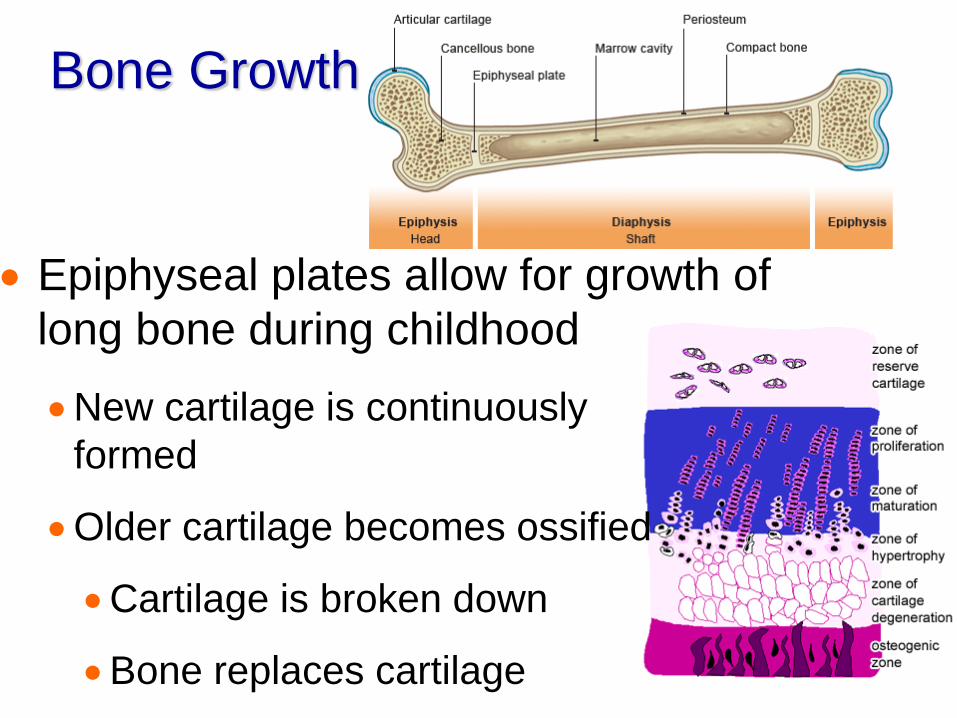
Bone Growth
Epiphyseal plates allow for growth of
long bone during childhood
New cartilage is continuously
formed
Older cartilage becomes ossified
Cartilage is broken down
Bone replaces cartilage
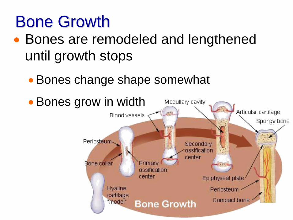
Bone Growth Bones are remodeled and lengthened
until growth stops
Bones change shape somewhat
Bones grow in width

Long Bone Formation and Growth

Long Bone Formation and Growth
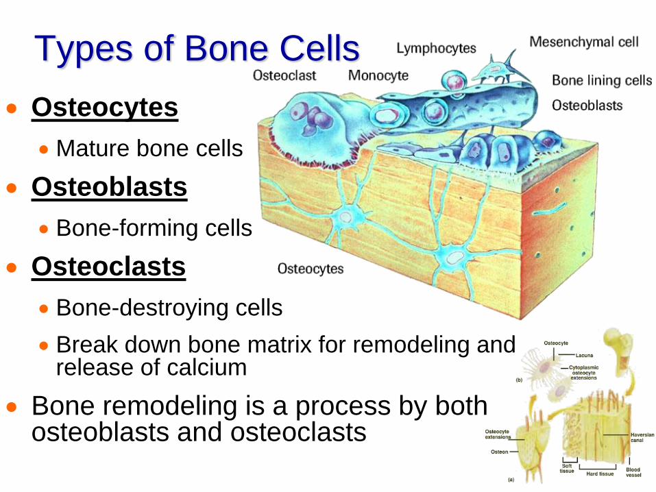
Types of Bone Cells
Osteocytes
Mature bone cells
Osteoblasts
Bone-forming cells
Osteoclasts
Bone-destroying cells
Break down bone matrix for remodeling and release of calcium
Bone remodeling is a process by both osteoblasts and osteoclasts
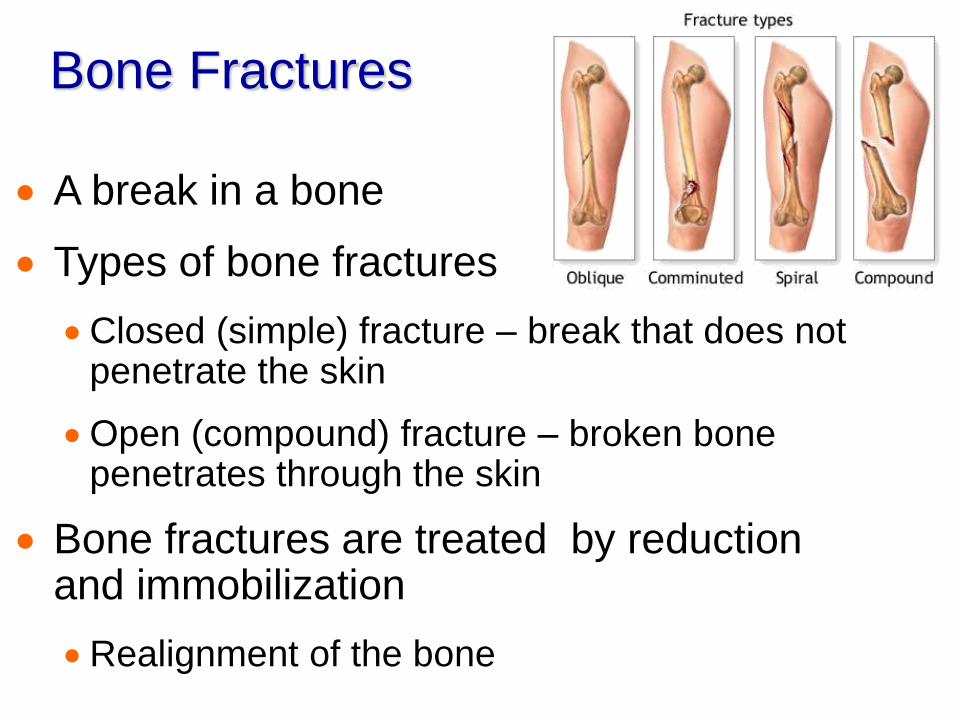
Bone Fractures
A break in a bone
Types of bone fractures
Closed (simple) fracture – break that does not penetrate the skin
Open (compound) fracture – broken bone penetrates through the skin
Bone fractures are treated by reduction and immobilization
Realignment of the bone
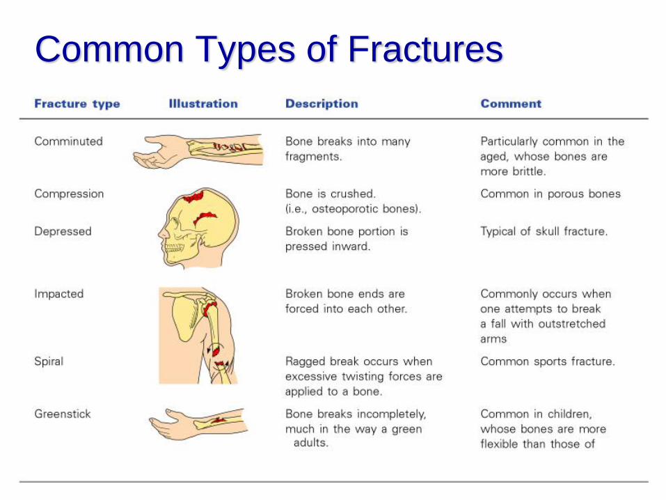
Common Types of Fractures
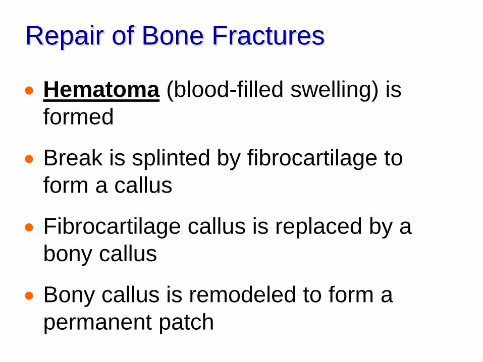
Repair of Bone Fractures
Hematoma (blood-filled swelling) is
formed
Break is splinted by fibrocartilage to
form a callus
Fibrocartilage callus is replaced by a
bony callus
Bony callus is remodeled to form a
permanent patch
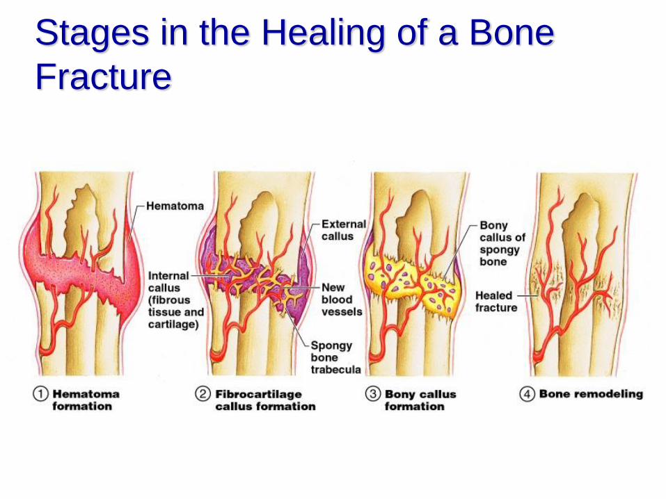
Stages in the Healing of a Bone
Fracture
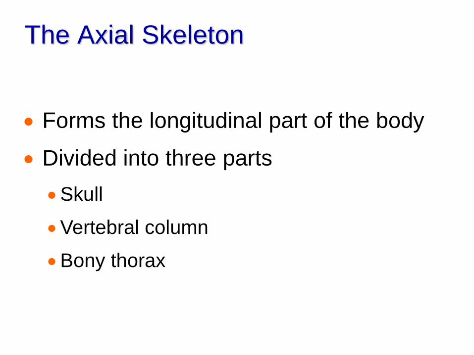
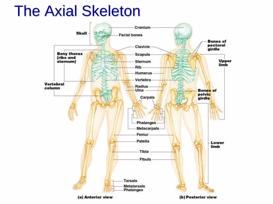
The Axial Skeleton
Forms the longitudinal part of the body
Divided into three parts
Skull
Vertebral column
Bony thorax
The Axial Skeleton
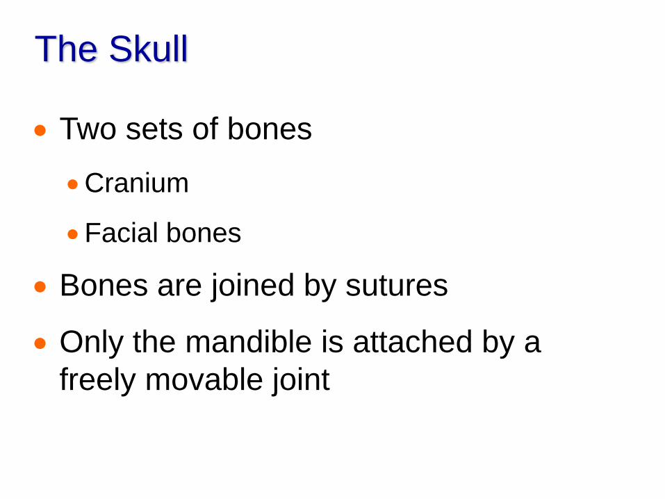
The Skull
Two sets of bones
Cranium
Facial bones
Bones are joined by sutures
Only the mandible is attached by a
freely movable joint
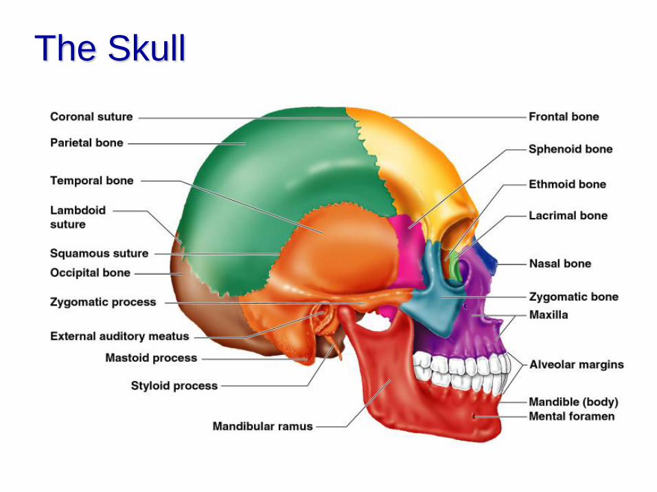
The Skull
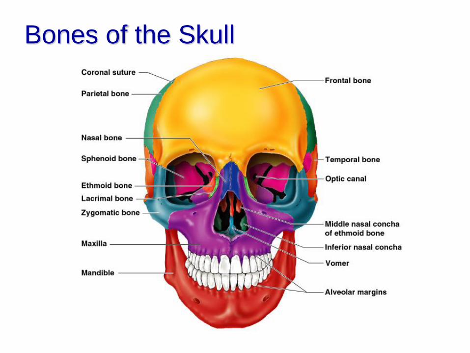
Bones of the Skull
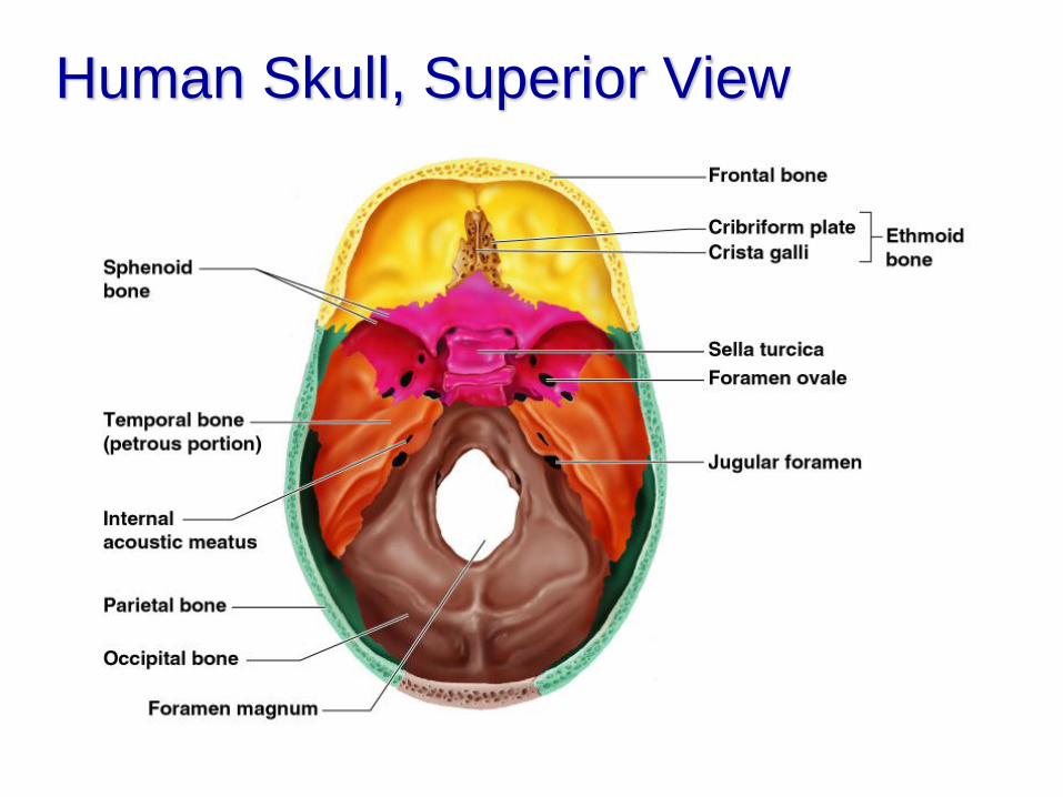
Human Skull, Superior View
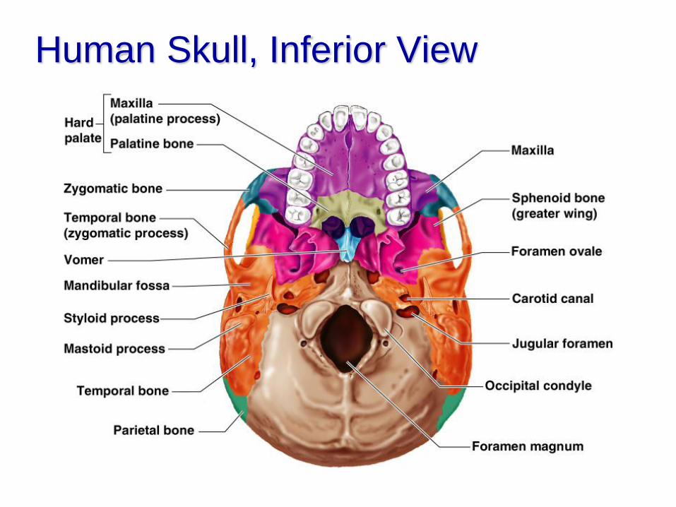
Human Skull, Inferior View
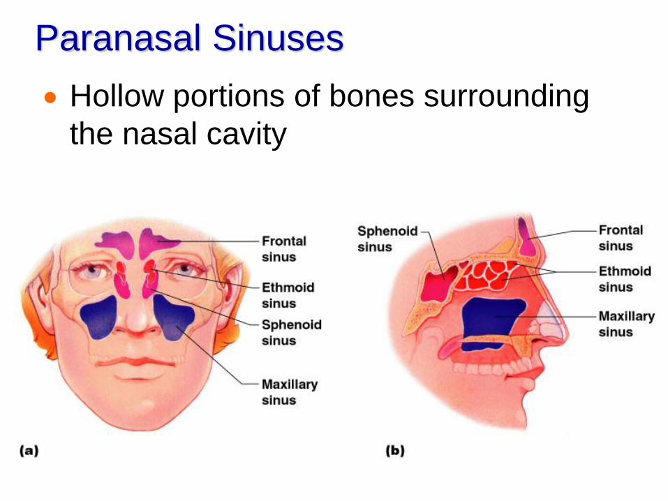
Paranasal Sinuses
Hollow portions of bones surrounding
the nasal cavity
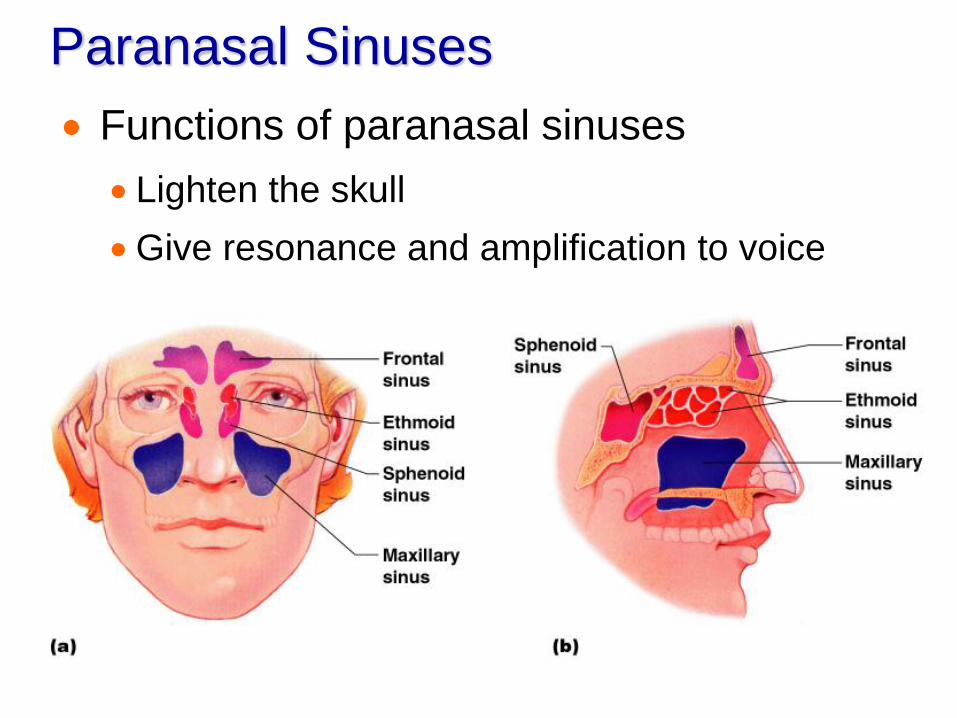
Paranasal Sinuses
Functions of paranasal sinuses
Lighten the skull
Give resonance and amplification to voice
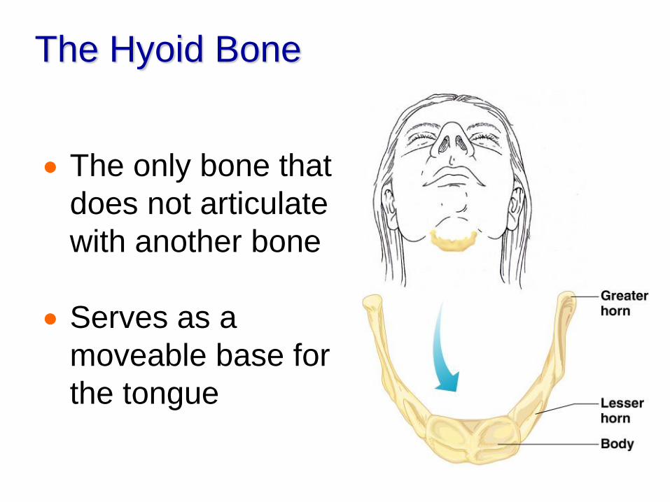
The Hyoid Bone
The only bone that
does not articulate
with another bone
Serves as a
moveable base for
the tongue
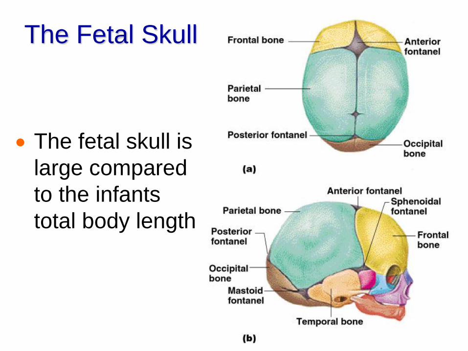
The Fetal Skull
The fetal skull is
large compared
to the infants
total body length
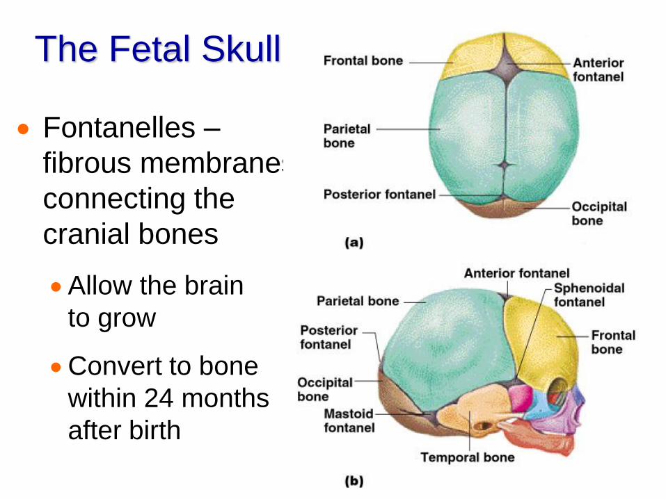
The Fetal Skull
Fontanelles –
fibrous membranes
connecting the
cranial bones
Allow the brain
to grow
Convert to bone
within 24 months
after birth
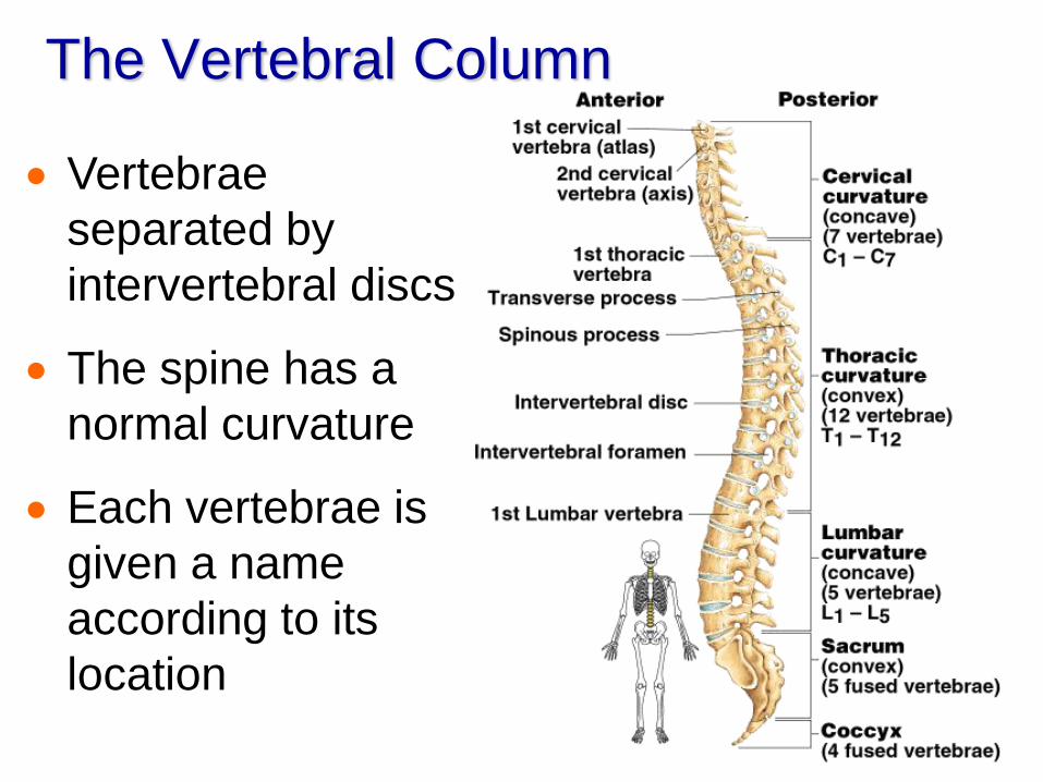
The Vertebral Column
Vertebrae
separated by
intervertebral discs
The spine has a
normal curvature
Each vertebrae is
given a name
according to its
location
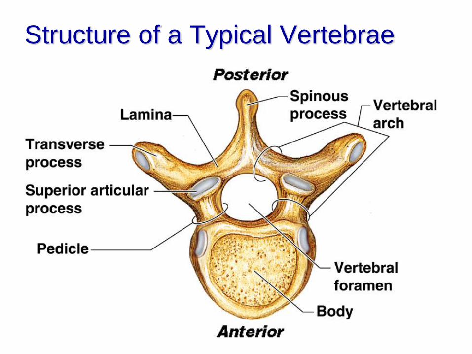
Structure of a Typical Vertebrae
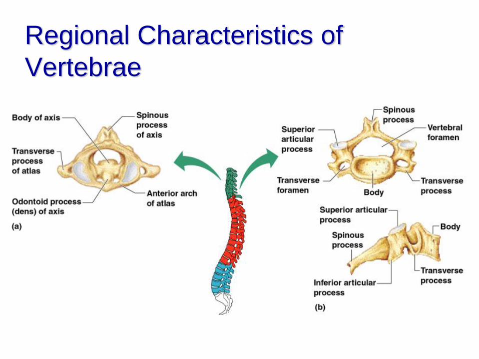
Regional Characteristics of
Vertebrae
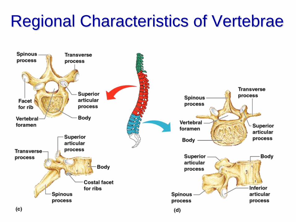
Regional Characteristics of Vertebrae
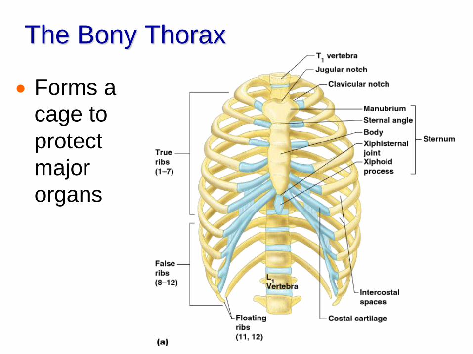
The Bony Thorax
Forms a
cage to
protect
major
organs
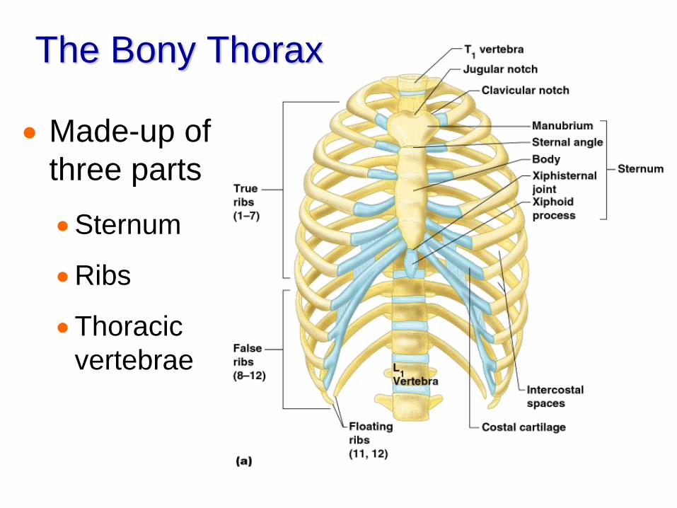
The Bony Thorax
Made-up of
three parts
Sternum
Ribs
Thoracic
vertebrae
The Appendicular Skeleton
Limbs (appendages)
Pectoral girdle
Pelvic girdle
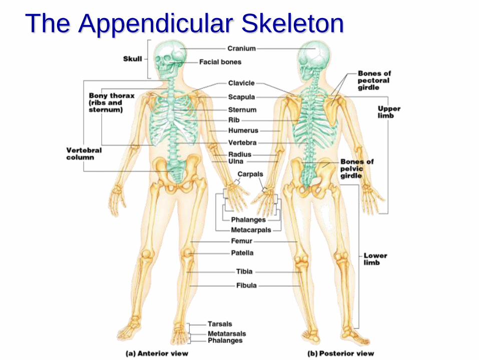
The Appendicular Skeleton
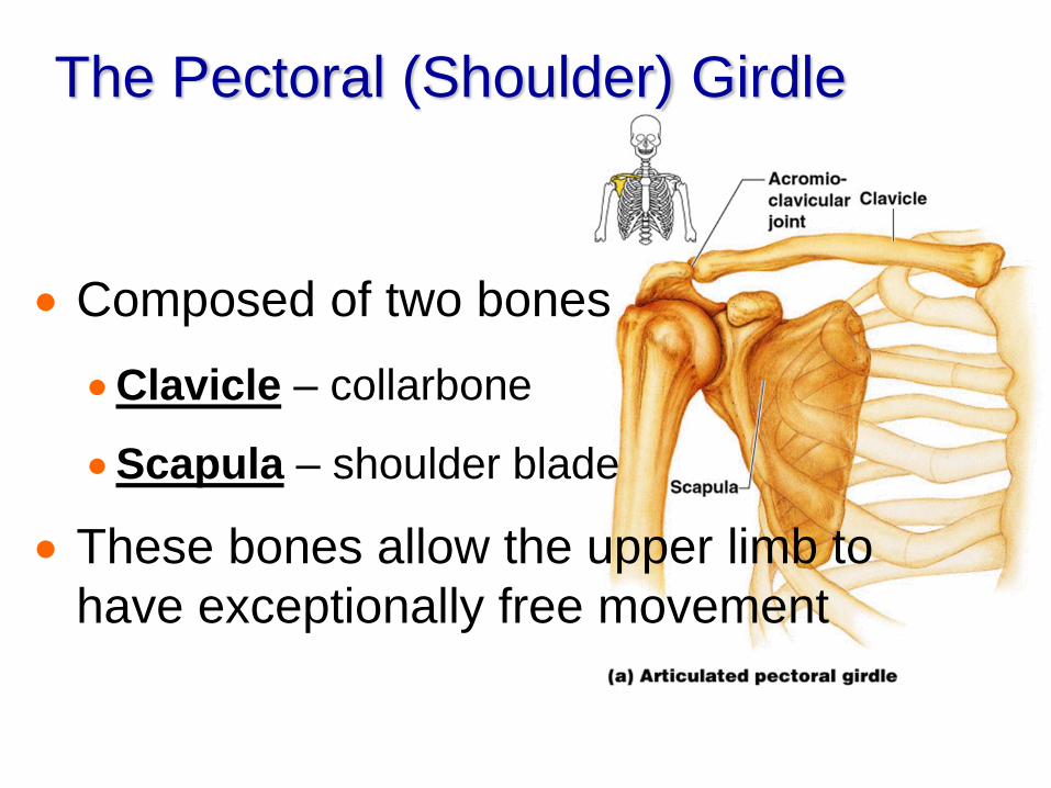
The Pectoral (Shoulder) Girdle
Composed of two bones
Clavicle – collarbone
Scapula – shoulder blade
These bones allow the upper limb to
have exceptionally free movement
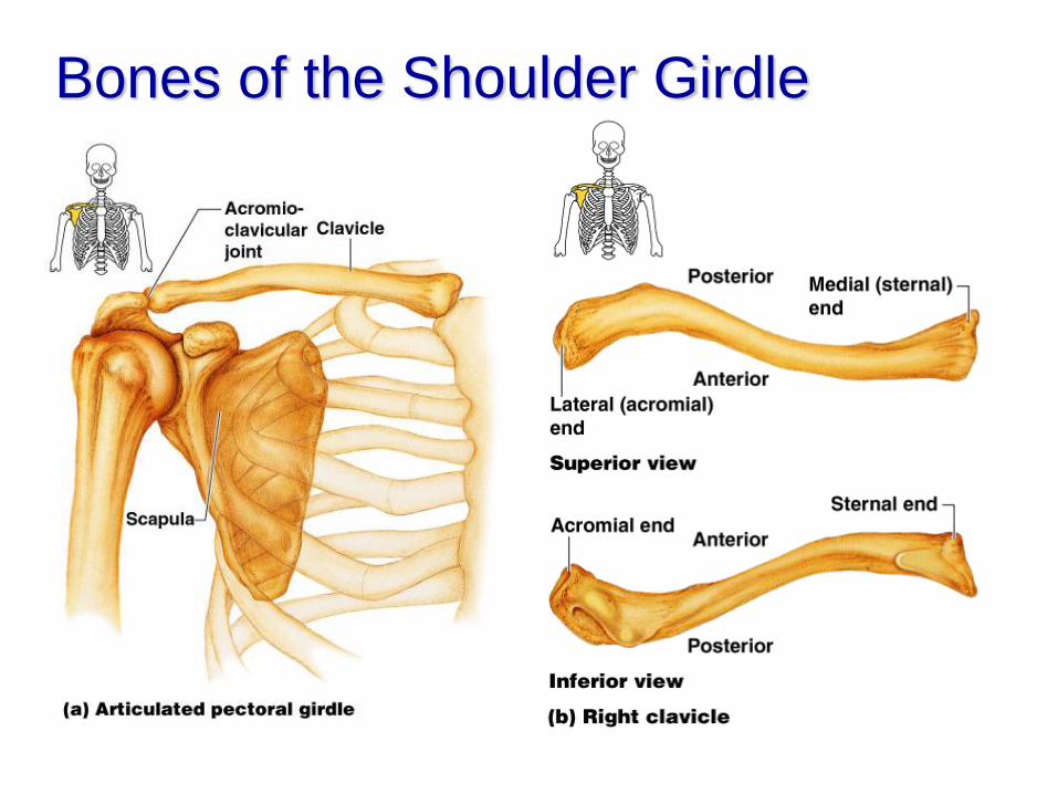
Bones of the Shoulder Girdle
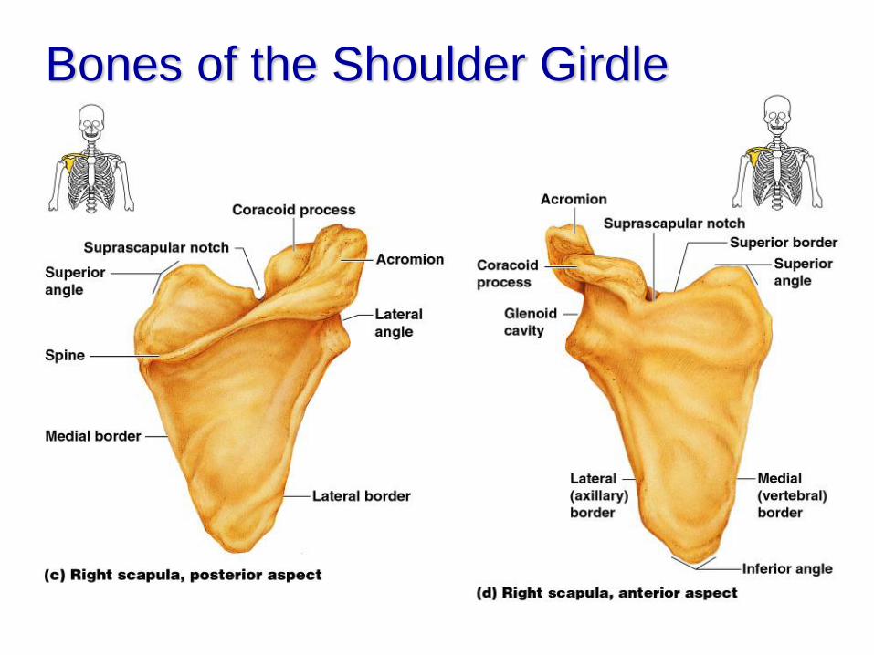
Bones of the Shoulder Girdle
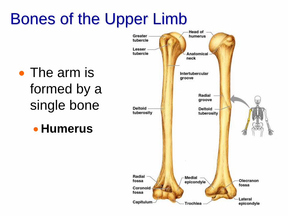
Bones of the Upper Limb
The arm is
formed by a
single bone
Humerus
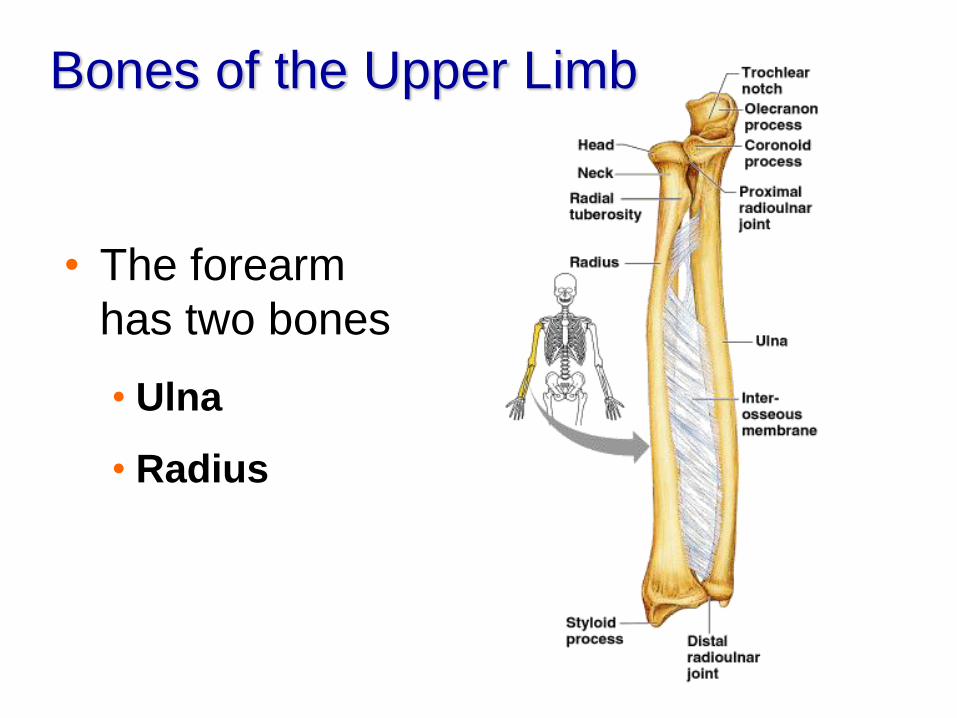
Bones of the Upper Limb
• The forearm
has two bones
• Ulna
• Radius

Bones of the Upper Limb
The hand
Carpals – wrist
Metacarpals –
palm
Phalanges –
fingers
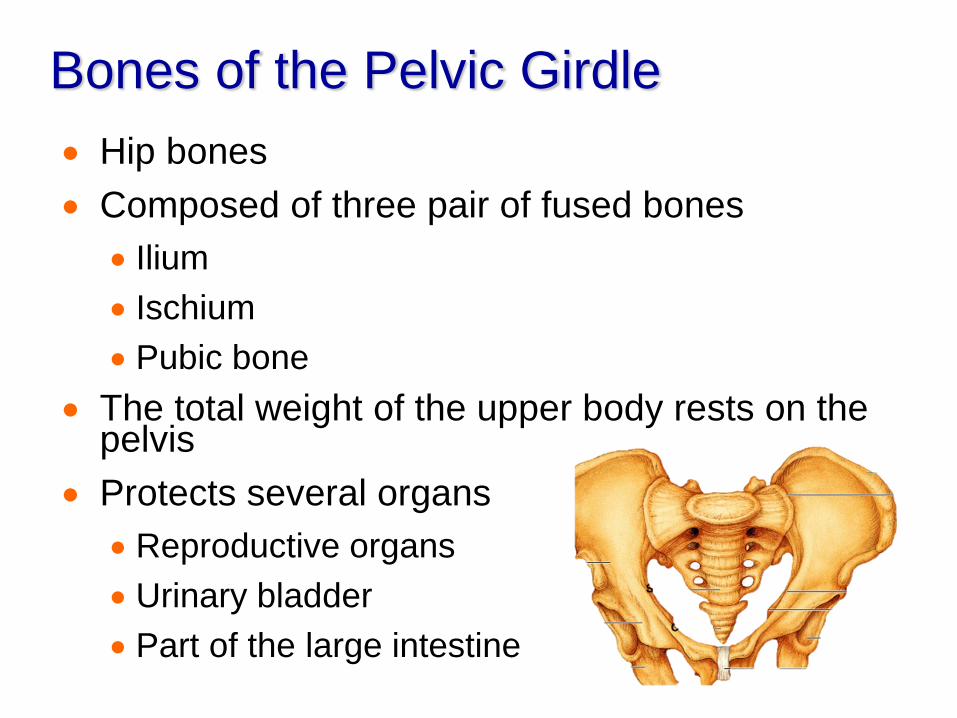
Bones of the Pelvic Girdle
Hip bones
Composed of three pair of fused bones
Ilium
Ischium
Pubic bone
The total weight of the upper body rests on the pelvis
Protects several organs
Reproductive organs
Urinary bladder
Part of the large intestine

The Pelvis
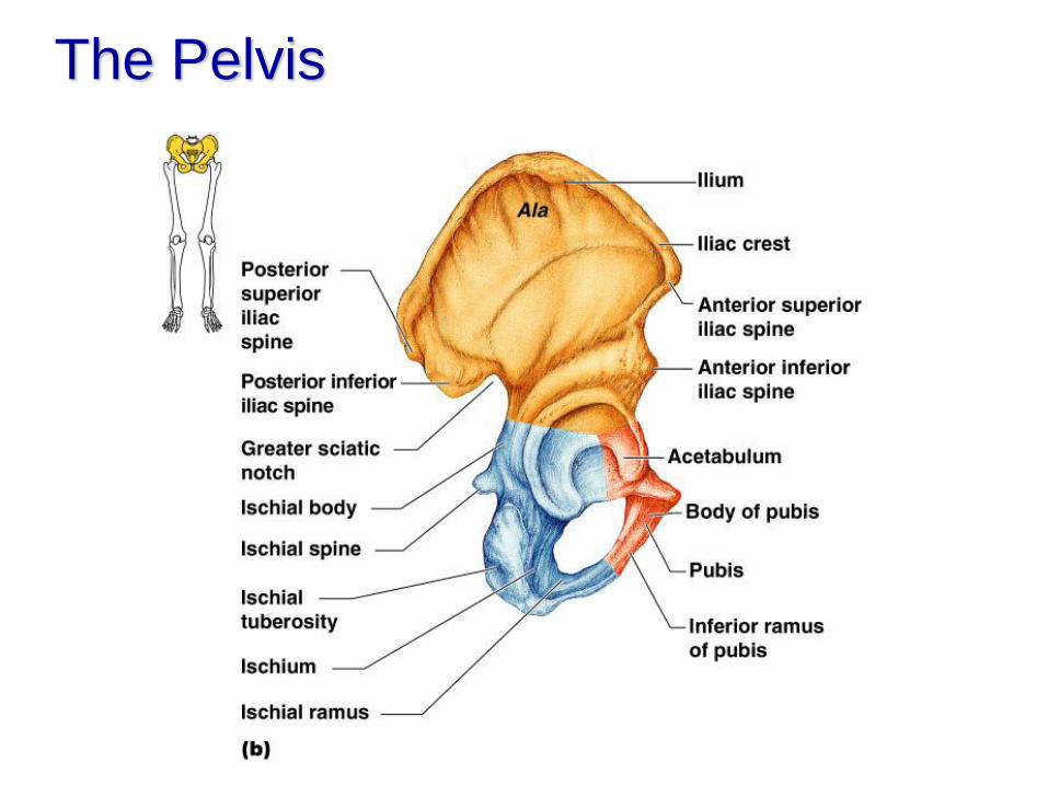
The Pelvis
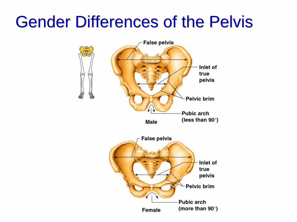
Gender Differences of the Pelvis

Bones of the Lower Limbs
The thigh has
one bone
Femur – thigh
bone
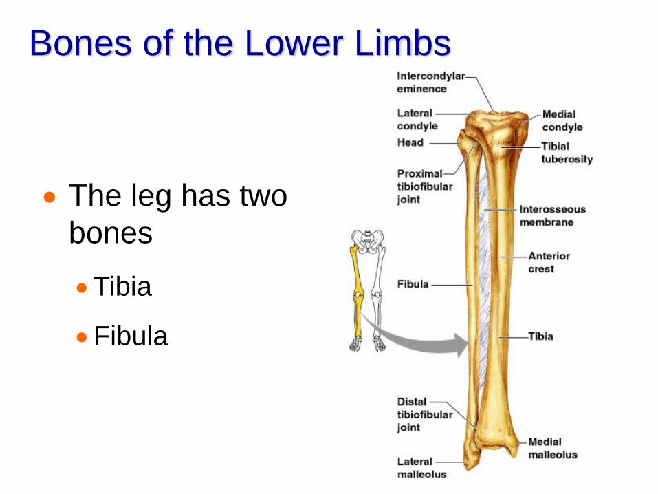
Bones of the Lower Limbs
The leg has two
bones
Tibia
Fibula
Bones of the Lower Limbs
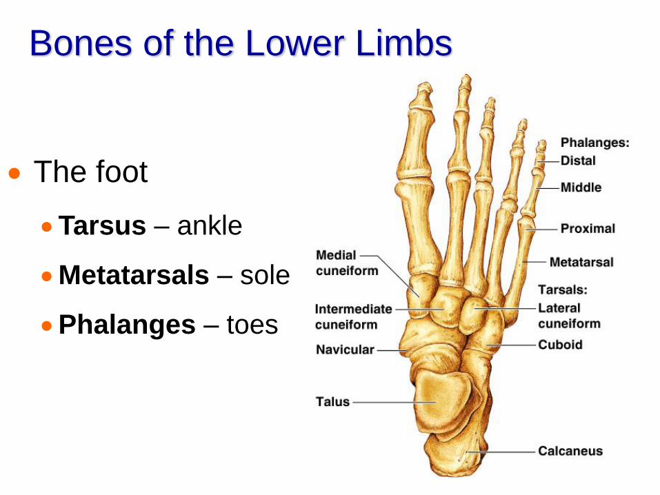
The foot
Tarsus – ankle
Metatarsals – sole
Phalanges – toes
Arches of the Foot
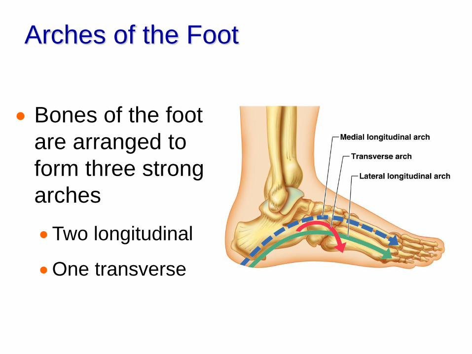
Bones of the foot
are arranged to
form three strong
arches
Two longitudinal
One transverse
Joints
Articulations of bones
Functions of joints
Hold bones together
Allow for mobility
Ways joints are classified
Functionally
Structurally
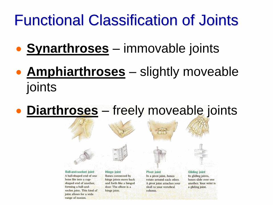
Functional Classification of Joints
Synarthroses – immovable joints
Amphiarthroses – slightly moveable
joints
Diarthroses – freely moveable joints
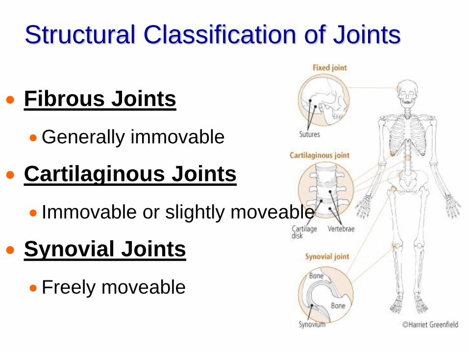
Structural Classification of Joints
Fibrous Joints
Generally immovable
Cartilaginous Joints
Immovable or slightly moveable
Synovial Joints
Freely moveable
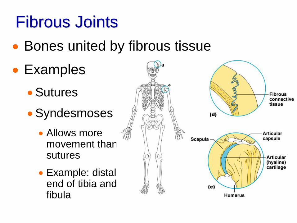
Fibrous Joints
Bones united by fibrous tissue
Examples
Sutures
Syndesmoses
Allows more movement than sutures
Example: distal end of tibia and fibula
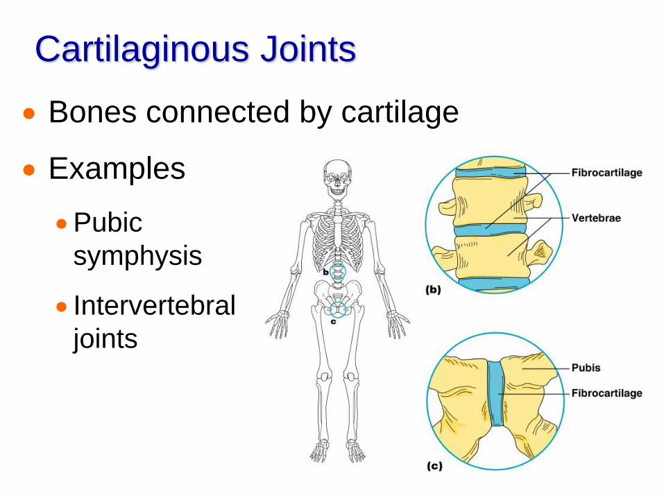
Cartilaginous Joints
Bones connected by cartilage
Examples
Pubic
symphysis
Intervertebral
joints
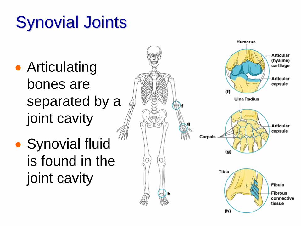
Synovial Joints
Articulating
bones are
separated by a
joint cavity
Synovial fluid
is found in the
joint cavity
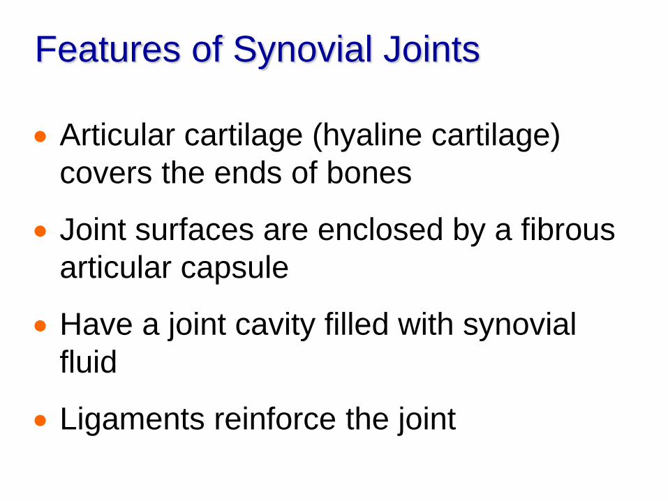
Features of Synovial Joints
Articular cartilage (hyaline cartilage)
covers the ends of bones
Joint surfaces are enclosed by a fibrous
articular capsule
Have a joint cavity filled with synovial
fluid
Ligaments reinforce the joint
Structures Associated with the
Synovial Joint
Bursae – flattened fibrous sacs
Lined with synovial membranes
Filled with synovial fluid
Not actually part of the joint
Tendon sheath
Elongated bursa that wraps around a tendon
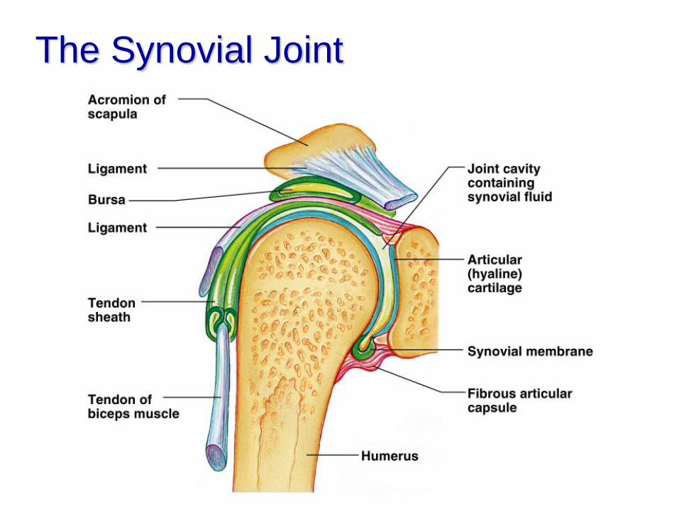
The Synovial Joint
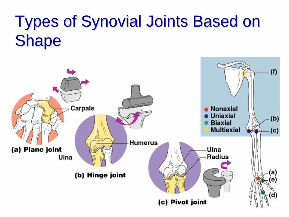
Types of Synovial Joints Based on
Shape
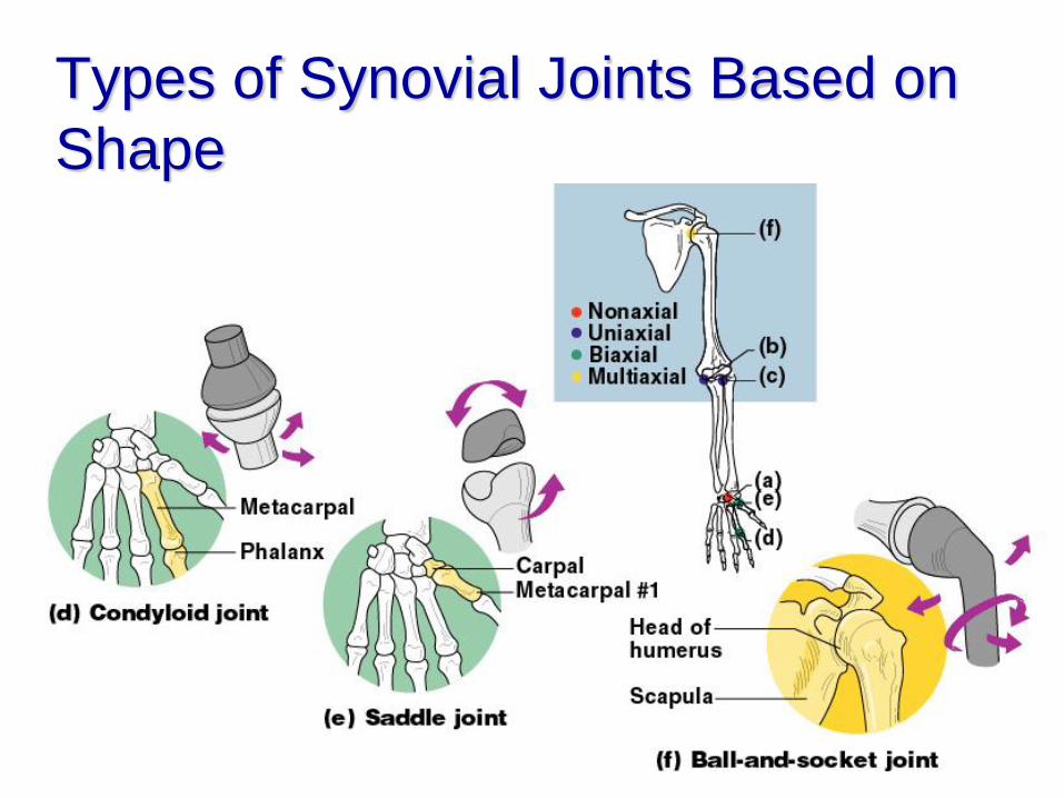
Types of Synovial Joints Based on
Shape
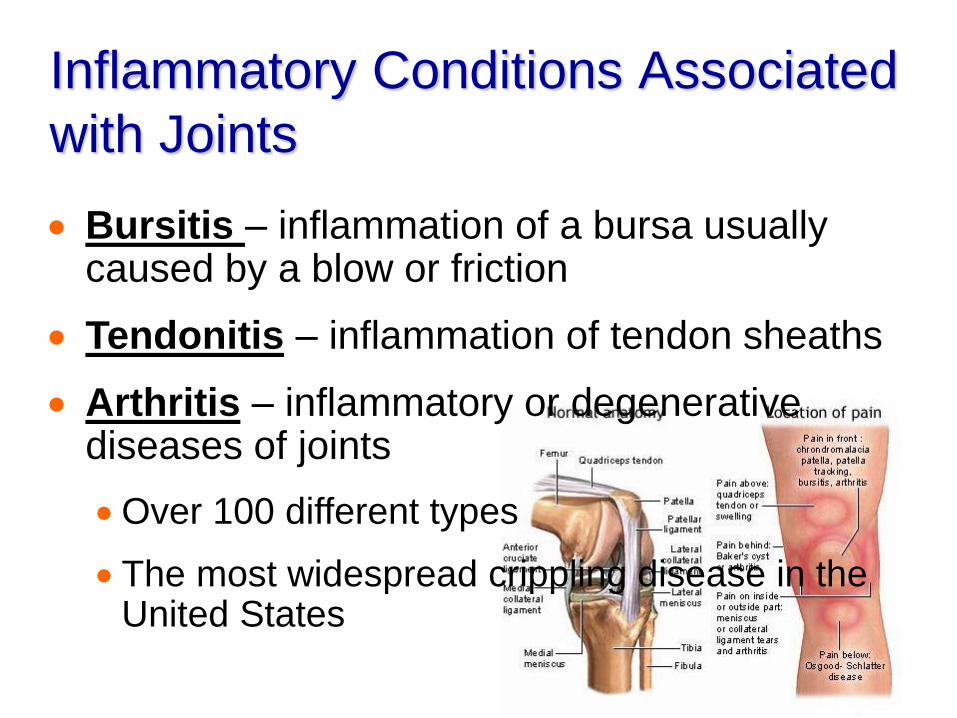
Inflammatory Conditions Associated
with Joints
Bursitis – inflammation of a bursa usually caused by a blow or friction
Tendonitis – inflammation of tendon sheaths
Arthritis – inflammatory or degenerative diseases of joints
Over 100 different types
The most widespread crippling disease in the United States
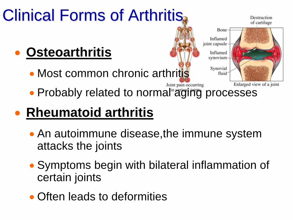
Clinical Forms of Arthritis
Osteoarthritis
Most common chronic arthritis
Probably related to normal aging processes
Rheumatoid arthritis
An autoimmune disease,the immune system attacks the joints
Symptoms begin with bilateral inflammation of certain joints
Often leads to deformities
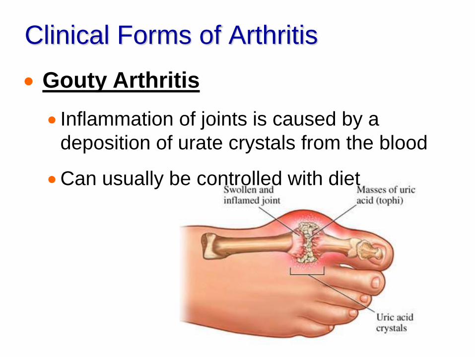
Clinical Forms of Arthritis
Gouty Arthritis
Inflammation of joints is caused by a
deposition of urate crystals from the blood
Can usually be controlled with diet

Developmental Aspects of the
Skeletal System
At birth, the skull bones are incomplete
Bones are joined by fibrous membranes
– fontanelles
Fontanelles are completely replaced
with bone within two years after birth