nerve endings nervous structures present in the body&classified according to the their functions...
TRANSCRIPT

Nerve Endings
Nervous structures present in the body&classified according to the their functions into
-Receptors
-Effectors

The receptors
Receive sensory impulses from out side the body or tissue
1-receptors for special sense asa-photoreceptors of vision by the retina of the eyesb-Audioreceptors of hearing by corti of earc-chemoreceptors of smell by epithelium of the nosed-chemoreceptor of taste by the taste buds of the tonguee-Reception of the different movement of head&body

2-Receptors for cutanouse sensibility( extereceptors)a-pain:sensation by free nerve endingsb-Temperature sensation by Krause bulb,Ruffinini organc-Touch sensation by Merkels disc,meissners corpuscle&free nerve endings
3-Receptors for deep pressure&vibration sense
a-from skin,muscle and wall of organs by pacinian corpus
b-from muscle,tendons&joints by muscle spindles&tendon

4-Receptor from the wall of viscera
To transmit autonomic sensation from the stomach,bladder

The exteroceptors
1-Receptors in the epithelial tissue
-Non-capsulated receptors
a-free or bare-nerve endings
these endings are formed of non myelinated sensory nerve fibres,branched in between the epithelial cells
site:epidermis of skin,cornea,conjunctival,oral cavity ,dental pulp(stratified sq.epithelium)
Function:responsible for pain&cold sensation
Respond to displacement of hair
b-merkels Disc
present in the deep layers of epidermis cells
Merkel cells:epidermal cells of the epidermis of the skin called Merkel cells
Polygonal in shape,irregular with cytoplasmic granules
Function:respond to light&deep pressure

c-plexus of bounet(peritrichial Endings)
present around the hair follicules of skin
function:responsible for the sensation of hair movement&act as mechanomoreceptors
d-other epithelial receptors around the neuro-epithelial cells
nerve fibres present around the neuroepithelial cells of the taste buds,around the epithelial receptors of the internal ear
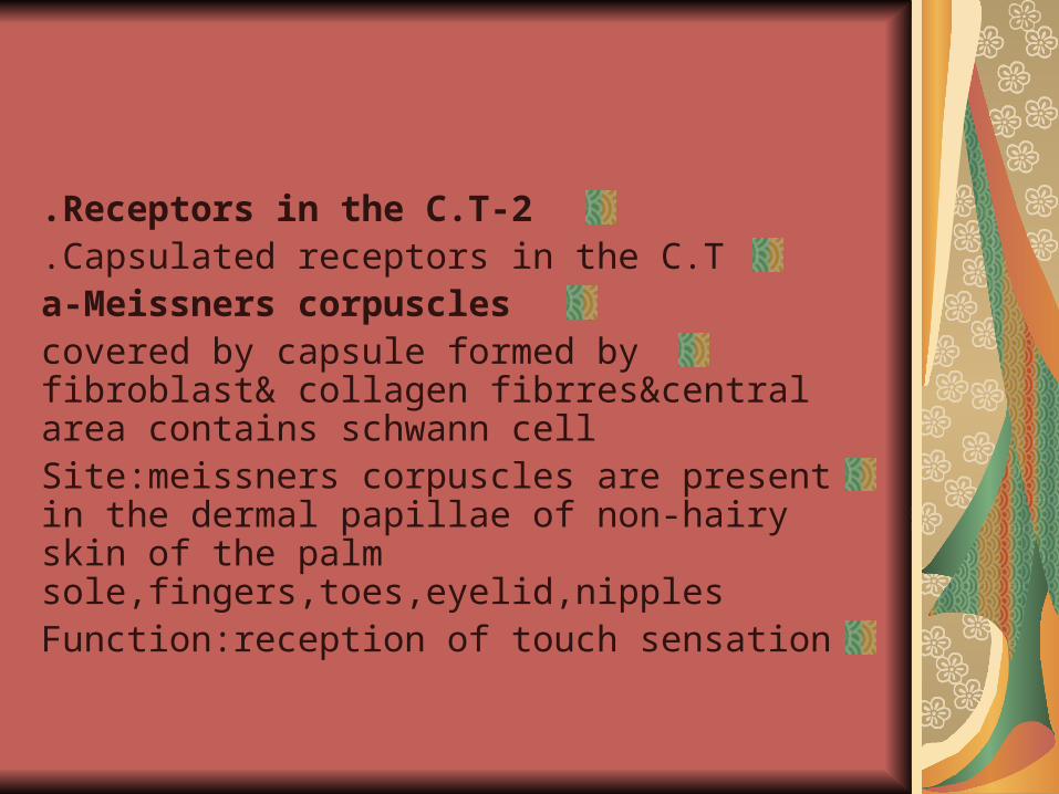
2-Receptors in the C.T.Capsulated receptors in the C.T.
a-Meissners corpusclescovered by capsule formed by fibroblast& collagen fibrres¢ral area contains schwann cellSite:meissners corpuscles are present in the dermal papillae of non-hairy skin of the palm sole,fingers,toes,eyelid,nipplesFunction:reception of touch sensation
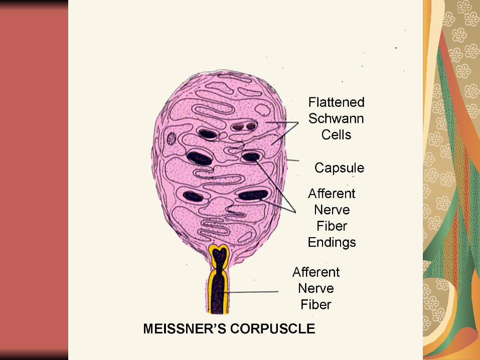

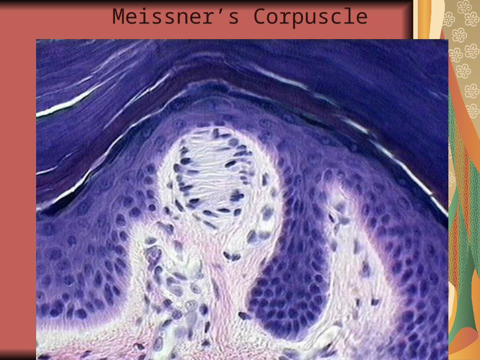
Meissner’s Corpuscle

MEISSNER’S CORPUSCLES
Tactile (light touch) Receptors
skin
dermal papilla
Meissner’s
corpuscles
b-Krauses end bulb
present in the C.T.dermis of the external genitalia,lip,tongue
function:receptors for deep pressure&cold sensation
C-the Ruffini end organ
Sensory nerve fibres branch to form dense clusters of nerve ending

Site:present in dermis&hypodermis of skin specially in the plantar surface of the feet
Function:they are mechanoreceptor for joint movements for deep pressure&may receive hot sensation

D-The pacinian corpuscle
Formed of numerous concentric lamellae
The concentric layers are formed of modified schwann cell
-the concentric layers are seprated from one another by collagenous fibres&by spaces filled with fluid
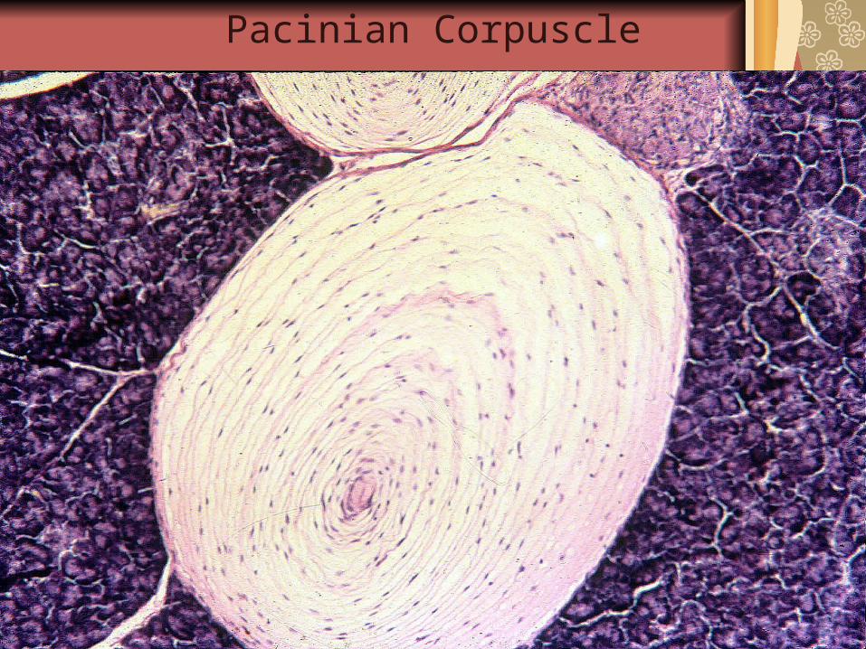
Pacinian Corpuscle
Site of pacinian corpuscles
Dermis of skin specially in palm&sole
-present in striated muscle,tendon,joints wall of large B.V.,wall of urinary bladder
Function: respond to changes in position,vibration sense,tactile localization they act as propriceptors
Blood Vascular System
There are 3 types of blood vesselsArteries,vens,&connecting vessels between arteries and veins
-Any Medium sized artery is made 3 layers from inside
1-Tunica Intima: the inner layer formed of three elementsa-simple Sq. epitheliumb-subendothelial layer of C.T.
c-internal elastic lamina
2-Tunica Media:it is the middle layer&formed of 3 element
a-circular smooth muscle fibres
b-few scattered elastic tissue
c-fine collagenous fibres
3-Tunica adventitia outer layer 3 elements
a-mostly collagenous fibres
b-some elastic fibres
c-some C.T. cells

General Structure of Blood Vessels
a common structural pattern
that can be seen in all blood vessels
with the exception of capillaries,
i.e. the division of the walls of the
blood vessels into three layers or
tunics .
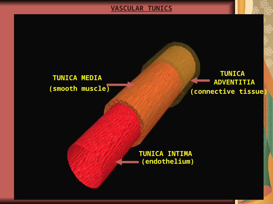
VASCULAR TUNICS
TUNICA MEDIATUNICA
ADVENTITIA
(endothelium)
(smooth muscle) (connective tissue)
TUNICA INTIMA
The medium sized vein also formed of 3 layers
Differences between of Medium sized of artery&medium sized of vein
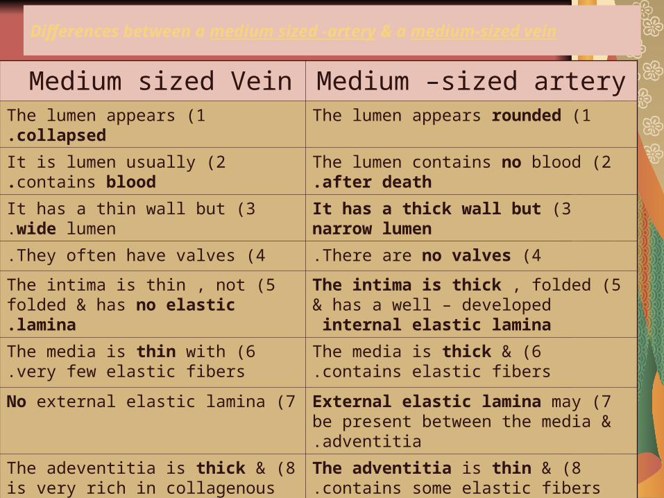
Differences between a medium sized -artery & a medium-sized vein
Medium –sized arteryMedium sized Vein 1 )The lumen appears rounded1 )The lumen appears collapsed.
2 )The lumen contains no blood after death.2 )It is lumen usually contains blood.
3 )It has a thick wall but narrow lumen3 )It has a thin wall but wide lumen.
4 )There are no valves.4 )They often have valves.
5 )The intima is thick , folded & has a well – developed internal elastic lamina
5 )The intima is thin , not folded & has no elastic lamina.
6 )The media is thick & contains elastic fibers.6 )The media is thin with very few elastic fibers.
7 )External elastic lamina may be present between the media & adventitia.
7 )No external elastic lamina
8 )The adventitia is thin & contains some elastic fibers.
8 )The adeventitia is thick & is very rich in collagenous fibers.
9 )No lymphatic capillaries in it is wall. 9 )Lymphatic capillaries may be present in it is adeventitia.
10 )It has a rapid flow of arterial blood.10 )It is a slow flow of venous blood.
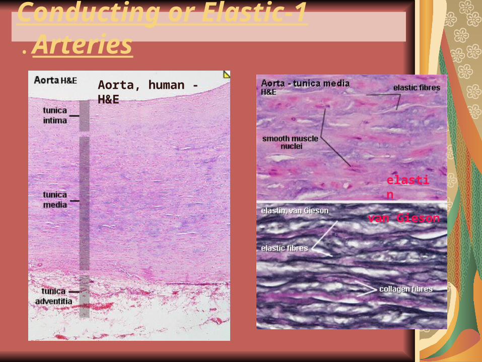
1-Conducting or Elastic Arteries.
Aorta, human - H&E
elastin
van Gieson
Arterioles:
They are small in diameters&small branches of the arteries
formed of three tunics.
with narrow lumens and are
1-Tunica Intima:
It consists of endlothelial cells rest on basal lamina with
very thin or without sub-endothelial C.T layer.
2 -Tunica Media:
It is formed of one or two layers of smooth muscle internal
elastic lamina if present it will be very thin,
3-Tunic Adventitia
:
It is formed of thick connective tissue, which is equal in
thickness to tunica media.
The Metarterioles are the terminal parts of the arterioles before their connections with blood capillaries

Large elastic Arteries
The large elastic arteries in the body are: aorta,the pulmonary,the subclavian.
Have thick wall ,very wide lumens,very rich in elastic fibres
1-The Aorta
Has avery thick wall&very wide lumenThe intima is thick&rich in elastic fibres
-media is very thick&made fenestrated elastic membranes enclosing between them smooth muscle,collagen&reticular fibresAdvintitia is thin,it contains collagenous and elastic fibresand also contain nerves and small blood vessels

2 -Basilar artery
:
This artery supplies brain with blood and is present inside
the skull so it is characterized by:
-Its wall is thin and both tunicae media and adventitia are
thin.
-it contains well developed internal elastic lamina are thick
3 -Coronary artery
It supplies the heart with blood and It is characterized by :
Thick sub-endothelial layer containing longitudinalsmooth muscle fibers.
-Tunica media is formed of smooth muscle fibres -Internal and external elastic laminae are well
developed.Adventitia is thin and formedoof areolar C.T.With collagenous&elastic fibres
Functions of the different types of Arteries
1-the Aorta allow aconstant flow of blood
2-The medium-sized arteries distribute the blood to the whole body
3-The arterioles control the blood pressure
Veins
Veins carry venous the blood from the tissue They are classified into:Large sized vein (inferior vena cava):
It is characterized by wide lumen, thin wall and presence ofvalves, which are folds from tunica intima including insideC.T to allow the blood to pass towards the heart. It consistsof three tunics.
1-Tunica Intema:
endothelial lining
2 -Tunica Media:
Formed of smooth muscles
3 -Tunica Adventitia:
Formed of areolar C.T.
Connections between arteries and veins
.
A- Capillaries
B-Sinusoids
C- Arteriovenous anastomoses
A- Capillaries:
The average diameter of capillaries from 8 microns indiameter.
They present evry where in the body
transversely lumens appear containing from one to 3
endothelial cells that rest on basal laminae
Function of the capillaries
:
1-selective exchange of materials between blood and tissues
2-secret prostaglandin which prevent formation of thrombus
3-convert angiotensin I into angiotensin II
Strucures of capillaries
1-simple sq.endothelium
2-basment membrane surrounding the endothelium,this basement membrane spilt to enclose small cells called Pericyte are undifferentiated cells,can differentiated into smooth muscle cells into fibroblast

CAPILLARY TYPES
ContinuousCapillary
TypicalLocations
fatmusclenervoussystem
Fenestrated
Capillary
TypicalLocations
intestinal villiendocrine glandskidney glomeruli
DiscontinuousCapillary
TypicalLocations
liverbone marrow
spleen
Types of blood capillaries
1-capillaries with continous endothelium and continuous basal laminaa-they present all over the bodyb-the are lined with contianous endothelial cells
2-capillaries which are lined with fenestrated endothelium.these fenestration are covered with diaphragmsThey are present in kidney,intestine,endocrine glands
3-capillaries lined tightly joined endothelial cells as brain capillaries
4-the sinusoidal capillaries they are present in bone marrow,liver and spleen
2-The blood sinusoids
-They have irregular wide lumen from 5-30 microns in diameter
-Lined with simple squamous endothelium,the sinusoids are surrounded by thin layer of reticular C.T

Sites and functions of blood sinusoids
a-present in the bone marrow to carry the formed blood cells,the slow circulation in the sinusoids causes alow oxygen tensionb-present in the spleen to store bloodthe phagocytic cells present in the sinusoidal wall can clean the blood from any foreign bodies

c-present in the liver to allow liver cells to be indirect contact with blood
d-present in the endocrine glands to carry the secreted hormones and to increase the blood supply to the endocrine secretory cells

Differences between capillaries & sinusoids
CapillariesSinusoids
1 )Present allover the body1 )Present in liver , spleen, bone marrow.
2 )Having narrow regular lumens.2 )Having wider irregular lumens.
3 )Their walls are formed of simple squamous cells & are surrounded by continuous basement membrane.
3 )Their walls are lined by fenestrated cells & are surrounded by reticular C.T.
4 )Their walls have no pores except in kidney & endocrine glands.
4 )Their walls contain pores.
5 )Undifferentiated pericyte cells are present in their walls.
5 )Phagocytic macrophage littoral cells are present outsides their walls.

3-Arterio-venous Anastomoses
A-V anastomoses arise as side branches from arterioles to venules without passing through the capillaries
They allow ashort and rapid circulation of blood to certain areas or organs of the body
Two types are present in arteriovenous anastomosis:
1 -Direct connection: through direct side branch fromarterioles to venules as in placenta. The side branch isstructurally similar to arterioles at the arterial side andsimilar to venules at the venous side.
2-Glomus:
Which is aspecialized organ present in genital organs,nailbed and ear
Structures of arterio-venous anastomosis
The wall of the connecting segment between an arteriole and venule has the structural characterstic of the vein at the venous side
The intermediate segment of the A-V anastomosis
characterized by:
1-lumen of these side branches decreases gradually towards the venous side
2-the internal elastic lamina disappears gradually towards the venous side
3-the media is well devolped at the venous side rich in longitudinal smooth muscles
4-Myoepithelial cells are present also in thck media at the venous side
5-the adventitia becomes thicker at the venous side

Site of Arteriovenous anastomoses
Tips of the fingers,toes, the external ear,the nose,in the external ear,nose,lip,tongue
Present also in the internal organs as stomach,intestine,liver,endocrine glands
Functions of A-V Anastomoses
1-they conserve the body temperature
2-Dilated in cold weather while in hot weather they constrict
Regulate the venous return
3-they regulate the blood flow to genital organs during erection
4-they regulate the uterin blood flow during menstruation and pregnancy

Lymphatic System
1-the lymphatic tissues which are:
a-lymph nodes
b-spleen
c-scattered lymphatic nodules
d-tonsils
2-the Lymphatic vessels: the lymphatic capillaries and lymph vessels;the carry the lymph
Formation of lymph
Tissue fluid which is filtered from the tissues and from the blood capillaries around the cells is drained by blind-ended lymphatic capillaries
The fluid when enters the lymphatic vessels called lymph

The lymph is filtred in lymph nodes&nodules
Lymph flows in one direction inside the lymphatic capillaries
The filtred lymph go again to the blood stream through alarge lymphatic vessels called thoracic duct

Differences between blood capillaries & lymphatic capillaries
Blood Capillaries Lymphatic capillaries1) Present superficial in position
under the skin & mucous membrane.1 )Present more deep in position
than blood capillaries
2)They are the branching vessels of arterioles & are connected with venules at the other side.
2)They start as blind – ended channels & are connected to lymph vessels at one side only.
3)They have uniform diameters3)Their diameters are irregular
4)The endothelium is fenestrated 4 )Non-fenestrated endothelium
5)They are surrounded with basement membranes & pericytes.
5)They have no basement membrane & no pericytes.
6)The lumen is usually patent6)The lumen may be collapsed.
7)No anchoring collagenous fibers outside their wall
7 )Anchoring collagenous fibers connect their wall to surrounding C.T.
8)They carry blood.8)They carry lymph.