neural mechanisms of action switching moderate the
TRANSCRIPT

University of New Orleans University of New Orleans
ScholarWorks@UNO ScholarWorks@UNO
University of New Orleans Theses and Dissertations Dissertations and Theses
Summer 8-10-2016
Neural Mechanisms of Action Switching Moderate the Neural Mechanisms of Action Switching Moderate the
Relationship Between Effortful Control and Aggression Relationship Between Effortful Control and Aggression
Eric L. Rawls University of New Orleans, [email protected]
Follow this and additional works at: https://scholarworks.uno.edu/td
Part of the Biological Psychology Commons, and the Cognitive Psychology Commons
Recommended Citation Recommended Citation Rawls, Eric L., "Neural Mechanisms of Action Switching Moderate the Relationship Between Effortful Control and Aggression" (2016). University of New Orleans Theses and Dissertations. 2234. https://scholarworks.uno.edu/td/2234
This Thesis is protected by copyright and/or related rights. It has been brought to you by ScholarWorks@UNO with permission from the rights-holder(s). You are free to use this Thesis in any way that is permitted by the copyright and related rights legislation that applies to your use. For other uses you need to obtain permission from the rights-holder(s) directly, unless additional rights are indicated by a Creative Commons license in the record and/or on the work itself. This Thesis has been accepted for inclusion in University of New Orleans Theses and Dissertations by an authorized administrator of ScholarWorks@UNO. For more information, please contact [email protected].

Neural Mechanisms of Action Switching Moderate the Relationship Between Effortful Control
and Aggression
A Thesis
Submitted to the Graduate Faculty of the
University of New Orleans
in partial fulfillment of the
requirements for the degree of
Master of Science
in
Psychology
Applied Biopsychology
by
Eric L. Rawls
B.S. Tulane University, 2012
August, 2016

ii
Table of Contents
List of Figures…………...…...……………………………………………………………..…….iii
List of Tables……………………………………………………………………………………..iv
Abstract…...……………………………………………………………………………………….v
Introduction…………………...………………………………………………………………...…1
Method..………...…………………………………………………………………...…………….5
Results………………..………………………………………………………….……………….13
Discussion…...………………………………………………………………….………………..24
References………………………..…………………………………………….………………...30
Vita……………………………………………………………………………………………….35

iii
List of Figures
Figure 1: Modified AX-CPT Task Diagram……………………………...…………………...…11
Figure 2: Grand average ERP waveforms……………………………..………………………...14
Figure 3: Morphology-based regions of interest (ROIs) generated using the Montreal
Neurological Institute average adult magnetic resonance image (MRI)…………..…………….16
Figure 4: Moderation plots: Interaction between N2 amplitudes and Effortful Control on
aggression…………………………………………..……………………………………………22
Figure 5: Moderation plots: Interaction between N2 amplitudes and Effortful Control on
aggression for source-space regions………………………………………..……………………27

iv
List of Tables
Table 1. Sex differences………...…………………......……..…………………………………..12
Table 2. Preliminary Pearson correlation analyses (r-values) between effortful control,
aggression, and brain measures…………………………………..………………………………16
Table 3. Regression Showing P2 Moderation Effects Predicting Aggression….………………..17
Table 4. Regression Showing N2 Moderation Effects Predicting Aggression…...…...................17
Table 5. Regression Showing P3 Moderation Effects Predicting Aggression…………………...18
Table 6. Pearson correlation analyses (r-values) between effortful control, aggression, and brain
& behavioral measures..………………………………………..………………………………...21
Table 7. Moderation Effects Showing Interactions Between Source Space Activation Underlying
the N2 and Effortful Control..………………………………..………..………………………....22

v
Abstract
Aggression and violence are social behaviors that exact a significant toll on human
societies. Individuals with aggressive tendencies display deficits in effortful control, particularly
in affectively charged situations. However, not all individuals with poor effortful control are
aggressive. This study uses event-related potentials (ERPs) to decompose the chronology of
cognitive functions underlying the link between effortful control and aggression. Specifically,
this study investigates which ERPs moderate the effortful control - aggression association. We
examined three successive ERP components (P2, N2 and P3) for stimuli that required effortful
control. Results indicated that N2 activation, but not P2 or P3 activation, moderated the
relationship between effortful control and aggression. These effects were present in negative and
neutral contexts. This moderating effect was consistent with previous studies linking neural
processing efficiency with reduced activation during cognitive control tasks. Our results suggest
that efficient cognitive processing moderates the association between effortful control and
aggression.
Effortful Control, Aggression, Emotion, Emotion Regulation, EEG, ERPs, LORETA

1
1, Introduction
Aggression and violence are complex social behaviors that exact a significant toll on
human societies (Mehta & Beer, 2007). Overly aggressive behaviors have been associated with
increased impulsivity and limited self-regulatory skills (Nelson & Trainor, 2007). In the context
of individuals who score high on measures of interpersonal aggression, particular deficits in
effortful control can be seen. Effortful control is defined in part as the process of inhibiting a
dominant response in favor of a subdominant response (Rothbart & Rueda, 2005). Since much of
human behavior consists of habitual or instinctive actions (Hikosaka & Isoda, 2008), effortful
control is necessary specifically in those situations when a “default mode” of behavior must be
overridden and different action initiated (Goldstein et al., 2007). Given the diversity and
complexity of computing different action plans, it is not surprising that many areas in the brain
are involved in effortful control (Rothbart et al., 2007; Bush et al., 2000). In addition, there is
considerable variability in the control strategies displayed by different individuals, and many
psychiatric disorders, such as substance abuse and ADHD, are thought to result partly from
effortful control impairments (Wiers et al., 2013; Vaidya et al., 2014; Siegle et al., 2007). People
with aggressive tendencies may have difficulty regulating their behavior during negative
emotional situations, resulting in harmful interpersonal behaviors (Lewis et al., 2007). However,
not all individuals who have poor effortful control are aggressive. The present study uses event-
related potentials (ERPs) to decompose the chronology of cognitive functions underlying
effortful control, and ascertains which of these cognitive functions contributes to the association
with aggressive behavior. More specifically, the current study investigates which ERPs moderate
the effortful control - aggression association. Additionally, the current study uses linear inverse
models to investigate which cortical regions underlying specific ERPs moderate the association

2
between effortful control and aggression. Furthermore, to ascertain how effortful control-related
activation patterns change depending on emotional context, this study uses an experimental
paradigm intended to capture participants’ control abilities during negative emotional contexts,
positive emotional contexts, and relatively unemotional contexts.
A key aspect of effortful control is to switch from a dominant response to a subdominant
response (Rothbart & Rueda, 2005), i.e., effectively switch action strategies. The ability to
flexibly switch action strategies likely requires a number of underlying cognitive functions (e.g.,
Badre & Wagner, 2006; Braver et al., 2009; Eslinger & Grattan, 1993). Given that any one of
these functions could show deficits that might contribute to aggressive behavior, understanding
the chronology underlying effective action switching is important to set the stage for targeted
treatment. Some cognitive processes that might contribute to flexible action switching are
attentional orienting (Weissman et al., 2002), cognitive control (Botvinick et al., 2001), and
context updating for subsequent behavioral action (Friedman et al., 2001). For example, a person
needs to orient their attention towards new environmental information in order to effectively
process this information. Additionally, a person needs to apply cognitive control to process or
monitor conflicting information, i.e., information that would have led to the previous action
strategy vs. information leading to the current (changed) action strategy. Lastly, the new
information needs to be encoded to prepare for the new action strategy. These three processes
can be measured using ERPs. To examine the time course of neural activation related to effective
action switching, we examined three successive ERP components for a stimulus that required
effortful control. P2 activation has been related to attentional orienting, and is thought to have
underlying neural sources in widespread areas including occipital, temporal, and frontal regions
(Britz & Pitts, 2011; Mulert et al., 2002; Vitacco et al., 2002). N2 activation has been related to

3
various aspects of cognitive control, such as conflict monitoring, inhibition, and emotion
regulation, and is thought to have underlying neural sources in prefrontal regions, including
DLPFC and ACC (Ladouceur et al., 2007; Bekker et al., 2005). P3 activation has been related to
motivational processing, novelty, and compilation of higher-level decision-making processes,
and is thought to have underlying neural sources in posterior regions, including occipital regions
(Barry & Rushby, 2006; Volpe et al., 2007). By examining the effects of this succession of ERP
components on the association between effortful control and aggression, we hope to gain a
greater understanding of the impact of these temporal processes on ineffective effortful control
and aggressive behavior. We hypothesize that temporally distinct patterns of neural activation
will moderate the association between participant scores of effortful control and aggression. In
order to elicit effortful control in the context of action switching, we used a modified AX
continuous-performance task (AX-CPT; Rosvold et al., 1956). This task consists of a cue, to
which participants have to provide a speeded response, then a delay period, and then a probe, to
which participants have to provide a second speeded response. A preponderance of one trial type
(A-X) creates a habitual response. Effortful control resources, on the other hand, are recruited
when action switching is necessary in order to adjust action strategies based on new contextual
information; that is, when the trial type changes (A-Y).
In addition, in line with the Rothbart model of emotional reactivity impacting effortful
control abilities (Rothbart & Sheese, 2007), we were interested in how this neural chronology
changes in the face of salient emotions. Previous studies that required aspects of effortful control
have yielded prefrontal cortical activation differences depending on the emotional context of the
task. For example, Monk et al. (2003) found greater ACC activation to fearful faces than neutral
faces during an attention task; Ochsner et al. (2004) found ACC, VLPFC, and DLPFC activation

4
during emotional up-regulation and down-regulation; and lastly, Lamm and Lewis (2010) found
elevated VMPFC activation for a negative condition compared to a neutral condition in a
motivated go/no-go task. In order to determine if moderating effects of neural activation differ
depending on emotional context, an emotional component was included in the form of neutral,
positive, or negative affectively charged pictures. Notably, the negative pictures shown in this
task were usually (75 percent) of a violent or threatening nature . A picture from one of these
categories was presented during the delay period, i.e., independent of task requirements, so that
neural activation underlying the process of action switching could be measured in the context of
neutral, positive, or negative (violent or threatening) affective stimuli. We hypothesize that in the
face of negatively valenced images compared to positive or neutral images, participants would
show greater neural activation underlying action-switching processes and thus reveal a stronger
moderating impact on aggressive behavior.

5
2, Method
2.1 Participants
The sample was recruited from undergraduate students taking psychology classes at the
University of New Orleans. Participants were 76 undergraduate students (35 male). Criteria for
exclusion from the study were current psychiatric diagnoses, current use of psychoactive
medication, and uncorrected visual impairments. All students were given extra credit to
compensate for their participation. All students were English speaking. Ethical approval for the
project was obtained from the University of New Orleans’ Institutional Review Board.
2.2 Procedure
Participants were briefly introduced to the testing environment, after which informed
consent was obtained. Participants were then seated in the testing room to complete
questionnaires. After completion of the questionnaire battery, participants were seated 67
centimeters in front of a computer monitor. The electrode sensor net was applied. They were
given a practice block of 16 trials, with the option to repeat the practice block, in order to ensure
proficiency with the task.
2.3 Measures & Task
2.3.1 The Adult Temperament Questionnaire Short Form (ATQ; Evans & Rothbart,
2007) is a 77-item reliable and valid self-report measure of emotional temperament and self-
regulatory capacity. The measure consists of 13 subscales, three of which comprise the effortful
control scale: attentional control, inhibitory control, and activation control. The effortful control
scale was used as a measure of an adult’s ability to effortfully regulate their actions.

6
2.3.2 The Buss Perry Aggression Scale (BPAS; Buss & Perry, 1992) is a 29-item
standardized, valid, and reliable self-report measure of aggression in adults. The overall score
(average of all items) was used to measure aggressive tendencies in this sample.
2.3.3 Action Switching Task. The task was a modified AX continuous performance task
(Rosvold, Mirsky, Sarason, Bransome, & Beck, 1956). Images were presented on a 17-in
monitor using E-prime Software (Psychology Software Tools, Inc., Pittsburgh, PA; Schneider,
Eschman, & Zuccolotto, 2002). Stimuli were shown on a black screen and consisted of negative
and neutral photos from the International Affective Picture System (IAPS; Lang, Bradley, &
Cuthbert, 2008) and single letters presented in either blue (cue) or white (probe). Negative,
positive, and neutral pictures were 11 cm wide by 8 cm tall and presented in black and white
(visual angle was 9.39 degrees). Letters were presented in 60-point size uppercase bold Courier
New font. Trials were roughly 3.7 seconds in duration and consisted of the following events (see
Figure 1): fixation (500 ms), cue (100-1000 ms), delay (1500 ms), probe (100-1000 ms), and
post probe fixation (500 ms). Cue and probe trial times were adjusted dynamically based on
participant performance (within each trial cue and probe trial times were always identical). The
delay period was comprised of fixation (500 ms), IAPS picture (800 ms), and fixation (200 ms).
Neutral, positive, and negative pictures were presented during the delay in pseudo-random order
(all participants received the same random order). A diagram of the task is shown in Figure 1.

7
Figure 1. Modified AX-CPT Task Diagram.
The task consisted of two trial types, AX and AY, distributed randomly throughout the
blocks. “A” stands for targeted cues and “X” stands for targeted probes while “Y” stands for any
nontargeted probes. AX trials were the propensity setting trial type (66% of trials) and required
participants to push a 2 after the cue and a 3 after the probe. AY trial types were presented less
frequently (33% of trials) and required participants to push the 2 button after both the cue and the
probe. Because AX trials were the propensity setting trial type, AY trials required participants to
alter their usual action plans from pushing a 3 after the probe to pushing a 2 after the probe. The
task was broken down into three blocks of 100 trials (300 hundred trials total) with opportunities

8
to rest in between each of the blocks. The task yielded two behavioral measures: performance
accuracy and reaction times.
Participants completed two practice blocks of 8 trials each in which no pictures were
displayed but task performance feedback was provided to ensure task proficiency. Feedback was
presented for erroneous cue/probe response patterns or late responding and consisted of a red
line, presented for 200 ms. Performance feedback was only provided during the practice block
and not during the actual test blocks.
2.4 EEG data collection and analyses
EEG was recorded using a 128-channel Geodesic Sensor Net and sampled at 250 Hz,
using EGI software (Net Station; Electrical Geodesic, Inc., Eugene, OR [data were also
processed using Net Station]). Once the impedance values for all EEG channels were reduced to
below 50 kΩ, data acquisition began. During recording, all channels were referenced to Cz and
after acquisition, data were re-referenced using an average reference.
Data were filtered using a FIR bandpass filter with a low-pass frequency of 50 Hz and a
high-pass frequency of .3 Hz. To best capture eye blink artifacts, the threshold was set to 140 µV
(peak-to-peak) and all trials in which this threshold was violated were excluded from analyses.
Furthermore, signal activation change (peak-to-peak) exceeding 150 µV across the entire
segment and fast transits exceeding a difference (peak-to-peak) of 140 µV were marked as bad
and interpolated. Trials with more than 10 bad channels were excluded from analyses.
2.4.2 Scalp data analyses. Waveforms for correct AX and AY trials were segmented into
epochs from 400 ms before to 600 ms after stimulus onset and baseline corrected for the 400 ms
preceding stimulus onset. Mediofrontal P2 activation was maximal between 160 and 270 ms
after stimulus onset, mediofrontal N2 activation was maximal between 270 and 390 ms after
9
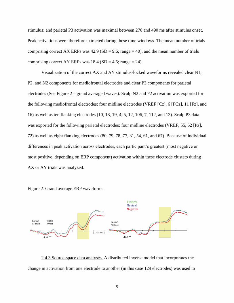
stimulus; and parietal P3 activation was maximal between 270 and 490 ms after stimulus onset.
Peak activations were therefore extracted during these time windows. The mean number of trials
comprising correct AX ERPs was 42.9 (SD = 9.6; range = 40), and the mean number of trials
comprising correct AY ERPs was 18.4 (SD = 4.5; range = 24).
Visualization of the correct AX and AY stimulus-locked waveforms revealed clear N1,
P2, and N2 components for mediofrontal electrodes and clear P3 components for parietal
electrodes (See Figure 2 – grand averaged waves). Scalp N2 and P2 activation was exported for
the following mediofrontal electrodes: four midline electrodes (VREF [Cz], 6 [FCz], 11 [Fz], and
16) as well as ten flanking electrodes (10, 18, 19, 4, 5, 12, 106, 7, 112, and 13). Scalp P3 data
was exported for the following parietal electrodes: four midline electrodes (VREF, 55, 62 [Pz],
72) as well as eight flanking electrodes (80, 79, 78, 77, 31, 54, 61, and 67). Because of individual
differences in peak activation across electrodes, each participant’s greatest (most negative or
most positive, depending on ERP component) activation within these electrode clusters during
AX or AY trials was analyzed.
Figure 2. Grand average ERP waveforms.
2.4.3 Source-space data analyses. A distributed inverse model that incorporates the
change in activation from one electrode to another (in this case 129 electrodes) was used to
Positive Neutral Negative

10
calculate the source-space activation. This type of algorithm estimates activation voxel-by-voxel
and sample-by-sample and does not require any dipoles to be “fit”, thereby limiting the influence
of user bias. The specific algorithm used in the current study was LORETA (Low Resolution
Brain Electromagnetic Tomography), which applies a constraint to the minimum-norm solution
in order to minimize the discrepancy between values of adjacent voxels (to achieve the most
realistic model) within the GeoSource interface (Electrical Geodesic, Inc., Eugene, OR). A
regularization constant (indicating how much noise is modeled) of 10-4 was applied. This amount
of regularization revealed current flow patterns that matched (via visual inspection) the grand-
averaged scalp topography better than other levels.
After the data were modeled (LORETA) for the entire cortex (2447 voxels), morphology-
based regions of interest (ROIs) were generated using the Montreal Neurological Institute (MNI)
average adult MRI (see Figure 3). We were interested in six ROIs: the left and right VLPFC ROI
(comprised of 22 voxels each; lateral part of BA 11 and 47), the dACC ROI (comprised of 50
voxels; dorsal part of BA 24 and 32), the left and right DLPFC ROI (comprised of 63 voxels
each; BA 9 and dorsal part of BA 46), and the VMPFC/OFC ROI (comprised of 147 voxels;
ventromedial parts of BAs 11, 10, 14, and 13). Source waveform amplitudes (nA) for all voxels
within an ROI were extracted for 400 ms before stimulus onset to 600 ms after stimulus onset
and baseline corrected using the 400 ms before stimulus onset. To ensure that each participant’s
maximal activation was analyzed, we chose the voxel and moment in time (within the time
period during which the scalp ERP component was maximal) that showed the most activation for
each ROI.

11
Figure 3. Morphology-based regions of interest (ROIs) generated using the Montreal
Neurological Institute average adult magnetic resonance image (MRI).
2.4.4 Statistical Analyses. All EEG data with values larger or smaller than 2 SD from the
mean were changed to show values of 2 SD from mean, thus preventing statistical analyses from
being skewed by outliers. Regression analyses were conducted in this study in order to examine
the moderating role of brain activation on the relation between effortful control and aggression.
A priori t-tests revealed sex differences for some independent and dependent variables (see Table
1); therefore, sex was entered as a covariate in all EEG analyses. Additionally, for data in the

12
negative condition, in order to capture only brain activation that was related to both cognitive
control and negative emotional state, negative AX and neutral AY brain data were entered as
covariates. For data in the positive condition, in order to capture only brain activation that was
related to both cognitive control and positive emotional state, positive AX and neutral AY brain
data were entered as covariates. For data in the neutral condition, in order to capture only brain
activation that was related to both cognitive control and a relatively unemotional context,
negative and positive AY and neutral AX brain data were entered as covariates.
Table 1. Sex Differences.

13
3, Results
3.1 Behavioral Results
In order to determine if there were any behavioral effects of emotional context (neutral,
negative, positive), two repeated-measures ANOVAs were conducted on AY trials: 1) with
performance accuracy as the dependent measure and 2) with reaction times as the dependent
measure. Results were not significant for performance accuracy, F(2,68) = .76, p = .47, η2 = .02,
or reaction times, F(2,68) = 1.52, p = .31, η2 = .04, suggesting that emotional context did not
influence performance accuracy or speed during action switching.
3.2 ERP Condition Differences
In order to determine if there were any ERP differences due to emotional context
(neutral, negative, positive), three repeated-measures ANOVAs were conducted: 1) P2 activation
as dependent variable, 2) N2 activation as dependent variable, and 3) P3 activation as dependent
variable. Since there were sex differences in the ERP data (see Table 1), sex was entered as a
between-subjects factor in these analyses. Additionally, trial count was entered in all analyses as
a covariate.
Results indicated that there was no main effect of emotion context on P2 amplitude,
F(2,122) = .89, p = .41, η2 = .01, nor was there a main effect of sex on P2 amplitude, F(1,61) =
2.86, p = .10, η2 = .05.
Results for N2 amplitude showed a significant interaction of emotional condition by sex,
F(2,122) = 4.36, p = .02, η2 = .07. This result indicates that males showed less negative N2
activation in negative trials than in positive trials (Mean difference = 1.04, p = .02), while
females showed less negative N2 activation in neutral trials than in negative trials (Mean
difference = .77, p = .04). Additionally, males showed significantly less negative N2 activation

14
than females in negative trials (Mean difference = 2.42, p = .004). All other contrasts were not
significant.
Results for P3 amplitude indicated a significant within-subjects effect of emotional
condition, F(2,122) = 5.42, p = .006, η2 = .08, as well as a significant main effect of sex, F(1,61)
= 9.14, p = .004, η2 = .13, with females showing less positive P3 amplitudes than males. Pairwise
comparisons for P3 amplitude indicated that subjects had a significantly less positive activation
in negative trials than in neutral trials (Mean difference = 1.43, p < .001) or in positive trials
(Mean difference = 1.79, p < .001); there was no difference in P3 activation between positive and
neutral conditions.
3.3 ERP Moderator Effects
Because not everyone with poor effortful control has aggressive tendencies, we also
conducted a number of linear regression analyses to test whether ERP amplitudes moderate the
association between effortful control and aggression. All covariates, outlined earlier, were
entered in step one of the regression model. Next, ERP amplitude and effortful control (centered
to decrease the possibility of multicollinearity influencing results, Aiken & West, 1991) were
entered in step two. Lastly, an interaction term of ERP amplitude and effortful control was
computed and entered in step 3 to test for moderation effects. Aggression was entered as the
dependent variable. Correlations between individual variables are summarized in Table 2, while
interaction (moderating) results are presented in Table 3. Results revealed that N2 amplitude in
both negative and neutral conditions significantly moderated the association between effortful
control and aggression. When probed at values of 1 SD above and below the mean, additional
regression analyses revealed that this moderating effect was driven by low (less negative) N2
activation. At low levels of N2 activation (less negative) effortful control was a significant

15
predictor of aggression scores in both negative conditions, β = -.59, t(58) = -3.62, p = .001,
and neutral conditions, β = -.53, t(58) = -3.45, p = .001. At high levels of N2 activation (more
negative), effortful control was not a significant predictor of aggression scores in negative
conditions, β = -.11, t(58) = -.55, p = .58, or neutral conditions, β = -.20, t(58) = -1.16, p = .25.
N2 amplitude in the positive emotional condition did not significantly moderate the association
between effortful control and aggression. Additionally, there were no significant moderation
effects for P2 or P3 amplitudes for any emotional conditions. Moderation plots are presented in
Figure 4.

16
Table 2. Preliminary Pearson correlation analyses (r-values) between effortful control, aggression, and brain measures.

17
Table 3. Regression Showing P2 Moderation Effects Predicting Aggression.
Table 4. Regression Showing N2 Moderation Effects Predicting Aggression.

18
Table 5. Regression Showing P3 Moderation Effects Predicting Aggression.
Figure 4. Moderation plots: Interaction between N2 amplitudes and Effortful Control on
aggression.
Negative Emotional Condition Unemotional Condition

19
3.2 N2 Source Space Analyses
Source space analyses were conducted to elucidate which cortical generators underlie the
moderating effects outlined above. Source space analyses were only conducted for the N2 in
negative and neutral conditions, because only this ERP in these conditions showed significant
moderation at the scalp level. Linear regression analyses were conducted separately for all ROIs.
Regression models were identical to the model structure outlined above, except that source space
activation was used rather than scalp ERP activation. In other words, step one consisted of all
covariates, step 2 consisted of main effects (source space activation underlying the N2 and
effortful control values), step 3 consisted of the interaction between source space activation and
effortful control, and the dependent variable was level of aggression. Source space activation
and effortful control values were centered to decrease the possibility of multicollinearity
influencing results (Aiken & West, 1991). Correlations between individual variables are
summarized in Table 4, while interaction (moderating) results are presented in Table 5. In the
negative emotion condition, source-space ROIs that significantly moderated the association
between effortful control and aggression included the right DLPFC, right VLPFC, VMPFC, and
ACC. When these interactions in the negative condition were probed at values of 1 SD above
and below the mean, additional regression analyses revealed that this moderating effect was
driven by low source space activation. At low levels of source space activation, effortful control
was a significant predictor of aggression scores for DLPFC right, β = -.80, t(58) = -4.76, p <
.001, VLPFC right, β = -.63, t(58) = -3.90, p < .001, VMPFC, β = -.75, t(58) = -4.18, p < .001,
and dACC, β = -.63, t(58) = -3.63, p < .001. At high levels of source space activation, effortful
control was not a significant predictor of aggression scores for DLPFC right, β = -.08, t(58) = -
.43, p = .67, VLPFC right, β = -.21, t(58) = -1.24, p = .22, VMPFC, β = .04, t(58) = .17, p = .87,

20
or dACC, β = -.06, t(58) = -.32, p = .75. In the neutral emotion condition, source-space ROIs that
significantly moderated the association between effortful control and aggression included the
right DLPFC, VMPFC, and ACC. When these interactions in the neutral condition were probed
at values of 1 SD above and below the mean, additional regression analyses revealed that this
moderating effect was also driven by low source space activation. At low levels of source space
activation, effortful control was a significant predictor of aggression scores for DLPFC right, β =
-.73, t(58) = -4.12, p < .001, VMPFC, β = -.69, t(58) = -4.09, p < .001, and dACC, β = -.68, t(58)
= -3.66, p = .001. At high levels of source space activation, effortful control was not a significant
predictor of aggression scores for DLPFC right, β = -.12, t(58) = -.56, p = .58, VMPFC, β = -.12,
t(58) = -.63, p = .53, or dACC, β = .14, t(58) = .48, p = .63. Moderation plots are presented in
Figure 5.

21
Table 6. Pearson correlation analyses (r-values) between effortful control, aggression, and brain & behavioral measures.

22
Table 7. Moderation Effects Showing Interactions Between Source Space Activation Underlying
the N2 and Effortful Control. Values are Regression Coefficients Predicting Aggression.

23
Figure 5. Moderation plots: Interaction between N2 amplitudes and Effortful Control on
aggression for source-space regions.
Negative Emotional Condition Unemotional Condition

24
4, Discussion
4.1 General Discussion
The present study examined the time course of neural activation underlying action
switching processes, a key aspect of effortful control, and how these patterns of activation
contribute to aggressive behavior. More specifically, we used ERPs and source-space activation
(LORETA) to examine whether patterns of neural activation moderate the relationship between
effortful control and aggression, and whether these moderating effects differed in emotionally
salient contexts compared to a relatively neutral context. As predicted, brain processes
underlying action switching significantly moderated the association between effortful control and
aggression at both the scalp and source-space levels.
Given that the ability to flexibly switch action strategies likely requires a number of
underlying cognitive functions (e.g., Badre & Wagner, 2006; Braver, Paxton, Locke, & Barch,
2009; Eslinger & Grattan, 1993) and that any one of these functions could show deficits that
might contribute to aggressive behavior, we decomposed the time course underlying action
switching. Our results indicate that only activation during the N2 window significantly
moderates the effortful control – aggression relationship. Given that the N2 has been associated
with aspects of cognitive control (Lamm, Zelazo, & Lewis, 2006) as well as aggression (Lamm,
Granic, Zelazo, & Lewis, 2011; Lewis, Granic, & Lamm, 2006), this suggests that activation
during the N2 time window might be a neural mechanism that influences self-control over
aggressive tendencies. However, it is not clear as to why the P2 and the P3 did not also moderate
the effortful control – aggression association. Given that P2 activation has been associated with
attentional orienting (Kanske, Plitschka, & Kotz, 2011; Eimer, Van Velzen, Gherri, & Press,
2006) and the fact that the current task presents all stimuli in the same location, it may be that
25
our task did not require enough attentional orienting. Thus, it may be that we had insufficient
attentional-orienting-related variance to reveal moderational effects. It is possible that in the
context of a task with active attentional orienting demands, P2 activation would play a
moderating role in the effortful control - aggression relationship.
P3 activation, which has generally been associated with novelty (Friedman, Cycowicz, &
Gaeta, 2001; Debener, Makeig, Delorme, & Engel, 2005), context updating (Donchin & Coles,
1988; Verleger, 1988), and motivation (Boksem et al., 2006; Potts, 2004), was not a significant
moderator of the effortful control – aggression relationship. While there has been research
linking P3 activation with aggression (Bartholow, Bushman, & Sestir, 2006) and externalizing
behavior (Lacono et al., 2002), research linking P3 activation to effortful control is much sparser.
For this reason, while P3 amplitude seems to be related to aggressive behavior, it does not seem
to specifically moderate the association between effortful control and aggression.
The direction of the moderating effect for N2 activation was consistent with previous
studies linking neural processing efficiency with reduced activation during cognitive control
tasks (e.g. Lamm, Pine, & Fox, 2013; Casey et al., 1997; Durston et al., 2006). For example,
Lamm, Pine, & Fox (2013) found that participants who successfully deployed a reactive (to the
environment) style of responding showed less prefrontal activation when required to execute
last-minute environmentally-triggered action switching. This decrease in prefrontal activation
was interpreted to reflect increased efficiency of cognitive-control-related cortical processing.
Thus, our results suggest that efficient cognitive control processing moderates the association
between effortful control and aggression. Follow-up analyses were conducted using source space
models (LORETA) to determine which neural generators drive the moderating effect. ROIs
moderating the association between effortful control and aggression included DLPFC, VLPFC,

26
dACC, and VMPFC. The direction of these effects is also consistent with an efficiency
hypothesis, in that lower (efficient) activation in these regions is associated with lower levels of
aggression at high levels of effortful control. This pattern of results supports the Lamm et al.
(2013) findings and suggests that improved control during the presence of last-minute
environmentally triggered information, as measured by an action-switching paradigm, is related
to low prefrontal cortical activation. More specifically, these findings, in combination with the
related extant literature, suggest a nascent theory of efficiency that may inform our
understanding of the neural underpinnings of cognitive control.
Interestingly, moderation effects existed only at low levels of N2 activation, which
suggests that low or efficient N2 activation during action switching, in conjunction with high
effortful control, contributes to less aggressive outcomes. We expected to find the converse as
well, that is, high N2 amplitude and poor effortful control should result in higher aggression.
Instead, the relationship between effortful control and aggression is flat at high levels of N2
activation. This suggests that our data might have an issue with restriction of range; that is, our
participants may not have shown enough variability in aggression. This argument is supported by
the fact that we recruited participants from a university environment. Recruitment from a less
preselected (academic) sample might reveal a greater range of aggression scores and thus
potentially reveal both the high effortful control – efficient processing – low levels of aggression
effect and the poor effortful control – inefficient processing – high levels of aggression effect.
Future research should replicate this study on a more diverse sample.
Previous literature has found emotion-specific increases in ERP activation for negative
emotional contexts compared to emotionally neutral contexts (e.g., Lamm, Pine, & Fox, 2013;
Lewis et al., 2006; Lamm & Lewis, 2010). However, because these studies did not include a

27
positive emotional condition, it is unclear if these effects were due to valence or arousal.
Similarly, Van Wouwe et al. (2010) found decreased N2 activation (less negative) for a positive
compared to a relatively neutral emotional condition in a similar AX-CPT task that showed
positive affective video clips. However, this study did not examine negative emotional
conditions. The current results add to the extant literature by examining this issue within a single
task, allowing direct comparison between positive, neutral, and negative contexts. Interestingly,
women showed the expected increased N2 activation in the negative condition while males did
not. This pattern of results is, however, in line with research by Lithari et al. (2010), who showed
that females demonstrate significantly more negative N2 amplitudes than males when passively
viewing negative affective stimuli. Additionally, there was a main effect of emotion for P3
amplitude, showing that following violent imagery participants had lower P3 activation than in
neutral or positive contexts. This finding might be informative for studies of emotion regulation,
and suggests that following negative emotional stimuli, we are left with fewer neural resources
with which to encode future actions. It is not clear why emotion differences were not found for
P2 amplitude, though the fact that emotional trials were presented randomly within each block
might have “watered down” the impact of the negative trials. Future research should compare
design differences, i.e., emotional random design vs. emotional block design, to ascertain if this
is indeed the case.
Additionally, results from the current study showed that N2 activation moderates the
effortful control – aggression relationship in the negative and neutral conditions but not in the
positive condition. These results suggest that in the face of negatively-charged (specifically,
violent or threatening) events, individuals with efficient cognitive control are less likely to
respond in an aggressive manner. Additionally, in the context of positively-charged events, likely
28
we require few cognitive-control-related resources to prevent us from lashing out aggressively.
Given that our neutral stimuli were not overly emotionally arousing, e.g., a chair, it is not clear
why we found significant moderation effects for this condition. It may be that our randomized
presentations of emotional trials lead to an emotional carryover effect from negative to neutral
trials, and that positive trials were arousing enough (positive valence) to override this emotional
carryover effect. Future research should replicate this study using an emotional block design.
4.2 Limitations
There are limitations to the current study. First, the use of source-space analyses allowed
us to ask region specific questions that scalp ERPs did not. However, activation patterns are
estimated effects and therefore should be interpreted with caution. Furthermore, since activation
is estimated, measuring activation differences for small ROIs or regions close together is
difficult.
Second, the current study used questionnaire-based proxies to measure both effortful
control and aggression. Questionnaire-based measures may be more subjective than biological or
behavioral measures, generally for reasons relating to social desirability (Sjöström & Holst,
2002; Richmond, Kiesler, Weisband, & Drasgow, 1999; Armitage & Conner, 1999). Therefore,
these results should be replicated using behavioral measures of aggression and effortful control.
Finally, the current study had a small age range of participants. Previous neuroimaging
research has shown that neural activation during cognitive control tasks differs between
adolescents and adults (Rubia et al., 2006; Luna, Padmanabhan, & O’Hearn, 2010; Eshel et al.,
2007), and therefore the adults included in this study might not be fully representative of the
adult range. This limits generalizability of results to other age ranges. Future work should expand

29
on the age range of participants, in order to determine if moderating effects differ throughout
development, including later adulthood.
4.3 Conclusions
These results suggest that neural mechanisms underlying flexible action switching
moderate the association between effortful control and aggression. Specifically, these results
suggest that low or efficient prefrontal cortical activation associated with effortful control
contributes to less aggressive outcomes. Future studies should build upon these results by
examining whether the converse is also true; that is, does high or inefficient activation during the
N2 time window contribute to more aggressive outcomes? These studies should prescreen
individuals to ensure that some participants are high in aggressive behavior so that there is
enough variability in aggression scores for these effects to be discernable. Additionally, future
research should incorporate longitudinal developmental data to ascertain whether inefficient use
of regulatory resources early in life predicts future aggressive behavior problems, thereby
highlighting a neural mechanism (or biomarker) that might be targeted by treatment approaches.

30
References
Aldao, A., Nolen-Hoeksema, S., & Schweizer, S. (2010). Emotion-regulation strategies across
psychopathology: A meta-analytic review. Clinical psychology review, 30(2), 217-237.
Anderson, M. C., Ochsner, K. N., Kuhl, B., Cooper, J., Robertson, E., Gabrieli, S. W., ... &
Gabrieli, J. D. (2004). Neural systems underlying the suppression of unwanted memories.
Science, 303(5655), 232-235.
Armitage, C. J., & Conner, M. (1999). The theory of planned behaviour: Assessment of
predictive validity and perceived control. British journal of social psychology, 38(1), 35-
54.
Banks, S. J., Eddy, K. T., Angstadt, M., Nathan, P. J., & Phan, K. L. (2007). Amygdala–frontal
connectivity during emotion regulation. Social cognitive and affective neuroscience, 2(4),
303-312.
Barry, R. J., & Rushby, J. A. (2006). An orienting reflex perspective on anteriorisation of the P3
of the event-related potential. Experimental brain research, 173(3), 539-545.
Bartholow, B. D., Bushman, B. J., & Sestir, M. A. (2006). Chronic violent video game exposure
and desensitization to violence: Behavioral and event-related brain potential data. Journal
of Experimental Social Psychology, 42(4), 532-539.
Bechara, A., Damasio, H., & Damasio, A. R. (2000). Emotion, decision making and the
orbitofrontal cortex. Cerebral cortex, 10(3), 295-307.
Bechara, A., Tranel, D., & Damasio, H. (2000). Characterization of the decision-making deficit
of patients with ventromedial prefrontal cortex lesions. Brain, 123(11), 2189-2202.
Bekker, E. M., Kenemans, J. L., & Verbaten, M. N. (2005). Source analysis of the N2 in a cued
Go/NoGo task. Cognitive Brain Research, 22(2), 221-231.
Best, M., Williams, J. M., & Coccaro, E. F. (2002). Evidence for a dysfunctional prefrontal
circuit in patients with an impulsive aggressive disorder. Proceedings of the National
Academy of Sciences, 99(12), 8448-8453.
Boksem, M. A., Tops, M., Wester, A. E., Meijman, T. F., & Lorist, M. M. (2006). Error-related
ERP components and individual differences in punishment and reward sensitivity. Brain
research, 1101(1), 92-101.
Braver, T. S., Paxton, J. L., Locke, H. S., & Barch, D. M. (2009). Flexible neural mechanisms of
cognitive control within human prefrontal cortex. Proceedings of the National Academy
of Sciences, 106(18), 7351-7356.
Britz, J., & Pitts, M. A. (2011). Perceptual reversals during binocular rivalry: ERP components
and their concomitant source differences. Psychophysiology, 48(11), 1490-1499.
Bush, G., Luu, P., & Posner, M. I. (2000). Cognitive and emotional influences in anterior
cingulate cortex. Trends in cognitive sciences, 4(6), 215-222.
Buss, A. H., & Perry, M. (1992). The aggression questionnaire. Journal of personality and social
psychology, 63(3), 452.
Casey, B. J., Trainor, R. J., Orendi, J. L., Schubert, A. B., Nystrom, L. E., Giedd, J. N., ... &
Rapoport, J. L. (1997). A developmental functional MRI study of prefrontal activation
during performance of a go-no-go task. Journal of cognitive neuroscience, 9(6), 835-847.
Davidson, R. J., Putnam, K. M., & Larson, C. L. (2000). Dysfunction in the neural circuitry of
emotion regulation--a possible prelude to violence. Science, 289(5479), 591-594.

31
Debener, S., Makeig, S., Delorme, A., & Engel, A. K. (2005). What is novel in the novelty
oddball paradigm? Functional significance of the novelty P3 event-related potential as
revealed by independent component analysis. Cognitive Brain Research, 22(3), 309-321.
Dennis, T. A., Malone, M. M., & Chen, C. C. (2009). Emotional face processing and emotion
regulation in children: An ERP study. Developmental neuropsychology, 34(1), 85-102.
Donchin, E., & Coles, M. G. (1988). Is the P300 component a manifestation of context
updating?. Behavioral and brain sciences, 11(03), 357-374.
Durston, S., Davidson, M. C., Tottenham, N., Galvan, A., Spicer, J., Fossella, J. A., & Casey, B.
J. (2006). A shift from diffuse to focal cortical activity with development. Developmental
science, 9(1), 1-8.
Eimer, M., Van Velzen, J., Gherri, E., & Press, C. (2006). Manual response preparation and
saccade programming are linked to attention shifts: ERP evidence for covert attentional
orienting and spatially specific modulations of visual processing. Brain research,
1105(1), 7-19.
Eshel, N., Nelson, E. E., Blair, R. J., Pine, D. S., & Ernst, M. (2007). Neural substrates of choice
selection in adults and adolescents: development of the ventrolateral prefrontal and
anterior cingulate cortices. Neuropsychologia, 45(6), 1270-1279.
Evans, D. E., & Rothbart, M. K. (2007). Developing a model for adult temperament. Journal of
Research in Personality, 41(4), 868-888.
Friedman, D., Cycowicz, Y. M., & Gaeta, H. (2001). The novelty P3: an event-related brain
potential (ERP) sign of the brain's evaluation of novelty. Neuroscience & Biobehavioral
Reviews, 25(4), 355-373.
Goldstein, M., Brendel, G., Tuescher, O., Pan, H., Epstein, J., Beutel, M., ... & Silbersweig, D.
(2007). Neural substrates of the interaction of emotional stimulus processing and motor
inhibitory control: an emotional linguistic go/no-go fMRI study. Neuroimage, 36(3),
1026-1040.
Gross, J. J. (2002). Emotion regulation: Affective, cognitive, and social consequences.
Psychophysiology, 39(3), 281-291.
Iacono, W. G., Carlson, S. R., Malone, S. M., & McGue, M. (2002). P3 event-related potential
amplitude and the risk for disinhibitory disorders in adolescent boys. Archives of General
Psychiatry, 59(8), 750-757.
Isoda, M., & Hikosaka, O. (2008). Role for subthalamic nucleus neurons in switching from
automatic to controlled eye movement. The Journal of Neuroscience, 28(28), 7209-7218.
Jackson, D. C., Mueller, C. J., Dolski, I., Dalton, K. M., Nitschke, J. B., Urry, H. L., ... &
Davidson, R. J. (2003). Now you feel it, now you don't: frontal brain electrical
asymmetry and individual differences in emotion regulation. Psychological science,
14(6), 612-617.
Kanske, P., & Kotz, S. A. (2007). Concreteness in emotional words: ERP evidence from a
hemifield study. Brain research, 1148, 138-148.
Kanske, P., Plitschka, J., & Kotz, S. A. (2011). Attentional orienting towards emotion: P2 and
N400 ERP effects. Neuropsychologia, 49(11), 3121-3129.
Ladouceur, C. D., Dahl, R. E., & Carter, C. S. (2007). Development of action monitoring
through adolescence into adulthood: ERP and source localization. Developmental
science, 10(6), 874-891.

32
Kochanska, G., & Knaack, A. (2003). Effortful control as a personality characteristic of young
children: Antecedents, correlates, and consequences. Journal of personality, 71(6), 1087-
1112.
Krug, M. K., & Carter, C. S. (2012). Proactive and reactive control during emotional interference
and its relationship to trait anxiety. Brain research, 1481, 13-36.
Lamm, C., Granic, I., Zelazo, P. D., & Lewis, M. D. (2011). Magnitude and chronometry of
neural mechanisms of emotion regulation in subtypes of aggressive children. Brain and
cognition, 77(2), 159-169.
Lamm, C., & Lewis, M. D. (2010). Developmental change in the neurophysiological correlates
of self-regulation in high-and low-emotion conditions. Developmental neuropsychology,
35(2), 156-176.
Lamm, C., Pine, D. S., & Fox, N. A. (2013). Impact of negative affectively charged stimuli and
response style on cognitive-control-related neural activation: An ERP study. Brain and
cognition, 83(2), 234-243.
Lamm, C., Zelazo, P. D., & Lewis, M. D. (2006). Neural correlates of cognitive control in
childhood and adolescence: Disentangling the contributions of age and executive
function. Neuropsychologia, 44(11), 2139-2148.
Lang, P. J., Bradley, M. M., & Cuthbert, B. N. (2008). International affective picture system
(IAPS): Affective ratings of pictures and instruction manual. Technical report A-8.
Lewis, M. D., Todd, R. M., & Honsberger, M. J. (2007). Event-related potential measures of
emotion regulation in early childhood. NeuroReport, 18(1), 61-65.
Lewis, M. D., Granic, I., & Lamm, C. (2006). Behavioral differences in aggressive children
linked with neural mechanisms of emotion regulation. Annals of the New York Academy
of Sciences, 1094(1), 164-177.
Lithari, C., Frantzidis, C. A., Papadelis, C., Vivas, A. B., Klados, M. A., Kourtidou-Papadeli, C.,
... & Bamidis, P. D. (2010). Are females more responsive to emotional stimuli? A
neurophysiological study across arousal and valence dimensions. Brain topography,
23(1), 27-40.
Luna, B., Padmanabhan, A., & O’Hearn, K. (2010). What has fMRI told us about the
development of cognitive control through adolescence?. Brain and cognition, 72(1), 101-
113.
Mehta, P. H., & Beer, J. (2010). Neural mechanisms of the testosterone–aggression relation: the
role of orbitofrontal cortex. Journal of Cognitive Neuroscience, 22(10), 2357-2368.
Miyake, A., Friedman, N. P., Emerson, M. J., Witzki, A. H., Howerter, A., & Wager, T. D.
(2000). The unity and diversity of executive functions and their contributions to complex
“frontal lobe” tasks: A latent variable analysis. Cognitive psychology, 41(1), 49-100.
Monk, C. S., McClure, E. B., Nelson, E. E., Zarahn, E., Bilder, R. M., Leibenluft, E., ... & Pine,
D. S. (2003). Adolescent immaturity in attention-related brain engagement to emotional
facial expressions. Neuroimage, 20(1), 420-428.
Mulert, C., Juckel, G., Augustin, H., & Hegerl, U. (2002). Comparison between the analysis of
the loudness dependency of the auditory N1/P2 component with LORETA and dipole
source analysis in the prediction of treatment response to the selective serotonin reuptake
inhibitor citalopram in major depression. Clinical Neurophysiology, 113(10), 1566-1572.
Mullin, B. C., & Hinshaw, S. P. (2007). Emotion Regulation and Externalizing Disorders in
Children and Adolescents.

33
Muris, P., van der Pennen, E., Sigmond, R., & Mayer, B. (2008). Symptoms of anxiety,
depression, and aggression in non-clinical children: Relationships with self-report and
performance-based measures of attention and effortful control. Child Psychiatry and
Human Development, 39(4), 455-467.
Nee, D. E., & Brown, J. W. (2012). Rostral–caudal gradients of abstraction revealed by multi-
variate pattern analysis of working memory. Neuroimage, 63(3), 1285-1294.
Nelson, R. J., & Trainor, B. C. (2007). Neural mechanisms of aggression. Nature Reviews
Neuroscience, 8(7), 536-546.
Ochsner, K. N., Ray, R. D., Cooper, J. C., Robertson, E. R., Chopra, S., Gabrieli, J. D., & Gross,
J. J. (2004). For better or for worse: neural systems supporting the cognitive down-and
up-regulation of negative emotion. Neuroimage, 23(2), 483-499.
Paxton, J. L., Barch, D. M., Racine, C. A., & Braver, T. S. (2008). Cognitive control, goal
maintenance, and prefrontal function in healthy aging. Cerebral Cortex, 18(5), 1010-
1028.
Posner, M. I., Rothbart, M. K., Sheese, B. E., & Tang, Y. (2007). The anterior cingulate gyrus
and the mechanism of self-regulation. Cognitive, Affective, & Behavioral Neuroscience,
7(4), 391-395.
Potts, G. F. (2004). An ERP index of task relevance evaluation of visual stimuli. Brain and
cognition, 56(1), 5-13.
Richman, W. L., Kiesler, S., Weisband, S., & Drasgow, F. (1999). A meta-analytic study of
social desirability distortion in computer-administered questionnaires, traditional
questionnaires, and interviews. Journal of Applied Psychology, 84(5), 754.
Rosvold, H. E., Mirsky, A. F., Sarason, I., Bransome Jr, E. D., & Beck, L. H. (1956). A
continuous performance test of brain damage. Journal of consulting psychology, 20(5),
343.
Rothbart, M. K., & Rueda, M. R. (2005). The development of effortful control. Developing
individuality in the human brain: A tribute to Michael I. Posner, 167-188.
Rothbart, M. K., Sheese, B. E., & Posner, M. I. (2007). Executive attention and effortful control:
Linking temperament, brain networks, and genes. Child Development Perspectives, 1(1),
2-7.
Rubia, K., Smith, A. B., Woolley, J., Nosarti, C., Heyman, I., Taylor, E., & Brammer, M. (2006).
Progressive increase of frontostriatal brain activation from childhood to adulthood during
event‐related tasks of cognitive control. Human brain mapping, 27(12), 973-993.
Rueda, M. R., Posner, M. I., & Rothbart, M. K. (2005). The development of executive attention:
Contributions to the emergence of self-regulation. Developmental neuropsychology,
28(2), 573-594.
Schneider, W., Eschman, A., & Zuccolotto, A. (2002). E-Prime reference guide. Psychology
Software Tools, Incorporated.
Siegle, G. J., Ghinassi, F., & Thase, M. E. (2007). Neurobehavioral therapies in the 21st century:
Summary of an emerging field and an extended example of cognitive control training for
depression. Cognitive Therapy and Research, 31(2), 235-262.
Sjöström, O., & Holst, D. (2002). Validity of a questionnaire survey: response patterns in
different subgroups and the effect of social desirability. Acta Odontologica Scandinavica,
60(3), 136-140.

34
Sterzer, P., Stadler, C., Krebs, A., Kleinschmidt, A., & Poustka, F. (2005). Abnormal neural
responses to emotional visual stimuli in adolescents with conduct disorder. Biological
psychiatry, 57(1), 7-15.
Stieben, J., Lewis, M. D., Granic, I., Zelazo, P. D., Segalowitz, S., & Pepler, D. (2007).
Neurophysiological mechanisms of emotion regulation for subtypes of externalizing
children. Development and Psychopathology, 19(02), 455-480.
Vaidya, C. J., Bunge, S. A., Dudukovic, N. M., Zalecki, C. A., Elliott, G. R., & Gabrieli, J. D.
(2014). Altered neural substrates of cognitive control in childhood ADHD: evidence from
functional magnetic resonance imaging. American Journal of Psychiatry.
van Wouwe, N. C., Band, G. P., & Ridderinkhof, K. R. (2011). Positive affect modulates
flexibility and evaluative control. Journal of Cognitive Neuroscience, 23(3), 524-539.
Verleger, R. (1988). Event-related potentials and cognition: A critique of the context updating
hypothesis and an alternative interpretation of P3. Behavioral and brain sciences, 11(3),
343-356.
Vitacco, D., Brandeis, D., Pascual‐Marqui, R., & Martin, E. (2002). Correspondence of
event‐related potential tomography and functional magnetic resonance imaging during
language processing. Human brain mapping, 17(1), 4-12.
Volpe, U., Mucci, A., Bucci, P., Merlotti, E., Galderisi, S., & Maj, M. (2007). The cortical
generators of P3a and P3b: a LORETA study. Brain research bulletin, 73(4), 220-230.
Wiers, R. W., Gladwin, T. E., Hofmann, W., Salemink, E., & Ridderinkhof, K. R. (2013).
Cognitive bias modification and cognitive control training in addiction and related
psychopathology mechanisms, clinical perspectives, and ways forward. Clinical
Psychological Science, 2167702612466547.

35
Vita
The author obtained his bachelor’s degree in mathematics and psychology from
Tulane University in 2012. He joined the University of New Orleans psychology graduate
program to pursue a PhD in Biopsychology, and became a member of Professor Connie
Lamm’s research group, the Developmental Cognitive Affective Psychophysiology Lab, in
2014.