neural mechanisms of affective interference in schizotypy€¦ · neural mechanisms of affective...
TRANSCRIPT
Neural Mechanisms of Affective Interference in Schizotypy
Aprajita Mohanty, John D. Herrington,Nancy S. Koven, Joscelyn E. Fisher,
Elizabeth A. Wenzel, Andrew G. Webb, andWendy Heller
University of Illinois at Urbana–Champaign
Marie T. BanichUniversity of Colorado at Boulder
Gregory A. MillerUniversity of Illinois at Urbana–Champaign
Negatively valenced stimuli foster cognitive impairment in schizotypy and schizophrenia. To identifyrelevant brain mechanisms, the authors had 16 positive-schizotypy and 16 control participants performan emotional Stroop task, judging the ink color of negative and neutral words during functional magneticresonance imaging (fMRI) of regional brain activity. Schizotypy individuals showed increased right anddecreased left activity in dorsolateral prefrontal cortex, indicating a deficit in maintenance of attentionalset in the presence of negative emotional distractors. They also showed abnormal activity in ventrallimbic areas, including decreased activity in nucleus accumbens and increased activity in hippocampusand amygdala, a circuit involved in the integration of cognitive and affective processes. These resultsindicate that aspects of emotion–cognition processes and the brain mechanisms that implement them aresimilar in schizotypy and schizophrenia.
Conceptualizations of schizophrenia often focus on cognitivedisturbances such as delusions and disorganized speech and on therole that cognitive mechanisms play in the development of thesesymptoms. However, extensive evidence indicates that affectivefactors play an important role in the development, clinical picture,prognosis, and treatment of schizophrenia. Several lines of inves-tigation, including life event, family environment, clinical, andlaboratory studies, provide evidence of symptom exacerbations inresponse to negative affect in schizophrenia (for review, see Do-cherty, 1996; Malla & Norman, 1992; Slade, 1972), particularlyfor positive symptoms (Docherty, Evans, Sledge, Seibyl, &
Krystal, 1994; Docherty & Hebert, 1997; Schwartz & Myers,1977). To better understand the role of affective disturbance in theformation and exacerbation of positive symptoms, it is importantto examine the role affective disturbance plays in the disruption ofbasic cognitive processes such as attention, perception, judgment,and language that may ultimately manifest as psychotic symptoms,as well as the brain mechanisms involved in this disruption.
Researchers have hypothesized that the interplay of cognitiveand affective processes leads to the formation and maintenance ofpositive symptoms including delusions and hallucinations (e.g.,Freeman, Garety, Kuipers, Fowler, & Bebbington, 2002; Garety,Kuipers, Fowler, Freeman, & Bebbington, 2001). For example,negative affect compromises basic areas of cognition in schizo-phrenia, including language (Burbridge & Barch, 2002; Docherty,1996; Grosh, Docherty, & Wexler, 1995), semantic processing(Kerns & Berenbaum, 2000), and size estimation (Asarnow, Crom-well, & Rennick, 1978). Although there is extensive researchdemonstrating selective attention deficits in schizophrenia andschizotypy (e.g., Braver, Barch, & Cohen, 1999; Lubow & Ger-wirtz, 1995), there is little research examining the impact ofemotion on attentional processing in individuals with schizophre-nia or schizotypy. Patients with predominantly positive symptomsshow more interference for threat-related words on an emotionalStroop task than do patients without active psychotic symptoms(Epstein, Stern, & Silbersweig, 1999). Similarly, perceptual aber-ration and magical ideation as measures of positive schizotypy areassociated with increased behavioral interference on an emotionalStroop task, whereas negative schizotypy (social and physicalanhedonia) shows no relationship (Mohanty et al., 2001). Thesefindings are in line with earlier research demonstrating that indi-viduals with predominantly positive symptoms appear to be moreresponsive to stressors (e.g., Schwartz & Myers, 1977) and showincreased affective reactivity of cognitive disturbance than do
Aprajita Mohanty, John D. Herrington, and Wendy Heller, Departmentof Psychology and Beckman Institute Brain Imaging Center, University ofIllinois at Urbana–Champaign; Nancy S. Koven, Joscelyn E. Fisher, andElizabeth A. Wenzel, Department of Psychology, University of Illinois atUrbana–Champaign; Andrew G. Webb, Department of Electrical andComputer Engineering and Beckman Institute Brain Imaging Center, Uni-versity of Illinois at Urbana–Champaign; Marie T. Banich, Department ofPsychology, University of Colorado at Boulder; Gregory A. Miller, De-partment of Psychology, Department of Psychiatry, and Beckman InstituteBrain Imaging Center, University of Illinois at Urbana–Champaign.
This research was supported by the National Institute of Drug AbuseGrant R21 DA14111, the National Institute of Mental Health Grants R01MH61358, T32 MH14257, and T32 MH19554, Carle Clinic, the BeckmanInstitute, and the University of Illinois Intercampus Research Initiative inBiotechnology.
We thank Joseph Barkmeier, Tracey Wszalek, and Holly Tracy for theircontributions to this project.
Correspondence concerning this article should be addressed to GregoryA. Miller, Biomedical Imaging Center, University of Illinois at Urbana–Champaign, 2100 South Goodwin, Urbana, IL 61801. E-mail: [email protected]
Journal of Abnormal Psychology Copyright 2005 by the American Psychological Association2005, Vol. 114, No. 1, 16–27 0021-843X/05/$12.00 DOI: 10.1037/0021-843X.114.1.16
16

individuals with negative symptoms (Docherty et al., 1994), afinding that has been replicated with positive schizotypy as well(Fernandes & Miller, 1995; Kerns & Berenbaum, 2000). It isunclear what brain mechanisms are involved in this association ofcognitive disturbance and negative affect in positive-symptomschizotypy and schizophrenia. The present study used an emo-tional Stroop paradigm and fMRI to examine brain regions in-volved in the adverse impact of emotion on attentional processingseen in positive schizotypy.
Schizotypy involves characteristics that predispose individualsto schizophrenia (Edell, 1995; Fernandes & Miller, 1995). Self-report measures of schizotypal features are associated with ele-vated risk for symptoms of psychosis (Chapman, Chapman,Kwapil, Eckblad, & Zinser, 1994; Erlenmeyer-Kimling et al.,1993; Fernandes & Miller, 1995; Freedman, Rock, Roberts, Corn-blatt, & Erlenmeyer-Kimling, 1998). High-schizotypy nonpatientsexhibit some of the psychological and biological abnormalitiesreported in individuals with schizophrenia yet generally lack ahistory of psychotropic medication, hospitalization, and othercomplications common in studies of schizophrenia (Dickey et al.,2000; Fernandes & Miller, 1995). Schizotypy factor dimensionsparallel symptom domains observed in patients with schizophre-nia, suggesting that schizotypal traits and schizophrenic symptomscan be viewed on a continuum (e.g., Vollema & Hoijtink, 2000).In summary, some but not all aspects of clinical schizophrenia arepresent in schizotypy, providing a relatively pure context forstudying selected symptoms and identifying associated brainmechanisms.
According to Braver, Barch, and Cohen (1999), the most con-sistently reported attentional impairments in schizophrenia involvethe exertion of attention in a selective and controlled manner tofacilitate processing of task-relevant information, to inhibit theprocessing of task-irrelevant information, or both. This impairmentis likely due to a failure in the maintenance of contextual infor-mation in the presence of salient distractors. The color–wordStroop task, referred to as the “gold standard” for the measurementof selective attention (MacLeod, 1992), has been used to demon-strate this impairment in schizophrenia (e.g., Barch, Carter, et al.,2001). Given that cognitive impairment in schizophrenia is exac-erbated by negative affect, it is likely that attentional functioningwill tend to deteriorate in the presence of aversive stimuli. Thepresent investigation used a variant of the color–word Stroop taskcalled the emotional Stroop task, which involves the simultaneouspresentation of task-relevant (color of letters) and task-irrelevant(emotional meaning) attributes. The task was to identify the inkcolor of a word as quickly as possible while ignoring the word’smeaning. Performance depends on selective attention to task-relevant versus task-irrelevant stimulus features and maintenanceof contextual information (J. D. Cohen & Servan-Schreiber, 1992;Servan-Schreiber, Cohen, & Steingard, 1996). On the basis ofprior literature (reviewed later), we proposed the involvement of aconstellation of brain regions, including dorsolateral prefrontalcortex (DLPFC), ventral and dorsal striatum, and limbic regions, toaccount for the increased interference for negative versus neutralwords in positive schizotypy.
DLPFC appears to play an important role in active maintenanceof contextual information (e.g., Banich et al., 2000, 2001; Barch etal., 1996; Braver et al., 1999), particularly in the face of interfer-ence (Miller, Erickson, & Desimone, 1996). It has been proposed
that the deficit in the ability to maintain contextual information inpatients with schizophrenia involves a disturbance in DLPFC (e.g.,Barch, Braver, et al., 2001). Studies integrating neuropsychologi-cal performance with neuroimaging data show that individualswith schizophrenia or those at risk for schizophrenia do not acti-vate DLPFC normally when performing a task that places demandson DLPFC processing resources (e.g., Andreasen et al., 1992;Barch, Braver, et al., 2001; Gold, Goldberg, & Weinberger, 1992;MacDonald, Johnson, Becker, & Carter, 2003; Weinberger, Ber-man, & Zec, 1986). This finding has contributed to the concept of“hypofrontality” in schizophrenia. However, there is increasingevidence that the traditional concept of hypofrontality in schizo-phrenia is incomplete (Ramsey et al., 2002; Weiss et al., 2003). Forexample, recent studies provide evidence for increased task-relatedactivity in either right (Callicott et al., 2000, 2003; Weiss et al.,2003) or left (Manoach et al., 1999, 2000; Weiss et al., 2003)DLPFC in individuals with schizophrenia or those at risk fordeveloping schizophrenia. These apparent inconsistencies mightbe attributed to many factors, including experimental design andbehavioral task complexity (e.g., Callicott et al., 1999), presence ofreward (Manoach et al., 2000), and data analytic techniques suchas examining group-averaged data (which underestimates DLPFCactivation because of heterogeneity of location within DLPFC)versus single-participant data (Manoach et al., 2000). Evidence forthe role of these factors in the inconsistencies is inconclusive.Although DLPFC dysfunction has been observed in relatives ofindividuals with schizophrenia (Calicott et al., 2003; MacDonaldet al., 2003), it remains to be studied in at-risk individuals who areelevated on measures of schizotypy. The primary hypothesis in thepresent study was that DLPFC dysfunction would be evident evenin nonpatient individuals with elevated positive schizotypy scoreswhen asked to perform a task requiring maintenance of context inthe presence of distracting, aversive information.
Despite considerable evidence demonstrating DLPFC dysfunc-tion in schizophrenia, it remains uncertain whether this dysfunc-tion arises from neural abnormalities primarily in DLPFC or as aresult of dysregulation of DLPFC by other structures (Callicott etal., 2000; Manoach et al., 2000). An important circuit that hasfrequently been implicated in cognitive and affective dysfunctionin schizophrenia is the “ventral limbic cortical–basal ganglia cir-cuit” (Grace & Moore, 1998). The ventral circuit includes theprefrontal cortex (PFC), limbic, and ventral striatal regions as wellas connections among them. As the main striatal link in the ventralcircuit, the nucleus accumbens receives inputs from hippocampus,amygdala, and PFC and can influence higher cognitive processingthrough efferents to a thalamocortical network that modulates PFC(Grace & Moore, 1998; Groenewegen et al., 1991; O’Donnell,Greene, Pabello, Lewis, & Grace, 1999). For example, single-cellstudies have shown that both hippocampal and amygdalar inputs tothe nucleus accumbens modulate or gate neurons in PFC (Grace,2000a). The firing of neurons in PFC results in the transmission ofaction potentials via the striatum only when there is concurrentinput to the striatum from the hippocampus (Grace, Moore, &O’Donnell, 1998). Besides this gating role, the hippocampus playsa very important role in contextual learning (N. J. Cohen &Eichenbaum, 1993; Rudy & Sutherland, 1995; Salzmann, Vid-yasagar, & Creutzfeldt, 1993) and recognition of context for anevent (Chun & Phelps, 1999). Together with the evidence thathippocampus can modulate activity in PFC, the latter findings
17AFFECTIVE INTERFERENCE IN SCHIZOTYPY

suggest a cardinal role that hippocampus may play in the devel-opment of positive symptoms (Liddle, Lane, & Ngan, 2000). Infact, there is considerable evidence implicating hippocampus andparahippocampal gyrus in positive symptoms of schizophrenia(Bogerts, 1997). More recent functional imaging studies have alsoreported that overactivity of hippocampus and parahippocampalgyrus is associated with positive symptoms (Epstein et al., 1999;Liddle et al., 1992; Silbersweig et al., 1995).
The amygdala, another important component of the ventralcircuit, is crucial in the evaluation of internal and external stimuliin terms of their motivational and emotional significance (e.g.,Cahill, Roozendaal, & McGaugh, 1997; Davidson & Irwin, 1999;LeDoux, 1995). The amygdala provides a different kind of neu-romodulation or gating: Stimulation of amygdala will facilitateprefrontal spiking only if it precedes PFC stimulation by less than40 ms (Grace & Moore, 1998). As a result, the amygdala providesa more event-related gating, rather than the more general, context-dependent gating from the hippocampus, a means by which salientemotional stimuli can override an otherwise context-limited re-sponse system. Studies examining the role of amygdala in emo-tional processing in schizophrenia have yielded mixed results,including reports of decreased (e.g., Schneider et al., 1998) andincreased (Epstein et al., 1999) amygdalar activity in response toemotional stimuli. An important factor that might account forvaried results is that most studies did not determine whether theschizophrenia sample included individuals with predominantlypositive or negative symptoms. This is particularly important be-cause positive symptoms tend to be more reactive to negativeaffect than are negative symptoms (Docherty et al., 1994). Al-though one study reported that positive-schizophrenia individualsshow less amygdala activity than control participants (M. L.Phillips et al., 1999), two others found a positive associationbetween positive symptoms and amygdala activity (Epstein et al.,1999; Taylor, Liberzon, Decker, & Koeppe, 2002). Clearly, thereis a need for more studies that delineate distinct symptom dimen-sions and examine their differential relationship with amygdalaactivity. Furthermore, it is also possible that the amygdala findingsin the schizophrenia literature are confounded with medicationstatus because ventral brain regions including amygdala are sitesof action for antipsychotic medication (Taylor et al., 2002). Thepresent study avoided these confounds by recruiting at-risk indi-viduals who were unmedicated and who scored high only onmeasures of positive schizotypy.
In summary, based on the architecture of the ventral circuit andthe roles its components play in cognitive and affective processing,it is plausible that abnormalities within any of the components ofthe ventral circuit are associated with positive symptoms in schizo-typy and schizophrenia. Despite evidence implicating the role ofdifferent components of this ventral circuit in cognitive and affec-tive dysfunction in schizophrenia, very few studies have examinedthe role that these components play in similar deficits amongat-risk populations such as those individuals who score high onmeasures of schizotypy. In the present study, it was hypothesizedthat positive-schizotypy individuals would show abnormal activa-tion in the network of regions involved in the ventral circuitincluding nucleus accumbens, hippocampus, amygdala, and PFCwhile performing the task for the negatively valenced words.
Another region that is functionally and anatomically well con-nected with the PFC is the dorsal striatum (basal ganglia, including
caudate and putamen; Middleton & Strick, 2000). Researchershave hypothesized that the basal ganglia plays an important role inworking memory by providing a gating mechanism that modulatesa prefrontal active maintenance system (Frank, Loughry, &O’Reilly, 2001). It is possible that working memory deficits inschizophrenia are related to a dysfunction in the dorsal frontos-triatal circuitry that implements this gating mechanism. Dysfunc-tion in frontostriatal circuitry in schizophrenia has been demon-strated using a variety of techniques (Buchsbaum et al., 1992;Manoach et al., 2000; Rubin et al., 1991; Siegel et al., 1993). Forexample, Rubin et al. (1991) showed that abnormal prefrontalactivity in schizophrenia is associated with a failure to suppressblood flow to the striatum during working memory performance.Severity of formal thought disorder in schizophrenia is positivelycorrelated with activity in right caudate nucleus (Kircher et al.,2001). Furthermore, dorsal striatal dopamine receptor abnormali-ties are implicated in thought and movement abnormalities as wellas in abnormal sensitivity to stress in schizophrenia (Walker,Neumann, Baum, & Davis, 1996).
Thus, another goal of the present study was to investigatewhether individuals who score high on a positive schizotypymeasure, like patients with schizophrenia, show abnormal activityin the basal ganglia while performing the Stroop task for negative-emotion stimuli. Use of a nonpatient sample to test these hypoth-eses regarding the emotional factors affecting the neural circuitsinvolved in cognitive impairment allowed a relatively uncompro-mised evaluation, especially with respect to alterations in dopami-nergic systems targeted by antipsychotic medication.
Method
Participants
Two groups of right-handed, native English speakers were recruitedfrom the university community, 17 scoring high on a measure of positive-symptom schizotypy (12 men and 5 women; mean age � 19.1, SD � 1.9)and 17 control participants (7 men and 10 women; mean age � 20.5, SD �3.9). Participants were screened for a history of neurologic damage, colorblindness, claustrophobia, or contraindications for fMRI participation andgave informed consent prior to participation. Positive-schizotypy individ-uals were selected from large groups of introductory psychology studentstested with a battery of questionnaires including the Chapman scales forpsychosis proneness. Individuals with a score at least 1.5 standard devia-tions above the mean on either the Perceptual Aberration Scale (Chapman,Chapman, & Raulin, 1978) or the Magical Ideation Scale (Eckblad &Chapman, 1983) were included in the positive-schizotypy group. Controlparticipants scored 0.5 standard deviation below the mean on both theMagical Ideation and the Perceptual Aberration Scales. The t tests con-firmed that the positive-schizotypy group scored higher than the compar-ison group on the Perceptual Aberration and Magical Ideation Scales ( p �.01). Anxiety measures were obtained using the Penn State Worry Ques-tionnaire (Meyer, Miller, Metzger, & Borkovec, 1990) from 15 of thepositive-schizotypy and 12 for the control participants.
Stimuli and Experimental Design
Stimulus presentation and collection of response times (RTs) wereaccomplished with MEL v2.0 PC software (Psychology Software Tools,Pittsburgh, PA). Each participant was involved in a single fMRI sessionconsisting of blocks of positive- or negative-emotion words alternatingwith blocks of neutral words. Four positive, four negative, and eight neutralblocks were presented in a session. Stimuli were blocked on emotional
18 MOHANTY ET AL.

valence for several reasons. Behavioral studies have demonstrated that theemotional quality of words is harder to ignore when they are blocked ratherthan intermixed with neutral words (Holle, Neely, & Heimberg, 1997; seealso Compton, Heller, Banich, Palmieri, & Miller, 2000; Dalgleish, 1995).The valence of real-world emotional information does not typically alter-nate rapidly. Finally, several behavioral pilot studies for this project dem-onstrated an interference effect with emotional words for blocked but notintermixed designs.
Each block consisted of 16 trials with 1 trial occurring every 2,000 ms.A trial began with the presentation of a word for 1,700 ms, followed by afixation cross for 300 ms. Each trial consisted of one word presented in oneof four ink colors (red, green, yellow, or blue), with each color occurringequally often with each word type (positive, neutral, or negative). The firstand third blocks were neutral words, and the second or fourth blocks werepositive or negative words, with this pattern repeated for the total of 16blocks. The valence order was counterbalanced across participants tominimize order effects. Negative words were chosen based on availablenorms of arousal and valence ratings (Bradley & Lang, 1998). Each neutralword list was matched closely to one of the emotion word lists onconcreteness, imageability, familiarity, and word length based on publishednorms (Toglia & Battig, 1983). Neutral words were chosen to be seman-tically related within a list, just as the emotion words were semanticallyrelated within list. Thus, one neutral word list consisted of time-relatedwords (e.g., decade, minute), and the other of math-related words (e.g.,add, subtract).
Procedure
Before the fMRI session, participants were thoroughly informed aboutthe nature of the study and the magnetic resonance environment; consentwas obtained. The experiment lasted approximately 45 min. Visual stimuliwere presented using a goggle system (Resonance Technology, Northridge,CA). Participants were instructed to identify the ink color of each word(red, yellow, green, or blue) while ignoring the word’s meaning. They wereasked to indicate their response by pressing one of four buttons corre-sponding to the ink colors. To reduce motion artifact, a manual responsewas used, consistent with previous studies in our laboratory and others’indicating successful elicitation of Stroop effects (e.g., Compton et al.,2003; Herrington et al., 2004). A short practice session was conductedoutside the magnet in order to familiarize the participants with the stimuliand to help them learn the button order on the response pad.
Echo-planar (EPI) images were acquired in a 1.5 Tesla GE Signa scanner(Milwaukee, WI). Head position was stabilized using a bite bar molded toeach participant’s dentition. For each participant, 445 EPI images wereacquired (time for repetition � 1,517 ms, echo time � 40 ms, and flipangle � 90°). Each image consisted of 15 contiguous slices (thickness �7 mm; no gap, in-plane voxel size � 3.75 � 3.75 mm) parallel to thehorizontal plane containing the anterior and posterior commissures. High-resolution, three-dimensional (3D) anatomical images (T1–weighted 3Dgradient echo images) of the whole brain were collected for each partici-pant for landmark selection purposes. T1–weighted anatomical images ofthe 15 functional acquisition slices were also collected for image registra-tion purposes.
Image Processing
Image processing and analyses was carried out using the Oxford Centerfor functional magnetic resonance of the brain (FMRIB) FEAT (FMRIB’seasy analysis toolVersion 5.00), part of FSL (FMRIB’s software library;www.fmrib.ox.ac.uk/fsl). The first six volumes from each participant werediscarded to allow the signal to reach steady state. Prior to analysis,functional data for each participant were motion corrected through the useof motion correction using FMRIB’s linear image registration tool(MCFLIRT; Jenkinson & Smith, 2001), intensity normalized, temporally
filtered with a nonlinear high-pass filter, and spatially smoothed using a 3DGaussian kernel (full width half maximum � 7 mm). MCFLIRT effec-tively adjusts for motion up to 2 mm (Jenkinson, Bannister, Brady, &Smith, 2002), so individuals showing head motion more than 2 mm wereexcluded (e.g., Schaefer et al., 2002). One participant in each groupexceeded this criterion for a final number of 16 per group.
Regression Analyses
Regression analyses were performed on the processed functional timeseries of each participant using FMRIB’s improved linear model (FILM).Four regressors, one each for the positive, negative, and two neutralconditions, were included in the regression model. For each regressor, thevector of assigned weights corresponding to word type was convolved witha gamma function to better approximate the temporal course of the blood-oxygen-dependent hemodynamic response. Each regressor yielded a per-voxel effect-size parameter (beta) estimate map representing the magnitudeof activity associated with that regressor. Because present hypothesesfocused specifically on the effect of negative stimuli on attentional pro-cessing, subsequent steps in the data analysis involved only the RT andfMRI measures derived from the negative and matched neutral wordcondition.
Thus, the beta values for the negative word condition were contrastedwith the neutral word condition, resulting in per-voxel contrast parameter(beta) estimate maps. These functional activation maps, as well as thecorresponding structural MRI maps, were morphed into a common stereo-taxic space (Talairach & Tournoux, 1988) using FMRIB’s linear imageregistration tool (FLIRT).
Prior to group comparisons, regions that were significantly more activefor negative than for neutral word conditions were identified within eachgroup via voxelwise, one-sample t tests on contrast beta maps. Probabilityvalues from the t tests were then converted to z scores. The z maps werethresholded for significance using a cluster-size algorithm (Forman et al.,1995) that reduces false positives in the context of multiple comparisons bytaking into account the spatial extent of the activation. Regions wereconsidered active if they contained at least 30 contiguous voxels, eachvoxel active above an alpha threshold of .05. That cluster-size thresholdprovides a corrected per-voxel, false-positive rate of .0005 (Forman et al.,1995). To minimize experiment-wise Type I error, group comparisonswere confined to voxels with significant activation for negative versusneutral words in at least one group. Voxelwise, two-tailed t tests comparedthe effect sizes (contrast betas) for the two groups. Once again, a cluster-size threshold of at least 30 contiguous voxels with each voxel above analpha of .05 (providing a corrected per-voxel, false-positive rate of .0005)was chosen to identify regions that differentiated the groups. Requiringconjunction of orthogonal within-group and between-groups tests yielded acorrected per-voxel error rate of p � .00000025 (.0005 � .0005; Barch,Braver, et al., 2002; Barch, Carter, et al., 2001; Braver, et al., 2001).
Mohanty et al. (2001) showed that anxiety and depression are stronglycorrelated with positive schizotypy. Thus, for the present independent dataset, a mediating variable analysis (Baron & Kenny, 1986) examinedwhether anxiety mediated the relationship between positive schizotypy andbrain activity in the regions identified in the cluster-threshold analyses.
RT data were not available for 2 schizotypy individuals because oftechnical difficulties. Although the present article evaluates responses tonegative emotional words, parallel analyses of positive versus neutralwords found no regionally overlapping group differences.
Results
Behavioral Data
Every participant showed choice-RT performance accuracy of atleast 80%. RTs for incorrect trials were excluded from behavioral
19AFFECTIVE INTERFERENCE IN SCHIZOTYPY

data analyses. Emotional Stroop interference was calculated as thedifference in RT for negative and neutral words. The group RTinterference effect was in the expected direction of greater inter-ference in the schizotypy group (5.7 ms, SD � 38.2) than in thecontrol group (1.7 ms, SD � 43.6), but RT interference and error-rate differences as a function of group did not approach signifi-cance. Results from a behavioral study using a very similar para-digm and a larger sample found a significant relationship betweencomposite schizotypy score and interference (Mohanty et al.,2001), supporting the effectiveness of the present experimentalmanipulation.
fMRI Data
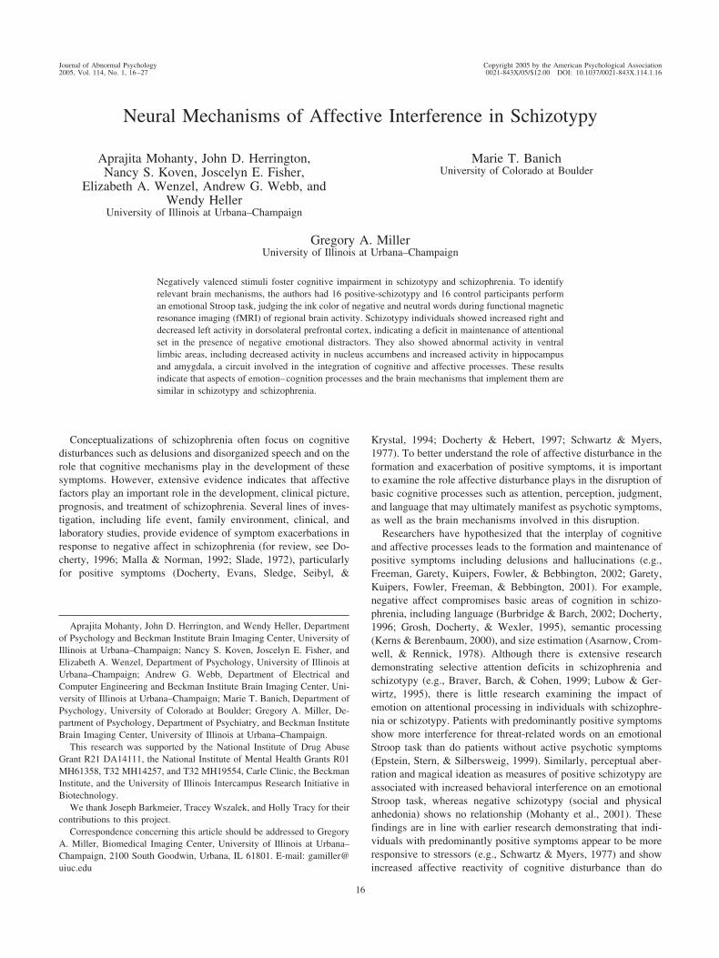
Table 1 reports regions showing greater activity for negativewords than neutral words, within each group. For both groups thisincluded DLPFC (Brodmann Area [BA] 9 and BA 46), inferiorfrontal gyrus (BA 46 and BA 47), and anterior cingulate (BA 32;see also Figure 1, lower right panel). Each group showed effects inseveral additional brain regions listed in Table 1.
Table 2 lists all brain regions showing group differences innegative-minus-neutral-condition activation. Figure 1 focuses ondifferences in regions that are part of the brain circuits hypothe-
sized to differentiate individuals with schizotypy. As predicted, thetwo groups differed in a network of regions believed to be in-volved in the implementation of attentional control, in mainte-nance of contextual information, and in emotional processing,including prefrontal, striatal, limbic, and cerebellar regions.
The schizotypy group showed greater activation in right middlefrontal gyrus within DLPFC (BA 9) and inferior frontal gyrus (BA46). The schizotypy group also showed differential activity in anetwork of regions constituting the ventral limbic cortical–basalganglia circuit. Specifically, the schizotypy group showed moreactivation in the right parahippocampal gyrus and in a regionaround and extending into the amygdala (a cluster for which thepeak was in the putamen per Table 1). Parallel analyses of deac-tivations showed that schizotypy individuals showed more deacti-vation in nucleus accumbens than did control participants. Outsideof these components of the ventral circuit, the schizotypy groupshowed greater activation in dorsal striatal regions including cau-date and putamen as well as left cerebellum. The control groupshowed greater activation in a number of areas, including the leftmiddle frontal gyrus (BA 46), left superior temporal gyrus (BA22), right inferior temporal gyrus (BA 21), and right middleoccipital gyrus (BA 18 and BA 19).
Table 1Brain Regions Showing More Activity for Negative Words Than for Neutral Words for EachGroup
Region Cluster size Mean Z
Location
X Y Z
Control
Left fusiform gyrus (BA 20) 33 2.16 �52 �34 �22Right inferior temporal gyrus (BA 21) 193 2.52 62 �8 �18Left and right inferior frontal gyrus (BA 47) 75 2.18 56 32 �4
63 2.08 �52 36 �4Anterior cingulate (BA 32) 79 2.24 0 36 �2Right middle occipital gyrus (BA 18 and BA 19) 155 2.28 30 �96 2
59 2.32 40 �82 12Left superior temporal gyrus (BA 22) 106 2.21 �56 �40 16Left and right middle frontal gyrus (BA 46) 180 2.26 58 34 22
34 2.18 �32 22 22
Positive schizotypy
Left cerebellum 615 2.38 �40 �40 �22Right hippocampus 204 2.28 30 �20 �18Left putamen 132 2.26 �20 4 �10Anterior cingulate (BA 32) 119 2.14 6 44 �2Left middle temporal gyrus (BA 22 and BA 21) 49 2.16 �62 �38 4
56 2.13 �64 �56 10Left and right inferior frontal gyrus (BA 46) 77 2.16 �54 48 4
137 2.52 40 42 6Right middle frontal gyrus (BA 9) 75 2.33 50 24 28Posterior cingulate (BA 23) 37 2.17 �12 �34 24Left inferior parietal lobule (BA 40) 339 2.27 �54 �58 40
Note. Activation was defined as significantly greater activity for negative than for neutral words within agroup. Regions having at least 30 contiguous voxels each active above z � 1.96 ( p � .05, two-tailed) are listed.The first column lists the names (Talairach & Tournoux, 1988) and Brodmann areas (BA) of brain regions thatshowed significant activation. The second and third columns provide the number of contiguously active voxelsin the cluster and mean z (effect size in standard deviation units) for the voxels in the cluster, respectively. Thelocation columns provide the cluster location in Talaraich coordinates.
20 MOHANTY ET AL.

Finally, as predicted from an earlier behavioral study (Mohantyet al., 2001), the positive schizotypy group had higher anxiousapprehension scores than the control group. However, a mediatingvariable analysis (Baron & Kenny, 1986) indicated that anxiousapprehension did not fully mediate the relationship between pos-itive schizotypy and brain activity in regions identified as moreactive in schizotypy individuals. Only the group difference in rightinferior frontal gyrus (BA 46) was mediated by the variance sharedbetween positive schizotypy and anxious apprehension.
Discussion
In light of the evidence indicating that negative affect fosterscognitive disturbance in individuals with positive symptoms ofschizotypy and schizophrenia, it was hypothesized that positiveschizotypy would be associated with an abnormal pattern of brainactivity in DLPFC and ventral–limbic–striatal regions when chal-lenged with negative emotion words in a selective attention task.
Results supported this hypothesis. The schizotypy group showedreduced left DLPFC and elevated right DLPFC activation. Theyalso produced larger changes in nucleus accumbens, hippocampus,amygdala, and basal ganglia activity. Although there is evidenceindicating that nonpatients who score high on measures of schizo-typy and individuals with schizophrenia show similar cognitiveabnormalities, this appears to be the first hemodynamic neuroim-aging study providing direct evidence that positive-schizotypyindividuals’ task-related brain activation patterns resemble thoseof patients with schizophrenia.
Studies investigating DLPFC function in schizophrenia haveyielded mixed results, with both hypoactivity and hyperactivityreported. DLPFC is involved in sustaining selective attention in thepresence of emotionally aversive and nonemotional distractors andis also sensitive to manipulations of intensity within each of thesecategories (Compton et al., 2003). Furthermore, DLPFC activityrelated to working memory can be influenced by affective vari-
Figure 1. The lower right panel illustrates greater activity in rostral–ventral anterior cingulate for negative thanfor neutral words for schizotypy (in orange) and control (in blue) groups. Dark pixels between orange and blueregions were significant in both groups. The remaining panels show hypothesized regions with significant groupdifferences in activation, with activation defined as greater activity for negative words than for neutral words.Orange depicts more activation for schizotypy participants than for control participants, and blue depicts moreactivation for control participants than for schizotypy participants. Following radiological convention, the rightside of the brain is on the left side in axial and coronal slices. For all panels, highlighted voxels are those withz scores �1.96 ( p � .05, two-tailed) meeting cluster-size threshold (see the Method section in text). Tables 1and 2 present Talairach coordinates of highlighted regions.
21AFFECTIVE INTERFERENCE IN SCHIZOTYPY

ables (Gray, Braver, & Raichle, 2002; Perlstein, Elbert, & Stenger,2002), and there is considerable literature documenting hemi-spheric differences in PFC activity as a function of emotionalvalence (e.g., Davidson, 1992; Heller, 1993). In the present study,participants selectively attended to task-relevant aspects in thepresence of distractors that were emotionally aversive or nonemo-tional in nature. The schizotypy group showed less left and moreright DLPFC activation for negative words. Present lateralizedDLPFC results provide some of the first fMRI support for theassociation of valence with frontal lateralization previously docu-mented with electroencephalogram studies and indicate that emo-tional aspects of tasks need to be considered when designing andinterpreting studies intended to investigate cognitive dysfunctionin psychopathology.
Evidence from fMRI and positron–emission tomography stud-ies indicates that during verbal working memory tasks, prefrontalactivity tends to be lateralized toward the left hemisphere (Cabeza& Nyberg, 2000). In particular, left inferior brain activity reflectsprocessing more specific to the Stroop task (Taylor, Kornblum,Lauber, Minoshima, & Koeppe, 1997). Similarly, negative emo-tional Stroop stimuli activate left DLPFC (Compton et al., 2003).In light of these findings, decreased left DLPFC activity in schizo-typy participants may be interpretable as a failure to fully engageoptimal executive processes leading to increased interference fromemotional stimuli on the task, as indicated by the performance datafrom a behavioral study with a large number of participants (Mo-hanty et al., 2001). Right PFC has been implicated in vigilance andresponse to threat (for reviews, see Nitschke & Heller, 2002;Nitschke & Heller, in press; Nitschke, Heller, & Miller, 2000).Thus, increased right DLPFC activity in schizotypy participantssuggests exaggerated attention to negative stimuli even though
they are task irrelevant. Indeed, there is evidence that individualswith schizophrenia (Blackwood, Howard, Bentall, & Murray,2001) or those at risk for schizophrenia (Green, Williams, &Davidson, 2001) preferentially attend to threat-related stimuli.Thus, present lateralized DLPFC findings sensitive to group andemotional valence suggest that uncontrolled task differences inemotion processing demands could account for a portion of theheterogeneity in findings about DLPFC patterns in schizophrenia.These results again underscore the importance of considering theemotional aspects of tasks when designing and interpreting studiesthat investigate cognitive dysfunction in psychopathology.
The schizotypy sample also showed more activation in rightinferior frontal gyrus. Inferior frontal cortex tends to be involvedin situations requiring resolution of interference among conflictingtask attributes (Nelson, Reuter-Lorenz, Sylvester, Jonides, &Smith, 2003). A number of studies have shown that the ability toinhibit processing of irrelevant or interfering stimuli is associatedwith right inferior frontal gyrus (e.g., Garavan, Ross, Murphy,Roche, & Stein, 2002; Garavan, Ross, & Stein, 1999; Konishi,Nakajima, Uchida, Sekihara, & Miyashita, 1998). In present re-sults, increased right inferior frontal gyrus activity in schizotypyindividuals may indicate a greater effort to inhibit strongly inter-fering emotional stimuli in order to achieve normal behavioralperformance. Researchers have hypothesized that anxiety, partic-ularly anxious apprehension, and positive symptoms have manythematic similarities (e.g., a bias toward threat-related stimuli) andthat anxiety makes a direct contribution to positive symptomdevelopment (Freeman et al., 2002). In light of that literature, thefinding in the present study that right inferior frontal gyrus acti-vation (BA 46) is mediated by the variance shared between posi-
Table 2Brain Regions Showing Group Differences in Negative-Minus, Neutral-Word Activation
Region Cluster size Mean Z
Location
X Y Z
Positive schizotypy � control
Right middle frontal gyrus (BA 9) 36 2.14 52 22 28Right inferior frontal gyrus (BA 46) 47 2.17 44 42 6Right hippocampus including parahippocampal gyrus 102 2.32 28 �18 �18Left putamen 104 2.53 �20 4 �10Left cerebellum 95 2.54 �22 �28 �22
58 2.20 �46 �50 �26
Control � positive schizotypy
Left middle frontal gyrus (BA 46) 31 2.25 �32 22 22Right inferior temporal gyrus (BA 21) 100 2.36 62 �8 �18Right middle occipital gyrus (BA 18 and BA 19) 77 2.44 30 �94 2
50 2.51 40 �82 12Left superior temporal gyrus (BA 22) 81 2.56 �56 �40 18
Note. Activation was defined as significantly greater activity for negative than for neutral words. A voxelshowed a significant group difference in activation if it exceeded a z threshold of 1.96 ( p � .05, two-tailed) andwas part of a cluster of at least 30 contiguous active voxels. The first column lists the names (Talairach &Tournoux, 1988) and Brodmann areas of brain regions that showed significant differential activation for the twogroups. The second and third columns characterize the number of contiguously active voxels in the cluster andthe group difference in terms of mean z (effect size in standard deviation units), respectively. The locationcolumns provide three-dimensional Talairach coordinates of these brain regions.
22 MOHANTY ET AL.

tive schizotypy and anxious apprehension suggests a brain mech-anism common to both.
Additional interesting findings emerged for regions constitutingthe ventral limbic cortical–basal ganglia circuit, which is believedto play a crucial role in the interaction and integration of cognitiveand affective processes. Dysfunction in any component of thiscircuit could contribute to symptoms in schizophrenia (Grace &Moore, 1998). Because the nucleus accumbens, a major compo-nent of this circuit, receives inputs from hippocampus, amygdala,and PFC, it may play a particularly important role in their modu-lation or integration. Thus, decreased activity in this region inschizotypy suggests a mechanism for dysregulation of inputs fromimportant brain regions in the face of aversive stimuli.
Greater activation in hippocampus and amygdala in the schizo-typy group is consistent with reports indicating that positive symp-toms in schizophrenia are associated with increased activity inhippocampal and amygdalar regions during performance on anemotional Stroop task (Epstein et al., 1999; Taylor et al., 2002) andother tasks (Blackwood et al., 2001). The amygdala is believed tobe central to the emotional evaluation of fear and threat-relatedsensory stimuli (LeDoux, 1995; see Zald, 2003, for review), andthe hippocampus is believed to mediate contextual aspects ofemotional evaluation (Grace & Moore, 1998; Kim & Fanselow,1992; R. G. Phillips & LeDoux, 1992). Abnormal inputs fromthese limbic structures to the ventral striatum are believed to playan important role in psychotic symptoms (e.g., Bogerts, 1997;Weinberger & Lipska, 1995). For example, there is evidence thathippocampal and amygdalar inputs to the nucleus accumbensmodulate activity in prefrontal brain regions in distinct ways(Grace & Moore, 1998). The hippocampus provides general,context-dependent modulation, whereas the amygdala provides amore discrete, event-related input, such that salient emotionalstimuli can override an otherwise context-dependent system. Ac-cordingly, if hippocampal neurons were to show abnormal firingpatterns that were due to a pathological condition, the result couldbe the reinforcement of a pattern of cerebral activity representinga contextually uncorroborated mental event—a mechanism poten-tially involved in delusions, hallucinations, magical ideation, andperceptual aberration (Liddle et al., 2000). Thus, abnormal activityin limbic and striatal regions prompted by negative stimuli sug-gests a mechanism through which negative affect is associatedwith an exacerbation of DLPFC-related cognitive disturbance inschizotypy and schizophrenia. That this disrupted mechanism isevident in a nonpatient sample merely reporting elevations inselected positive symptoms indicates that the mechanism and itsdisruption can be in place prior to the onset of the diagnoseddisorder.
In addition to support for the two major a priori hypothesesabout DLPFC and ventral limbic areas, the present study obtainedother findings consistent with the schizophrenia literature. Schizo-typal individuals showed more activation in basal ganglia andcerebellum than did control participants. Some of these regionsmay contribute to the DLPFC and ventral limbic relationships onwhich the study focused. The findings of increased activity in basalganglia are similar to those in schizophrenia, in which positivesymptoms are associated with increased task-related activity in thedorsal striatum (e.g., Kircher et al., 2001). Furthermore, decreasedsuppression of striatal activity during working memory perfor-mance is associated with abnormal PFC activity in schizophrenia
(Rubin et al., 1991), indicating that a frontostriatal circuitry dys-function may be involved in cognitive deficits in schizophrenia(Manoach et al., 2000).
The finding of increased cerebeller activation in the schizotypygroup is in line with considerable clinical and neuroimaging evi-dence supporting cerebellar involvement in schizophrenia. Thecerebellum and its connections to PFC have been implicated inthought abnormalities in schizophrenia (Andreasen et al., 1996,1999; Andreasen, Paradiso, & O’Leary, 1998). More specifically,the fluid coordination of motor activity and thought may involverapid feedback between cerebral cortex and cerebellum mediatedthrough thalamus, and its disruption has been proposed as a fun-damental deficit in schizophrenia called “cognitive dysmetria”(Andreasen et al., 1996, 1998, 1999). Evidence from neuroimagingstudies using a variety of tasks, including recall of complex nar-ratives, episodic memory, memory for word lists, and randomepisodic silent thought, indicates abnormal activity in variouselements of a cortico–cerebellar–thalamic–cortical circuit inschizophrenia. Present findings in the lateral cerebellum suggestthat negative affect, or the distraction it can provide from task-relevant cognitive activity, is another means to foster cognitivedysmetria. This may contribute to the exacerbation of cognitivedisturbance associated with negative affect in schizophrenia andschizotypy.
Another interesting finding in the present study involved theanterior cingulate, a region that plays an important role in cogni-tive and emotional processing. Both schizotypy and control groupsshowed more activity in rostral–ventral anterior cingulate for neg-ative than for neutral words. This finding is consistent with liter-ature showing that rostral–ventral anterior cingulate is activated byaffect-related tasks, including the emotional Stroop task, for nor-mal control participants and psychiatric patients (e.g., Shin et al.,2001; Whalen et al., 1998; for review, see Bush, Luu, & Posner,2000). The rostral–ventral division of the anterior cingulate differsfrom the dorsal division in terms of its anatomical connectivity andthe functions it implements (Bush et al., 2000). The dorsal subdi-vision has been shown to play an important role in a variety ofcognitive functions, including response selection, competitionmonitoring, error detection, and working memory. In contrast, therostral–ventral subdivision (referred to as the “affective subdivi-sion” by Bush et al., 2000) is involved in assessment of thesalience of emotional information and regulation of emotionalresponses. The rostral–ventral subdivision is connected to amyg-dala, nucleus accumbens, hypothalamus, hippocampus, and orbito-frontal cortex (Devinsky, Morrell, & Vogt, 1995). Researchershave hypothesized that rostral–ventral cingulate regulates or in-hibits other brain regions involved in response to threat such asamygdala (e.g., Shin et al., 2001). In present data, both groupsactivated rostral–ventral cingulate when processing emotionallynegative stimuli, indicating a regulatory process. However, com-pared with the control group, the schizotypal group also activatedamygdala and hippocampus and deactivated nucleus accumbens(all connected to rostral–ventral anterior cingulate), suggesting afailure to fully engage optimal regulatory processes, a possiblemechanism by which emotional stimuli disrupt cognitivefunctioning.
There is little research examining the impact of emotion onattentional processing in individuals with schizotypy or schizo-phrenia. The present investigation is the first hemodynamic neu-
23AFFECTIVE INTERFERENCE IN SCHIZOTYPY

roimaging study examining such processing in an at-risk popula-tion, and the replication of some findings from the schizophrenialiterature is noteworthy (Barch, Carter, et al., 2001; Callicott et al.,2000; Epstein et al., 1999; Kircher et al., 2001; Liddle, Friston,Frith, & Frackowiack, 1992; Rubin et al., 1991; Silbersweig et al.,1995; Taylor et al., 2002; Weinberger & Lipska, 1995). Appar-ently, at least some of the functional brain phenomena noted inschizophrenia are observable in a relatively well-functioning non-patient sample, suggesting that they reflect predisposing mecha-nisms in schizophrenia rather than the sequelae of the disease or itstreatment. That the present sample is unmedicated avoids seriousconfounds that are often present in studies of individuals withschizophrenia. This is particularly important because limbic re-gions of the forebrain and the amygdala are sites of action ofantipsychotic mediation. Chronic schizophrenia, but not schizo-typy, is associated with illness-related epiphenomena driven byantipsychotic medication and hospitalization. Moreover, the cog-nitive performance of individuals with schizotypy is less likely tobe confounded with or compromised by the often-substantial def-icits in motivation, task comprehension, and task performanceassociated with the disease process, antipsychotic medication, andhospitalization.
Although there were more similiarities than differences in re-gional brain activation observed for positive-symptom schizotypalindividuals and control participants, most of the observed regionaldifferences were supportive of (a) continuity between this nonpa-tient sample and some of the schizophrenia literature, (b) neuralcircuitry proposed to play a central role in schizophrenia, and (c)emotional factors in putatively cognitive tasks substantially affect-ing regional brain patterns. Mechanisms contributing to this mod-ulation of cognitive dysfunction by emotional processing mayprove central to understanding schizophrenia.
References
Andreasen, N. C., Nopoulos, P., O’Leary, D. S., Miller, D. D., Wassink, T.,& Flaum, M. (1999). Defining the phenotype of schizophrenia: Cogni-tive dysmetria and its neural mechanisms. Biological Psychiatry, 46,908–920.
Andreasen, N. C., O’Leary, D. S., Cizadlo, T., Arndt, S., Rezai, K., BolesPonto, L. L., et al. (1996). Schizophrenia and cognitive dysmetria: Apositron–emission tomography study of dysfunctional prefrontal–thalamic–cerebellar circuitry. Proceedings of the National Academy ofSciences, USA, 93, 9985–9990.
Andreasen, N. C., Paradiso, S., & O’Leary, D. S. (1998). Cognitivedysmetria as an integrative theory of schizophrenia: A dysfunction incortical–cerebellar circuitry? Schizophrenia Bulletin, 24, 203–218.
Andreasen, N. C., Rezai, K., Alliger, R., Swayze, V. W., Flaum, M.,Kirchner, P., et al. (1992). Hypofrontality in neuroleptic-naı̈ve andchronic schizophrenic patients: Assessment with xenon-133 single-photon emission computed tomography and the Tower of London.Archives of General Psychiatry, 49, 943–958.
Asarnow, R. F., Cromwell, R. L., & Rennick, P. M. (1978). Cognitive andevoked response measures of information processing in schizophrenicswith and without a family history of schizophrenia. Journal of Nervous& Mental Diseases, 166, 719–730.
Banich, M. T., Milham, M. P., Atchley, R. A., Cohen, N. J., Webb, A.,Wszalek, T., et al. (2000). Prefrontal regions play a predominant role inimposing an attentional “set”: Evidence from fM.R.I. Cognitive BrainResearch, 10, 1–9.
Banich, M. T., Milham, M. P., Jacobson, B. L., Webb, A., Wszalek, T.,
Cohen, N. J., et al. (2001). Attentional selection and the processing oftask-irrelevant information: Insights from fM.R.I. examinations of theStroop task. Progress in Brain Research, 134, 459–470.
Barch, D. M., Braver, T. S., Akbudak, E., Conturo, T., Ollinger, J., &Snyder, A. (2001). Anterior cingulate cortex and response conflict:Effects of response modality and processing domain. Cerebral Cortex,11, 837–848.
Barch, D. M., Braver, T. S., Nystrom, L. E., Forman, S. D., Noll, D. C., &Cohen, J. D. (1996). Dissociating working memory from task difficultyin human prefrontal cortex. Neuropsychologia, 35, 1373–1380.
Barch, D. M., Carter, C. S., Braver, T. S., Sabb, F. W., MacDonald, A., III,Noll, D. C., et al. (2001). Selective deficits in prefrontal cortex functionin medication-naive patients with schizophrenia. Archives of GeneralPsychiatry, 58, 280–288.
Baron, R. M., & Kenny, D. A. (1986). The moderator–mediator variabledistinction in social psychological research: Conceptual, strategic, andstatistical considerations. Journal of Personality and Social Psychology,51, 1173–1182.
Blackwood, N. J., Howard, R. J., Bentall, R. P., & Murray, R. M. (2001).Cognitive neuropsychiatric models of persecutory delusions. AmericanJournal of Psychiatry, 158, 527–539.
Bogerts, B. (1997). The temporolimbic system theory of positive schizo-phrenic symptoms. Schizophrenia Bulletin, 23, 423–435.
Bradley, M. M., & Lang, P. J. (1998). Affective norms for English words(ANEW). Gainesville, FL: University of Florida, The National Instituteof Mental Health Center for the Study of Emotion and Attention.
Braver, T. S., Barch, D. M., & Cohen, J. D. (1999). Cognition and controlin schizophrenia: A computational model of dopamine and prefrontalfunction. Biological Psychiatry, 46, 312–328.
Braver, T. S., Barch, D. M., Gray, J. R., Molfese, D. L., & Snyder, A.(2001). Anterior cingulate cortex and response conflict: Effects of re-sponse modality and processing domain. Cerebral Cortex,11, 837–848.
Buchsbaum, M. S., Haier, R. J., Potkin, S. G., Nuechterlein, K., Bracha,H. S., Katz, M., et al. (1992). Frontostriatal disorder of cerebral metab-olism in never-medicated schizophrenics. Archives of General Psychi-atry, 49, 935–942.
Burbridge, J. A., & Barch, D. M. (2002). Emotional valence and referencedisturbance in schizophrenia. Journal of Abnormal Psychology, 111,186–191.
Bush, G., Luu, P., & Posner, M. I. (2000). Cognitive and emotionalinfluences in anterior cingulate cortex. Trends in Cognitive Sciences, 4,215–222.
Cabeza, R., & Nyberg, L. (2000). Imaging cognition: II. An empiricalreview of 275 PET and fMRI studies. Journal of Cognitive Neuro-science, 12, 1–47.
Cahill, L., Roozendaal, B., & McGaugh, J. L. (1997). The neurobiology ofmemory for aversive emotional events. In M. E. Bouton & M. S.Fanselow (Eds.), Learning, motivation, and cognition: The functionalbehaviorism of Robert C. Bolles (pp. 369–384). Washington, DC: Amer-ican Psychological Association.
Callicott, J. H., Bertolino, A., Mattay, V. S., Langheim, F. J. P., Duyn, J.,Coppola, R., et al. (2000). Physiological dysfunction of the dorsolateralprefrontal cortex in schizophrenia revisited. Cerebral Cortex, 10, 1078–1092.
Callicott, J. H., Egan, M. F., Mattay, V. S., Bertolino, A., Bone, A. D.,Verchinski, et al. (2003). Abnormal fM.R.I. response of the dorsolateralprefrontal cortex in cognitively intact siblings of patients with schizo-phrenia. American Journal of Psychiatry, 160, 709–719.
Callicott, J. H., Mattay, V. S., Bertolino, A., Finn, K., Coppola, R., Frank,J. A., et al. (1999). Physiological characteristics of capacity constraintsin working memory as revealed by functional MRI. Cerebral Cortex, 9,20–26.
Chapman, L. J., Chapman, J. P., Kwapil, T. R., Eckblad, M., & Zinser,
24 MOHANTY ET AL.

M. C. (1994). Putatively psychosis-prone subjects 10 years later. Journalof Abnormal Psychology, 103, 171–183.
Chapman, L. J., Chapman, J. P., & Raulin, M. L. (1978). Body-imageaberration in schizophrenia. Journal of Abnormal Psychology, 87, 399–407.
Chun, M. M., & Phelps, E. A. (1999). Memory deficits for implicitcontextual information in amnesic subjects with hippocampal damage.Nature Neuroscience, 2, 844–847.
Cohen, J. D., & Servan-Schreiber, D. (1992). Context, cortex, and dopa-mine: A connectionist approach to behavior and biology in schizophre-nia. Psychological Review, 99, 45–77.
Cohen, N. J., & Eichenbaum, H. (1993). Memory, amnesia, and thehippocampal system. Cambridge, MA: MIT Press.
Compton, R. J., Banich, M. T., Mohanty, A., Milham, M. P., Herrington,J., Miller, G. A., et al. (2003). Paying attention to emotion: An fM.R.I.investigation of cognitive and emotional Stroop tasks. Cognitive, Affec-tive & Behavioral Neuroscience, 3, 81–96.
Compton, R. J., Heller, W., Banich, M. T., Palmieri, P. A., & Miller, G. A.(2000). Responding to threat: Hemispheric asymmetries and interhemi-spheric division of input. Neuropsychology, 14, 254–264.
Dalgleish, T. (1995). Performance on the emotional Stroop task in groupsof anxious, expert, and control subjects: A comparison of computer andcard presentation formats. Cognition & Emotion, 9, 341–362.
Davidson, R. J. (1992). Anterior cerebral asymmetry and the nature ofemotion. Brain and Cognition, 20, 125–151.
Davidson, R. J., & Irwin, W. (1999). The functional neuroanatomy ofemotion and affective style. Trends in Cognitive Sciences, 3, 11–21.
Devinsky, O., Morrell, M. J., & Vogt, B. A. (1995). Contributions ofanterior cingulate cortex to behaviour. Brain, 118, 279–306.
Dickey, C. C., Shenton, M. E., Hirayasu, Y., Fischer, I., Voglmaier, M. M.,Niznikiewicz, M. A., et al. (2000). Large C.S.F. volume not attributableto ventricular volume in schizotypal personality disorder. AmericanJournal of Psychiatry, 157, 48–54.
Docherty, N. M. (1996). Affective reactivity of symptoms as a processdiscriminator in schizophrenia. Journal of Nervous & Mental Diseases,184, 535–541.
Docherty, N. M., Evans, I. M., Sledge, W. H., Seibyl, J. P., & Krystal, J. H.(1994). Affective reactivity of language in schizophrenia. Journal ofNervous & Mental Disease, 182, 98–102.
Docherty, N. M., & Hebert, A. S. (1997). Comparative affective reactivityof different types of communication disturbances in schizophrenia. Jour-nal of Abnormal Psychology, 106, 325–330.
Eckblad, M., & Chapman, L. J. (1983). Magical ideation as an indicator ofschizotypy. Journal of Consulting and Clinical Psychology, 51, 215–225.
Edell, W. S. (1995). The psychometric measurement of schizotypy usingthe Wisconsin scales of psychosis-proneness. In G. A. Miller (Ed.), Thebehavioral high-risk paradigm in psychopathology (pp. 3–46). NewYork: Springer-Verlag.
Epstein, J., Stern, E., & Silbersweig, D. (1999). Mesolimbic activityassociated with psychosis in schizophrenia: Symptom-specific P.E.T.studies. In J. F. McGinty (Ed.), Advancing from the ventral striatum tothe extended amygdala: Implications for neuropsychiatry and drug use:In honor of Lennart Heimer (pp. 562–574). New York: New YorkAcademy of Sciences.
Erlenmeyer-Kimling, L., Cornblatt, B. A., Rock, D., Roberts, S., Bell, M.,&West, A. (1993). The New York High-Risk Project: Anhedonia, at-tentional deviance, and psychopathology. Schizophrenia Bulletin, 19,141–153.
Fernandes, L., & Miller, G. A. (1995). Compromised performance andabnormal psychophysiology associated with the Wisconsin scales ofpsychosis proneness. In G. A. Miller (Ed.), The behavioral high-riskparadigm in psychopathology (pp. 47–87). New York: Springer-Verlag.
Forman, S. D., Cohen, J. D., Fitzgerald, M., Eddy, W. F., Mintun, M. A.,
& Noll, D. C. (1995). Improved assessment of significant activation infunctional magnetic resonance imaging (fMRI): Use of a cluster-sizethreshold. Magnetic Resonance in Medicine, 33, 636–647.
Frank, M. J., Loughry, B., & O’Reilly, R. C. (2001). Interactions betweenfrontal cortex and basal ganglia in working memory: A computationalmodel. Cognitive Affective and Behavioral Neuroscience, 2, 137–160.
Freedman, L. R., Rock, D., Roberts, S. A., Cornblatt, B. A., & Erlenmeyer-Kimling, L. (1998). The New York High-Risk Project: Attention, anhe-donia and social outcome. Schizophrenia Research, 30, 1–9.
Freeman, D., Garety, P. A., Kuipers, E., Fowler, D., & Bebbington, P. E.(2002). A cognitive model of persecutory delusions. British Journal ofClinical Psychology, 41, 331–347.
Garavan, H., Ross, T. J., Murphy, K., Roche, R. A., & Stein, E. A. (2002).Dissociable executive functions in the dynamic control of behavior:Inhibition, error detection, and correction. Neuroimage, 17, 1820–1829.
Garavan, H., Ross, T. J., & Stein, E. A. (1999). Right hemisphere domi-nance of inhibitory control: An event-related functional MRI study.Proceedings of the National Academy of Sciences, USA, 96, 8301–8306.
Garety, P. A., Kuipers, E., Fowler, D., Freeman, D., & Bebbington, P. E.(2001). A cognitive model of the positive symptoms of psychosis.Psychological Medicine, 31, 195–198.
Gold, J. M., Goldberg, T. E., & Weinberger, D. R. (1992). Prefrontalfunction and schizophrenic symptoms. Neuropsychiatry, Neuropsychol-ogy, and Behavioral Neurology, 5, 253–261.
Grace, A. A. (2000). Gating of information flow within the limbic systemand the pathophysiology of schizophrenia. Brain Research—Brain Re-search Reviews, 31, 330–341.
Grace, A. A., & Moore, H. (1998). Regulation of information flow in thenucleus accumbens: A. model for the pathophysiology of schizophrenia.In F. Lenzenweger & R. H. Dworkin (Eds.), Origins and development ofschizophrenia: Advances in experimental psychopathology (pp. 123–157). Washington, DC: American Psychological Association.
Grace, A. A., Moore, H., & O’Donnell, P. (1998). The modulation ofcorticoaccumbens transmission by limbic afferents and dopamine: A.model for the pathophysiology of schizophrenia. Advances in Pharma-cology, 42, 721–724.
Gray, J. R., Braver, T. S., & Raichle, M. E. (2002). Integration of emotionand cognition in the lateral prefrontal cortex. Proceedings of the Na-tional Academy of Sciences, USA, 99, 4115–4120.
Green, M. J., Williams, L. M., & Davidson, D. J. (2001). Processing ofthreat-related affect is delayed in delusion-prone individuals. BritishJournal of Clinical Psychology, 40, 157–165.
Groenewegen, H. J., Berendse, H. W., Meredith, G. E., Haber, S. N.,Voorn, P., Wolters, J. G., et al. (1991). Functional anatomy of ventral,limbic system-innervated striatum. In P. Willner & J. Scheel-Kruger(Eds.), The mesolimbic dopamine system: From motivation to action (pp.19–60). Chichester, England: Wiley.
Grosh, E. S., Docherty, N. M., & Wexler, B. E. (1995). Abnormal lateralityin schizophrenic patients and their parents. Schizophrenia Research, 14,155–160.
Heller, W. (1993). Neuropsychological mechanisms of individual differ-ences in emotion, personality, and arousal. Neuropsychology, 7, 476–489.
Herrington, J. D., Mohanty, A., Koven, N. S., Fisher, J. E., Stewart, J. L.,Banich, M. T., et al. (2004). Pleasant stimuli enhance performance andincrease activity in left dorsolateral prefrontal cortex. Manuscript sub-mitted for publication.
Holle, C., Neely, J. H., & Heimberg, R. G. (1997). The effects of blockedversus random presentation and semantic relatedness of stimulus wordson response to a modified Stroop task among social phobics. CognitiveTherapy & Research, 21, 681–697.
Jenkinson, M., Bannister, P., Brady, J., & Smith, S. (2002). Improvedoptimisation for the robust and accurate linear registration and motioncorrection of brain images. NeuroImage, 17, 825–841.
25AFFECTIVE INTERFERENCE IN SCHIZOTYPY

Jenkinson, M., & Smith, S. M. (2001). A global optimization method forrobust affine registration of brain images. Medical Image Analysis, 2,143–156.
Kerns, J. G., & Berenbaum, H. (2000). Aberrant semantic and affectiveprocessing in people at risk for psychosis. Journal of Abnormal Psy-chology, 109, 728–732.
Kim, J. J., & Fanselow, M. S. (1992, May 1). Modality-specific retrogradeamnesia of fear. Science, 256, 675–677.
Kircher, T. T. J., Liddle, P. F., Brammer, M. J., Williams, S. C. R., Murray,R. M., & McGuire, P. K. (2001). Neural correlates of formal thoughtdisorder in schizophrenia: Preliminary findings from a functional mag-netic resonance imaging study. Archives of General Psychiatry, 58,769–774.
Konishi, S., Nakajima, K., Uchida, I., Sekihara, K., & Miyashita, Y.(1998). No-go dominant brain activity in human inferior prefrontalcortex revealed by functional magnetic resonance imaging. EuropeanJournal of Neuroscience, 10, 1209–1213.
LeDoux, J. E. (1995). Emotion: Clues from the brain. Annual Review ofPsychology, 46, 209–235.
Liddle, P. F., Friston, K. J., Frith, C. D., & Frackowiak, R. S. (1992).Cerebral blood flow and mental processes in schizophrenia. Journal ofResearch in Social Medicine, 85, 224–227.
Liddle, P. F., Friston, K., Frith, C., Hirsch, S. R., Jones, T., & Frackowiak,R. S. (1992). Patterns of cerebral blood flow in schizophrenia. BritishJournal of Psychiatry, 160, 179–186.
Liddle, P. F., Lane, C. J., & Ngan, E. T. C. (2000). Immediate effects ofrisperidone on cortico–striato–thalamic loops and the hippocampus.British Journal of Psychiatry, 177, 402–407.
Lubow, R. E., & Gewirtz, J. C. (1995). Latent inhibition in humans: Data,theory, and implications for schizophrenia. Psychological Bulletin, 117,87–103.
MacDonald, A. W., III., Johnson, M. K., Becker, T. M., & Carter, C. S.(2003, March). Context processing deficits associated with hypofrontal-ity in the healthy relatives of schizophrenia patients: An event relatedfMRI study. Paper presented at the IXth meeting of International Con-gress of Schizophrenia Research, Colorado Springs, CO.
MacLeod, C. M. (1992). The Stroop task: The “gold standard” of atten-tional measures. Journal of Experimental Psychology: General, 121,12–14.
Malla, A. K., & Norman, R. M. (1992). Relationship of major life eventsand daily stressors to symptomatology in schizophrenia. Journal ofNervous & Mental Disease, 180, 664–667.
Manoach, D. S., Gollub, R. L., Benson, E. S., Searl, M. M., Goff, D. C.,Halpern, E., et al. (2000). Schizophrenic subjects show aberrant fMRIactivation of dorsolateral prefrontal cortex and basal ganglia duringworking memory performance. Biological Psychiatry, 48, 99–109.
Manoach, D. S., Press, D. Z., Thangaraj, V., Searl, M. M., Goff, D. C.,Halpern, E., et al. (1999). Schizophrenic subjects activate dorsolateralprefrontal cortex during a working memory task, as measured by fM.R.I.Biological Psychiatry, 45, 1128–1137.
Meyer, T. J., Miller, M. L., Metzger, R. L., & Borkovec, T. D. (1990).Development and validation of the Penn State Worry Questionnaire.Behavior Research and Therapy, 28, 487–495.
Middleton, F. A., & Strick, P. L. (2000). Basal ganglia and cerebellarloops: Motor and cognitive circuits. Brain Research—Brain ResearchReviews, 31, 236–250.
Miller, E. K., Erickson, C. A., & Desimone, R. (1996). Neural mechanismsof visual working memory in prefrontal cortex of the macaque. Journalof Neuroscience, 16, 5154–5167.
Mohanty, A., Koven, N. S., Fisher, J. E., Herrington, J. H., Stewart, J. L.,Heller, W., et al. (2001, November). Emotion regulation and positiveand negative symptoms in psychosis proneness. Paper presented at theannual meeting of the Society for Research in Psychopathology, Madi-son, WI.
Nelson, J. K., Reuter-Lorenz, P. A., Sylvester, C.-Y. C., Jonides, J., &Smith, E. E. (2003). Dissociable neural mechanisms underlyingresponse-based and familiarity-based conflict in working memory. Pro-ceedings of the National Academy of Sciences, USA, 100, 11171–11175.
Nitschke, J. B., & Heller, W. (2002). The neuropsychology of anxietydisorders: Affect, cognition, and neural circuitry. In H. D’Haenen, J. A.den Boer, H. Westenberg, & P. Willner (Eds.), Textbook of biologicalpsychiatry (pp. 975–988). Chichester, England: Wiley.
Nitschke, J. B., & Heller, W. (in press). Distinguishing neural substrates ofheterogeneity among anxiety disorders. In D. Barch (Ed.), Cognitive andaffective neuroscience of psychopathology. New York: Oxford Univer-sity Press.
Nitschke, J. B., Heller, W., & Miller, G. A. (2000). Anxiety, stress, andcortical brain function. In J. C. Borod (Ed.), The neuropsychology ofemotion: Series in affective science (pp. 298–319). New York: OxfordUniversity Press.
O’Donnell, P., Greene, J., Pabello, N., Lewis, B. L., & Grace, A. A. (1999).Modulation of cell firing in the nucleus accumbens. In J. F. McGinty(Ed.), Advancing from the ventral striatum to the extended amygdala:Implications for neuropsychiatry and drug use: In honor of LennartHeimer (pp. 157–175). New York: New York Academy of Sciences.
Perlstein, W. M., Elbert, T., & Stenger, V. A. (2002). Dissociation inhuman prefrontal cortex of affective influences on working memory-related activity. Proceedings of the National Academy of Sciences, USA,99, 1736–1741.
Phillips, M. L., Williams, L., Senior, C., Bullmore, E. T., Brammer, M. J.,Andrew, C., et al. (1999). A differential neural response to threateningand non-threatening negative facial expressions in paranoid and non-paranoid schizophrenics. Psychiatry Research: Neuroimaging, 92, 11–31.
Phillips, R. G., & LeDoux, J. E. (1992). Differential contribution ofamygdala and hippocampus to cued and contextual fear conditioning.Behavioral Neuroscience, 106, 274–285.
Ramsey, N. F., Koning, H. A., Welles, P., Cahn, W., Van Der Linden,J. A., & Kahn, R. S. (2002). Excessive recruitment of neural systemssubserving logical reasoning in schizophrenia. Brain, 125, 1793–1807.
Rubin, P., Holm, S., Friberg, L., Videbech, P., Andersen, H. S., Bendsen,B. B., et al. (1991). Altered modulation of prefrontal and subcorticalbrain activity in newly diagnosed schizophrenia and schizophreniformdisorder: A regional cerebral blood flow study. Archives of GeneralPsychiatry, 48, 987–995.
Rudy, J. W., & Sutherland, R. J. (1995). Configural association theory andthe hippocampal formation: An appraisal and reconfiguration. Hip-pocampus, 5, 375–389.
Salzmann, E., Vidyasagar, T. R., & Creutzfeldt, O. D. (1993). Functionalcomparison of neuronal properties in the primate posterior hippocampusand parahippocampus (area TF/TH) during different behavioural para-digms involving memory and selective attention. Behavioural BrainResearch, 53, 133–149.
Schaefer, S. M., Jackson, D. C., Davidson, R. J., Aguirre, G. K., Kimberg,D. Y., & Thompson-Schill, S. L. (2002). Modulation of amygdalaractivity by the conscious regulation of negative emotion. Journal ofCognitive Neuroscience, 14, 913–921.
Schneider, F., Weiss, U., Kessler, C., Salloum, J. B., Posse, S., & Muller-Gartner, H. M. (1998). Differential amygdala activation in schizophreniaduring sadness. Schizophrenia Research, 34, 133–142.
Schwartz, C. C., & Myers, J. K. (1977). Life events and schizophrenia: II.Impact of life events on symptom configuration. Archives of GeneralPsychiatry, 34, 1242–1245.
Servan-Schreiber, D., Cohen, J. D., & Steingard, S. (1996). Schizophrenicdeficits in the processing of context: A test of a theoretical model.Archives of General Psychiatry, 53, 1105–1112.
Shin, L. M., Whalen, P. J., Pitman, R. K., Bush, G., Macklin, M. L., Lasko,
26 MOHANTY ET AL.

N. B., et al. (2001). An fMRI study of anterior cingulate function isposttraumatic stress disorder. Biological Psychiatry, 50, 932–942.
Siegel, B. V., Buchsbaum, M. S., Bunney, W. E., Gottschalk, L. A., Haier,R. J., Lohr, J. B., et al. (1993). Cortical–striatal–thalamic circuits andbrain glucose metabolic activity in 70 unmedicated male schizophrenicpatients. American Journal of Psychiatry, 150, 1325–1336.
Silbersweig, D. A., Stern, E., Frith, C. D., Cahill, C., Hommes, A.,Grootoonk, S., et al. (1995). A functional neuroanatomy of hallucina-tions in schizophrenia. Nature, 378, 176–179.
Slade, P. D. (1972). The effects of systematic desensitization on auditoryhallucinations. Behaviour Research & Therapy, 10, 85–91.
Talairach, J., & Tournoux, P. (1988). Co-planar stereotaxic atlas of thehuman brain. New York: Thieme.
Taylor, S. F., Kornblum, S., Lauber, E. L., Minoshima, S., & Koeppe, R. A.(1997). Isolation of specific interference processing in the Stroop task:PET activation studies. Neuroimage, 6, 81–92.
Taylor, S. F., Liberzon, I., Decker, L. R., & Koeppe, R. A. (2002). Afunctional anatomic study of emotion in schizophrenia. SchizophreniaResearch, 58, 159–172.
Toglia, M. P., & Battig, W. F. (1983). Handbook of semantic word norms.Hillsdale, NJ: Erlbaum.
Vollema, M. G., & Hoijtink, H. (2000). The multidimensionality of self-report schizotypy in a psychiatric population: An analysis using multi-dimensional Rasch models. Schizophrenia Bulletin, 26, 565–575.
Walker, E. F., Neumann, C. C., Baum, K., & Davis, D. M. (1996). The
developmental pathways to schizophrenia: Potential moderating effectsof stress. Development & Psychopathology, 8, 647–665.
Weinberger, D. R., Berman, K. F., & Zec, R. F. (1986). Physiologicaldysfunction of dorsolateral prefrontal cortex in schizophrenia: I. Re-gional cerebral blood flow (rC.B.F.) evidence. Archives of GeneralPsychiatry, 43, 114–124.
Weinberger, D. R., & Lipska, B. K. (1995). Cortical maldevelopment,anti-psychotic drugs, and schizophrenia: A search for common ground.Schizophrenia Research, 16, 87–110.
Weiss, E. M., Golaszewski, S. M., Mottaghy, F. M., Hofer, A., Hausmann,A., Kemmler, G., et al. (2003). Brain activation patterns during aselective attention test—A functional M.R.I. study in healthy volunteersand patients with schizophrenia. Psychiatry Research: Neuroimaging,123, 1–15.
Whalen, P. J., Bush, G., McNally, R. J., Wilhelm, S., McInerney, S. C.,Jenike, M. A., & Rauch, S. L. (1998). The emotional counting Stroopparadigm: A functional magnetic resonance imaging probe of the ante-rior cinguilate affective division. Biological Psychiatry, 44, 1219–1228.
Zald, D. H. (2003). The human amygdala and the emotional evaluation ofsensory stimuli. Brain Research Reviews, 41, 88–123.
Received July 10, 2003Revision received March 18, 2004
Accepted March 23, 2004 �
27AFFECTIVE INTERFERENCE IN SCHIZOTYPY
All in-text references underlined in blue are linked to publications on ResearchGate, letting you access and read them immediately.