neural mechanisms of mother-infant bonding and pair
TRANSCRIPT
Neural mechanisms of mother-infant bonding andpair bonding: Similarities, differences, andbroader implicationsMichael Numan, Rio RanchoLarry Young, Emory University
Journal Title: Hormones and BehaviorVolume: Volume 77Publisher: Elsevier | 2016-01-01, Pages 98-112Type of Work: Article | Post-print: After Peer ReviewPublisher DOI: 10.1016/j.yhbeh.2015.05.015Permanent URL: https://pid.emory.edu/ark:/25593/rwft6
Final published version: http://dx.doi.org/10.1016/j.yhbeh.2015.05.015
Copyright information:© 2015 Elsevier Inc.
Accessed November 13, 2021 8:39 PM EST

Neural mechanisms of mother-infant bonding and pair bonding: Similarities, differences, and broader implications
Michael Numan1,* and Larry J. Young2
1Rio Rancho, NM 87144
2Center for Translational Social Neuroscience, Silvio O. Conte Center for Oxytocin and Social Cognition, Yerkes National Primate Research Center, Department of Psychiatry and Behavioral Sciences, Emory University, Atlanta GA 30329
Abstract
Mother-infant bonding is a characteristic of virtually all mammals. The maternal neural system
may have provided the scaffold upon which other types of social bonds in mammals have been
built. For example, most mammals exhibit a polygamous mating system, but monogamy and pair
bonding between mating partners occurs in ∼5% of mammalian species. In mammals, it is
plausible that the neural mechanisms that promote mother-infant bonding have been modified by
natural selection to establish the capacity to develop a selective bond with a mate during the
evolution of monogamous mating strategies. Here we compare the details of the neural
mechanisms that promote mother-infant bonding in rats and other mammals with those that
underpin pair bond formation in the monogamous prairie vole. Although details remain to be
resolved, remarkable similarities and a few differences between the mechanisms underlying these
two types of bond formation are revealed. For example, amygdala and nucleus accumbens-ventral
pallidum (NA-VP) circuits are involved in both types of bond formation, and dopamine and
oxytocin action within NA appears to promote the synaptic plasticity that allows either infant or
mating partner stimuli to persistently activate NA-VP attraction circuits, leading to an enduring
social attraction and bonding. Further, although the medial preoptic area is essential for maternal
behavior, its role in pair bonding remains to be determined. Our review concludes by examining
the broader implications of this comparative analysis, and evidence is provided that the maternal
care system may have also provided the basic neural foundation for other types of strong social
relationships, beyond pair bonding, in mammals, including humans.
Keywords
oxytocin; vasopressin; dopamine; medial preoptic area; nucleus accumbens; ventral pallidum; amygdala; social attachment; monogamy
*Corresponding Author: Michael Numan, 2409 Desert View Road, NE, Rio Rancho, NM 87144, [email protected], 1-505-369-3716.
Publisher's Disclaimer: This is a PDF file of an unedited manuscript that has been accepted for publication. As a service to our customers we are providing this early version of the manuscript. The manuscript will undergo copyediting, typesetting, and review of the resulting proof before it is published in its final form. Please note that during the production process errors may be discovered which could affect the content, and all legal disclaimers that apply to the journal pertain.
HHS Public AccessAuthor manuscriptHorm Behav. Author manuscript; available in PMC 2017 January 01.
Published in final edited form as:Horm Behav. 2016 January ; 77: 98–112. doi:10.1016/j.yhbeh.2015.05.015.
Author M
anuscriptA
uthor Manuscript
Author M
anuscriptA
uthor Manuscript

Introduction
The two most common prosocial behaviors in mammals are the sexual interactions that
occur during mating and mother-infant interactions. In most mammals, sexual interactions
are short lasting, while the mother-infant bond is long-term, persisting, at least, through
weaning. The mother-infant bond, therefore, is the most common enduring social bond in
mammals.
More specifically, about 95% of mammalian species exhibit a polygamous mating system
(Kleiman, 1977), and although the two sexes are attracted to each other during mating, once
mating is complete the two sexes leave each other. In these cases, the pregnant female gives
birth and cares for her offspring by herself, giving rise to the typical mammalian maternal
care system. In contrast, about 5% of mammalian species exhibit a monogamous mating
system where one male engages in sexual activity preferentially with one female, and then
the male-female pair remains together and both parents contribute to raising offspring
(McGraw & Young, 2010; Young et al., 2011). Such a pair bond represents the formation of
a selective and a long-term social attraction between two specific adults who recognize each
other and remain together after sexual activity has terminated. Monogamy usually evolves
under conditions where biparental care is necessary for infant survival (Brown, 1975).
Since the strong attraction between a mother and her infant(s) is common to all mammals,
while the enduring pair bond in monogamy is rare in mammals, it can be suggested that the
neural circuitry and mechanisms that underpin the long-term mother-infant attraction may
have provided a primordial neural scaffold upon which other types of strong social bonds,
such as pair bonds, have been built. Although many researchers have made this point (for a
review, see Numan, 2015), the purpose of this paper is to compare the details of the neural
circuitry and mechanisms that contribute to the formation of the mother-infant bond and the
pair bond, where remarkable similarities (and some important differences) exist. In
particular, in this review we will show that the amygdala and nucleus accumbens-ventral
pallidum (NA-VP) circuits are involved in both types of bond formation, and that dopamine
(DA) and oxytocin (OT) action within NA appears to promote the synaptic plasticity that
allows either infant or mating partner stimuli to persistently activate NA-VP attraction
circuits, leading to an enduring social attraction and bonding. Further, we will present
evidence that OT effects on the connections between the olfactory bulbs and the amygdala
may play a role in the selective recognition processes that occur during maternal bonding in
certain species and during pair bond formation. Such processes would allow for individual
recognition of one's offspring or mating partner.
Most of the research we will review concerns maternal behavior in rats and sheep and pair
bond formation in prairie voles. We will analyze the maternal bonding system first and then
follow with the pair bond system because the former is proposed to be the primordial
system. We will conclude with a comparative analysis of the two systems, along with
broader implications.
Numan and Young Page 2
Horm Behav. Author manuscript; available in PMC 2017 January 01.
Author M
anuscriptA
uthor Manuscript
Author M
anuscriptA
uthor Manuscript

Maternal Behavior and Mother-Infant Attachment
The development of a mother-infant bond consists of a two-step process: a recognition
process and a persistent attraction process (Numan, 2015). The recognition process allows
infant stimuli to gain access to those neural mechanisms that promote attraction and
maternal care rather than rejection and avoidance of infant stimuli, while the persistent
attraction process is the result of brain plasticity mechanisms that cause an enduring or long-
lasting attraction to develop between a mother and her infant(s).
With respect to the recognition process, it can be either nonselective or selective. A
nonselective recognition process typically occurs in mothers that give birth to altricial
young, such as rats and many other rodents: maternal care is directed toward a generic infant
stimulus rather than to particular infants, and mothers will care for any conspecific infant
throughout the postpartum period. As a result, general infant stimuli are recognized as
positive, rather than negative, social stimuli. In contrast, a selective recognition process
operates in mothers that give birth to precocial young, such as sheep and other ungulates, or
semi-mobile young, such as primates, where selective maternal care is ultimately directed
toward the particular offspring that the mother gives birth to, while other (alien) young are
rejected (Insel & Young, 2001; Nowak et al., 2011; Numan & Insel, 2003). Experimental
manipulations show that postpartum rats will care for their own pups or pups from another
mother, while sheep learn the olfactory characteristics of their lamb at the time of birth and
will subsequently care for their own lamb while rejecting the advances of alien young. This
difference in the selectivity of the recognition mechanism and the mother-infant bond is the
result of evolutionary forces: selective recognition mechanisms are adaptive for sheep but
have no adaptive significance for most rodents (Numan & Insel, 2003). However, the long-
term attraction of a mother toward her infant(s) is an essential component of the mother-
infant bond in all mammals.
It is worth pointing out that when one compares the mother-infant bond with the pair bond
in monogamous species, the overall characteristics of the pair bond closely match the
selective maternal bond that forms in such species as sheep. In both cases, a selective and
long-term attraction is formed to a particular individual. However, please note that two
processes are occurring during bond formation: one governing the nature of the stimuli that
are relayed to an attraction mechanism (specific or generic stimuli), and one controlling the
operation and modification of the attraction mechanism so that a persistent attraction
develops to the relevant stimuli. Research on the maternal bond in species with precocial
young, when compared to those with altricial young, will be shown to inform us about both
processes. In particular, research on sheep has highlighted the mechanisms underlying the
selective recognition process, while research on rats has provided significant insights into
the mechanisms that underpin the development of an enduring attraction between a mother
and her young. The research on pair bond formation in prairie voles, in the context of the
maternal bonding data, provides hypotheses on how these two mechanisms are combined so
that a selective and enduring attraction develops between mating partners.
In most mammals, the endocrine events associated with the end of pregnancy and
parturition, in conjunction with OT release into the brain that results from the vaginocervical
Numan and Young Page 3
Horm Behav. Author manuscript; available in PMC 2017 January 01.
Author M
anuscriptA
uthor Manuscript
Author M
anuscriptA
uthor Manuscript

stimulation associated with the birth process, act on the brain to stimulate the immediate
onset of maternal behavior at parturition, but after maternal behavior becomes established
during the early postpartum hours, its maintenance until weaning becomes independent of
hormonal control (Numan et al., 2006). Therefore, hormonal action on the brain, in
conjunction with an initial maternal experience with infants, results in a long-lasting
modification of brain function so that an enduring attraction between a mother and her
infant(s) persists after the hormonal events that activated the behavior have waned. This
process shares similarities with pair bond formation, where gonadal hormones trigger sexual
motivation and attraction, but then the attraction between mating partners persists after
sexual behavior and the hormonal events that triggered it have waned.
Maternal Behavior in Rats: The Response of Virgin Females
Given that pregnancy hormones trigger the onset of maternal behavior, it is not surprising
that naive virgin female rats initially avoid young pups. However, the classic research of
Rosenblatt (1967) showed that when virgins are continuously exposed to healthy pups
(obtained from lactating donor females), pup-stimulated or sensitized maternal behavior
occurs: the virgin will begin to retrieve pups to a single location and adopt a nursing posture
over them, even though the virgin cannot lactate. This sensitization process occurs after
about seven days of pup exposure, and the exact number of days of pup exposure needed to
induce maternal behavior in a particular female is referred to as the female's sensitization
latency. Therefore, although maternal behavior can occur in virgins, the hormonal events
occurring at the end of pregnancy allow for immediate maternal responsiveness at
parturition. If such immediate responsiveness did not occur, infant pups would die of
neglect. Research indicates that hormonal and neuropeptide (OT) factors act on the brain
near the time of parturition to depress neural circuits that promote avoidance and rejection of
infant stimuli while at the same time stimulating those neural circuits which promote
attraction and prosocial responses toward infant stimuli (Numan, 2015; 2007; 2006; Sheehan
et al., 2001). In other words, parturient physiological events promote a critical shift in the
processing of infant stimuli, allowing such stimuli to gain access to attraction mechanisms
while depressing their access to avoidance mechanisms. The focus of this review will be on
the former process. Since postpartum rats give birth to altricial young and will care for any
rat pup, this process would be an example of a nonselective recognition mechanism.
Maternal Memory in Rats
Maternal memory refers to the fact that although primiparous rats require hormonal
stimulation to initiate prompt maternal behavior, once a critical duration of maternal
experience occurs, subsequent episodes of maternal behavior, even after long periods of
mother-infant separation, become emancipated from hormones (Bridges, 1975; Orpen &
Fleming, 1987). In a typical experiment, one group of primiparous parturient rats is allowed
to interact with pups for 1 hr after birth and then the pups are removed, while a second group
has its pups removed immediately at birth. A retention test is then conducted 10 or more
days later, at which time all females are continuously exposed to young foster pups until
sensitized maternal behavior occurs. The parturient females with the 1-hr maternal
experience exhibit sensitization latencies of 1-2 days during the retention test (even when
such females are ovariectomized: Bridges, 1975), while the parturient females without
Numan and Young Page 4
Horm Behav. Author manuscript; available in PMC 2017 January 01.
Author M
anuscriptA
uthor Manuscript
Author M
anuscriptA
uthor Manuscript
maternal experience show the typical naïve virgin sensitization latency of about 7 days. The
processes underlying maternal memory formation are likely similar to those that underpin
the maintenance of maternal behavior during the postpartum period, both representing the
formation of an enduring bond between a mother and a general pup stimulus. These
processes indicate that maternal neural mechanisms can be modified by hormonal events,
OT, and maternal experience in first time mothers, allowing the maternal circuit to
subsequently be activated relatively easily by pup stimuli alone.
An important question relates to the exact mechanisms that modify particular neural circuits
so that an enduring attraction forms between a mother and her infant(s). A guiding principle
is that sites of neural plasticity should be located within the circuits that regulate maternal
behavior and maternal attraction to infant stimuli (Numan, 2015). Certain circuits that are
active during the hormonally stimulated onset of maternal behavior in primiparous females
might become strengthened, resulting in the formation of a persistent mother-infant
attraction that remains intact without an essential continuance of hormonal involvement.
Given this perspective, the neural circuits regulating maternal motivation will be presented
first, followed by research that shows that maternal memory likely results from experience-
induced modifications within this circuitry.
Brain Circuits Regulating Maternal Motivation in Rats
The medial preoptic area (MPOA) and the adjoining ventral bed nucleus of the stria
terminalis (vBST) are essential components of the neural circuitry regulating maternal
behavior (Bridges et al., 1990; Numan, 2015; Numan et al., 1988; Numan & Numan, 1996;
Numan et al., 1977; Numan & Stolzenberg, 2009). Lesions of MPOA/vBST disrupt
maternal behavior and estradiol and prolactin action on MPOA promotes the onset of
maternal behavior. OT action on the MPOA, presumably derived from neural inputs from
the paraventricular nucleus of the hypothalamus (PVN) that are activated by vaginocervical
and suckling stimulation (Numan et al., 2006), is also essential for the onset of maternal
behavior at parturition in rats (Insel & Harbaugh, 1989; Pedersen et al., 1994). Indeed, one
of the functions of estradiol action within MPOA neurons may be to stimulate the
expression of oxytocin receptors (OTRs) so that the critical neurons become responsive to
OT, allowing OT to activate MPOA efferents that regulate maternal motivation (Numan &
Stolzenberg, 2009). Significantly, MPOA neural activity is necessary for all phases of
maternal behavior in rats: its onset and maintenance in primiparous and multiparous females
and its occurrence in sensitized virgins (Numan, 2015; Numan & Stolzenberg, 2009).
Research has indicated that MPOA/vBST efferents regulate maternal motivation by
interacting with the mesolimbic DA system (Numan & Stolzenberg, 2009; Stack et al.,
2002). Within the mesolimbic DA system, activation of ventral tegmental area (VTA) DA
inputs to the NA facilitates an organism's behavioral reactivity to a variety of biologically
significant sensory stimuli that are relayed to the NA and adjoining ventral pallidum (VP) by
afferents from the amygdala, hippocampus, and allocortical parts of the prefrontal cortex
(Numan, 2015; Stolzenberg & Numan, 2011). MPOA stimulation of the mesolimbic DA
system appears to trigger the ability of the NA-VP circuit to respond to infant-related
stimuli.
Numan and Young Page 5
Horm Behav. Author manuscript; available in PMC 2017 January 01.
Author M
anuscriptA
uthor Manuscript
Author M
anuscriptA
uthor Manuscript

Mogenson (1987) presented a model of how the mesolimbic DA system operates: DA action
on NA was proposed to depress the activity of NA, and since the GABAergic medium spiny
output neurons (MSNs) of NA send inhibitory projections to VP, the action of DA on NA
MSNs would release VP from inhibition. It was the output of VP that was proposed to
promote goal-directed motivated responses. Recent research has affirmed the involvement of
VP in motivational processes (Smith et al., 2009). In contrast to Mogenson, other research
suggests that DA action stimulates the output of NA GABAergic MSNs in order to facilitate
goal-directed responses (Ambroggi et al., 2008). The organization of the NA, composed of a
medial shell (NAs) and lateral core (NAc), is complex: NAs and NAc GABAergic efferents
project to areas other than VP, and some MSNs contain the D1-type DA receptor while
others contain the D2-type DA receptor (Humphries & Prescott, 2010; Smith et al., 2013). It
is probably best to conceive of the NA as containing functionally discrete modules, each
with unique inputs and outputs, with these modules regulating different motivational
processes via different and specialized operating mechanisms (Nelson & Kreitzer, 2014;
Pennartz et al., 1994).
Given these caveats, the involvement of the mesolimbic DA system in maternal motivation
is very consistent with Mogenson's (1987) model. A simplified model of the interaction
between the MPOA/vBST and the mesolimbic DA system in the regulation of maternal
motivation in rats is shown in Fig. 1 (Numan, 2015; Numan & Stolzenberg, 2009). It is
proposed that when MPOA/vBST is primed by hormones and OT it becomes responsive to
multimodal pup stimuli derived from the medial prefrontal cortex [mPFC] (see Balfour,
Brown, Yu, & Coolen, 2006; Mattson & Morrell, 2005), which activate MPOA/vBST
projections to VTA, resulting in DA release into NAs. DA is presumed to depress the
inhibitory GABAergic projections from NAs to VP, resulting in VP being more easily
excitable. In the model, glutamatergic excitatory efferents from the basomedial and
basolateral amygdala (BMA/BLA) relay sensory inputs from pups to both NA and VP. Such
stimuli are conceived as representing a generic pup stimulus. In the absence of DA release
triggered by pup stimuli, as would occur in naive nulliparous females (see Afonso et al.,
2013), the responsiveness of VP is suppressed by NA inhibition and appetitive maternal
responses, such as retrieval of pups, do not occur. Due to the proposed depressing action of
DA on NA, triggered by pup stimuli at the level of the hormone-primed MPOA/vBST in
parturient rats, VP becomes more easily excited by BMA/BLA glutamate input, and the
stimulated output of VP then promotes maternal behavior. The site(s) to which VP projects
to influence maternal motivation is an important question for future research.
Some evidence supporting the model shown in Fig. 1 is: (a) MPOA/vBST neurons project to
VTA (Numan & Numan, 1997; 1996), and VTA neural activity is necessary for the
occurrence of the appetitive aspects of maternal behavior (Numan et al., 2009); (b) DA is
released into NAs during maternal behavior (Afonso et al., 2009; Champagne et al., 2004);
(c) DA receptor antagonist action on NAs disrupts, and DA receptor agonist action on NAs
stimulates, maternal behavior (Keer & Stern, 1999; Numan et al., 2005a; Stolzenberg et al.,
2007; Stolzenberg et al., 2010); (d) Asymmetrical lesion disconnection studies show that
MPOA/vBST is linked to mesolimbic DA system in the control of maternal behavior
(Numan et al., 2005b; Numan & Smith, 1984); (e) Suppression of VP activity disrupts
Numan and Young Page 6
Horm Behav. Author manuscript; available in PMC 2017 January 01.
Author M
anuscriptA
uthor Manuscript
Author M
anuscriptA
uthor Manuscript

maternal behavior in postpartum rats while similar suppression of NA activity has negligible
effects, which fits with the idea that NA activity needs to be depressed and VP activity needs
to be upregulated for normal maternal responsiveness to occur (Numan et al., 1988, Numan
et al., 2005b); (f) BMA/BLA is anatomically positioned to relay olfactory, gustatory, and
somatic sensory stimuli (which are important types of stimuli that a mother receives from
her infants) to both the NA and VP (Perry & McNally, 2013; Petrovich et al., 1996), and
suppression of BMA/BLA neural activity and its input to VP disrupts maternal behavior
(Numan, 2015; Numan et al., 2010); (g) Depression of mPFC neural activity interferes with
the appetitive aspects of maternal behavior in postpartum rats (Febo et al., 2010; Pereira &
Morrell, 2011).
Research shows that it is DA action on D1 receptors in NAs that stimulates the onset of
maternal behavior and is also necessary for the maintenance of maternal motivation, when
examined over postpartum days 3-7 (Numan et al., 2005a; Stolzenberg et al., 2007;
Stolzenberg et al., 2010). Interestingly, research on the dorsal striatum (caudate nucleus and
putamen) indicates that DA action of D1 receptors located on MSNs stimulates these
neurons (Humphries & Prescott, 2010). If this occurred in the NAs module that regulates
maternal motivation, DA would act to suppress the VP, which would contradict the
mechanism depicted in Fig 1. However, in comparison to the dorsal striatum, in the NA
(ventral striatum) of rats, D1 receptors are not only located on MSNs, but are also located on
the axon terminals of glutamatergic inputs to NAs (Dumartin et al., 2007), and DA action on
such receptors results in presynaptic inhibition of glutamate release, which can act to
selectively depress BMA/BLA excitation of NAs MSNs, while leaving such input to the VP
intact (Charara & Grace, 2003). As a hypothesis, we propose that this may be the
mechanism of action of DA within the NA-to-VP module that regulates maternal behavior,
which would decrease the excitatory drive on NAs inhibitory input to VP (refer to Fig. 3).
Significantly, OT not only acts on MPOA to stimulate the onset of maternal behavior in rats,
but also stimulates VTA-DA neurons to influence maternal behavior (Pedersen et al., 1994;
Shahrokh et al., 2010). Interestingly, in female prairie voles, OT acts on OTRs in NA to
stimulate allomaternal behavior (Olazabal & Young, 2006a). Since MPOA/vBST projects to
PVN (Csaki et al., 2000; Numan & Numan, 1996; Simerly & Swanson, 1988), and since
PVN OT neurons project to MPOA, VTA, and NA (as well as other sites such as the
amygdala) (Bosch et al., 2010; Knobloch et al., 2012; Ross et al., 2009a; Shahrokh et al.,
2010), it is possible that during the onset of maternal behavior the MPOA participates in the
activation of OT input to each node in the crucial MPOA-to-VTA-to-NAs pathway that is
necessary for maternal motivation. This process, of course, would occur in conjunction with
OT release in the brain that is triggered by vaginocervical and suckling stimulation near the
time of parturition.
Although much data support the proposed model shown in Fig.1, it is still a work in progress
and needs to be further validated by future research.
The Mechanisms of Maternal Memory in Rats
Using the occurrence of maternal memory as a model for the long-term persistence of a
mother-infant bond, in conjunction with the principle that maternal memory should be
Numan and Young Page 7
Horm Behav. Author manuscript; available in PMC 2017 January 01.
Author M
anuscriptA
uthor Manuscript
Author M
anuscriptA
uthor Manuscript
related to synaptic plasticity within the neural circuit that regulates maternal behavior, it is
significant that Fleming's group has shown that essential neurochemical inputs to NAs
during an initial maternal experience in primiparous parturient rats are involved in maternal
memory formation (D'Cunha et al., 2011; Li & Fleming, 2003; Parada et al., 2008). Some of
these results are shown in Fig. 2, which indicate that DA and OT action on NAs are each
necessary for maternal memory formation (as we will subsequently show, a similar process
is also essential of pair bond formation in prairie voles). In these studies, primiparous rats
were allowed 1 hr of postpartum maternal experience and then the pups were removed.
Immediately after this 1 hr experience, DA receptor antagonists, an OTR antagonist (OTA),
or control solutions were injected into NAs. Ten days later, females were re-exposed to
young foster pups on a daily basis and sensitization latencies for the re-initiation of maternal
behavior were recorded. Fig. 2A shows that blockade of both D1 and D2 receptors in NAs
effectively disrupted maternal memory, while the blockade of either receptor alone was
relatively ineffective (cf. Byrnes et al., 2002). Fig. 2B shows that blockade of OT action on
OT receptors in NAs also effectively disrupted maternal memory formation.
Research on the onset and maintenance of maternal motivation has shown that DA action on
D1, but not D2 receptors, in NAs is essential for maternal behavior (Numan et al., 2005a;
Stolzenberg et al., 2007). But Parada et al. (2008) have shown that DA action on D1 and D2
receptors in NAs is involved in maternal memory formation. Therefore, the DA mechanisms
involved in the synaptic plasticity that underpins maternal memory are not necessarily
identical to those that regulate ongoing maternal motivation. That is, DA motivational
mechanisms within NAs may be at least partially distinct from DA reinforcement
mechanisms (that is, mechanisms that strengthen synapses). Finally, with respect to OT
action at the level of NAs and maternal memory, the OTA used by D'Cunha et al. (2011) is
relatively nonselective and also blocks V1a vasopressin receptors (Manning et al., 2008),
leaving open the possibility that these receptors might also be involved in maternal memory
formation (see Nephew & Bridges, 2008).
Based on these very preliminary data, a hypothetical model of maternal memory formation
in rats, which has been influenced by the research on pair bond formation in prairie voles, is
presented in Fig. 3 (Numan, 2015). This speculative model represents a proposed
mechanism for the development of a persistent attraction by an experienced mother to
general pup stimuli and is meant to serve as a foundation for future research, which will test
its various elements. The rat NAs contains OTRs (Olazabal & Young, 2006b; Veinante &
Freund-Mercier, 1997), but their exact cellular location has not been specified in detail.
Recent research suggests that OTRs may form heteromers with D2 receptors on MSNs and
that OT binding to such OTRs increases the affinity of DA to the associated D2 receptor
(Romero-Fernandez et al., 2013). Based on the assumption, derived from research on the
dorsal striatum, that DA action of D2 receptors depresses the activity of MSNs (Humphries
& Prescott, 2010), the findings of Romero-Fernandez et al. suggest that OT could potentiate
such inhibitory effects. The model shown in Fig. 3 incorporates this mechanism of OT
action. The model shows that during an initial maternal experience in parturient rats, DA
action on D1 and D2 receptors and OT action on OTRs in NAs all act to depress MSN
inhibitory output to VP. Note that both DA and OT inputs to NAs are shown as being
Numan and Young Page 8
Horm Behav. Author manuscript; available in PMC 2017 January 01.
Author M
anuscriptA
uthor Manuscript
Author M
anuscriptA
uthor Manuscript

directed, in part, by active MPOA efferent projections. According to this hypothetical NA-
VP module, D1 receptors are located presynaptically on glutamatergic axon terminals within
NAs that are derived from BMA/BLA, while D2-OTR heteromers are located on MSN
projections to VP. The resultant depression of NAs inhibitory input to VP by each of these
factors allows for a supernormal excitation of BMA/BLA inputs to VP, which then
strengthens those particular synapses (shown by a dashed circle) via a Hebbian-like activity-
dependent facilitation. Subsequently, when pregnancy hormones are no longer acting on
MPOA, and even after long periods of mother-infant separation, it is proposed that a lower-
level activation of the MPOA by pup stimuli (presumably due to the absence of hormonal
stimulation of MPOA) is compensated for by the strengthened BMA/BLA-to-VP synapses
so that MPOA output can still effectively activate mesolimbic DA stimulation of VP output.
Much more research is needed to lend support to the model shown in Fig. 3. (a) Are D2-
OTR heteromers actually present in NAs of parturient rats, and if one were to selectively
disrupt such heteromer formation, would maternal memory be disrupted? (b) Is the action of
DA on presynaptic D1 receptors the actual mechanism by which D1 DA receptors promote
maternal motivation and memory? (c) Further, it would be important to provide evidence
that BMA/BLA inputs to VP are indeed strengthened by maternal experience, with such a
process being necessary for maternal memory formation.
In addition to the hypothesized synaptic plasticity within the NA-VP circuit, it is likely that
maternal memory is mediated by experience-induced modifications at other neural sites. For
example, maternal experience-induced modifications within MPOA neurons may allow such
neurons to be more easily activated by pup stimuli in the absence of hormonal priming
(Stolzenberg et al., 2012). As another example, maternal experience may not only cause an
enduring potentiation of the ability of pup stimuli to access prosocial circuits, but may also
cause a long-term attenuation of the ability of infant stimuli to access those neural circuits
which mediate avoidance and rejection responses toward pups (Numan & Insel, 2003).
Moderate numbers of OTRs are located in the NA of rats (Olazabal & Young, 2006b).
Further, Stolzenberg and Numan (2011) have reviewed the evidence that MPOA interactions
with the mesolimbic DA system are not only involved in maternal motivation, but also
appear to contribute to the appetitive aspects of male and female sexual behaviors in rats.
Therefore, why do rats form enduring mother-infant bonds but not pair bonds? One
possibility is that the hormonal and OT changes associated with the end of pregnancy
coupled with maternal interaction with pups in some way boosts the number of OTRs in
NAs to levels much higher than those that are typically expressed in nonpregnant females
and males. Perhaps epigenetic mechanisms are involved in this effect (Stolzenberg et al.,
2014; 2012; cf. Wang et al., 2013). This is certainly and important area for future research.
Maternal Motivation and Maternal Selectivity in Sheep
Research on maternal behavior in sheep indicates that at parturition a selective recognition
mechanism and an enduring bond is formed between a maternal ewe and her lamb (Levy &
Keller, 2008; Nowak et al., 2011; Numan, 2015). Relevantly, these dual characteristics are
similar to the bond that forms between monogamous mating partners. As in naïve virgin
rats, estrous cycling ewes avoid and reject the advances of lambs. As in rats, the hormonal
Numan and Young Page 9
Horm Behav. Author manuscript; available in PMC 2017 January 01.
Author M
anuscriptA
uthor Manuscript
Author M
anuscriptA
uthor Manuscript

events occurring at the end of pregnancy coupled with the central release of OT, derived
from PVN projections that are activated by parturition-induced vaginocervical stimulation,
stimulate the onset of maternal behavior, and recently parturient ewes will accept and
nurture any newborn lamb. However, as a result of interacting with a particular lamb during
the first few postpartum hours, the mother learns its specific olfactory characteristics, and
subsequently she will only care for this lamb while rejecting the advances of other lambs.
Further, the MPOA is essential for maternal motivation in ewes as depression of MPOA
activity blocks the onset and maintenance of maternal behavior (Perrin et al., 2007). In
contrast to rats, it is not yet known whether MPOA interacts with the mesolimbic DA
system and the NA-VP circuit to influence the appetitive aspects of maternal responsiveness
in sheep.
Concerning the neural mechanisms regulating selective maternal recognition, research
indicates that neural plasticity within both the olfactory bulbs and the amygdala are
involved. The vaginocervical stimulation that occurs during parturition in sheep activates
norepinephrine (NE) and OT release into the olfactory bulbs, which may influence the
particular types of lamb stimuli that are relayed to the amygdala (Levy et al., 1995; Numan
& Insel, 2003). Although NE release into the olfactory bulbs has been shown to be necessary
for the development of maternal selectivity, such data is not yet available with respect to the
role of OT release at this site. Note that the olfactory bulbs have significant projections to
the cortical and medial amygdala [CoA, MeA, respectively], and that CoA and MeA are
reciprocally connected to one another, and also project to the BMA/BLA and to MPOA
(Meurisse et al., 2009). Importantly, lidocaine injections into CoA and MeA of parturient
ewes have been found to disrupt the development of maternal selectivity without interfering
with ongoing maternal motivation (Keller et al., 2004). Lidocaine-treated ewes showed
maternal behavior toward their own and alien lambs, while control females only cared for
their own lamb. Since OTRs are located in MeA of sheep (Broad et al., 1999), and since OT
action in MeA influences the formation of olfactory memories involved in individual
recognition in rodents (Ferguson et al., 2001; Gur et al., 2014), an interesting hypothesis is
that interactions between OT, MeA/CoA, and BMA during an initial maternal experience
participate in switching a parturient ewe from responding to a general lamb stimulus to
responding to the specific olfactory characteristics of a familiar lamb. As a result, a specific
lamb stimulus may ultimately acquire the ability to activate VP attraction mechanisms,
replacing the ability of a general lamb stimulus to do so. If olfactory bulb, CoA, MeA, and
OT interactions and plasticity mechanisms are disrupted, BMA input to VP may allow the
mother to form an enduring attraction to a general lamb stimulus (as described for rats).
Another possibility is that plasticity processes within CoA/MeA may result in specific lamb
stimuli acquiring the ability to directly activate MPOA output pathways (see Numan, 2015).
It would certainly be interesting to determine whether OT antagonism within the olfactory
bulbs and/or amygdala would block selective recognition in ewes without interfering with
ongoing maternal behavior, resulting in a nonselective recognition mechanism.
Summary and Conclusions: Comparisons Between Rats and Sheep
At this point, it is interesting to emphasize the potentially diverse roles that OT plays in
mother-infant bonding. Activation of the PVN in parturient mammals causes the release of
Numan and Young Page 10
Horm Behav. Author manuscript; available in PMC 2017 January 01.
Author M
anuscriptA
uthor Manuscript
Author M
anuscriptA
uthor Manuscript

OT into several different brain regions (Numan et al., 2006), and OT appears to exert a
variety of functional effects at these sites. OT release into MPOA, VTA, and NA boosts
maternal motivation and attraction to young at parturition. OT release into NA, in
conjunction with DA, appears to promote the synaptic plasticity within the NA-VP circuit so
that maternal attraction to young persists throughout the postpartum period in the absence of
continued pregnancy hormone stimulation. Finally, in those species, such as sheep, that form
selective attachments to particular young, OT action on the olfactory bulbs and MeA/CoA
may participate in the neural plasticity mechanisms that regulate the development of
selective recognition, with this process limiting the types of stimuli that gain access to NA-
VP reward and attraction circuits.
The research we have described for sheep has focused on the mechanisms underlying the
development of the selective recognition process, while the research we have described for
rats has focused on the mechanisms that may be responsible for the development an
enduring attraction between a mother and her young. Future research on sheep might
examine how a persistent attraction is formed between a ewe and her lamb, using the rodent
research as a guide. Explorations of the role of MPOA interactions with the mesolimbic DA
system, and amygdala input to VP, in the development of the maternal bond in sheep should
be illuminating. It would also be interesting to determine whether OT and DA interact in
NAs to influence maternal bond formation in sheep.
In comparing sheep with rats, the types of stimuli that either directly or indirectly activate
VP attraction mechanisms may determine whether a mother forms a selective or a
nonselective long-term attraction to young. Under natural conditions in sheep, neural
plasticity mechanisms within the olfactory bulbs and amygdala limit the types of infant
stimuli that are hypothesized to activate VP attraction mechanisms; only the specific
olfactory signature of the lamb that the ewe was exposed to at birth acquires to ability to
persistently activate the attraction mechanism. In contrast, for rats, a generic pup stimulus,
presumably composed of general olfactory, gustatory, auditory, and tactile properties,
acquires the ability to persistently activate the attraction system. Significantly, however, the
processes through which generic pup stimuli are shifted from promoting avoidance in
virgins to promoting approach in parturient females is part of the nonselective recognition
process that occurs in parturient rats, and there is some indirect evidence that such a shift
may be promoted by OT action on the amygdala (and olfactory bulbs). Although we have
not described this nonselective recognition process in detail, a more detailed discussion of
this issue can be found in Numan (2015; 2012) and Numan and Insel (2003).
In conclusion, the recognition process and the persistent attraction process are the result of
neural plasticity mechanisms that occur in two interacting neural systems, one regulating the
types of infant stimuli that acquire a positive valence and the other regulating the relay of
those stimuli to maternal attraction/motivational mechanisms and the strengthening of that
neural connection. These processes will be elaborated in Fig. 5, which provides a model of
pair bond formation, a model that might be quite similar to what occurs during sheep
maternal bonding to their lambs.
Numan and Young Page 11
Horm Behav. Author manuscript; available in PMC 2017 January 01.
Author M
anuscriptA
uthor Manuscript
Author M
anuscriptA
uthor Manuscript

Maternal Behavior in Mice and Allomaternal Behavior
Feral virgin female house mice act like virgin female laboratory rats when first exposed to
pups: they ignore or attack pups; the hormonal and other physiological events associated
with the end of pregnancy stimulate the immediate onset of maternal behavior in feral
parturient mice (Numan & Insel, 2003). In contrast, as a result of the inbreeding and
selective breeding used to create laboratory strains of mice, virgin female laboratory mice
typically show spontaneous maternal behavior, retrieving and adopting a nursing posture
over pups within minutes after the pups are placed in the female's home cage (Numan, 2015;
Numan & Insel, 2003). Therefore, the onset of maternal behavior in laboratory mice is
relatively emancipated from the control of pregnancy hormones. However, the hormonal
events of pregnancy do boost maternal motivation, which results in primiparous postpartum
mice being more likely than virgin females to enter a novel (fear-inducing) environment in
order to retrieve displaced pups back to their home cages (Numan & Insel, 2003).
Importantly, after four days of pup exposure, virgin female laboratory mice are also more
willing to retrieve pups from a novel environment back to their home cages (Stolzenberg &
Rissman, 2011). Therefore, maternal experience can influence the future maternal behavior
of laboratory mice. It would be interesting to perform the following maternal memory-like
experiment on lab mice: allow one group of primiparous mice 1 hr of maternal experience
with pups in the home cage and then remove the pups. For the other group of postpartum
mice, remove the pups as soon as they are born. Ten days later, test the degree to which
these females are willing to retrieve foster pups from a novel T-maze back to their home
cages. It is predicted that the brief 1 hr maternal experience will increase the tendency of
females to retrieve pups from the novel environment.
MPOA neural activity is essential for maternal motivation in virgin and postpartum
laboratory mice (Akther et al., 2014; Tsuneoka et al., 2013; Wu et al., 2014), as is the OTR,
since mutant female mice with a knockout mutation of the OTR show deficits in the onset of
maternal behavior (Rich et al., 2014; Takayanagi et al., 2005). There is also evidence that
the MPOA may promote BMA/BLA input to VP in the regulation of maternal behavior in
mice (Akther et al., 2014; Okabe et al., 2013; also see Lee & Brown, 2007), and that OT
may act at the level of NA to influence such behavior (Akther et al., 2013; Jin et al., 2007;
cf. Dolen et al., 2013). Therefore, given that maternal experience influences the future
maternal behavior of mice, it is likely that some of the neural plasticity changes proposed for
rats also occur in mice.
Allomaternal behavior, where adult virgin females care for conspecific pups, occurs
naturally in some mammalian species (Numan & Insel, 2003). In prairie voles, about 50% of
virgins are spontaneously maternal (Olazabal & Young, 2006a; cf. Lonstein & De Vries,
1999). Note that the remaining 50% of virgins are not maternal, and that when an OTR
antagonist is injected into NAs, 0% of virgin female prairie voles are spontaneously
maternal (Olazabal & Young, 2006a). Interestingly, using systemic injections of a
nonselective DA receptor antagonist, Lonstein (2002) has provided evidence that DA neural
systems promote maternal motivation in postpartum prairie voles. Therefore, since 100% of
primiparous female prairie voles are naturally maternal at parturition (Hayes & De Vries,
2007), the combined experimental results suggest that the physiological events of pregnancy
Numan and Young Page 12
Horm Behav. Author manuscript; available in PMC 2017 January 01.
Author M
anuscriptA
uthor Manuscript
Author M
anuscriptA
uthor Manuscript
termination, coupled with OT, and perhaps DA, action on NA, promote full maternal
motivation (cf. Ross et al., 2009b). Surprisingly, the involvement of the MPOA in vole
maternal behavior has not been directly examined. In order to distinguish maternal
motivation from maternal memory and the formation of a persistent maternal bond in prairie
voles, it would be interesting to determine whether 100% of primiparous females that
receive a brief maternal experience at parturition would subsequently respond maternally to
foster pups that are presented to them 10 or more days later, after the influences of
pregnancy hormones have waned. As in rats, perhaps DA and OT action on NA would
contribute to the formation of such a long-lasting mother-infant bond.
Insights from allomaternal behavior in non-human mammals are clearly relevant to humans.
Hrdy (2009) has proposed that allomaternal behavior was essential for infant survival during
early human evolution in hunter-gatherer type societies, and therefore favored by natural
selection. The evolution of brain mechanisms that promote allomaternal behavior may
explain why maternal behavior is relatively emancipated from endocrine control in women,
allowing for the adoption of infants and perfectly normal maternal behavior. As in mice,
however, the hormonal and neuropeptide (OT) events associated with parturition boost
maternal motivation and mother-infant bonding in women (Numan & Insel, 2003; Rilling,
2013), and the maternal motivation of nulliparous women who adopt infants may also
improve with experience, suggesting that neural plasticity mechanisms similar to those
involved in rodent maternal memory may also occur in the human maternal brain (cf.
Scanlan et al., 2006).
Partner Preferences and the Pair Bond
Socially monogamous prairie voles (Microtus ochrogaster) have provided a wonderful
opportunity to explore the neural mechanisms of social attachment between adults in a
mating pair, and this research has revealed remarkable parallels between the neural
mechanisms of mother-infant bonding and pair bonding (Johnson & Young, 2015; McGraw
& Young, 2010; Ross & Young, 2009; Young & Wang, 2004). In fact, it is highly plausible
that the neural mechanisms present in virtually all mammalian species to promote maternal
motivation and mother-infant bonding have been modified by natural selection to establish
the capacity to develop a selective bond with a mate during the evolution of monogamous
mating strategies. The formation of a pair bond in prairie voles in the laboratory is assessed
by using the partner preference test (Williams et al., 1994). In this test, after a period of
cohabitation, during which experimental manipulations can occur, the partner of the
experimental subject is tethered to one chamber of a 3-chambered testing arena, restricting
its movement to that chamber. A novel opposite sex “stranger” is tethered to the opposite
chamber, and the experimental subject is placed in the center chamber and is free to
associate with either stimulus animal. A partner preference, which is considered to be a
proxy for a pair bond, is typically defined as the experimental animals spending twice as
much time in contact or close proximity with partner compared to with the stranger. Mating
facilitates the development of a partner preference in both male and female prairie voles.
However, longer exposures to an opposite sex partner without mating can also result in a
partner preference, perhaps analogous to the onset of maternal care in virgin female rats
following repeated exposure to pups. We will focus our attention in this review on mating-
Numan and Young Page 13
Horm Behav. Author manuscript; available in PMC 2017 January 01.
Author M
anuscriptA
uthor Manuscript
Author M
anuscriptA
uthor Manuscript

induced partner preference formation as the neurochemical processes underlying mating-
induced partner preferences are best understood and share a number of features with the
onset of maternal care following parturition and with the establishment of maternal memory.
As is the case for maternal responsiveness, once the partner preference has been established,
it endures in the absence of further partner stimulation. Following the establishment of a
partner preference, the pair can be separated for two weeks and still display a partner
preference when they are reunited (Insel & Hulihan, 1995). Thus the partner preference is
thought to involve neural plasticity resulting in selective neural responsiveness to the unique
social stimuli of the partner (e.g. olfactory signature) much like that proposed to give rise to
maternal selectivity in sheep. An apparent difference between maternal behavior and pair
bonding is that both male and female prairie voles develop partner preferences following
mating. Although there may be subtle differences in the regulation of partner preference
formation between the sexes, common neural pathways are engaged during pair bonding in
males and females. However, in those species where paternal behavior occurs, the brain
mechanisms for paternal behavior match those regulating maternal behavior (Dulac et al.,
2014; Numan, 2015; Wu et al., 2014). Therefore, it is highly likely that there is also
significant overlap between the mechanisms of paternal-infant bonding and male pair
bonding to a female partner.
Brain Mechanisms of Pair Bonding
As mentioned above, the proposed neural circuitry of maternal motivation, maternal
recognition processes, and maternal memory outlined here and elsewhere (Numan, 2015)
has been influenced by our understanding of the neural mechanisms underlying pair
bonding. The opposite is true as well. While the roles of OT, vasopressin, and DA in the
NAs and VP in regulating pair bonding have been well documented (Young et al., 2011;
Young & Wang, 2004), the precise neural mechanisms by which these molecules lead to the
formation of a selective social preference for the partner has yet to be determined, and is the
subject of speculation. However, one thing is clear. There are many remarkable parallels
between maternal bond formation and pair bond formation. Here we will present the
findings from experimental manipulations and then propose a neural network model
highlighting the parallels with the regulation of maternal behavior. We will show that pair
bond formation represents an amalgam of the sheep and rat maternal literature: the
mechanisms regulating the selective recognition of one's mating partner map on to the
selective recognition mechanisms that develop in maternal sheep, and the mechanisms
regulating a mate's persistent attraction to his/her mating partner map on to the persistent
attraction of a rat mother to her pups (maternal memory).
Oxytocin, Nucleus Accumbens, and the Mesolimbic Reward System
With the knowledge that OT plays a role in maternal bonding, Sue Carter and colleagues
hypothesized that OT may also play a role in the development of the pair bond. This
hypothesis was first tested in females and similar results were later found in males (Cho et
al., 1999; Williams et al., 1994). In the initial study, virgin female prairie voles were given
central (intracerebroventricular) infusions of OT or vehicle and cohabitated with a male for
6 hr without mating. Vehicle control females failed to display a partner preference.
Numan and Young Page 14
Horm Behav. Author manuscript; available in PMC 2017 January 01.
Author M
anuscriptA
uthor Manuscript
Author M
anuscriptA
uthor Manuscript

However, those females receiving OT displayed robust partner preferences and this effect
was blocked with co-administration of an OT antagonist. Centrally infused OT antagonist
also prevented mating-induced partner preferences in females, suggesting that the
endogenous OT system plays a role in partner preference formation (Insel & Hulihan, 1995).
The vast majority of studies examining the role of OT on partner preference formation have
been performed in female prairie voles because of the incorrect assumption that OT does not
play a role in male partner preference formation based on the results of early seminal studies
(Insel & Hulihan, 1995; Winslow et al., 1993). However, we now know that the endogenous
OT system plays an important role in both males and females (A.C Keebaugh, Z.R. Johnson
and L.J. Young, unpublished data), but our discussion here will be restricted to studies in
female prairie voles that have been published.
Clues to the neuroanatomical site of OT action in the formation of partner preferences came
from comparative analysis of OTR distribution in prairie voles compared to vole species that
do not form pair bonds. Prairie voles had higher densities of OTR in the mPFC, NA and
lateral amygdala than nonmonogamous montane and meadow voles (Fig. 4; Insel & Shapiro,
1992). Indeed, OTR is virtually absent in the NA of nonmonogamous vole species. This
robust species difference in OTR density in the mesolimbic reward pathway led to
speculation that these receptor populations may be involved in mating-induced partner
preference formation. Indeed, infusion of an OT antagonist into the NA or mPFC, but not
into the adjacent caudate-putamen, prevents mating-induced partner preferences in female
prairie voles (Young et al., 2001). As already noted, similar infusions of OT antagonists into
the NA also disrupt allomaternal care in female prairie voles (Olazabal & Young, 2006a)
and maternal memory formation in rats (D'Cunha et al., 2011), consistent with the notion of
parallel processes being involved in both behaviors. The effects of such infusions on
paternal behavior in voles would be interesting to explore.
There is significant individual variation in the density of OTR in the NA of prairie voles
(Young, 1999). Female prairie voles with higher density of OTR in the NA are more likely
to display spontaneous allomaternal behavior toward novel pups. However, the regulation of
allomaternal behavior and partner preference formation by the OT system can be dissociated
to some degree. For example, increasing OTR expression in the NA of female prairie voles
in adulthood using viral vector gene transfer accelerates partner preference formation but
does not increase allomaternal nurturing (Ross et al., 2009b). In contrast, increasing OTR
expression by infusing the viral vector at weaning (21 days of age) accelerates partner
preference formation and allomaternal nurturing when the subject becomes an adult
(Keebaugh & Young, 2011). Similarly, suppressing OTR expression in the NA using viral
vector mediated RNA interference beginning at weaning inhibits both partner preference
formation and alloparental behavior (Keebaugh et al., 2015). This suggests that OTR
signaling in the NA may have an organizational role in developing the neural pathways
involved in spontaneous allomaternal behavior that is not necessary for enhancing partner
preference formation. Nevertheless, OT action on NA is involved in both behaviors.
In mothers, OT is released in the brain during parturition and nursing due to vaginocervical
and suckling stimulation, respectively. What is the stimulus leading to OT release within the
mesolimbic dopamine system of prairie voles during sexual interactions? This question was
Numan and Young Page 15
Horm Behav. Author manuscript; available in PMC 2017 January 01.
Author M
anuscriptA
uthor Manuscript
Author M
anuscriptA
uthor Manuscript
addressed by placing an in vivo microdialysis probe into the NA of female prairie voles in
order to monitor OT release during social interactions and mating with a male. Peak
extracellular OT concentrations were detected in females during mating bouts (Ross et al.,
2009a). Thus, while vaginocervical stimulation during delivery stimulates OT release in the
parturient mother, similar stimulation during mating induces OT release in the NA to
facilitate the formation of partner preferences in females. Although not examined in male
prairie voles, mating has also been shown to stimulate central OT release in male rats
(Waldherr & Neumann, 2007). As we shall see, OT does not act alone, but rather interacts
with DA in the NA to facilitate partner preference formation.
Vasopressin and the Ventral Pallidum
Early research in male prairie voles suggested that arginine vasopressin (AVP, which is
structurally similar to OT), but not OT, regulated the development of partner preferences
following mating (Winslow et al., 1993). However, there is evidence that blocking OTR in
the NA (and mPFC) prevents partner preference formation in males just as it does in females
(A.C Keebaugh, Z.R. Johnson and L.J. Young, unpublished data). However, in addition to
activation of the OTR system, males require AVP activation of V1a receptors (V1aR) for
both the formation and the expression of mating-induced partner preferences (Donaldson et
al., 2010; Winslow et al., 1993). Significantly, there is also evidence that central AVP
systems may be important for pair bonding in female prairie voles (Cho et al., 1999).
As was the case for OTR, comparison of the distribution of the V1aR in the brains of prairie
voles and nonmonogamous montane and meadow voles revealed robust species differences
in expression patterns (Fig. 4; Young & Wang, 2004). Of particular interest, prairie voles
have a higher density of V1aR binding in the VP compared to meadow and montane voles,
although differences are also found in the amygdala and thalamus. To identify which of
these regions is responsible for AVP-dependent partner preference formation, a selective
V1aR antagonist was infused into each site in virgin male prairie voles prior to cohabitation
and mating. Blocking V1aR in the VP, but not the amygdala or thalamus, prevented partner
preference formation (Lim and Young, 2004). It is likely that the species differences in
V1aR expression in the VP may be partly responsible for the species differences in the
ability to form a partner preference. When V1aR is over-expressed in the VP of male
meadow voles to resemble the expression pattern of prairie voles, the meadow vole males
develop partner preferences (Lim et al., 2004). Furthermore, reducing V1aR density in the
VP of prairie voles using viral vector mediated RNA interference inhibited mating induced
partner preferences in male prairie voles (Barrett et al., 2013). A separate study found that
blocking V1aR in the lateral septum (LS) also prevented mating-induced partner preferences
in male prairie voles (Liu et al., 2001). Furthermore, genetic variation in the 5′ flanking
region of the V1aR gene (avpr1a) is associated with variation in V1aR density in the LS and
the probability of forming a partner preference following a brief cohabitation with a female
(Hammock & Young, 2005). Interestingly, AVP signaling in the LS has also been shown to
play a role in paternal behavior in prairie vole males (Wang et al., 1994), providing another
intriguing example of parallels between parental nurturing and pair bonding.
Numan and Young Page 16
Horm Behav. Author manuscript; available in PMC 2017 January 01.
Author M
anuscriptA
uthor Manuscript
Author M
anuscriptA
uthor Manuscript

Given the importance of the NA-VP circuit for pair bond formation in voles, with OT action
on OTRs in NA being important in both males and females, and for AVP action on V1aR in
VP being essential for the development of male partner preferences, it would be interesting
to determine whether AVP acts on the VP of female voles to influence their pair bonding. In
drawing parallels between maternal bond formation in rats and pair bonding in voles, it
would also be interesting to examine whether AVP action on VP influences maternal
memory.
In other rodents, AVP has been implicated in both social recognition and territorial behavior
and aggression (Albers, 2012; Bielsky et al., 2005). This has led to speculation that pair
bonding in male prairie voles may have developed, in part, by modifying neural systems
typically involved in territorial behaviors (Young & Alexander, 2012). In this context, it is
interesting to note that pair bonding in male pair voles involves not only a selective
preference to associate with the partner that requires both OT and AVP signaling in the NA-
VP circuitry, but also a robust induction in selective aggressive behavior (mate guarding)
towards unfamiliar male and female voles that is regulated in part by V1aR signaling in the
anterior hypothalamus (Gobrogge et al., 2007; Gobrogge et al., 2009). The fact that AVP
acts at different neural sites in male prairie voles to promote pair bond formation (VP) and
mate guarding (anterior hypothalamus) shows that the bonding system and the aggression
system are at least partly independent. Relevant to our discussion of parallels between the
maternal system and the pair bonding system, there has been growing evidence that AVP
also plays a role in vigilance and protective maternal aggression toward male intruders in rat
dams (Bosch, 2013; Bosch & Neumann, 2012).
Dopamine, the Nucleus Accumbens and Partner Preference Formation and Maintenance
The discovery that OTR and V1aR signaling in the NA and VP, respectively, is necessary
for partner preference formation strongly suggested that DA may also be involved in pair
bonding. Indeed DA signaling is critical for partner preference formation in both sexes.
Peripheral administration of a non-selective DA antagonist prevented mating-induced
partner preference formation following a 24 hr cohabitation period, while administration of
apomorphine, a non-selective agonist facilitated partner preferences following a 6 hr
cohabitation in the absence of mating (Aragona et al., 2003; Wang et al., 1999). Female
prairie voles have a 50% increase in extracellular DA in the NA relative to baseline during
mating, while males that mate are reported to have a 33% increase in DA turnover in the NA
compared to males that do not mate (Aragona et al., 2003; Gingrich et al., 2000). DA acting
on D2 receptors, but not D1 receptors, is necessary for partner preference formation since a
selective D2 receptor antagonist prevents mating-induced partner preferences while selective
D1 antagonists do not (Young et al., 2011). The neuroanatomical localization of D2-
dependent partner preference formation was further explored using site-specific infusions of
selective DA receptor antagonists or agonists into the NA. Microinjections of the D2
antagonist, eticlopride, into the NA, but not the mPFC, prevented mating-induced partner
preferences in female prairie voles (Gingrich et al., 2000). Likewise, microinfusion of the
D2 agonist, quinpirole, into the NAs, but not the NAc, facilitated partner preferences in male
prairie vole in the absence of mating (Aragona et al., 2006).
Numan and Young Page 17
Horm Behav. Author manuscript; available in PMC 2017 January 01.
Author M
anuscriptA
uthor Manuscript
Author M
anuscriptA
uthor Manuscript

In female prairie voles, simultaneous activation of D2 and OTR in the NA is required for
partner preference formation. Infusion of a D2 antagonist into the NA prevented OT-induced
partner preferences, while infusion of an OTR antagonist prevented D2 agonist-induced
partner preferences (Liu & Wang, 2003). Thus neither OT nor DA is sufficient to develop a
social attachment with a partner, but rather the simultaneous activation of both receptors is
absolutely required for the development of a pair bond.
D1 and D2 DA receptors actually have opposing actions in the regulation of partner
preference formation in male prairie voles. A selective D1 agonist prevents D2 agonist-
dependent partner preference formation as well as mating-induced partner preference
formation (Aragona et al., 2006). It has been proposed that the D1 receptor may play an
important role in the maintenance of the pair bond in male prairie voles. Male prairie voles
experience a reorganization in their NA DA receptor systems, such that during the first 2
weeks after the pair bond is formed, the density of D1 receptor increases substantially.
Consequently, while in virgin males, mating induced DA release may result in higher D2
signaling in the NA relative to D1 signaling, once the bond is formed, D1 signaling becomes
more prominent and therefore acts to prevent subsequent pair bonds with novel partners.
Indeed, D1 antagonists significantly reduce the expression of selective aggression in pair
bonded males (Aragona et al., 2006).
In comparing the roles of DA action in NA in pair bond formation in voles and maternal
memory in rats, note that DA action on D1 and D2 receptors in NA promotes maternal
memory. Unlike pair bonding in voles, DA action on D1 receptors does not act to depress
maternal bonding. It would be interesting to determine whether in voles D1 receptors are
only located on MSNs, while in rats, D1 receptors are also located presynaptically on
incoming amygdala afferents to NA, as hypothesized in the model shown in Fig. 3.
The Amygdala, the Transmission of Social Information, and the Development of Selective Recognition
The role of the BMA/BLA in partner preference formation has yet to be explored, but given
their roles in maternal motivation and their role in transmitting multimodal social
information to the NA and VP, these nuclei may play a significant role in pair bond
formation. There is one study that provides indirect evidence for this proposal (Kirkpartick,
Carter, Newman, & Insel, 1994). Male prairie voles were paired with an ovariectomized
female for 2 days, allowing the males to become familiar with the female's stimulus
characteristics. Subsequently, the males received either large neuron-specific lesions of the
amygdala that included MeA, CoA, and BMA, or sham lesions. Males were then placed in a
two-compartment cage, with one compartment containing the familiar female, while the
other compartment was empty. When given a choice, control males spent most of their time
in physical contact with the familiar female, and this time was significantly greater than that
exhibited by the amygdala-lesioned males. Although this test is not partner preference test, it
is a social attraction test. Perhaps, as described for sheep, the BMA relays general sensory
qualities to NA-VP circuits that mediate social attraction, while MeA and CoA relay specific
olfactory stimuli to these attraction mechanisms.
Numan and Young Page 18
Horm Behav. Author manuscript; available in PMC 2017 January 01.
Author M
anuscriptA
uthor Manuscript
Author M
anuscriptA
uthor Manuscript
In the rat mother, the hormones of pregnancy termination prime the MPOA/vBST to
maximally respond to OT release that is triggered by vaginocervical stimulation during
parturition so that, in response to sensory input from the pups, MPOA/vBST projections
stimulate the VTA to release DA in the NA. In prairie voles, one hypothesis is that sex
steroids prime the MPOA to generate sexual motivation, and it is mating itself that
stimulates the release of dopamine from VTA projection into the NA. Alternatively, it has
been proposed that MPOA projections to VTA are involved not only in maternal motivation,
but also in male and female sexual motivation (Stolzenberg & Numan, 2011). Therefore, it
should not be excluded that the MPOA participates in the release of DA into NA during
mating in prairie voles. During mating, OT and D2 receptors are being simultaneously
activated in the NAs, perhaps suppressing the inhibition of the VP. In males there is also
V1aR activation in the VP. The release of the VP from inhibition is proposed to lead to
appetitive affiliative responses to the partner. Yet, for a selective bond to be formed, neural
encoding representing the identity of the partner must also be transmitted to the NA and/or
VP according to our hypothesis. Olfactory social information representing the partner's
identity is transmitted through olfactory amygdala nuclei (e.g. MeA, CoA), which then relay
that neural encoding either directly or via the BMA/BLA to the NA and VP. Studies in mice
demonstrate that both OTR and V1aR signaling are required for individual recognition
(Bielsky et al., 2005; Ferguson et al., 2001). Most importantly, OT signaling in the MeA is
critical for the formation of a social memory. OT knockout mice have social amnesia after a
social encounter, and microinfusion of OT into MeA restores social recognition ability
(Ferguson et al., 2001). We hypothesize that the convergence of the simultaneous activation
of OTR/V1aR and D2 receptor in the NA-VP circuit combined with OTR activation in MeA
allows the neural signature of the social cues from the partner to be transmitted from the
MeA/BMA/BLA to the NA-VP, resulting in synaptic plasticity within this amygdala to NA-
VP circuitry. This plasticity leads to enhanced mesolimbic reward system activation by the
neural olfactory signature of the partner even in the absence of further OT and DA
stimulation, perhaps caused by a heightened stimulation of VP output by the partner's
stimuli.
In comparing the development of selective recognition mechanisms for pair bonding in
voles and maternal bonding in sheep, it is interesting to speculate that OT release into the
brain caused by the vaginocervical stimulation or other mating stimuli in voles and with
vaginocervical stimlation associated with parturition in ewes acts on both the olfactory bulbs
and MeA/CoA to restrict that nature of the olfactory stimuli relayed to the VP so that
selective recognition for one's mate or lamb develops. As mentioned for sheep, experimental
research should be aimed at determining whether antagonism of OTRs in the amygdala, and
perhaps olfactory bulbs, is actually capable of blocking the development of selective
recognition in prairie voles.
A Hypothetical Neural Model for Pair Bonding in Voles
This research and analysis strongly supports the view that the neural networks involved in
sexual motivation, processing of social cues, and bonding in prairie voles overlap in
significant ways with the models proposed for maternal motivation and mother-infant
bonding (Figs. 2 and 3). Fig. 5 presents a hypothetical model of pair bond formation in
Numan and Young Page 19
Horm Behav. Author manuscript; available in PMC 2017 January 01.
Author M
anuscriptA
uthor Manuscript
Author M
anuscriptA
uthor Manuscript

prairie voles that highlights these similarities. The model proposes that during mating, both
VTA-DA input to NAs and PVN-OT input to the NAs and to the olfactory bulbs and
amygdala are stimulated. In addition, olfactory and other stimuli from the mating partner
activate amygdala glutamatergic input to NA-VP and AVP input, proposed to arise from
MeA (Lim & Young, 2004), to VP. The mechanisms of pair bond formation are proposed to
involve a two-step plasticity process: an individual recognition stage and a persistent
attraction phase. OT action at the level of the amygdala, and perhaps also at the level of the
olfactory bulbs, is shown as promoting selective social recognition and memory by
strengthening synapses in the amygdala (outlined with a dashed circle) that then allow
olfactory and other stimuli from the mating partner to activate amygdala input to NA-VP.
For the development of a persistent attraction to one's mating partner, DA action on D2
receptors and OT action on OTRs in NA (shown as D2-OTR heteromers) are proposed to
depress the activity of NA GABAergic MSN input to VP, resulting in a disinhibition of VP.
The disinhibited VP is now strongly activated by the partner-induced glutamate and AVP
input that it receives from the amygdala, and these synapses are strengthened (outlined with
dashed circles). Once mating is completed, and a mate is given a partner preference test, the
specific olfactory and other stimulus characteristics of the partner, but not of a stranger, are
now capable of strongly activating VP over long periods of time, creating an enduring and
selective social bond. As described in the maternal section, mechanisms similar to these may
underlie selective mother-infant bonding in sheep.
This detailed model is supported by much research, but it clearly contains many hypothetical
elements that are in need of future research support. As some examples: (a) On which
neurons within NAs are OTRs located? More specifically, are D2-OTR heteromers present
on MSNs in the NAs of prairie voles and are they necessary for bond formation? (b) Is MeA
the source of AVP input to VP, and is AVP input to VP only important for pair bonding in
males, or is it also important for females? (c) Is the output of VP necessary for pair bond
formation and maintenance? For example, would inactivation of VP prevent bond formation
or disrupt its maintenance? (d) Do OT and DA act to depress NA and activate VP during a
mating bout in prairie voles as has been hypothesized for the maternal system? (e) Is OT
action on MeA involved in the development of selective recognition?
Comparative Analysis of the Neural Regulation of Maternal-Infant Bonding
and Pair Bonding
In comparing Fig. 3 with Fig. 5, note the crucial similarities in the mechanisms proposed to
regulate mother-infant bonding and pair bonding. Activation of the mesolimbic DA and
oxytocinergic systems is involved in both bonding processes. Further, DA and OT action on
NA is proposed to depress NA input to VP. This depression allows amygdala input to VP,
which relays either general or specific social stimuli to VP, to be strengthened, allowing for
the development of a persistent attraction to those particular social stimuli. Also, although
not shown in Fig. 3, which represents a model for maternal memory in rats, OT action in the
olfactory bulbs and/or amygdala may promote the development of selective recognition in
maternal sheep and in pair-bonded voles (see Fig. 5).
Numan and Young Page 20
Horm Behav. Author manuscript; available in PMC 2017 January 01.
Author M
anuscriptA
uthor Manuscript
Author M
anuscriptA
uthor Manuscript

While there are remarkable similarities in the neural systems involved in maternal behavior
and pair bonding, there are some notable differences that may be informative for
understanding the uniqueness of each behavior. D2 facilitates and D1 inhibits pair bonding,
while it is clear that D1 enhances maternal responsiveness and that DA action on both D1
and D2 receptors in NAs contributes to maternal memory formation. This difference may be
related to differences in the NA-VP circuitry that regulates each kind of bonding. Perhaps
presynaptic D1 receptors are present in the rat NAs (Fig. 3) but not in the vole NAs (Fig. 5).
Another apparent difference is in the role of the MPOA. In rat dams, the MPOA orchestrates
the activity of the VTA and PVN to release DA and OT into the NA in response to pup
stimuli, while in prairie voles, it is hypothesized that sensory input (e.g. vaginocervical
stimulation in females) during mating is stimulating release of these neuromodulators.
However, it should be noted that no experimental manipulations have investigated a role for
the MPOA in partner preference formation and maintenance, and this structure is likely
involved in regulating the motivation to engage in sexual activity (see Stolzenberg &
Numan, 2011) that leads to the pair bond. Further, since mating stimuli activate the MPOA
in male and female rodents (Lim & Young, 2004; Pfaus et al., 2006), it is certainly possible
that the MPOA activates VTA and PVN in prairie voles, in this way further enhancing DA
and OT release into NAs. Since neural inactivation of the MPOA disrupts the established
mother-infant bond in rats and sheep, it would be interesting to determine whether such
inactivation would disrupt an established pair bond in male and female prairie voles. With
respect to pair bond formation in female prairie voles, since MPOA inactivation in rodents
does not disrupt lordosis (a consummatory female sexual response which allows the male to
have a successful intromission and which provides the female with vaginocervical
stimulation), an interesting question is whether such inactivation would disrupt pair bond
formation in females.
There are undoubtedly other important differences in the regulation of mother-infant
bonding and pair bonding, although a basic neural scaffold may be common to both
behaviors. Maternal nurturing is evolutionarily very ancient and present in all mammalian
species, and under heavy selection pressure to be maintained. By contrast, pair bonding is
rare in mammals and has evolved multiple times independently, and we can only speculate
that in each species where it exists common pathways with maternal bonding are present in
general form, but that specific neural differences between species are likely to exist as well,
with such differences contributing to variations in the strengths of the particular social bonds
that actually form (cf. Dolen et al., 2013).
Future Directions
Since the pioneering studies of Jay Rosenblatt and his colleagues, we have learned a great
deal about the hormonal and neural regulation of maternal behavior, which has guided, and
has been guided by, our understanding of the underlying neural mechanisms of pair bonding.
However, there are areas where our models presented in this review are built upon informed
speculation, and there are many gaps in our understanding of precisely how the various
neurochemicals modulate connectivity between regions within the circuit. As already noted,
we do not know whether OTRs are localized on neurons expressing D1 or D2 receptors, or
Numan and Young Page 21
Horm Behav. Author manuscript; available in PMC 2017 January 01.
Author M
anuscriptA
uthor Manuscript
Author M
anuscriptA
uthor Manuscript
both. Sensitive molecular techniques such as in situ hybridization and single-cell
transcriptomics will likely help us test our models and better understand the interaction of
these receptors. We do not understand how OT or AVP is affecting the neural activity of the
NA and VP, respectively. Nor do we completely understand the role of the MPOA in driving
neural changes in NA and VP, particularly in prairie voles. Simultaneous
electrophysiological recordings in multiple brain regions (e.g. PFC, BMA/BLA, MeA,
MPOA, NA, VP) in free moving animals following exposure to pups or mating partners,
combined with pharmacological manipulations would allow us a better understanding of the
mechanisms through which OT, DA, AVP and their receptors modulate the communication
across these brain regions.
Finally, optogenetics is a powerful tool for dissecting out the role of subpopulations of
neurons in a selected brain region in the regulation of behavior. A recent report used
optogenetics to identify the role of MPOA galanin-containing neurons in the transition to
paternal responsiveness in male mice (Wu et al., 2014). Optogenetics and electrophysiology
could be a powerful combination to detect patterns of neural communication associated with
parenting or pair bonding behavior and then to demonstrate causality by mimicking that
communication experimentally with laser stimulation.
Broader Implications
The neural models outlined in this chapter are derived predominantly from laboratory
studies in rats, sheep, and voles. Are these findings relevant to our own species? Clearly we
have become somewhat emancipated from the strict regulation of parental behavior and pair
bonding by hormones and neurochemistry. However, there is growing evidence that OT and
many of the neural systems outlined in this chapter are engaged in both parental behavior
and other social relationships in humans (Numan, 2015; Rilling, 2013; Rilling & Young,
2014; Scheele et al., 2013; Strathearn et al., 2009; Walum et al., 2012; Walum et al., 2008).
Indeed, a case has been made that MPOA interactions with the OT and mesolimbic DA
systems are involved in mother-infant bonds in humans (Numan, 2015), although more
research needs to focus on the septal-MPOA-hypothalamic regions (cf. Moll et al., 2012).
With respect to the involvement of OT neural systems in human parents, intranasal OT leads
to increased parental synchrony and engagement with the child, which in turn increases
salivary OT in the child (Weisman et al., 2012). Intranasal OT also decreases aversive
responses from infant stimuli (e.g. crying) and increases activation of reward systems
(Rilling, 2013). With respect to pair bonding, intranasal OT given to men in romantic
relationships causes the men to rate their partner, but not other females, as being more
attractive than after placebo, and results in a heighted BOLD response in the NA specifically
when viewing images of the partner (Scheele et al., 2013).
Since maternal behavior can be viewed as the most primordial caregiving system in
mammals, that system has likely provided the rudimentary neural foundation for the broader
types of prosocial bonds and aid-giving responses that form in human societies, even beyond
those involved in monogamous pair bonds. Indeed OT and many of the other neural systems
described in this review have been implicated in a wide range of human social cognition,
emotion, and behavior, including empathy, trust, cooperation, and altruism, that may well
Numan and Young Page 22
Horm Behav. Author manuscript; available in PMC 2017 January 01.
Author M
anuscriptA
uthor Manuscript
Author M
anuscriptA
uthor Manuscript

have evolutionary roots in their roles in regulating maternal responsiveness. In other words,
the neural systems regulating maternal responsiveness may not only serve as a foundation
for pair bonds, but also for other strong social bonds that form in human societies, where
cooperation and altruism play such important roles in fostering social cohesion. In humans,
neocortical brain regions, cognitive factors, and emotional feeling states are clearly
important for mother-infant bonding and for other types of social relationships (Numan,
2015; Parsons et al., 2013; Rilling, 2013), but such mechanisms likely interact with some
form of the primarily subcortical mechanisms regulating parental responsiveness and pair
bonding that were described in this review. Such an interaction may allow thinking and
feeling to be translated into prosocial aid-giving behavioral responses. As an example, the
anterior insular cortex (AI) is a prominent part of the neural circuitry underlying empathic
feeling states in humans (Craig, 2009; Engen & Singer, 2013), and AI has direct and indirect
neural connections with the amygdala, PFC, NA, and hypothalamus. Such projections may
allow empathic emotions to promote aid-giving responses not only to one's offspring or
spouse, but also to other individuals in one's larger social group (for reviews, see Brown et
al., 2012; Numan, 2015).
Understanding the neurobiology of mother-infant bonding and pair bonding may also have
important clinical implications for improving social functioning in psychiatric disorders.
Common features of maternal behavior and pair bond formation include the neural
processing of social information by the amygdala and PFC, and the transmission of that
social information to the mesolimbic DA pathway to lead to directed prosocial responses to
another individual. Both processes involve neurotransmission at OTR, V1aR and DA
receptors within the neural network presented in this chapter. There is increasing evidence
that activating the OT system in human subjects increases the salience of social stimuli and
the reinforcing value of those stimuli, as it does in a rat dam (Young, 2015). These
properties could be incredibly useful for improving social functioning in psychiatric
disorders such as autism spectrum disorder [ASD] (Penagarikano et al., 2015; Modi et al.,
2015; Modi & Young, 2012). There is already promising evidence that intranasal OT can
improve some aspects of social functioning in ASD (Andari et al., 2010; Young & Barrett,
2015).
Therefore, the transformations that occur in the maternal brain of rats and sheep, and neural
mechanisms that lead to the intense and enduring maternal motivation to nurture and protect
offspring, are a powerful model for understanding many aspects of social relationships in
our own species, with implications for human parenting, romantic partnerships, as well as
healthy functioning in a complex social environment.
Acknowledgments
The authors would like to acknowledge support from National Institutes of Health grants R01MH096983 and 1P50MH100023 to LJY and OD P51OD11132 to YNPRC.
Numan and Young Page 23
Horm Behav. Author manuscript; available in PMC 2017 January 01.
Author M
anuscriptA
uthor Manuscript
Author M
anuscriptA
uthor Manuscript
References
Afonso VM, King S, Chatterjee D, Fleming AS. Hormones that increase maternal responsiveness affect accumbal dopaminergic responses to pup- and food-stimuli in the female rat. Horm Behav. 2009; 56:11–23. [PubMed: 19248782]
Afonso VM, Shams WM, Jin D, Fleming AS. Distal pup cues evoke dopamine responses in hormonally primed rats in the absence of pup experience or ongoing maternal behavior. J Neurosci. 2013; 33:2305–2312. [PubMed: 23392661]
Akther S, Fakhrul AAKM, Higashida H. Effects of electrical lesions of the medial preoptic area and the ventral pallidum on mate-dependent paternal behavior in mice. Neurosci Lett. 2014; 570:21–25. [PubMed: 24721669]
Akther S, Korshnova N, Zhong J, Liang M, Cherepanov SM, Lopatina O, Komleva YK, Salmina AB, Nishimura T, Fakhrul AA, Hirai H, Kato I, Yamamoto S, Takasawa S, Okamoto H, Higashida H. CD38 in the nucleus accumbens and oxytocin are related to paternal behavior in mice. Mol Brain. 2013; 6:41. [PubMed: 24059452]
Albers HE. The regulation of social recognition, social communication and aggression: vasopressin in the social behavior neural network. Horm, Behav. 2012; 61:283–292. [PubMed: 22079778]
Ambroggi F, Ishikawa A, Fields HL, Nicola SM. Basolateral amygdala neurons reward-seeking behavior by exciting nucleus accumbens neurons. Neuron. 2008; 59:648–661. [PubMed: 18760700]
Andari E, Duhamel JR, Zalla T, Herbrecht E, Leboyer M, Sirigu A. Promoting social behavior with oxytocin in high-functioning autism spectrum disorders. Proc Natl Acad Sci USA. 2010; 107:4389–4394. [PubMed: 20160081]
Aragona BJ, Liu Y, Curtis JT, Stephan FK, Wang Z. A critical role for nucleus accumbens dopamine in partner-preference formation in male prairie voles. J Neurosci. 2003; 23:3483–3490. [PubMed: 12716957]
Aragona BJ, Liu Y, Yu YJ, Curtis JT, Detwiler JM, Insel TR, Wang Z. Nucleus accumbens dopamine differentially mediates the formation and maintenance of monogamous pair bonds. Nat Neurosci. 2006; 9:133–139. [PubMed: 16327783]
Balfour ME, Brown JL, Yu L, Coolen LM. Potential contribution of efferents from medial prefrontal cortex to neural activation following sexual behavior in the male rat. Neuroscience. 2006; 137:1259–1276. [PubMed: 16388908]
Barrett CE, Keebaugh AC, Ahern TH, Bass CE, Terwilliger EF, Young LJ. Variation in vasopressin (Avpr1a) expression creates diversity in behaviors related to monogamy in prairie voles. Horm Behav. 2013; 63:518–526. [PubMed: 23370363]
Bielsky IF, Hu SB, Ren X, Terwilliger EF, Young LJ. The V1a vasopressin receptor is necessary and sufficient for normal social recognition: a gene replacement study. Neuron. 2005; 47:503–513. [PubMed: 16102534]
Bosch OJ. Maternal aggression in rodents: brain oxytocin and vasopressin mediate pup defence. Philos Trans R Soc Lond B Biol Sci. 2013; 368:20130085. [PubMed: 24167315]
Bosch OJ, Neumann ID. Both oxytocin and vasopressin are mediators of maternal care and aggression in rodents: from central release to sites of action. Horm Behav. 2012; 61:293–303. [PubMed: 22100184]
Bosch OJ, Pfortsch J, Beiderbeck DI, Landgraf R, Neumann ID. Maternal behaviour is associated with vasopressin release into the medial preoptic area and bed nucleus of the stria terminalis. J Neuroendocrinol. 2010; 22:420–429. [PubMed: 20163514]
Bridges RS. Long-term effects of pregnancy and parturition upon maternal responsiveness in the rat. Physiol Behav. 1975; 14:245–49. [PubMed: 1169782]
Bridges RS, Numan M, Ronsheim PM, Mann PE, Lupini CE. Central prolactin infusions stimulate maternal behavior in steroid-treated, nulliparous female rats. Proc Natl Acad Sci USA. 1990; 87:8003–8007. [PubMed: 2236014]
Broad KD, Levy F, Kimura T, Keverne EB, Kendrick KM. Previous maternal experience potentiates the effect of parturition on oxytocin receptor mRNA expression in the paraventricular nucleus. Eur J Neurosci. 1999; 11:3725–3737. [PubMed: 10564379]
Brown, JL. The Evolution of Behavior. Norton; New York: 1975.
Numan and Young Page 24
Horm Behav. Author manuscript; available in PMC 2017 January 01.
Author M
anuscriptA
uthor Manuscript
Author M
anuscriptA
uthor Manuscript
Brown, SL.; Brown, RM.; Penner, LA., editors. Moving Beyond Self-Interest: Perspectives from Evolutionary Biology, Neuroscience, and the Social Sciences. Oxford; New York: 2012.
Byrnes EM, Rigero BA, Bridges RS. Dopamine receptor antagonists during parturition disrupt maternal care and the retention of maternal behavior in rats. Pharmacol Biochem Behav. 2002; 73:869–875. [PubMed: 12213533]
Champagne FA, Chretien P, Stevenson PW, Zhang TY, Gratton A, Meaney MJ. Variations in nucleus accumbens dopamine associated with individual differences in maternal behavior in the rat. J Neurosci. 2004; 24:4113–4123. [PubMed: 15115806]
Charara A, Grace AA. Dopamine receptor subtypes selectively modulate excitatory afferents from the hippocampus and amygdala in rat nucleus accumbens neurons. Neuropsychopharmacology. 2003; 28:1412–1421. [PubMed: 12799620]
Cho MM, DeVries AC, Williams JR, Carter CS. The effects of oxytocin and vasopressin on partner preferences in male and female prairie voles (Microtus ochrogaster). Behav Neursoci. 1999; 113:1071–1079.
Craig AA. How do you feel now? The anterior insula and human awareness. Nat Rev Neurosci. 2009; 10:59–70. [PubMed: 19096369]
Csaki A, Kocsis K, Halasz B, Kiss J. Localization of glutamtergic/aspartatergic neurons projecting to the hypothalamic paraventricular nucleus studied by retrograde transport of [3H]D-aspartate autoradiography. Neuroscience. 2000; 101:637–655. [PubMed: 11113313]
D'Cunha TM, King SJ, Fleming AS, Levy F. Oxytocin receptors in the nucleus accumbens shell are involved in the consolidation of maternal memory in postpartum rats. Horm Behav. 2011; 59:14–21. [PubMed: 20932839]
Dolen G, Darvishzadeh A, Huang KW, Malenka RC. Social reward requires coordinated activity of nucleus accumbens oxytocin and serotonin. Nature. 2013; 501:179–184. [PubMed: 24025838]
Donaldson ZR, Spiegel L, Young LJ. Central vasopressin V1a receptor activation is independently necessary for both partner preference formation and expression in socially monogamous male prairie voles. Behav Neursoci. 2010; 124:159–163.
Dulac C, O'Connell LA, Wu Z. Neural control of maternal and paternal behaviors. Science. 2014; 345:765–770. [PubMed: 25124430]
Dumartin B, Doudnikoff E, Gonon F, Bloch B. Differences in ultrastuctural localization of D1 receptors between the dorsal striatum and nucleus accumbens in the rat. Neurosci Lett. 2007; 419:273–277. [PubMed: 17498877]
Engen HG, Singer T. Empathy circuits. Curr Opin Neurobiol. 2013; 23:275–283. [PubMed: 23219409]
Febo M, Felix-Ortiz AC, Johnson TR. Inactivation or inhibition of neural activity in the medial prefrontal cortex largely reduces pup retrieval and grouping in maternal rats. Brain Res. 2010; 1325:77–88. [PubMed: 20156425]
Ferguson JN, Aldag JM, Insel TR, Young LJ. Oxytocin in the medial amygdala is essential for social recognition in the mouse. J Neurosci. 2001; 21:8278–8285. [PubMed: 11588199]
Gingrich B, Liu Y, Cascico C, Wang Z, Insel TR. Dopamine D2 receptors in the nucleus accumbens are important for social attachment in female prairie voles (Microtus ochrogaster). Behav Neursci. 2000; 114:173–183.
Gobrogge KL, Liu Y, Jia X, Wang Z. Anterior hypothalamic neural activation and neurochemical associations with aggression in pair-bonded male prairie voles. J Com Neurol. 2007; 502:1109–1122.
Gobrogge KL, Liu Y, Young LJ, Wang Z. Anterior hypothalamic vasopressin regulates pair-bonding and drug-induced aggression in a monogamous rodent. Proc Natl Acad Sci USA. 2009; 106:19144–19149. [PubMed: 19858480]
Gur R, Tendler A, Wagner S. Long-term social recognition memory is mediated by oxytocin-dependent synaptic plasticity in the medial amygdala. Biol Psychiatry. 2014; 76:377–386. [PubMed: 24787950]
Hammock EA, Young LJ. Microsatellite instability generates diversity in brain and sociobehavioral traits. Science. 2005; 308:1630–1634. [PubMed: 15947188]
Hayes UL, De Vries GJ. Role of pregnancy and parturition in induction of maternal behavior in prairie voles (Microtus ochrogaster). Horm Behav. 2007; 51:265–272. [PubMed: 17174957]
Numan and Young Page 25
Horm Behav. Author manuscript; available in PMC 2017 January 01.
Author M
anuscriptA
uthor Manuscript
Author M
anuscriptA
uthor Manuscript

Hrdy, SB. Mothers and Others: The Evolutionary Origins of Mutual Understanding. Belknap Press; Cambridge: 2009.
Humphries MD, Prescott TJ. The ventral basal ganglia, a selection mechanism at the crossroads of space, strategy, and reward. Prog Neurobiol. 2010; 90:385–417. [PubMed: 19941931]
Insel TR, Harbaugh CR. Lesions of the hypothalamic paraventricular nucleus disrupt the initiation of maternal behavior. Physiol Behav. 1989; 45:1033–1041. [PubMed: 2780864]
Insel TR, Hulihan TJ. A gender-specific mechanism for pair bonding: oxytocin and partner preference formation in monogamous voles. Behav Neurosci. 1995; 109:782–789. [PubMed: 7576222]
Insel TR, Shapiro LE. Oxytocin receptor distribution reflects social organization in monogamous and polygamous voles. Proc Natl Acad Sci USA. 1992; 89:5981–5985. [PubMed: 1321430]
Insel TR, Young LJ. The neurobiology of attachment. Nat Rev Neurosci. 2001; 2:129–136. [PubMed: 11252992]
Jin D, Liu HX, Hirai H, Torashima T, Nagai T, Lopatina O, Shnayder NA, Yamada K, Noda M, Seike T, Fujita K, Takasawa S, Yokoyama S, Koizumi K, Shiraishi Y, Tanaka S, Hashii M, Yoshihara T, Higashida K, Islam MS, Yamada N, Hayashi K, Noguchi N, Kato I, Okamoto H, Matsushima A, Salmina A, Munesue T, Shimizu N, Mochida S, Asano M, Higashida H. CD38 is critical for social behavior by regulating oxytocin secretion. Nature. 2007; 446:41–45. [PubMed: 17287729]
Johnson ZV, Young LJ. Neurobiological mechanisms of social attachment and pair bonding. Curr Opin Behav Sci. 2015; 3:38–44. [PubMed: 26146650]
Keebaugh AC, Young LJ. Increasing oxytocin receptor expression in the nucleus accumbens of pre-pubertal female prairie voles enhances alloparental responsiveness and partner preference formation in adults. Horm Behav. 2011; 60:498–504. [PubMed: 21851821]
Keebaugh AC, Barrett CE, LaPrairie JL, Jenkins JJ, Young LJ. RNAi knockdown of oxytocin receptor in the nucleus accumbens inhibits social attachment and parental care in monogamous female prairie voles. Social Neurosci. 2015 Online ahead of print. 10.1080/17470919.2015.1040893
Keer SE, Stern JM. Dopamine receptor blockade in the nucleus accumbens inhibits maternal retrieving and licking, but enhances nursing behavior in lactating rats. Physiol Behav. 1999; 67:659–669. [PubMed: 10604835]
Keller M, Perrin G, Meurisse M, Ferreira G, Levy F. Cortical and medial amygdala are both involved in the formation of olfactory memory in sheep. Eur J Neurosci. 2004; 20:3433–3441. [PubMed: 15610176]
Kirkpartick B, Carter CS, Newman SW, Insel TR. Axon-sparing lesions of the medial nucleus of the amygdala decrease affiliative behaviors in the prairie vole (Microtus ochrogaster): behavioral and anatomical specificity. Behav Neurosci. 1994; 108:501–513. [PubMed: 7917044]
Kleiman DG. Monogamy in mammals. Q Rev Biol. 1977; 52:39–69. [PubMed: 857268]
Knobloch HS, Charlet A, Hoffmann LC, Eliava M, Khrulev S, Cetin AH, Osten P, Schwarz MK, Seeburg PH, Stoop R, Grinevich V. Evoked axonal oxytocin release in the central amygdala attenutates fear response. Neuron. 2012; 73:553–566. [PubMed: 22325206]
Lee AW, Brown RE. Comparison of medial preoptic, amygdala, and nucleus accumbens lesions on parental behavior in California mice (Peromyscus californicus). Physiol Behav. 2007; 92:617–628. [PubMed: 17610916]
Levy, F.; Keller, M. Neurobiology of maternal behavior in sheep. In: Brockman, HJ.; Roper, TJ.; Naguib, M.; Wynne-Edwards, KE.; Barnard, C.; Mitani, M., editors. Advances in the Study of Behavior. Vol. 38. Elsevier; Amsterdam: 2008. p. 399-437.
Li M, Fleming AS. Differential involvement of the nucleus accumbens shell and core subregions in maternal memory in postpartum female rats. Behav Neurosci. 2003; 117:426–445. [PubMed: 12802872]
Lim MM, Wang Z, Olazabal DE, Ren X, Terwilliger EF, Young LJ. Enhanced partner preference in promiscuous species by manipulating expression of a single gene. Nature. 2004; 429:754–757. [PubMed: 15201909]
Lim MM, Young LJ. Vasopressin-dependent neural circuits underlying pair bond formation in the monogamous prairie vole. Neuroscience. 2004; 25:35–45. [PubMed: 15051143]
Liu Y, Curtis JT, Wang Z. Vasopressin in the lateral septum regulates pair bond formation in male prairie voles (Microtus ochrogaster). Behav Neurosci. 2001; 115:910–919. [PubMed: 11508730]
Numan and Young Page 26
Horm Behav. Author manuscript; available in PMC 2017 January 01.
Author M
anuscriptA
uthor Manuscript
Author M
anuscriptA
uthor Manuscript

Liu Y, Wang ZX. Nucleus accumbens dopamine and oxytocin interact to regulate pair bond formation in female prairie voles. Neuroscience. 2003; 121:537–544. [PubMed: 14568015]
Lonstein JS. Effects of dopamine receptor antagonism with haloperidol on nuturing behavior in biparental prairie voles. Pharm Biochem Behav. 2002; 74:11–19.
Lonstein JS, De Vries GJ. Sex differences in the parental behaviour of adult virgin prairie voles: independence from gonadal hormones and vasopressin. J Neuroendocrinol. 1999; 11:441–449. [PubMed: 10336725]
Manning M, Stoev S, Chini B, Durroux T, Mouillac B, Guillon G. Peptide and non-peptide agonists and antagonists for vasopressin and oxytocin V1a, V1b, V2, and OT receptors: research tools and potential therapeutic agents. Prog Brain Res. 2008; 170:473–512. [PubMed: 18655903]
McGraw LA, Young LJ. The prairie vole: an emerging model organism for understanding the social brain. Trends Neurosci. 2010; 33:103–109. [PubMed: 20005580]
Mattson BJ, Morrell JI. Preference for cocaine- versus pup-associated cues differentially activates neurons expressing either Fos or cocaine- and amphetamine-regulated transcript in lactating, maternal rodents. Neuroscience. 2005; 135:315–328. [PubMed: 16112474]
Meurisse M, Chaillo E, Levy F. Afferent and efferent connections of the cortical and medial nuclei of the amygdala in sheep. J Chem Neuroanat. 2009; 37:87–97. [PubMed: 18835351]
Modi ME, Inoue K, Barrett CE, Kittelberger KA, Smith DG, Landgraf R, Young LJ. The effect of melanocortin receptor agonists on oxytocin-dependent partner preference formation in the prairie vole. Neuropsychopharmacology. 2015 online ahead of print. 10.1038/npp.2015.35.
Modi ME, Young LJ. The oxytocin system in drug discovery for autism: Animal models and novel therapeutic strategies. Horm Behav. 2012; 61:340–350. [PubMed: 22206823]
Mogenson GJ. Limbic-motor integration. Prog Psychobiol Physiol Psych. 1987; 12:117–170.
Moll J, Bado P, de Oliveira-Souza R, Bramati IE, Lima DO, Paiva FF, Sato JR, Tovar-Moll F, Zahn R. A neural signature of affiliative emotion in the human septohypothalamic area. J Neurosci. 2012; 32:12499–12505. [PubMed: 22956840]
Nelson AB, Kreitzer AC. Reassessing models of basal ganglia function and dysfunction. Annu Rev Neurosci. 2014; 37:117–135. [PubMed: 25032493]
Nephew BC, Bridges RS. Arginine vasopressin V1a receptor antagonist impairs maternal memory in rats. Physiol Behav. 2008; 95:182–186. [PubMed: 18620713]
Nowak R, Keller M, Levy F. Mother-young relationships in sheep: a model for a multidisciplinary approach of the study of attachment in mammals. J Neuroendocrinol. 2011; 23:1042–1053. [PubMed: 21827554]
Numan, M. Neurobiology of Social Behavior: Toward an Understanding of the Prosocial and Antisocial Brain. Elsevier; Amsterdam: 2015.
Numan M. Maternal behavior: Neural circuits, stimulus valence, and motivational processes. Parenting: Science and Practice. 2012; 12:105–114.
Numan M. Motivational systems and the neural circuitry of maternal behavior in the rat. Dev Psychobiol. 2007; 49:12–21. [PubMed: 17186513]
Numan M. Hypothalamic neural circuits regulating maternal responsiveness toward infants. Behav Cogn Neurosci Rev. 2006; 5:163–190. [PubMed: 17099111]
Numan M, Bress JA, Ranker LR, Gary AJ, Denicola AL, Bettis JK, Knapp SE. The importance of the basolateral/basomedial amygdala for goal-directed maternal responses in postpartum rats. Behav Brain Res. 2010; 214:368–376. [PubMed: 20542062]
Numan M, Corodimas KP, Numan MJ, Factor EM, Piers WD. Axon-sparing lesions of the preoptic region and substantia innominata disrupt maternal behavior in rats. Behav Neurosci. 1988; 102:381–396. [PubMed: 3395448]
Numan, M.; Fleming, AS.; Levy, F. Maternal behavior. In: Neill, JD., editor. Physiology of Reproduction. Vol. 2. Academic Press; San Diego: 2006. p. 1921-1993.
Numan, M.; Insel, TR. The Neurobiology of Parental Behavior. Springer; New York: 2003.
Numan M, Numan MJ. Projection sites of medial preoptic area and ventral bed nucleus of the stria terminalis neurons that express Fos during maternal behavior in female rats. J Neuroendocrinol. 1997; 9:369–384. [PubMed: 9181491]
Numan and Young Page 27
Horm Behav. Author manuscript; available in PMC 2017 January 01.
Author M
anuscriptA
uthor Manuscript
Author M
anuscriptA
uthor Manuscript

Numan M, Numan MJ. A lesion and neuroanatomical tract-tracing analysis of the role of the bed nucleus of the stria terminalis in retrieval behavior and other aspects of maternal responsiveness in rats. Dev Psychobiol. 1996; 29:23–51. [PubMed: 8719181]
Numan M, Numan MJ, Pliakow N, Stolzenberg DS, Mullins OJ, Murphy JM, Smith CD. The effects of D1 and D2 dopamine receptor antagonism in the medial preoptic area, ventral pallidum, or nucleus accumbens on the maternal retrieval response and other aspects of maternal behavior in rats. Behav Neurosci. 2005a; 119:1588–1604. [PubMed: 16420162]
Numan M, Numan MJ, Schwarz JM, Neuner CM, Flood TF, Smith CD. Medial preoptic area interactions with the nucleus accumbens-ventral pallidum circuit and maternal behavior in rats. Behav Brain Res. 2005b; 158:53–68. [PubMed: 15680194]
Numan M, Rosenblatt JS, Komisaruk BR. Medial preopitc area and onset of maternal behavior in the rat. J Comp Physiol Psych. 1977; 91:146–164.
Numan M, Smith HG. Maternal behavior in rats: evidence for the involvement of preoptic projections to the ventral tegmental area. Behav Neurosci. 1984; 98:712–727. [PubMed: 6087844]
Numan M, Stolzenberg DS. Medial preoptic area interactions with dopamine neural systems in the control of the onset and maintenance of maternal behavior in rats. Front Neuroendocrinol. 2009; 30:46–64. [PubMed: 19022278]
Numan M, Stolzenberg DS, Dellivigne AA, Correnti CM, Numan MJ. Temporary inactivation of ventral tegmental area neurons with either muscimol or baclofen reversibly disrupts maternal behavior in rats through different underlying mechanisms. Behav Neurosci. 2009; 123:740–751. [PubMed: 19634931]
Okabe S, Nagasawa M, Kihara T, Kato M, Harada T, Koshida N, Mogi K, Kikusui T. Pup odor and ultrasonic vocalizations synergistically stimulate maternal attention in mice. Behav Neurosci. 2013; 127:432–438. [PubMed: 23544596]
Olazabal DE, Young LJ. Oxytocin receptors in the nucleus accumbens shell facilitate “spontaneous” maternal behavior in adult female prairie voles. Neuroscience. 2006a; 141:559–568. [PubMed: 16725274]
Olazabal DE, Young LJ. Species and individual differences in juvenile female alloparental care are associated with oxytocin receptor density in the striatum and the lateral septum. Horm Behav. 2006b; 49:681–687. [PubMed: 16442534]
Orpen BG, Fleming AS. Experience with pups sustains maternal responding in postpartum rats. Physiol Behav. 1987; 40:47–54. [PubMed: 3615654]
Parada M, King S, Li M, Fleming AS. The roles of accumbal dopamine D1 and D2 receptors in maternal memory in rats. Behav Neurosci. 2008; 122:368–376. [PubMed: 18410175]
Parsons CE, Stark EA, Young KS, Stein A, Kringelbach ML. Understanding the human parental brain: a critical role of the orbitofrontal cortex. Soc Neurosci. 2013; 8:525–543. [PubMed: 24171901]
Pedersen CA, Caldwell JD, Walker C, Ayers G, Mason GA. Oxytocin activates the postpartum onset of rat maternal behavior in the ventral tegmental and medial preoptic areas. Behav Neurosci. 1994; 108:1163–1171. [PubMed: 7893408]
Penagarikano O, Lazaro MT, Lu XH, Gordon A, Dong H, Lam HA, Peles E, Maidment NT, Murphy NP, Yang XW, Golshani P, Geschwind DH. Exogenous and evoked oxytocin restores social behavior in the Cntnap2 mouse model of autism. Sci Transl Med. 2015; 7:271ra278.
Pennartz CM, Groenewegen HJ, Lopes da Silva FH. The nucleus accumbens as a complex of functionally distinct neuronal ensembles: an integration of behavioral, electrophysiological and anatomical data. Prog Neurobiol. 1994; 42:719–761. [PubMed: 7938546]
Pereira M, Morrrell JI. Functional mapping of the neural circuitry of rat maternal motivation: effects of site-specific neural inactivation. J Neuroendocrinol. 2011; 23:1020–1035. [PubMed: 21815954]
Perrin G, Meurisse M, Levy F. Inactivation of the medial preoptic area or the bed nucleus of the stria terminalis differentially disrupts maternal behavior in sheep. Horm, Behav. 2007; 52:461–473. [PubMed: 17678928]
Perry CJ, McNally GP. A role for the ventral pallidum in context-induced and primed reinstatement of alcohol seeking. Eur J Neurosci. 2013; 38:2762–2773. [PubMed: 23773238]
Petrovich GD, Risold PY, Swanson LW. Organization of projections from the basomedial nucleus of the amygdala: a PHAL study in the rat. J Comp Neurol. 1996; 374:387–420. [PubMed: 8906507]
Numan and Young Page 28
Horm Behav. Author manuscript; available in PMC 2017 January 01.
Author M
anuscriptA
uthor Manuscript
Author M
anuscriptA
uthor Manuscript

Pfaus JG, Manitt C, Coopersmith CB. Effects of pelvic, pudendal, or hypogastric nerve cuts on Fos induction in the rat brain following vaginocervical stimulation. Physiol Behav. 2006; 89:627–636. [PubMed: 16959279]
Rich ME, deCardenas EJ, Lee HJ, Caldwell HK. Impairments in the initiation of maternal behavior in oxytocin receptor knockout mice. PLoS One. 2014; 9:e98839. [PubMed: 24892749]
Rilling RK. The neural and hormonal bases of human parental care. Neuropsychologia. 2013; 51:731–747. [PubMed: 23333868]
Rilling RK, Young LJ. The biology of mammalian parenting and its effects on offspring social development. Science. 2014; 345:771–776. [PubMed: 25124431]
Romero-Fernandez W, Borroto-Escuela DO, Agnati LF, Fuxe K. Evidence for the existence of dopamine d2-oxytocin heteromers in the ventral and dorsal striatum with facilitatory receptor-receptor interactions. Mol Psychiatry. 2013; 18:849–850. [PubMed: 22824810]
Rosenblatt JS. Nonhormonal basis of maternal behavior in the rat. Science. 1967; 156:1512–1514. [PubMed: 5611028]
Ross HE, Cole CD, Smith Y, Neumann ID, Landgraf R, Murphy AZ, Young LJ. Characterization of the oxytocin system regulating affiliative behavior in female prairie voles. Neuroscience. 2009a; 162:892–903. [PubMed: 19482070]
Ross HE, Freeman SM, Spiegel LL, Ren X, Terwilliger EF, Young LJ. Variation in oxytocin receptor density in the nucleus accumbens has differential effects on affiliative behaviors in monogamous and polygamous voles. J Neurosci. 2009b; 29:1312–1318. [PubMed: 19193878]
Ross HE, Young LJ. Oxytocin and the neural mechanisms regulating social cognition and affiliative behavior. Front Neuroendocrinol. 2009; 30:534–547. [PubMed: 19481567]
Scanlan VF, Byrnes EM, Bridges RS. Reproductive experience and activation of maternal memory. Behav Neurosci. 2006; 120:676–686. [PubMed: 16768619]
Scheele D, Wille A, Kendrick KM, Stoffel-Wagner B, Becker B, Güntürkün O, Maier W, Hurlemann R. Oxytocin enhances brain reward system responses in men viewing the face of their female partner. Proc Natl Acad Sci USA. 2013; 110:20308–20313. [PubMed: 24277856]
Shahrokh DK, Zhang TY, Diorio J, Gratton A, Meaney MJ. Oxytocin-dopamine interactions mediate variations in maternal behavior in the rat. Endocrinology. 2010; 151:2276–2286. [PubMed: 20228171]
Sheehan T, Paul M, Amaral E, Numan MJ, Numan M. Evidence that the medial amygdala projects to the anterior/ventromedial hypothalamic nuclei to inhibit maternal behavior in rats. Neuroscience. 2001; 106:341–356. [PubMed: 11566505]
Simerly RB, Swanson LW. Projections of the medial preoptic nucleus: a Phaseolus vulgaris leucoagglutinin anterograde tract-tracing in the rat. J Comp Neurol. 1988; 270:209–242. [PubMed: 3259955]
Smith RJ, Lobo MK, Spencer S, Kalivas PW. Cocaine-induced adaptations in D1 and D2 accumbens projection neurons (a dichotomy not necessarily synonymous with direct and indirect pathways). Curr Opin Neurobiol. 2013; 23:546–552. [PubMed: 23428656]
Smith KS, Tindell AJ, Aldridge JW, Berridge KC. Ventral pallidum roles in reward and motivation. Behav Brain Res. 2009; 196:155–167. [PubMed: 18955088]
Stack EC, Balakrishnan R, Numan MJ, Numan M. A functional neuroanatomical investigation of the role of the medial preoptic area in neural circuits regulating maternal behavior. Behav Brain Res. 2002; 131:17–36. [PubMed: 11844569]
Stolzenberg DS, McKenna JB, Keough S, Hancock R, Numan MJ, Numan M. Dopamine receptor D1 receptor stimulation of the nucleus accumbens or the medial preoptic area promotes the onset of maternal behavior in pregnancy-terminated rats. Behav Neurosci. 2007; 121:907–919. [PubMed: 17907823]
Stolzenberg DS, Numan M. Hypothalamic interactions with the mesolimbic DA system in the control of maternal and sexual behaviors in rats. Neurosci Biobehav Rev. 2011; 35:826–847. [PubMed: 20955733]
Stolzenberg DS, Rissman EF. Oestrogen-independent, experience-induced maternal behavior in female mice. J Neuroendocrinol. 2011; 23:345–354. [PubMed: 21276101]
Numan and Young Page 29
Horm Behav. Author manuscript; available in PMC 2017 January 01.
Author M
anuscriptA
uthor Manuscript
Author M
anuscriptA
uthor Manuscript

Stolzenberg DS, Stevens JS, Rissman EF. Histone deacetylase inhibition induces long-lasting changes in maternal behavior and gene expression in female mice. Endocrinology. 2014; 155:3674–3683. [PubMed: 24932804]
Stolzenberg DS, Stevens JS, Rissman EF. Experience-facilitated improvements in pup retrieval; evidence for an epigenetic effect. Horm Behav. 2012; 62:128–135. [PubMed: 22687346]
Stolzenberg DS, Zhang KY, Luskin K, Ranker L, Bress J, Numan M. Dopamine D(1) receptor activation of adenylyl cyclase, not phospholipase C, in the nucleus accumbens promotes maternal behavior onset in rats. Horm Behav. 2010; 57:96–104. [PubMed: 19799904]
Strathearn L, Fonagy P, Amico J, Montague PR. Adult attachment predicts maternal brain and oxytocin response to infant cues. Neuropsychopharmacology. 2009; 34:2655–2666. [PubMed: 19710635]
Takayanagi Y, Yoshida M, Bielsky IF, Ross HE, Kawamata M, Onaka T, Yanagisawa T, Kimura T, Matzuk MM, Young LJ, Nishimori K. Pervasive social deficits, but normal parturition, in oxytocin receptor-deficient mice. Proc Natl Acad Sci USA. 2005; 102:16096–16101. [PubMed: 16249339]
Terenzi MG, Ingram CD. Oxytocin-induced excitation of neurons in the central and medial amygdaloid nuclei. Neuroscience. 2005; 134:345–354. [PubMed: 15961240]
Tsuneoka Y, Maruyama T, Yoshida S, Nishimori K, Kato T, Numan M, Kuroda KO. Functional, anatomical, and neurochemical differentiation of medial preoptic area subregions in relation to maternal behavior in the mouse. J Comp Neurol. 2013; 521:1633–1663. [PubMed: 23124836]
Veinante P, Freund-Mercier MJ. Distribution of oxytocin- and vasopressin-binding sites in the rat extended amygdala: a histoautoradiographic study. J Comp Neurol. 1997; 383:305–325. [PubMed: 9205043]
Waldherr M, Neumann ID. Centrally released oxytocin mediates mating-induced anxiolysis in male rats. Proc Natl Acad Sci USA. 2007; 104:16681–16684. [PubMed: 17925443]
Walum H, Lichtenstein P, Neiderhiser JM, Reiss D, Ganiban JM, Spotts EL, Pedersen NL, Anckarsater H, Larsson H, Westberg L. Variation in the oxytocin receptor gene is associated with pair-bonding and social behavior. Biol Psychiatry. 2012; 71:419–426. [PubMed: 22015110]
Walum H, Westberg L, Henningsson S, Neiderhiser JM, Reiss D, Igl W, Ganiban JM, Spotts EL, Pedersen NL, Eriksson E, Lichtenstein P. Genetic variation in the vasopressin receptor 1a gene (AVPR1A) associates with pair-bonding in humans. Proc Natl Acad Sci USA. 2008; 105:14153–14156. [PubMed: 18765804]
Wang H, Duclot F, Liu Y, Wang Z, Kabbaj M. Histone deacetylase inhibitors facilitate partner preference formation in female prairie voles. Nat Neurosci. 2013; 16:919–924. [PubMed: 23727821]
Wang Z, Ferris CF, De Vries GJ. Role of septal vasopressin innervation in paternal behavior in prairie voles (Microtus ochrogaster). Proc Natl Acad Sci USA. 1994; 91:400–404. [PubMed: 8278401]
Wang Z, Yu GZ, Cascio C, Liu Y, Gingrich B, Insel TR. Dopamine D2 receptor-mediated regulation of partner preferences in female prairie voles: a mechanism for pair bonding? Behav Neurosci. 1999; 113:602–611. [PubMed: 10443786]
Weisman O, Zagoory-Sharon O, Feldman R. Oxytocin administration to parent enhances infant physiological and behavioral readiness for social engagement. Biol Psychiatry. 2012; 72:982–989. [PubMed: 22795645]
Williams JR, Insel TR, Harbaugh CR, Carter CS. Oxytocin administered centrally facilitates the formation of a partner preference in prairie voles (Microtus ochrogaster). J Neuroendocrinol. 1994; 6:47–250. [PubMed: 8025568]
Winslow J, Hastings N, Carter CS, Harbaugh CR, Insel TR. A role for central vasopressin in pair bonding in monogamous prairie voles. Nature. 1993; 365:545–548. [PubMed: 8413608]
Wu Z, Autry AE, Bergan JF, Watabe-Uchida M, Dulac CG. Galanin neurons in the medial preoptic area govern parental behaviour. Nature. 2014; 509:325–330. [PubMed: 24828191]
Young KA, Gobrogge KL, Liu Y, Wang Z. The neurobiology of pair bonding: insights from a socially monogamous rodent. Front Neuroendocrinol. 2011; 32:53–69. [PubMed: 20688099]
Young, L.; Alexander, B. The Chemistry Between Us: Love, Sex, and the Science of Attraction. Current; New York: 2012.
Numan and Young Page 30
Horm Behav. Author manuscript; available in PMC 2017 January 01.
Author M
anuscriptA
uthor Manuscript
Author M
anuscriptA
uthor Manuscript

Young LJ. Oxytocin, social cognition and psychiatry. Neuropsychopharmacology. 2015; 40:243–244. [PubMed: 25482173]
Young LJ. Frank A. Beach Award: Oxytocin and vasopressin receptors and species-typcial social behaviors. Horm Behav. 1999; 36:212–221. [PubMed: 10603285]
Young LJ, Barrett CE. Can oxytocin treat autism? Science. 2015; 347:825–826. [PubMed: 25700501]
Young LJ, Lim MM, Gingrich B, Insel TR. Cellular mechanisms of social attachment. Horm Behav. 2001; 40:133–138. [PubMed: 11534973]
Young LJ, Wang Z. The neurobiology of pair bonding. Nat Neurosci. 2004; 7:1048–1054. [PubMed: 15452576]
Numan and Young Page 31
Horm Behav. Author manuscript; available in PMC 2017 January 01.
Author M
anuscriptA
uthor Manuscript
Author M
anuscriptA
uthor Manuscript

Highlights
• Mother-infant bonding is typical of all mammals but monogamy with pair
bonding is rare.
• Remarkable similarities exist in the neural mechanisms of mother-infant
bonding and pair bonding.
• Dopamine and oxytocin action on the nucleus accumbens-ventral pallidum
circuit is involved.
• The maternal neural circuitry may have provided the foundation for other types
of strong social bonds in mammals.
Numan and Young Page 32
Horm Behav. Author manuscript; available in PMC 2017 January 01.
Author M
anuscriptA
uthor Manuscript
Author M
anuscriptA
uthor Manuscript
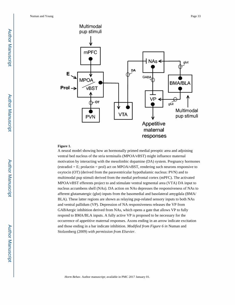
Figure 1. A neural model showing how an hormonally primed medial preoptic area and adjoining
ventral bed nucleus of the stria terminalis (MPOA/vBST) might influence maternal
motivation by interacting with the mesolimbic dopamine (DA) system. Pregnancy hormones
(estradiol = E; prolactin = prol) act on MPOA/vBST, rendering such neurons responsive to
oxytocin (OT) (derived from the paraventricular hypothalamic nucleus: PVN) and to
multimodal pup stimuli derived from the medial prefrontal cortex (mPFC). The activated
MPOA/vBST efferents project to and stimulate ventral tegmental area (VTA) DA input to
nucleus accumbens shell (NAs). DA action on NAs depresses the responsiveness of NAs to
afferent glutamatergic (glut) inputs from the basomedial and basolateral amygdala (BMA/
BLA). These latter regions are shown as relaying pup-related sensory inputs to both NAs
and ventral pallidum (VP). Depression of NA responsiveness releases the VP from
GABAergic inhibition derived from NAs, which opens a gate that allows VP to fully
respond to BMA/BLA inputs. A fully active VP is proposed to be necessary for the
occurrence of appetitive maternal responses. Axons ending in an arrow indicate excitation
and those ending in a bar indicate inhibition. Modified from Figure 6 in Numan and
Stolzenberg (2009) with permission from Elsevier.
Numan and Young Page 33
Horm Behav. Author manuscript; available in PMC 2017 January 01.
Author M
anuscriptA
uthor Manuscript
Author M
anuscriptA
uthor Manuscript

Figure 2. Dopamine action on D1 and D2 receptors (A), and oxytocin action on oxytocin receptors
(B), in the nucleus accumbens shell (NAs) are involved in the establishment of maternal
memory in rats. Primiparous rats are allowed one hour of postpartum experience with their
young, after which the pups are removed. Immediately thereafter, various neurochemicals
are microinjected into NAs in different groups of rats. Ten days later, the rats are re-exposed
to pups and sensitization latencies are measured. As shown in A, blockade of both D1 and
D2 receptors in NAs with flupenthixol disrupts maternal memory formation (Parada et al.,
2008). As shown in B, injection of an oxytocin receptor antagonist (OTA) into NAs also
blocks maternal memory formation (D'Cunha et al., 2011). Other abbreviations: CSF =
artificial cerebrospinal fluid. In comparing these findings with the development of pair
bonds in prairie voles, as shown in Fig. 5, a crucial similarity is that DA action on D2
receptors and OT action on OT receptors within NA are involved in both types of bond
formation. Reproduced from Figure 5.16 in Numan (2015) with permission from Elsevier.
Numan and Young Page 34
Horm Behav. Author manuscript; available in PMC 2017 January 01.
Author M
anuscriptA
uthor Manuscript
Author M
anuscriptA
uthor Manuscript
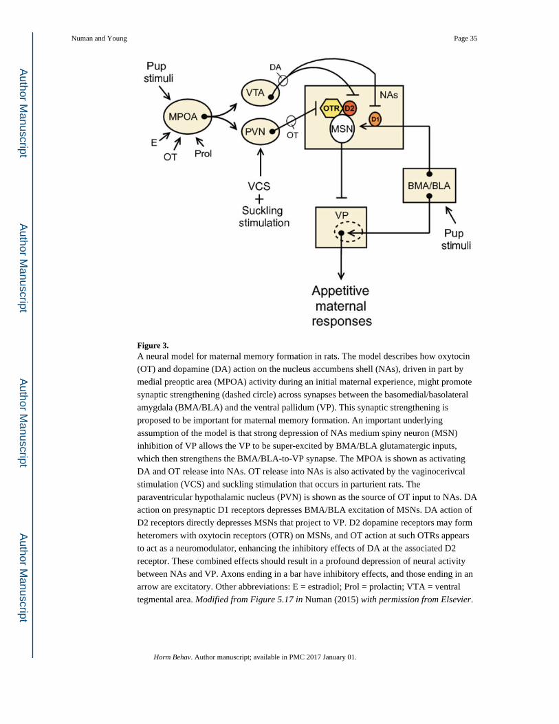
Figure 3. A neural model for maternal memory formation in rats. The model describes how oxytocin
(OT) and dopamine (DA) action on the nucleus accumbens shell (NAs), driven in part by
medial preoptic area (MPOA) activity during an initial maternal experience, might promote
synaptic strengthening (dashed circle) across synapses between the basomedial/basolateral
amygdala (BMA/BLA) and the ventral pallidum (VP). This synaptic strengthening is
proposed to be important for maternal memory formation. An important underlying
assumption of the model is that strong depression of NAs medium spiny neuron (MSN)
inhibition of VP allows the VP to be super-excited by BMA/BLA glutamatergic inputs,
which then strengthens the BMA/BLA-to-VP synapse. The MPOA is shown as activating
DA and OT release into NAs. OT release into NAs is also activated by the vaginocerivcal
stimulation (VCS) and suckling stimulation that occurs in parturient rats. The
paraventricular hypothalamic nucleus (PVN) is shown as the source of OT input to NAs. DA
action on presynaptic D1 receptors depresses BMA/BLA excitation of MSNs. DA action of
D2 receptors directly depresses MSNs that project to VP. D2 dopamine receptors may form
heteromers with oxytocin receptors (OTR) on MSNs, and OT action at such OTRs appears
to act as a neuromodulator, enhancing the inhibitory effects of DA at the associated D2
receptor. These combined effects should result in a profound depression of neural activity
between NAs and VP. Axons ending in a bar have inhibitory effects, and those ending in an
arrow are excitatory. Other abbreviations: E = estradiol; Prol = prolactin; VTA = ventral
tegmental area. Modified from Figure 5.17 in Numan (2015) with permission from Elsevier.
Numan and Young Page 35
Horm Behav. Author manuscript; available in PMC 2017 January 01.
Author M
anuscriptA
uthor Manuscript
Author M
anuscriptA
uthor Manuscript
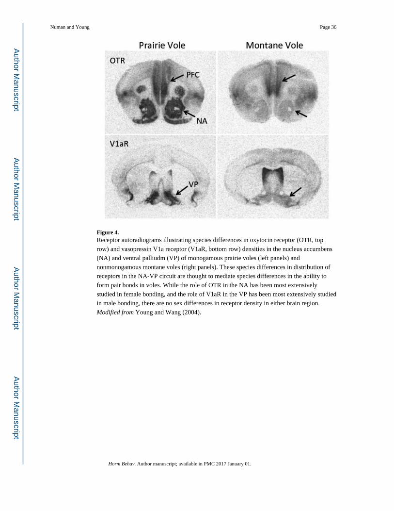
Figure 4. Receptor autoradiograms illustrating species differences in oxytocin receptor (OTR, top
row) and vasopressin V1a receptor (V1aR, bottom row) densities in the nucleus accumbens
(NA) and ventral palliudm (VP) of monogamous prairie voles (left panels) and
nonmonogamous montane voles (right panels). These species differences in distribution of
receptors in the NA-VP circuit are thought to mediate species differences in the ability to
form pair bonds in voles. While the role of OTR in the NA has been most extensively
studied in female bonding, and the role of V1aR in the VP has been most extensively studied
in male bonding, there are no sex differences in receptor density in either brain region.
Modified from Young and Wang (2004).
Numan and Young Page 36
Horm Behav. Author manuscript; available in PMC 2017 January 01.
Author M
anuscriptA
uthor Manuscript
Author M
anuscriptA
uthor Manuscript
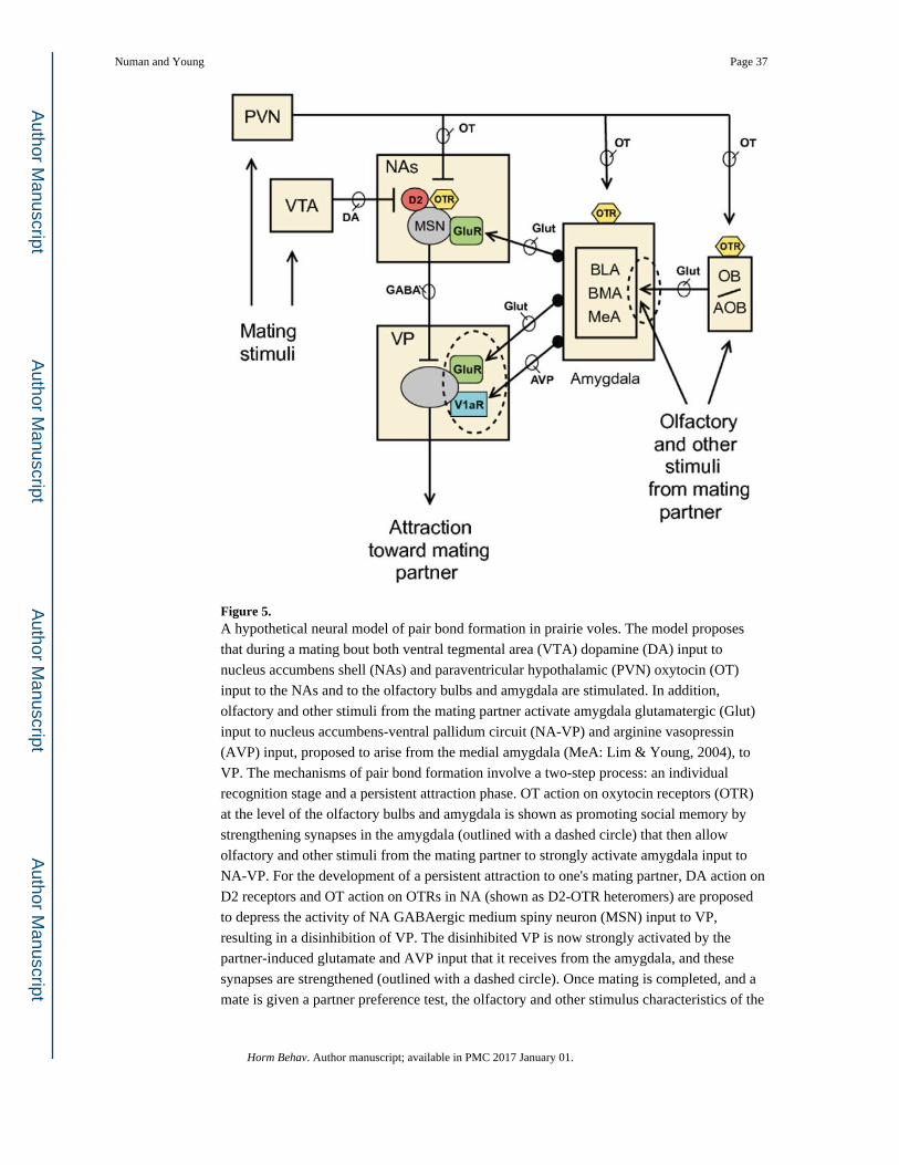
Figure 5. A hypothetical neural model of pair bond formation in prairie voles. The model proposes
that during a mating bout both ventral tegmental area (VTA) dopamine (DA) input to
nucleus accumbens shell (NAs) and paraventricular hypothalamic (PVN) oxytocin (OT)
input to the NAs and to the olfactory bulbs and amygdala are stimulated. In addition,
olfactory and other stimuli from the mating partner activate amygdala glutamatergic (Glut)
input to nucleus accumbens-ventral pallidum circuit (NA-VP) and arginine vasopressin
(AVP) input, proposed to arise from the medial amygdala (MeA: Lim & Young, 2004), to
VP. The mechanisms of pair bond formation involve a two-step process: an individual
recognition stage and a persistent attraction phase. OT action on oxytocin receptors (OTR)
at the level of the olfactory bulbs and amygdala is shown as promoting social memory by
strengthening synapses in the amygdala (outlined with a dashed circle) that then allow
olfactory and other stimuli from the mating partner to strongly activate amygdala input to
NA-VP. For the development of a persistent attraction to one's mating partner, DA action on
D2 receptors and OT action on OTRs in NA (shown as D2-OTR heteromers) are proposed
to depress the activity of NA GABAergic medium spiny neuron (MSN) input to VP,
resulting in a disinhibition of VP. The disinhibited VP is now strongly activated by the
partner-induced glutamate and AVP input that it receives from the amygdala, and these
synapses are strengthened (outlined with a dashed circle). Once mating is completed, and a
mate is given a partner preference test, the olfactory and other stimulus characteristics of the
Numan and Young Page 37
Horm Behav. Author manuscript; available in PMC 2017 January 01.
Author M
anuscriptA
uthor Manuscript
Author M
anuscriptA
uthor Manuscript

partner, but not those of a stranger, are now capable of strongly activating VP over long
periods of time, creating an enduring and selective social bond. In fact, stranger-related
stimuli activate AVP release into anterior hypothalamus (not shown) leading to aggression
and rejection. Mechanisms similar to these may be involved in maternal bonding and
selectivity in sheep. Note that although OT typically has excitatory effects in neurons
(Terenzi & Ingram, 2005), when it acts at sites composed of D2-OTR heteromers, it
functions to potentiate the inhibitory effects of DA at the D2 receptor. In order to simplify
the diagram and show its similarities with Fig. 3, the presence of D1 receptors on NA MSNs
is not shown although, as indicated in the text, DA action at this site suppresses pair bond
formation. Perhaps OT not only suppresses the output of MSNs with D2 receptors, but also
those with D1 receptors (see Numan, 2015). Other abbreviations: AOB = accessory
olfactory bulb; BLA = basolateral amygdala; BMA = basomedial amygdala; GluR =
glutamate receptor; OB = olfactory bulb; V1aR = vasopressin V1a receptor. Modified from
Figures 6.2 and 6.3 in Numan (2015) with permission from Elsevier.
Numan and Young Page 38
Horm Behav. Author manuscript; available in PMC 2017 January 01.
Author M
anuscriptA
uthor Manuscript
Author M
anuscriptA
uthor Manuscript