neural plate morphogenesis during mouse neurulation is...
TRANSCRIPT
DEVELO
PMENT
3203DEVELOPMENT AND DISEASE RESEARCH ARTICLE
INTRODUCTIONNeurulation is the process in which the neural plate bends and fusesto form the neural tube – the developmental forerunner of the brainand spinal cord. Understanding the molecular regulation ofneurulation is significant not only because of its pivotal importancein establishing the CNS primordium, but also because defects ofneurulation result in clinically important congenital malformations,termed neural tube defects (NTDs). Failure of brain closure(anencephaly) and low spinal closure (open spina bifida) occur witha high prevalence in humans, around 1 per 1000 pregnancies(Mitchell, 2005), and form part of the phenotype of over 100 mutantmouse strains (Copp et al., 2003).
Mouse primary neurulation is characterised by a stereotypicalpattern of neural plate bending along the spinal neuraxis (Shum andCopp, 1996). Neural tube closure begins at the level of thecervical/hindbrain boundary at embryonic day (E)8.5 (6-somitestage), and finishes at E10.5 (30-somite stage) when closure iscompleted at the upper sacral level. In the intervening 48-hour period,a wave of neural tube closure propagates in a cranio-caudal directiondown the spine. Just caudal to the closure propagation front, a regionof open, elevating neural folds comprises the ‘posterior neuropore’(PNP). This represents the next axial region to undergo neural tubeclosure (Van Straaten et al., 1992). Previously, we divided thecontinuous process of spinal neurulation into three modes, according
to the morphology of neural plate bending within the PNP (Shum andCopp, 1996). In mode 1 (E8.5-E9), the closing neural plate has a V-shaped cross section, with bending solely at the median hinge point(MHP), overlying the notochord. As neurulation progresses to lowerspinal levels, mode 2 (E9-E9.75) becomes recognisable, in which theclosing neural folds adopt a different morphology with paireddorsolateral hinge points (DLHPs) in addition to the MHP. At themost-caudal level of the spinal axis, just prior to completion of spinalneurulation, MHP bending disappears and the neural plate bendssolely at the DLHPs (mode 3; E9.75-E10.5).
Although the cell shape changes that comprise bending of theneuroepithelium at MHP and DLHPs have been documented(Schoenwolf, 1985; Smith et al., 1994), the identity of thedorsoventral molecular signals that regulate these cell shape changesremains unknown. Previously, we and others showed that MHPbending requires the influence of the adjacent notochord:suppression of notochordal development results in the absence ofmidline bending (Davidson et al., 1999; Smith and Schoenwolf,1989; Ybot-Gonzalez et al., 2002). In an analogous way, removal ofthe surface ectoderm, which normally covers the outer aspect of thedorsal neural fold, results in the absence of DLHPs, whereas just asmall surface ectodermal remnant is capable of inducing a DLHP(Jacobson and Moury, 1995; Moury and Schoenwolf, 1995; Ybot-Gonzalez et al., 2002). Hence, signals from the notochord andsurface ectoderm are required for MHP and DLHP formation,respectively. By contrast, the paraxial mesoderm can be removedwithout influencing MHP or DLHP formation (Ybot-Gonzalez etal., 2002). Further studies have demonstrated a negative influence ofShh, emanating from the notochord, on the presence of DLHPs. Shhis both necessary and sufficient to inhibit DLHP formation, asdemonstrated by the occurrence of DLHPs at an abnormally rostrallevel in Shh–/– mice, and the inhibition of DLHP formation by beadsreleasing N-terminal Shh (Shh-N) peptide implanted adjacent to thedorsolateral neural plate at low spinal levels (Ybot-Gonzalez et al.,2002).
Neural plate morphogenesis during mouse neurulation isregulated by antagonism of Bmp signallingPatricia Ybot-Gonzalez1,*, Carles Gaston-Massuet1, Gemma Girdler1,†, John Klingensmith2, Ruth Arkell3,Nicholas D. E. Greene1 and Andrew J. Copp1,*
Dorsolateral bending of the neural plate, an undifferentiated pseudostratified epithelium, is essential for neural tube closure in themouse spinal region. If dorsolateral bending fails, spina bifida results. In the present study, we investigated the molecular signalsthat regulate the formation of dorsolateral hinge points (DLHPs). We show that Bmp2 expression correlates with upper spinalneurulation (in which DLHPs are absent); that Bmp2-null embryos exhibit premature, exaggerated DLHPs; and that the local releaseof Bmp2 inhibits neural fold bending. Therefore, Bmp signalling is necessary and sufficient to inhibit DLHPs. By contrast, the Bmpantagonist noggin is expressed dorsally in neural folds containing DLHPs, noggin-null embryos show markedly reduced dorsolateralbending and local release of noggin stimulates bending. Hence, Bmp antagonism is both necessary and sufficient to inducedorsolateral bending. The local release of Shh suppresses dorsal noggin expression, explaining the absence of DLHPs at high spinallevels, where notochordal expression of Shh is strong. DLHPs ‘break through’ at low spinal levels, where Shh expression is weaker.Zic2 mutant embryos fail to express Bmp antagonists dorsally and lack DLHPs, developing severe spina bifida. Our findings reveal amolecular mechanism based on antagonism of Bmp signalling that underlies the regulation of DLHP formation during mouse spinalneural tube closure.
KEY WORDS: Neurulation, Morphogenesis, Neural tube defects, Noggin, Sonic hedgehog, Mouse, Zic genes
Development 134, 3203-3211 (2007) doi:10.1242/dev.008177
1Neural Development Unit, Institute of Child Health, University College London, 30Guilford Street, London WC1N 1EH, UK. 2Department of Cell Biology, DukeUniversity Medical Center, Durham, NC 27710, USA. 3Molecular Genetics andEvolution Group, Research School of Biological Sciences, Australian NationalUniversity, Canberra ACT 0200, Australia.
*Authors for correspondence (e-mails: [email protected];[email protected])†Present address: Department of Anatomy and Developmental Biology, UniversityCollege London, Gower Street, London, WC1E 6BT, UK
Accepted 27 June 2007

DEVELO
PMENT
3204
Here, we identify the molecular interactions regulatingdorsolateral bending during mouse neurulation. Analysis of embryoslacking function of Bmp2 or noggin, and of wild-type embryosexposed to the local release of Bmp2 and noggin peptides, identifiesBmp2 as an inhibitor of DLHP formation. By contrast, noggin andprobably another Bmp antagonist, neuralin (also called chordin-like1), induce DLHP formation. DLHPs are restricted to low levels ofthe spinal neuraxis because, at upper levels, Shh concentrations arehigh and noggin expression is inhibited. DLHPs are absent fromhomozygous Zic2Ku embryos, which later develop severe spinabifida owing to the absence of Bmp antagonists in the dorsal neuralplate. Hence, this study reveals a molecular mechanism of regulationof neural tube closure, based on inhibition of Bmp signalling, in thespinal region of the mouse embryo.
MATERIALS AND METHODSMouse strains, embryo culture and insertion of beadsNon-mutant embryos were obtained from CD1 random-bred mice. Micewith mutations in Bmp2 (Zhang and Bradley, 1996), noggin (McMahon etal., 1998), Shh (Chiang et al., 1996) and Zic2 (Elms et al., 2003) weregenotyped as described previously. Whole-embryo culture was performedas described (Copp et al., 1999), with opening of the yolk sac and amnion toa minimum extent compatible with access to the PNP region for beadinsertion. AffiGel blue beads (BioRad, Cat. No. 153-7302) were soaked forat least 2 hours at 4°C in 0.5 �g/�l Bmp2 (R&D Systems), 1 �g/�l noggin(R&D Systems), 1 �g/�l Shh-N peptide (R&D Systems) or in PBS as avehicle control. Beads were held by suction on the end of a mouth-controlledglass micropipette, inserted singly or in pairs through a slit in the surfaceectoderm overlying the neural fold and positioned as closely as possible tothe dorsolateral neural fold region. Following a variable period in culture,depending on the experiment, embryos were removed from theirextraembryonic membranes, rinsed in PBS, and fixed in either Bouin’s fluidfor haematoxylin and eosin (H&E) staining, 4% paraformaldehyde (PFA) inPBS for in situ hybridisation, and either PFA or Sainte-Marie’s fixative (95%ethanol, 1% acetic acid) for immunohistochemistry. Presence or absence ofa DLHP was determined in transverse embryo sections using anglemeasurement criteria as described previously (Ybot-Gonzalez et al., 2002).
In situ hybridisationPreviously published probes were: Bmp2, Bmp4 and Bmp7 (Furuta et al.,1997), cadherin 6 (Henderson et al., 1997), chordin (Klingensmith et al.,1999), Msx1 (Mackenzie et al., 1991), Msx2 (Monaghan et al., 1991),neuralin (Coffinier et al., 2001) and Zic2 (Gaston-Massuet et al., 2005).Additional Msx1 and Msx2 probes were as described (Catron et al., 1996).To prepare a cDNA probe for noggin, forward 5�-CCAGCACT AT -CTACACATCC-3� and reverse 5�-ACTTGGATGGCTTACACACC-3�primers were used to amplify a 518 bp fragment corresponding tonucleotides 327-845 of the noggin cDNA sequence (GenBank accessionnumber u79163). Reverse transcriptase (RT)-PCR was performed on totalRNA from E10.5 CBA/Ca embryos using TRIzol reagent (Gibco BRL). Theamplified fragment was cloned into the pGEM-T vector (Promega, UK) andsequenced to confirm its identity. Whole-mount in situ hybridisation withpreparation of 50 �m transverse vibratome sections was as described (Coppet al., 1999). In situ hybridisation on paraffin-embedded sections wasperformed using digoxigenin-labelled cRNA probes (Breitschopf et al.,1992). Sense-strand cRNA probes were tested for all genes with no specifichybridisation.
Immunohistochemistry, cell counting and statistical analysisFixed embryos were dehydrated, embedded in paraffin wax and sectionedat 7 �m. Sections were rehydrated and antigen retrieval was performed usingDeclere (Cell Marque). Antibodies to the phosphorylated forms of Smad1,Smad5 and Smad8 combined (phospho-Smad1,5,8; dilution 1:100; CellSignalling Technology); caspase 3 (dilution 1:1000; Cell SignalingTechnology); phospho-histone H3 (dilution 1:250; Upstate Biotechnology);noggin (dilution 1:7; R&D systems); and Bmp2 (dilution 1:10; R&DSystems) were diluted in 5% goat or rabbit serum, 0.15% glycine and 2
mg/ml BSA in Tris-buffered saline. Primary antibodies were detected witha biotinylated goat anti-rabbit or biotinylated rabbit anti-goat (both 1:250,DAKO), using a Vectastain ABC kit (Vector) and diaminobenzidine(peroxidase substrate kit DAB, Vector). Omission of primary antibodyserved as a negative control. Embryos for comparison of combined phospho-Smad1, -Smad5 and -Smad8 staining were processed together in the sameparaffin block and on the same slide, to enable accurate comparison.Sections were counter-stained with methylene green. For analysis ofapoptotic cell frequency, the number of cells positive for caspase 3 wasdetermined in the dorsal and ventral halves of the neural plate in 7-12sections through the PNP of three to four embryos at each of modes 1 and 3.The mean number of apoptotic cells per section was compared by two-wayanalysis of variance, using neurulation mode and neural plate region asvariables. For analysis of cell proliferation, cells positive for phospho-histone H3 were counted in the dorsal two thirds of the neural plate in fivesections of three embryos at each of modes 1 and 3. The mean number ofmitotic cells per section was compared by t-test.
Scanning electron microscopyEmbryos were rinsed in PBS and fixed overnight in 2% glutaraldehyde, 2%PFA in PBS. Tissues were rinsed in phosphate buffer and post-fixed in 1%OsO4 for 1 hour. Samples were dehydrated through an ascending alcoholseries, using three changes of acetone to displace the alcohol, then CO2
critical point dried, mounted on specimen stubs, gold sputter-coated andexamined in a JEOL SEM 5410 LV scanning electron microscope.
RESULTSIn order to investigate the molecular regulation of DLHP formation,we first examined the expression of genes in the Bmp signallingpathway, because our previous studies had suggested a possible rolefor Bmp2 in this process (Ybot-Gonzalez et al., 2002).
Diminished Bmp signalling in association withDLHP formationWe compared mode 1, in which DLHPs are absent, with mode 3, inwhich DLHPs are present (Fig. 1A). Bmp2 and Bmp7 mRNAtranscripts occurred with similar intensity in the surface ectodermoverlying the spinal neural folds of mode 1 (Fig. 1B,D) and mode 3(Fig. 1C,E and data not shown) embryos. Bmp2 was localisedspecifically to the dorsal-most ectoderm, whereas Bmp7 wasexpressed throughout the surface ectoderm at this axial level. Bmp4,Bmp5 and Bmp6 were expressed in more-ventral or anteriorembryonic regions, but not in the vicinity of the neural folds (datanot shown). In the absence of any apparent difference in Bmp2 orBmp7 expression between modes 1 and 3, we questioned whetherthe presence or absence of DLHPs might be associated withdifferences in the activity of downstream Bmp signalling. Cadherin6, a gene regulated by Bmp signalling (Sela-Donenfeld andKalcheim, 1999), was expressed intensely throughout the neuralplate in mode 1 (Fig. 1F), but appeared downregulated at mode 3(Fig. 1G and data not shown). By contrast, Msx1, which is alsoregulated by Bmps in some systems, was not detectable in the neuralfolds of mode 1 embryos (Fig. 1H), and was only weakly expressedat the neural fold tips in mode 3 (Fig. 1I and data not shown). Msx2was not detected in the PNP region (data not shown).Immunohistochemistry for phospho-Smad1,5,8, which areimmediately downstream of Bmp receptor activation (Massague andWotton, 2000), revealed markedly stronger expression in the dorsalneural folds at mode 1 than at mode 3 (Fig. 1J,K), consistent withthe pattern of cadherin 6 expression. It seems, therefore, that,although Bmp gene expression per se does not differ along theneuraxis, downstream Bmp signalling is strongest in locations whereDLHPs are absent. This suggests an inhibitory effect of Bmps onneural plate bending.
RESEARCH ARTICLE Development 134 (17)

DEVELO
PMENT
Next, we studied whether cell death or cell proliferation differin the neural plate of mode 1 and mode 3 embryos.Immunohistochemistry for activated caspase 3 revealedconsiderable numbers of dying cells dorsally in the neural foldsof mode 1, whereas cell death was only rarely observed in themode 3 neural plate (Fig. 1L,M). The incidence of cell death wassignificantly increased in the dorsal region of the mode 1 neuralplate (2.6±0.5 cells positive for caspase 3 per section) comparedwith the ventral region at mode 1 (0.3±0.1 cells per section;P<0.05), the dorsal region at mode 3 (0.4±0.01 cells per section;P<0.05) and the ventral region at mode 3 (0.2±0.1 cellsper section; P<0.05). By contrast, phospho-histone H3immunostaining showed no difference in the frequency ofproliferating cells in the dorsal two thirds of the neural plate atmode 1 and 3 (Fig. 1N,O; mean number of H3-positive cells:1.5±0.2 for mode 1; 1.2±0.1 for mode 3; P>0.05). Hence,programmed cell death, which is associated with active Bmpsignalling in the early embryonic hindbrain (Graham et al., 1994;Yokouchi et al., 1996; Jernvall et al., 1998), also appears tocorrelate specifically with mode 1 neurulation in the mouseembryo, consistent with the idea that, at mode 1, Bmp signallingis strongest in the dorsal neural plate, in which DLHPs are absent.
Bmp2 is necessary and sufficient to inhibitdorsolateral neural plate bendingIn order to examine the causal relationship between Bmp signallingand dorsolateral neural plate bending, we compared the spinalneural folds of wild-type embryos with those of embryos
homozygous for a null mutation in Bmp2. Whereas Bmp2–/–
embryos die at around mid-gestation (Zhang and Bradley, 1996),we found that neural fold morphology at the PNP was clearlyidentifiable in both E8.5 and E9.5 embryos. At the 7-somite stage,immediately after neural tube closure was initiated, Bmp2–/–
embryos exhibited mode 1 neurulation (n=5), as did wild-typelittermates (Fig. 2A,B). Just a few hours later, at the 9-somite stage,whereas wild-type embryos continued in mode 1 (Fig. 2C), allBmp2–/– embryos studied (n=7) were found to exhibit strikingDLHPs, indicating premature entry into mode 2/3 (Fig. 2D). Thisis despite the growth retardation that characterises null embryos atthis stage (Zhang and Bradley, 1996). At 15 somites, the latest stagethat could be examined owing to their imminent demise, Bmp2–/–
embryos (n=4) exhibited an entirely closed spinal neural tube,consistent with the premature formation of DLHPs (Fig. 2F) (Yipet al., 2002). By contrast, wild-type littermates with 15 somiteswere in mode 2, with prominent DLHPs (Fig. 2E). We concludethat Bmp2 is necessary for the inhibition of DLHPs early in mousespinal neurulation and that, in its absence, null embryos showpremature, exaggerated formation of DLHPs.
To determine whether Bmp2 is sufficient for the inhibition ofDLHP formation, we implanted AffiGel blue beads, soaked in eitherBmp2 or PBS, within the presomitic mesoderm adjacent to onespinal neural fold, in wild-type embryos at either E8.5 (mode 1) orE9.5 (mode 2/3). After 5 hours culture, embryos were harvested andsectioned transversely to determine whether DLHPs had beeninduced (at mode 1, when DLHPs are normally absent) or inhibited(at mode 2/3, when DLHPs are normally present). Local release of
3205RESEARCH ARTICLEBmp signalling and neural tube closure
Fig. 1. Bmp signalling during mouse spinal neurulation.(A) Diagrammatic transverse sections through the PNP ofembryos at modes 1 (E8.5-E9), 2 (E9-E9.75) and 3 (E9.75-E10.5) of spinal neurulation. Yellow triangles, median hingepoint (MHP); red triangles, dorsolateral hinge points (DLHPs).(B-I) In situ hybridisation for Bmp2 (B,C), Bmp7 (D,E), cadherin6 (F,G) and Msx1 (H,I) during mode 1 (E8.5; B,D,F,H) and mode3 (E9.5; C,E,G,I) neurulation as seen in transverse sectionsthrough the posterior neuropore (PNP) of whole-mountembryos. Arrows show intense in situ hybridisation signal.(J-O) Immunohistochemistry for phospho-Smad1, -Smad5 and-Smad 8 combined (phospho-Smad1,5,8; J,K); activatedcaspase 3 (L,M); and phospho-histone H3 (N,O) during mode 1(J,L,N) and mode 3 (K,M,O) neurulation. (J) Arrows indicatehigh levels of phospho-Smad1,5,8 in dorsal neural folds.(L,M) Caspase 3-positive cells were accumulated over tenconsecutive sections of the same embryo and projected onto asingle section. (L) Arrow indicates intensive apoptosis in thedorsal neural fold at mode 1. (N,O; insets) Controls with theomission of the primary antibody. (P,Q) Diagrammatic right-sided view (P) of the PNP at mode 3. Transverse section (Q) atthe level of the dotted line in P summarises the embryonictissues that are visible in C,E,G,I,K,M,O: hg, hindgut; mes,mesoderm; np, neural plate; no, notochord; se, surfaceectoderm; ver, ventral ectoderm ridge. Scale bars: 0.1 mm in B(also C-I), in K (also J), in M (also L), in O (also N) and in inset O(also inset N).

DEVELO
PMENT
3206
either Bmp2 or PBS had no discernible effect on the mode 1 neuralfold (Fig. 2G,I,K,M), whereas, in the mode 2/3 neuropore, Bmp2inhibited dorsolateral bending in 76% of cases (n=25; Fig. 3Q)compared with PBS beads, which had no effect (compare Fig. 2H,Lwith Fig. 2J,N). Immunohistochemistry on sections of embryosimplanted with Bmp2 beads showed a striking upregulation ofphospho-Smad1,5,8 in tissues on the same side as the bead, but notcontralaterally (Fig. 2K,L). By contrast, embryos with implantedPBS beads demonstrated only background Smad staining (Fig.2M,N). Hence, local release of Bmp2 stimulates downstreamsignalling and is sufficient to inhibit dorsolateral bending of theneural plate in mouse spinal neurulation.
Noggin is necessary and sufficient for theinduction of DLHPsIn view of the finding that both Bmp2 dorsally (Fig. 2), and Shhventrally (Ybot-Gonzalez et al., 2002), are necessary and sufficientto inhibit DLHPs, we next considered the hypothesis that Bmpantagonists including noggin, neuralin (Coffinier et al., 2001) andchordin might be in vivo inducers of dorsolateral bending duringmouse neurulation. In situ hybridisation showed that neuralin wasexpressed in the neural plate at modes 1, 2 and 3 of spinalneurulation (Fig. 3A,B and data not shown), whereas chordin wasexpressed solely in the notochord (Fig. 3C,D and data not shown).Noggin was expressed in the notochord, but also in the neural foldtips, immediately underlying the Bmp2- and Bmp7-positive surfaceectoderm. Strikingly, in mode 1, noggin was expressed most-intensely in the notochord, but only weakly at the neural fold tips(Fig. 3E), whereas, in mode 3, the reverse was true: noggintranscripts were intense at the neural fold tips but weak in thenotochord (Fig. 3F and data not shown). Mode 2 gave anintermediate result, in which noggin expression was strong in bothneural folds tips and notochord (Fig. 3E,F, inset). Neuralin was alsoexpressed more-intensely at the tips of the neural folds in mode 3than it was in mode 1, and also had an intermediate appearance inmode 2 (Fig. 3A,B). Hence, the dorsal neural plate expression ofnoggin and neuralin correlates with the formation of DLHPs atmode 3.
To test whether Bmp antagonism is necessary for DLHPformation, we examined the PNP region of embryos homozygousfor a loss-of-function allele of noggin, in the presence of which spinabifida is observed at high frequency (McMahon et al., 1998;Stottmann et al., 2006). On this genetic background, normallittermates displayed prominent DLHPs at mode 2 (17- to 18-somitestage), whereas stage-matched noggin–/– (Nog–/–) embryos exhibitedmarkedly reduced dorsolateral bending, with neural platemorphology closely resembling mode 1 (Fig. 3G,H). This finding isthe opposite of that observed with Bmp2–/– embryos and is consistentwith the idea that noggin is required for dorsolateral bending duringPNP development.
To determine whether noggin is sufficient to induce DLHPformation, we inserted noggin-soaked beads adjacent to the neuralfold in wild-type embryos. After 4-5 hours culture, in 43% of cases(n=58; Fig. 3Q), an ectopic DLHP was observed in the mode 1neural plate (Fig. 3I,K,M), whereas noggin beads had nodiscernible effect on mode 2/3 neural folds (Fig. 3J,L,O).Immunohistochemistry for phospho-Smad1,5,8 confirmed that thelocal release of noggin markedly diminished downstream Bmpactivation (Fig. 3M-P), although with variation between embryos. Itis possible that this variable inhibition of phospho-Smad signallingby exogenous noggin can explain why noggin is not able to inductDLHPs in all bead-implanted embryos. We conclude that noggin isboth necessary and sufficient to induce dorsolateral bending in mode1 neural plate during mouse spinal neurulation.
Interestingly, exogenous noggin induced a contralateral DLHP insome embryos (13/58; Fig. 3Q), although it more frequentlyproduced an ipsilateral DLHP (16/58; Fig. 3I,K,M). By contrast, thelocal release of Bmp2 inhibited DLHP formation and activateddownstream Smad signalling solely on the same side as theimplanted bead. Because the release of peptides from implantedbeads is difficult to quantitate, we cannot rule out differential loadingor diffusion of noggin and Bmp2 as an explanation for thisobservation. Alternatively, the strong expression of the Bmpantagonist chordin in the notochord (Fig. 3C,D) might serve toneutralise Bmp2 diffusing to the midline, whereas noggin is able tocross the midline unopposed.
RESEARCH ARTICLE Development 134 (17)
Fig. 2. Bmp2 is necessary and sufficient to inhibitDLHPs. (A-F) Transverse H&E-stained sections through theposterior neuropore (PNP) region of E8.5 (A-D) and E9 (E,F)embryos. At 7 somites (7 s), when closure is first initiated,dorsolateral hinge points (DLHPs) are absent from wild-type(WT, A) and Bmp2–/– (B) embryos. Whereas wild-typeembryos at 9 somites continue in mode 1 neurulation (C),Bmp2–/– embryos show marked DLHPs, characteristic ofmodes 2/3 (arrows in D). At 15 somites, wild-type embryoshave entered mode 2 neurulation with clear DLHPs (E),whereas 15-somite Bmp2–/– embryos have undergonepremature closure of the neural tube (F). (G-N) Transversesections through the PNP of mode 1 (G,I,K,M) and mode 2/3(H,J,L,N) embryos stained with H&E (G-J) or afterimmunohistochemistry for phospho-Smad1, -Smad5 and-Smad8 combined (K-N). A bead soaked in Bmp2 (G,H,K,L)or PBS (I,J,M,N) has been implanted next to one neural fold.Exposure to Bmp2 inhibits DLHP formation in mode 2/3(arrows in H,L), whereas DLHPs remain in PBS-treated controlembryos (arrowheads in J,N). Note that the period ofphospho-Smad staining was longer in PBS control embryos(M,N) than in those implanted with a Bmp2 bead (K,L) inorder to reveal endogenous expression, as a positive control.Scale bars: 0.1 mm in F (also A-E); 0.1 mm in J (also G-I) andN (also K-M).

DEVELO
PMENT
Regulation of noggin expression by Bmp2 andShh during spinal neurulationThe analysis of Bmp2- and noggin-null embryos, and of wild-typeembryos implanted with Bmp2 and noggin beads, suggests amechanism in which Bmp signalling negatively regulates DLHPformation, with alleviation of this inhibition by noggin. At mode 1,noggin is expressed at low intensity in the dorsal neural plate so thatBmp signalling is dominant, preventing DLHP formation. Bycontrast, at mode 2/3, noggin expression is upregulated, preventingBmp-mediated DLHP inhibition and enabling dorsolateral bendingto occur. Two further questions arise: first, how is noggin expressionregulated spatially, so that transcripts are found specifically at thetips of the neural folds? Second, how is noggin expression regulatedtemporally, so that noggin transcripts are more plentiful in the dorsalneural plate at mode 2/3 than at mode 1?
Our previous work demonstrated that surface ectodermattachment is required for the formation and maintenance of DLHPs(Ybot-Gonzalez et al., 2002). Perhaps Bmp2 and/or Bmp7 in thesurface ectoderm are responsible for inducing noggin expression,thereby explaining this dependence on surface ectoderm attachment.To investigate this possibility, we examined the expression of noggin
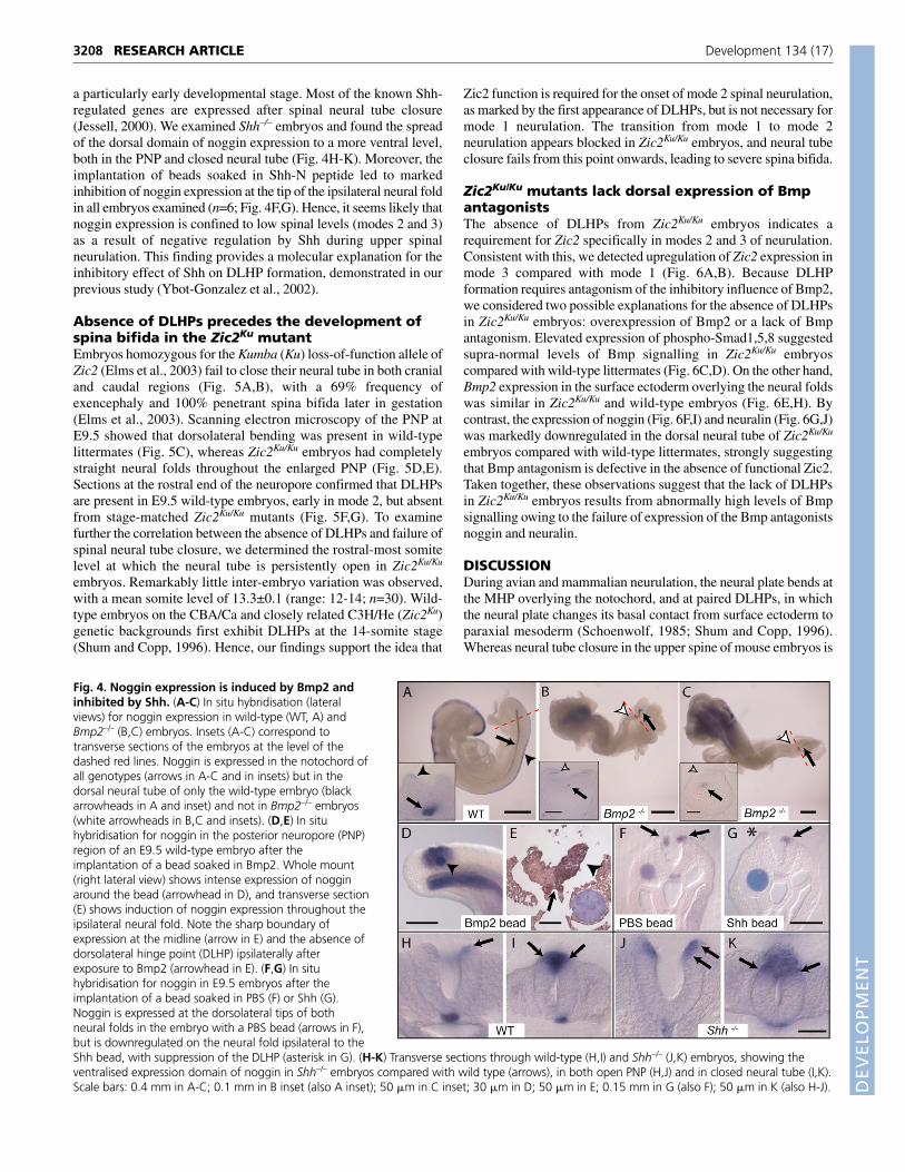
mRNA in Bmp2–/– embryos compared with non-mutant littermates.At all stages prior to the completion of PNP closure, at around the15-somite stage, we observed marked downregulation of nogginexpression in Bmp2–/– embryos, with a total absence of transcriptsfrom the dorsal neural plate (Fig. 4A-C). By contrast, notochordalexpression of noggin continued to be detected in Bmp2–/– embryos,although its expression was less-intense caudally. We also implantedBmp2 beads into wild-type embryos, and observed a massiveupregulation of noggin transcripts ipsilateral to the bead (Fig. 4D,E).Hence, dorsal neuroepithelial expression of noggin is stimulated byBmp2 from the overlying surface ectoderm, whereas notochordalnoggin seems likely to be regulated by factors other than Bmp2.
The expression of neither Bmp2 nor Bmp7 varied along the spinalaxis (Fig. 1B-E) suggesting that other factor(s) must be responsiblefor the temporal regulation of noggin expression from mode 1 to 3.We found previously that the strength of Shh signalling from thenotochord diminishes as the wave of spinal neurulation passes downthe body axis (Ybot-Gonzalez et al., 2002), raising the possibilitythat Shh might negatively regulate noggin expression in the dorsalneural plate. If confirmed, this would be an example of Shh-mediated dorsoventral regulation of neural tube gene expression at
3207RESEARCH ARTICLEBmp signalling and neural tube closure
Fig. 3. Antagonism of Bmps is necessary and sufficientto induce DLHPs. (A-F) In situ hybridisation for neuralin(A,B), chordin (C,D) and noggin (E,F) gene expression inmode 1 (A,C,E), mode 2 (insets in A-F) and mode 3 (B,D,F),as seen in transverse sections through the posteriorneuropore (PNP) of whole-mount embryos. Arrows indicatedetection of a strong signal in the dorsal neural plate ornotochord (B-F); arrowheads indicate detection of adiminished signal in the dorsal neural plate or notochord(E,F). (G,H) Transverse sections, stained with H&E, throughthe PNP of E9.5 Nog+/+ (G) and Nog–/– (H) embryos. Wild-type embryos show exaggerated dorsolateral hinge points(DLHPs; G, black arrows) whereas DLHPs in mutant embryosare absent (H, left white arrow) or markedly reduced (H,right white arrow). (I-P) Transverse sections through the PNPof mode 1 (I,K,M,N) and mode 2/3 (J,L,O,P) embryos afterthe implantation of beads soaked in noggin peptide.Sections were stained with H&E (I,J), or byimmunohistochemistry for noggin (K,L) or phospho-Smad1,5,8 (M-P). Arrows, induction of DLHPs ipsilaterally(I,K,M) after exposure to exogenous noggin. Notice thereduced phospho-Smad staining around the noggin beads(arrowheads in M,O) compared with slightly more-rostralsections of the same embryos (N,P), which were notexposed to exogenous noggin and show normalendogenous levels of phospho-Smad1,5,8. (Q) Summary ofexperiments showing the induction of DLHPs by noggin(mode 1, second column, asterisk) and inhibition of DLHPsby Bmp2 (mode 2/3, second column, asterisk). PBS:implantation of saline control beads. Percentages ofembryos with particular morphology are shown, togetherwith total number (n) receiving bead implants. Out of 25(43.1%) mode 1 embryos with noggin-induced DLHPs, 12had an ipsilateral DLHP, nine had a contralateral DLHP andfour had bilateral DLHPs. The DLHP was contralateral inboth of the affected PBS-treated mode 1 embryos. Thefrequency of DLHP induction in mode 1 is significantlygreater in noggin-treated embryos than in PBS controls(Fisher exact test; P=0.02). The frequency of DLHP inhibitionin mode 2/3 is significantly greater in Bmp2-treated embryosthan in PBS controls (P<0.001). Scale bars: 0.1 mm in B (alsoA,C-F and insets); 0.05 mm in H (also G); 0.1 mm in I (also J)in K, in L and in P (also M-O).
DEVELO
PMENT
3208
a particularly early developmental stage. Most of the known Shh-regulated genes are expressed after spinal neural tube closure(Jessell, 2000). We examined Shh–/– embryos and found the spreadof the dorsal domain of noggin expression to a more ventral level,both in the PNP and closed neural tube (Fig. 4H-K). Moreover, theimplantation of beads soaked in Shh-N peptide led to markedinhibition of noggin expression at the tip of the ipsilateral neural foldin all embryos examined (n=6; Fig. 4F,G). Hence, it seems likely thatnoggin expression is confined to low spinal levels (modes 2 and 3)as a result of negative regulation by Shh during upper spinalneurulation. This finding provides a molecular explanation for theinhibitory effect of Shh on DLHP formation, demonstrated in ourprevious study (Ybot-Gonzalez et al., 2002).
Absence of DLHPs precedes the development ofspina bifida in the Zic2Ku mutantEmbryos homozygous for the Kumba (Ku) loss-of-function allele ofZic2 (Elms et al., 2003) fail to close their neural tube in both cranialand caudal regions (Fig. 5A,B), with a 69% frequency ofexencephaly and 100% penetrant spina bifida later in gestation(Elms et al., 2003). Scanning electron microscopy of the PNP atE9.5 showed that dorsolateral bending was present in wild-typelittermates (Fig. 5C), whereas Zic2Ku/Ku embryos had completelystraight neural folds throughout the enlarged PNP (Fig. 5D,E).Sections at the rostral end of the neuropore confirmed that DLHPsare present in E9.5 wild-type embryos, early in mode 2, but absentfrom stage-matched Zic2Ku/Ku mutants (Fig. 5F,G). To examinefurther the correlation between the absence of DLHPs and failure ofspinal neural tube closure, we determined the rostral-most somitelevel at which the neural tube is persistently open in Zic2Ku/Ku
embryos. Remarkably little inter-embryo variation was observed,with a mean somite level of 13.3±0.1 (range: 12-14; n=30). Wild-type embryos on the CBA/Ca and closely related C3H/He (Zic2Ku)genetic backgrounds first exhibit DLHPs at the 14-somite stage(Shum and Copp, 1996). Hence, our findings support the idea that
Zic2 function is required for the onset of mode 2 spinal neurulation,as marked by the first appearance of DLHPs, but is not necessary formode 1 neurulation. The transition from mode 1 to mode 2neurulation appears blocked in Zic2Ku/Ku embryos, and neural tubeclosure fails from this point onwards, leading to severe spina bifida.
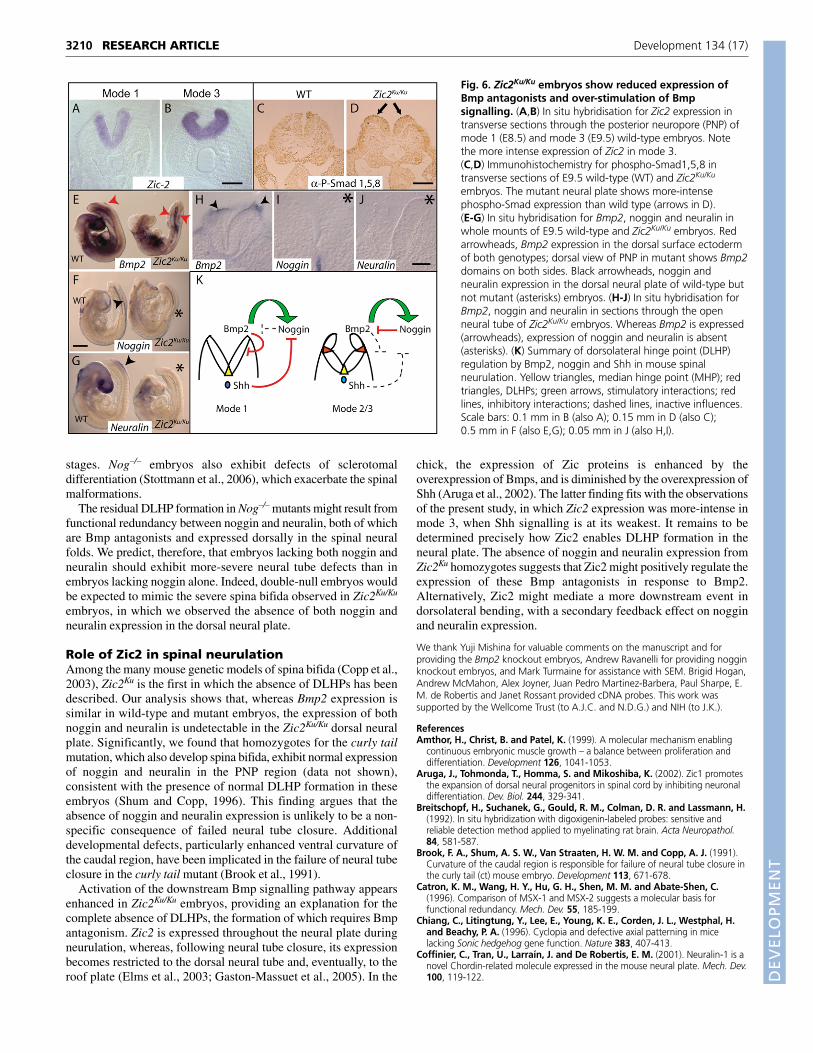
Zic2Ku/Ku mutants lack dorsal expression of BmpantagonistsThe absence of DLHPs from Zic2Ku/Ku embryos indicates arequirement for Zic2 specifically in modes 2 and 3 of neurulation.Consistent with this, we detected upregulation of Zic2 expression inmode 3 compared with mode 1 (Fig. 6A,B). Because DLHPformation requires antagonism of the inhibitory influence of Bmp2,we considered two possible explanations for the absence of DLHPsin Zic2Ku/Ku embryos: overexpression of Bmp2 or a lack of Bmpantagonism. Elevated expression of phospho-Smad1,5,8 suggestedsupra-normal levels of Bmp signalling in Zic2Ku/Ku embryoscompared with wild-type littermates (Fig. 6C,D). On the other hand,Bmp2 expression in the surface ectoderm overlying the neural foldswas similar in Zic2Ku/Ku and wild-type embryos (Fig. 6E,H). Bycontrast, the expression of noggin (Fig. 6F,I) and neuralin (Fig. 6G,J)was markedly downregulated in the dorsal neural tube of Zic2Ku/Ku
embryos compared with wild-type littermates, strongly suggestingthat Bmp antagonism is defective in the absence of functional Zic2.Taken together, these observations suggest that the lack of DLHPsin Zic2Ku/Ku embryos results from abnormally high levels of Bmpsignalling owing to the failure of expression of the Bmp antagonistsnoggin and neuralin.
DISCUSSIONDuring avian and mammalian neurulation, the neural plate bends atthe MHP overlying the notochord, and at paired DLHPs, in whichthe neural plate changes its basal contact from surface ectoderm toparaxial mesoderm (Schoenwolf, 1985; Shum and Copp, 1996).Whereas neural tube closure in the upper spine of mouse embryos is
RESEARCH ARTICLE Development 134 (17)
Fig. 4. Noggin expression is induced by Bmp2 andinhibited by Shh. (A-C) In situ hybridisation (lateralviews) for noggin expression in wild-type (WT, A) andBmp2–/– (B,C) embryos. Insets (A-C) correspond totransverse sections of the embryos at the level of thedashed red lines. Noggin is expressed in the notochord ofall genotypes (arrows in A-C and in insets) but in thedorsal neural tube of only the wild-type embryo (blackarrowheads in A and inset) and not in Bmp2–/– embryos(white arrowheads in B,C and insets). (D,E) In situhybridisation for noggin in the posterior neuropore (PNP)region of an E9.5 wild-type embryo after theimplantation of a bead soaked in Bmp2. Whole mount(right lateral view) shows intense expression of nogginaround the bead (arrowhead in D), and transverse section(E) shows induction of noggin expression throughout theipsilateral neural fold. Note the sharp boundary ofexpression at the midline (arrow in E) and the absence ofdorsolateral hinge point (DLHP) ipsilaterally afterexposure to Bmp2 (arrowhead in E). (F,G) In situhybridisation for noggin in E9.5 embryos after theimplantation of a bead soaked in PBS (F) or Shh (G).Noggin is expressed at the dorsolateral tips of bothneural folds in the embryo with a PBS bead (arrows in F),but is downregulated on the neural fold ipsilateral to theShh bead, with suppression of the DLHP (asterisk in G). (H-K) Transverse sections through wild-type (H,I) and Shh–/– (J,K) embryos, showing theventralised expression domain of noggin in Shh–/– embryos compared with wild type (arrows), in both open PNP (H,J) and in closed neural tube (I,K).Scale bars: 0.4 mm in A-C; 0.1 mm in B inset (also A inset); 50 �m in C inset; 30 �m in D; 50 �m in E; 0.15 mm in G (also F); 50 �m in K (also H-J).

DEVELO
PMENT
achieved via bending solely at the MHP (mode 1), closure in the lowspine also involves dorsolateral bending (modes 2 and 3). Ouranalysis of the Zic2Ku mutant shows that the complete absence ofDLHPs is incompatible with the progression of spinal neural tubeclosure beyond the level of the 14th somite. Hence, the formation ofDLHPs is an obligatory part of low spinal neurulation and, in itsabsence, severe spina bifida results.
Molecular regulation of DLHP formationWe have identified an inhibitory influence of Bmp signalling onDLHP formation, with abrogation of this inhibitory Bmp effect bythe action of co-existing Bmp antagonists, particularly noggin butalso probably neuralin. As summarised in Fig. 6K, DLHPs areabsent from mode 1 neurulation because of the unopposed inhibitionof dorsolateral bending by Bmp2. Although the transcription ofnoggin is stimulated by Bmp2 at all levels of the body axis, Shhexpression from the notochord is strong during mode 1 neurulation,
inhibiting noggin expression. Hence, the inhibitory influence ofBmp2 on DLHP formation is not counteracted by noggin, which isexpressed less-intensely in mode 1. By contrast, at lower levels ofthe neuraxis, in which mode 2 and 3 spinal neurulation occur, theinfluence of Shh is reduced because, as shown in our previousstudies, the notochord is largely Shh-negative until after neural tubeclosure (Ybot-Gonzalez et al., 2002). Noggin expression is de-inhibited and antagonises the negative influence of Bmp2 onDLHPs, allowing bending to occur. Shh-null embryos exhibit mode3-type neurulation along the entire body axis (Ybot-Gonzalez et al.,2002), demonstrating that DLHPs are the ‘default’ neural platebehaviour, in the absence of Shh influence. Hence, the transitionfrom mode 1 to mode 2 to mode 3 in mouse spinal neurulation isregulated by Shh, with Bmp and its antagonists playing down-streamregulatory roles.
Evidence from other systems supports the existence of anegative-feedback loop in which Bmp antagonists are induced byBmps, but then serve to limit the intensity of Bmp signalling. Forexample, Bmp4 induces noggin expression in chick muscle(Amthor et al., 1999) and somites (Sela-Donenfeld and Kalcheim,2002), whereas, in the mouse embryo, Bmp2 from the ventralectodermal ridge induces noggin expression in the adjacent ventralmesoderm (Goldman et al., 2000). Overexpression of noggin in thechick neural tube downregulates Bmp4 activity and delays neuralcrest induction (Sela-Donenfeld and Kalcheim, 2000; Sela-Donenfeld and Kalcheim, 1999; Liem et al., 1995; Liem, Jr et al.,1997), demonstrating the quantitative nature of the effect of nogginon Bmp signalling activity. Moreover, noggin behaves as a typicalexample of a dorsal neural tube gene that is repressed, in aquantitative manner, by the ventralising activity of Shh (Jessell,2000).
Our findings provide a striking parallel to the well-establisheddorsoventral regulation of specific neuronal and glial cell types inthe spinal cord, in which Shh ventralises and Bmps dorsalise theneural tube, promoting or inhibiting specific classes of downstreamgenes (Jessell, 2000; Rowitch, 2004). The neuroepithelium at thestage of neural tube closure is pseudostratified, with all cellsremaining in the proliferative pool and no differentiated cells typesbeing present. The MHP and DLHPs, although morphologicallydistinct, do not contain specific differentiated cell types. Therefore,the present study shows that diffusible factors, including Bmps andShh, can act over a distance of several cell diameters to regulate thecell shape changes (Schoenwolf, 1985; Smith et al., 1994) thatmediate neural plate bending, prior to the onset of differentiation ofspecific cell types.
Effect of loss of noggin function on spinalneurulationEmbryos homozygous for a null mutation of noggin show over-activation of Bmp signalling (McMahon et al., 1998), markeddiminution of DLHPs (this study) and defects of neural tube closure,with failure of cranial neurulation and late-appearing spina bifida(McMahon et al., 1998; Stottmann et al., 2006). We observedresidual DLHP activity in Nog–/– embryos, probably explaining theability of the spinal neural tube to close in most homozygousembryos (Stottmann et al., 2006). Nevertheless, spinal neural tubeclosure in Nog–/– embryos is only temporary, with re-opening in allfetuses by E14 (Stottmann et al., 2006). Our findings suggest thatNog–/– embryos undergo ‘pseudo-mode 1’ closure, with minimalformation of DLHPs, even at low levels of the spinal neuraxis. Thistype of closure is probably unstable, in the highly curved lower body,and subject to a high risk of re-opening to yield spina bifida at later
3209RESEARCH ARTICLEBmp signalling and neural tube closure
Fig. 5. DLHPs are absent from Zic2Ku/Ku embryos during thedevelopment of spina bifida. (A-E) Scanning electron micrographs ofthe E9.5 caudal region of wild-type (A,C) and Zic2Ku/Ku (B,D,E) embryos.(A) The wild-type embryo has a closed brain and an open posteriorneuropore (PNP), as expected at this stage of development. (B) TheZic2Ku/Ku embryo has exencephaly (arrowheads) and a greatly enlargedPNP (arrow), indicative of incipient spina bifida. Higher-magnificationviews (C-E; see bracketed areas in A,B) show indentations, representingdorsolateral hinge points (DLHPs), in the dorso-medial region of wild-type neural folds (arrow in C), whereas these are absent from mutantneural folds (asterisks), both at rostral (D) and more caudal (E) levels ofthe PNP. (F,G) H&E-stained sections through the PNP show early DLHPsin a wild-type embryo (arrows in F) but entirely straight neural folds,with no DLHPs (asterisks in G), in a Zic2Ku/Ku embryo. The level ofsections in F and G are indicated by dotted red lines in C and D,respectively. Scale bars: 0.2 mm (A,B); 50 �m (C,E); 25 �m (D); 0.2 mm(F,G).
DEVELO
PMENT
3210
stages. Nog–/– embryos also exhibit defects of sclerotomaldifferentiation (Stottmann et al., 2006), which exacerbate the spinalmalformations.
The residual DLHP formation in Nog–/– mutants might result fromfunctional redundancy between noggin and neuralin, both of whichare Bmp antagonists and expressed dorsally in the spinal neuralfolds. We predict, therefore, that embryos lacking both noggin andneuralin should exhibit more-severe neural tube defects than inembryos lacking noggin alone. Indeed, double-null embryos wouldbe expected to mimic the severe spina bifida observed in Zic2Ku/Ku
embryos, in which we observed the absence of both noggin andneuralin expression in the dorsal neural plate.
Role of Zic2 in spinal neurulationAmong the many mouse genetic models of spina bifida (Copp et al.,2003), Zic2Ku is the first in which the absence of DLHPs has beendescribed. Our analysis shows that, whereas Bmp2 expression issimilar in wild-type and mutant embryos, the expression of bothnoggin and neuralin is undetectable in the Zic2Ku/Ku dorsal neuralplate. Significantly, we found that homozygotes for the curly tailmutation, which also develop spina bifida, exhibit normal expressionof noggin and neuralin in the PNP region (data not shown),consistent with the presence of normal DLHP formation in theseembryos (Shum and Copp, 1996). This finding argues that theabsence of noggin and neuralin expression is unlikely to be a non-specific consequence of failed neural tube closure. Additionaldevelopmental defects, particularly enhanced ventral curvature ofthe caudal region, have been implicated in the failure of neural tubeclosure in the curly tail mutant (Brook et al., 1991).
Activation of the downstream Bmp signalling pathway appearsenhanced in Zic2Ku/Ku embryos, providing an explanation for thecomplete absence of DLHPs, the formation of which requires Bmpantagonism. Zic2 is expressed throughout the neural plate duringneurulation, whereas, following neural tube closure, its expressionbecomes restricted to the dorsal neural tube and, eventually, to theroof plate (Elms et al., 2003; Gaston-Massuet et al., 2005). In the
chick, the expression of Zic proteins is enhanced by theoverexpression of Bmps, and is diminished by the overexpression ofShh (Aruga et al., 2002). The latter finding fits with the observationsof the present study, in which Zic2 expression was more-intense inmode 3, when Shh signalling is at its weakest. It remains to bedetermined precisely how Zic2 enables DLHP formation in theneural plate. The absence of noggin and neuralin expression fromZic2Ku homozygotes suggests that Zic2 might positively regulate theexpression of these Bmp antagonists in response to Bmp2.Alternatively, Zic2 might mediate a more downstream event indorsolateral bending, with a secondary feedback effect on nogginand neuralin expression.
We thank Yuji Mishina for valuable comments on the manuscript and forproviding the Bmp2 knockout embryos, Andrew Ravanelli for providing nogginknockout embryos, and Mark Turmaine for assistance with SEM. Brigid Hogan,Andrew McMahon, Alex Joyner, Juan Pedro Martinez-Barbera, Paul Sharpe, E.M. de Robertis and Janet Rossant provided cDNA probes. This work wassupported by the Wellcome Trust (to A.J.C. and N.D.G.) and NIH (to J.K.).
ReferencesAmthor, H., Christ, B. and Patel, K. (1999). A molecular mechanism enabling
continuous embryonic muscle growth – a balance between proliferation anddifferentiation. Development 126, 1041-1053.
Aruga, J., Tohmonda, T., Homma, S. and Mikoshiba, K. (2002). Zic1 promotesthe expansion of dorsal neural progenitors in spinal cord by inhibiting neuronaldifferentiation. Dev. Biol. 244, 329-341.
Breitschopf, H., Suchanek, G., Gould, R. M., Colman, D. R. and Lassmann, H.(1992). In situ hybridization with digoxigenin-labeled probes: sensitive andreliable detection method applied to myelinating rat brain. Acta Neuropathol.84, 581-587.
Brook, F. A., Shum, A. S. W., Van Straaten, H. W. M. and Copp, A. J. (1991).Curvature of the caudal region is responsible for failure of neural tube closure inthe curly tail (ct) mouse embryo. Development 113, 671-678.
Catron, K. M., Wang, H. Y., Hu, G. H., Shen, M. M. and Abate-Shen, C.(1996). Comparison of MSX-1 and MSX-2 suggests a molecular basis forfunctional redundancy. Mech. Dev. 55, 185-199.
Chiang, C., Litingtung, Y., Lee, E., Young, K. E., Corden, J. L., Westphal, H.and Beachy, P. A. (1996). Cyclopia and defective axial patterning in micelacking Sonic hedgehog gene function. Nature 383, 407-413.
Coffinier, C., Tran, U., Larraín, J. and De Robertis, E. M. (2001). Neuralin-1 is anovel Chordin-related molecule expressed in the mouse neural plate. Mech. Dev.100, 119-122.
RESEARCH ARTICLE Development 134 (17)
Fig. 6. Zic2Ku/Ku embryos show reduced expression ofBmp antagonists and over-stimulation of Bmpsignalling. (A,B) In situ hybridisation for Zic2 expression intransverse sections through the posterior neuropore (PNP) ofmode 1 (E8.5) and mode 3 (E9.5) wild-type embryos. Notethe more intense expression of Zic2 in mode 3.(C,D) Immunohistochemistry for phospho-Smad1,5,8 intransverse sections of E9.5 wild-type (WT) and Zic2Ku/Ku
embryos. The mutant neural plate shows more-intensephospho-Smad expression than wild type (arrows in D).(E-G) In situ hybridisation for Bmp2, noggin and neuralin inwhole mounts of E9.5 wild-type and Zic2Ku/Ku embryos. Redarrowheads, Bmp2 expression in the dorsal surface ectodermof both genotypes; dorsal view of PNP in mutant shows Bmp2domains on both sides. Black arrowheads, noggin andneuralin expression in the dorsal neural plate of wild-type butnot mutant (asterisks) embryos. (H-J) In situ hybridisation forBmp2, noggin and neuralin in sections through the openneural tube of Zic2Ku/Ku embryos. Whereas Bmp2 is expressed(arrowheads), expression of noggin and neuralin is absent(asterisks). (K) Summary of dorsolateral hinge point (DLHP)regulation by Bmp2, noggin and Shh in mouse spinalneurulation. Yellow triangles, median hinge point (MHP); redtriangles, DLHPs; green arrows, stimulatory interactions; redlines, inhibitory interactions; dashed lines, inactive influences.Scale bars: 0.1 mm in B (also A); 0.15 mm in D (also C);0.5 mm in F (also E,G); 0.05 mm in J (also H,I).

DEVELO
PMENT
Copp, A., Cogram, P., Fleming, A., Gerrelli, D., Henderson, D., Hynes, A.,Kolatsi-Joannou, M., Murdoch, J. and Ybot-Gonzalez, P. (1999).Neurulation and neural tube closure defects. In Developmental BiologyProtocols. Vol. 1 (ed. R. S. Tuan and C. W. Lo), pp. 135-160. Totowa, NJ:Humana Press.
Copp, A. J., Greene, N. D. E. and Murdoch, J. N. (2003). The genetic basis ofmammalian neurulation. Nat. Rev. Genet. 4, 784-793.
Davidson, B. P., Kinder, S. J., Steiner, K., Schoenwolf, G. C. and Tam, P. P. L.(1999). Impact of node ablation on the morphogenesis of the body axis and thelateral asymmetry of the mouse embryo during early organogenesis. Dev. Biol.211, 11-26.
Elms, P., Siggers, P., Napper, D., Greenfield, A. and Arkell, R. (2003). Zic2 isrequired for neural crest formation and hindbrain patterning during mousedevelopment. Dev. Biol. 264, 391-406.
Furuta, Y., Piston, D. W. and Hogan, B. L. M. (1997). Bone morphogeneticproteins (BMPs) as regulators of dorsal forebrain development. Development124, 2203-2212.
Gaston-Massuet, C., Henderson, D. J., Greene, N. D. E. and Copp, A. J.(2005). Zic4, a zinc finger transcription factor, is expressed in the developingmouse nervous system. Dev. Dyn. 233, 1110-1115.
Goldman, D. C., Martin, G. R. and Tam, P. P. L. (2000). Fate and function of theventral ectodermal ridge during mouse tail development. Development 127,2113-2123.
Graham, A., Francis-West, P., Brickell, P. and Lumsden, A. (1994). Thesignalling molecule BMP4 mediates apoptosis in the rhombencephalic neuralcrest. Nature 372, 684-686.
Henderson, D. J., Ybot-Gonzalez, P. and Copp, A. J. (1997). Over-expression ofthe chondroitin sulphate proteoglycan versican is associated with defectiveneural crest migration in the Pax3 mutant mouse (splotch). Mech. Dev. 69, 39-51.
Jacobson, A. G. and Moury, J. D. (1995). Tissue boundaries and cell behaviorduring neurulation. Dev. Biol. 171, 98-110.
Jernvall, J., Åberg, T., Kettunen, P., Keränen, S. and Thesleff, I. (1998). Thelife history of an embryonic signaling center: BMP- 4 induces p21 and isassociated with apoptosis in the mouse tooth enamel knot. Development 125,161-169.
Jessell, T. M. (2000). Neuronal specification in the spinal cord: inductive signalsand transcriptional codes. Nat. Rev. Genet. 1, 20-29.
Klingensmith, J., Ang, S. L., Bachiller, D. and Rossant, J. (1999). Neuralinduction and patterning in the mouse in the absence of the node and itsderivatives. Dev. Biol. 216, 535-549.
Liem, K. F., Tremml, G., Roelink, H. and Jessell, T. M. (1995). Dorsaldifferentiation of neural plate cells induced by BMP-mediated signals fromepidermal ectoderm. Cell 82, 969-979.
Liem, K. F., Jr, Tremml, G. and Jessell, T. M. (1997). A role for the roof plate andits resident TGF�-related proteins in neuronal patterning in the dorsal spinalcord. Cell 91, 127-138.
Mackenzie, A., Leeming, G. L., Jowett, A. K., Ferguson, M. W. J. and Sharpe,P. T. (1991). The homeobox gene Hox 7.1 has specific regional and temporalexpression patterns during early murine craniofacial embryogenesis, especiallytooth development in vivo and in vitro. Development 111, 269-285.
Massague, J. and Wotton, D. (2000). Transcriptional control by the TGF-beta/Smad signaling system. EMBO J. 19, 1745-1754.
McMahon, J. A., Takada, S., Zimmerman, L. B., Fan, C. M., Harland, R. M.and McMahon, A. P. (1998). Noggin-mediated antagonism of BMP signaling isrequired for growth and patterning of the neural tube and somite. Genes Dev.12, 1438-1452.
Mitchell, L. E. (2005). Epidemiology of neural tube defects. Am. J. Med. Genet. C135, 88-94.
Monaghan, A. P., Davidson, D. R., Sime, C., Graham, E., Baldock, R.,Bhattacharya, S. S. and Hill, R. E. (1991). The Msh-like homeobox genesdefine domains in the developing vertebrate eye. Development 112, 1053-1061.
Moury, J. D. and Schoenwolf, G. C. (1995). Cooperative model of epithelialshaping and bending during avian neurulation: autonomous movements of theneural plate, autonomous movements of the epidermis, and interactions in theneural plate epidermis transition zone. Dev. Dyn. 204, 323-337.
Rowitch, D. H. (2004). Glial specification in the vertebrate neural tube. Nat. Rev.Neurosci. 5, 409-419.
Schoenwolf, G. C. (1985). Shaping and bending of the avian neuroepithelium:morphometric analyses. Dev. Biol. 109, 127-139.
Sela-Donenfeld, D. and Kalcheim, C. (1999). Regulation of the onset of neuralcrest migration by coordinated activity of BMP4 and Noggin in the dorsal neuraltube. Development 126, 4749-4762.
Sela-Donenfeld, D. and Kalcheim, C. (2000). Inhibition of noggin expression inthe dorsal neural tube by somitogenesis: a mechanism for coordinating thetiming of neural crest emigration. Development 127, 4845-4854.
Sela-Donenfeld, D. and Kalcheim, C. (2002). Localized BMP4-noggininteractions generate the dynamic patterning of noggin expression in somites.Dev. Biol. 246, 311-328.
Shum, A. S. W. and Copp, A. J. (1996). Regional differences in morphogenesis ofthe neuroepithelium suggest multiple mechanisms of spinal neurulation in themouse. Anat. Embryol. 194, 65-73.
Smith, J. L. and Schoenwolf, G. C. (1989). Notochordal induction of cellwedging in the chick neural plate and its role in neural tube formation. J. Exp.Zool. 250, 49-62.
Smith, J. L., Schoenwolf, G. C. and Quan, J. (1994). Quantitative analyses ofneuroepithelial cell shapes during bending of the mouse neural plate. J. Comp.Neurol. 342, 144-151.
Stottmann, R. W., Berrong, M., Matta, K., Choi, M. and Klingensmith, J.(2006). The BMP antagonist Noggin promotes cranial and spinal neurulation bydistinct mechanisms. Dev. Biol. 295, 647-663.
Van Straaten, H. W. M., Hekking, J. W. M., Copp, A. J. and Bernfield, M.(1992). Deceleration and acceleration in the rate of posterior neuropore closureduring neurulation in the curly tail (ct) mouse embryo. Anat. Embryol. 185, 169-174.
Ybot-Gonzalez, P., Cogram, P., Gerrelli, D. and Copp, A. J. (2002). Sonichedgehog and the molecular regulation of neural tube closure. Development129, 2507-2517.
Yip, G. W., Ferretti, P. and Copp, A. J. (2002). Heparan sulphate proteoglycansand spinal neurulation in the mouse embryo. Development 129, 2109-2119.
Yokouchi, Y., Sakiyama, J., Kameda, T., Iba, H., Suzuki, A., Ueno, N. andKuroiwa, A. (1996). BMP-2/-4 mediate programmed cell death in chicken limbbuds. Development 122, 3725-3734.
Zhang, H. B. and Bradley, A. (1996). Mice deficient for BMP2 are nonviable andhave defects in amnion chorion and cardiac development. Development 122,2977-2986.
3211RESEARCH ARTICLEBmp signalling and neural tube closure