neurobiologyofdisease anatomicallydefinedneuron
TRANSCRIPT

Neurobiology of Disease
Anatomically Defined Neuron-Based Rescue ofNeurodegenerative Niemann–Pick Type C Disorder
Manuel E. Lopez,1,2,3,4 Andres D. Klein,1,2,3,4 Ubah J. Dimbil,1,2,3,4 and Matthew P. Scott1,2,3,4
Departments of 1Developmental Biology, 2Genetics, and 3Bioengineering and 4Howard Hughes Medical Institute, Stanford University School of Medicine,Stanford, California 94305-5439
Niemann–Pick type C disease is a fatal lysosomal storage disorder caused by loss of NPC1 function. The disorder severely affects multiplebody systems, particularly the nervous system. To test whether rescue of NPC1 activity in neurons, astrocytes, or other cell types cancorrect the neurological defects, a Tet-inducible Npc1-YFP transgene was introduced into Npc1 �/� mice for the cell type-specific rescueof NPC1 loss. NPC1-YFP produced in neurons prevented neuron degeneration, slowed reactive glial activity, and ameliorated the disease.NPC1-YFP produced in astrocytes or in cells of visceral tissue did not. These results suggest that loss of NPC1 activity from neurons is theprimary cause of the neuropathology and that rescue of NPC1 function in neurons is sufficient to mitigate the disease. The ability ofneurons to survive and function in a cell-autonomous fashion allowed the use of this newly engineered rescue system to further define thebrain regions or neuron populations required to ameliorate a neurological symptom. NPC1-YFP produced specifically in cerebellarPurkinje neurons reduced ataxia, increased weight, and prolonged life, but it did not prevent the eventual decline and premature death ofNpc1 �/� mice. Significant increase in lifespan correlated with sustained reduction of inflammation in the thalamus. Neuron rescue ofother forebrain areas provided little benefit. Future work targeting increasingly discrete neuronal networks should reveal which CNSareas are critical for survival. This work may have broad implications for understanding the anatomical and cellular basis of neurologicalsigns and symptoms of other neurodegenerative and lysosomal disorders.
IntroductionNiemann–Pick disease, type C (NPC), is an inherited cholesterolstorage disorder. Clinical presentations mostly relate to the pro-gressive widespread neurological deterioration (Imrie et al., 2007).Symptoms include, but are not limited to, dementia, ataxia, dysar-thria, dystonia, and epilepsy (Fink et al., 1989). Patients suffer symp-toms for many years before death, and the increasing severity ofneurological problems adds to the health care burden.
The majority (95%) of NPC cases result from loss-of-functionmutations in the NPC1 gene (Park et al., 2003). NPC1 encodes asterol-binding 13-pass membrane protein required to preventthe intracellular accumulation of lipids (Infante et al., 2008). Al-though the genetic cause of the disease has been identified, anunderstanding of the cellular basis of the neurological symptomsremains limited.
Previous attempts to identify the cell types responsible forthe disease pathology have used a mouse model with a mutant
Npc1 allele, npcnih (Npc1�) (Loftus et al., 1997). Considerabledisease correction was observed when functional NPC1 pro-tein was produced in Npc1�/� mice using a prion (Loftus et al.,2002) or a glial fibrillary acidic protein (GFAP) promoter-driven transgene (Zhang et al., 2008; Donohue et al., 2009;Kapur et al., 2009). The prion promoter directed high expres-sion of Npc1 in the brain, both in neurons and glia (Loftus etal., 2002). Although GFAP can be found in neurons (Hol et al.,2003), the GFAP promoter has been extensively used to drivetransgene expression in astrocytes (Lee et al., 2008). Priormammalian studies have revealed NPC1 predominantly local-ized in glia (Patel et al., 1999), and both astrocytes and micro-glia have been suggested to mediate inflammation andneurodegeneration in NPC mice (Baudry et al., 2003; Chen etal., 2007). Based on these data, it is reasonable to infer thatglial loss of NPC1 function is the primary cause of theneuropathology.
Although glial dysfunction has been implicated in the diseaseprocess, two independent studies using a mouse chimera or aconditional knock-out of Npc1 have demonstrated the cell-autonomous death of NPC1-deficient neurons (Ko et al., 2005;Elrick et al., 2010). However, neuron-specific NPC1 rescue in anotherwise Npc1�/� animal had not been tested. We hypothesizedthat neuron survival could also be cell-autonomous, in whichcase, NPC1 function in neurons would be sufficient to correctneurological disorder. To test our hypothesis, we created a novelNPC rescue model that takes advantage of the powerful and ver-satile Tet system for inducible and cell type-specific gene expres-sion in mice (Zhu et al., 2002).
Received Nov. 14, 2010; revised Jan. 4, 2011; accepted Jan. 27, 2011.Continuous funding and support from the Ara Parseghian Medical Research Foundation (APMRF) made this work
possible. M.E.L. was supported by grants from the National Institutes of Health (Training Grants GM07790 andGM007276) and APMRF, A.D.K. was supported by fellowships from the Pew Foundation and APMRF, and M.P.S. is anInvestigator of the Howard Hughes Medical Institute. We thank L. Milenkovic for edits to the manuscript and J. Hongfor helpful discussions. We also acknowledge, with admiration, the children and families whose experiences withNPC disease, and perseverance, provided the strongest possible motivation for our research.
The authors declare no competing financial interests.Correspondence should be addressed to Matthew P. Scott, Departments of Developmental Biology, Genetics, and
Bioengineering and Howard Hughes Medical Institute, Stanford University School of Medicine, Clark Center, 318Campus Drive, Stanford, CA 94305-5439. E-mail: [email protected].
DOI:10.1523/JNEUROSCI.5981-10.2011Copyright © 2011 the authors 0270-6474/11/314367-12$15.00/0
The Journal of Neuroscience, March 23, 2011 • 31(12):4367– 4378 • 4367

To identify the cell type responsible for disease pathogenesis,we produced traceable and functional NPC1 protein in CNS neu-rons, astrocytes, or visceral tissue of Npc1�/� mice. We foundthat neuronal NPC1 function is sufficient for neuron survival andthat neuron-specific rescue corrected neurological dysfunctionin anatomically defined brain regions. Differences in the signsand symptoms of the disease and its rate of progression dependedon which neuron populations were rescued.
Materials and MethodsEngineered tetO-Npc1-YFP reporter mouse line. To build the tetO-Npc1-YFP transgene construct, the Npc1-FP (Ko et al., 2001) coding region ina pEYFP-N3 vector (Clontech) was inserted into a pcDNA3.1� vector(Invitrogen) between the hCMV promoter and the BGH poly(A) site.The hCMV promoter was then replaced with a Tet-O CMVmin sequencefrom pUHD 10 –3 (Gossen and Bujard, 1992). The FVB-Tg(tetO-Npc1-YFP) strain was produced in-house (Stanford Transgenic Research Cen-ter) by DNA microinjection into FVB/N embryos. Nine founders wereidentified, and only the one selected for this work exhibited good induc-tion of NPC1-YFP specifically in the Purkinje neuron (PN) layer of themouse cerebellum, when crossed with a Pcp2-tTA driver line.
Acquired mouse lines and backcross onto the FVB strain. FVB/N, BALB/c-Npc1�/�, FVB-Tg(Pcp2-tTA), B6;CBA-Tg(Camk2a-tTA), B6.Cg-Tg(Eno2-tTA), B6.Cg-Tg(GFAP-tTA), and B6.Cg-Gt(ROSA)26Sortm1(rtTA*M2 ) wereobtained from The Jackson Laboratory. The BALB/c-Npc1�/� strain wasbackcrossed for �10 generations to develop a congenic FVB-Npc1�/�. TheB6 transgenic lines were backcrossed five generations to develop incipientcongenic FVB lines. For all congenic lines, strain coat color consistentlyremained albino as expected for a FVB stock, and PCR tests verified homozy-gosity for the retinal degeneration allele Pde6brd1/rd1 (Gimenez and Monto-liu, 2001), which is unique to FVB.
Mouse genotyping. Ear clips (1–2 mM) were dissolved in 75 �l of 50 mM
NaOH at 95°C for 30 min. Two microliters were used to run standard PCR:35 cycles of 95°C for 30 s denaturing, 56°C for 30 s annealing, and 72°C for30 s extension. For identifying homozygotes, 0.05 �g of genomic DNA wasused in a 21-cycle PCR. Novel primer combinations used for genotyping arelisted 5� to 3�. Primers ACTTACAGATCGCCATTGAAAGCATC, CGAT-GCACATCTGGTTCCATCTAC, and TGTGTCTTTCAAGGTTGTTC-CAGA produced a 325 bp amplicon for Npc1� and 199 bp forNpc1�. GACGTAAACGGCCACAAGTTC and CTTCAGCTCGATGCG-GTTCAC generated a 300 bp amplicon for YFP, and CAACCCGTA-AACTCGCCCAG and GGCTCTGCACCTTGGTGATC generated a 450 bpamplicon for tTA. For ROSA-rtTA, see Jackson Laboratory genotyping pro-tocols for strain name B6.Cg-Gt(ROSA)26Sortm1(rtTA*M2 )Jae/J.
Mouse care, handling, and behavioral data analysis. Under U.S. federallaw concerning animal welfare, all procedures are in compliance withregulations set by Stanford’s Institutional Animal Care and Use Commit-tee. The behavioral assays undertaken have been chosen for their mini-mum mouse handling. Prism5 GraphPad software was used to generategraphs and perform statistics. Only the data from mice with verifiedNPC1-YFP expression at endpoint were incorporated into the analysis.To induce reporter expression with rtTA, Dox was administered viadrinking water at 1 mg/ml in 5% sucrose every 3 d.
Parallel rod walkway test for ataxia. The parallel rod floor test for ataxia(Kamens and Crabbe, 2007) was modified. A transparent narrow tunnelwas suspended 30 cm above the floor. The floor of the tunnel was com-posed of evenly spaced parallel rods similar to the original floor test. Themouse, while inside the tunnel, traveled back and forth from one end tothe other traversing the rods. Foot slips and distance traveled were re-corded using a Sony HDR-HC9 camcorder. The data are reported aserrors/cm � 100 as previously described (Kamens et al., 2005).
Weight and survival measurements. Mouse weight was recorded ev-ery 2–7 d. Lifespan was calculated as the average age of death for maleand female mice from a particular strain. Day of death was noted asthe day the mice were killed. A mouse was killed when body weightdropped below 14 g, the mouse exhibited a slow response to stimuli,and the mouse had a dehydrated, frail appearance. The Gehan–
Breslow–Wilcoxon test was used to evaluate statistical significancebetween mean lifespans.
Nest-building performance. A nestlet (Ancare), a 5 � 5 � 0.65 cmsquare of compressed cotton fiber, was placed on the center of the homecage of 58- to 60-d-old single-housed male mice. Mice were not exposedto nestlets before that age. On the third day, the quality of nest built wasscored as described in Figure 7 and based on previous methods (Deacon,2006).
Dystonic features. To observe hindlimb posture, mice were placed in anopen Nalgene 500 ml screw cap container and filmed while rearing. Thefoot positions in relation to the pelvis and trunk were scored as describedin Figure 7. The best score achievable was recorded for each mouse.
Immunoblot analysis. Whole or half portions of the cerebellum, brain-stem, interbrain, and cortex were isolated and snap-frozen. Frozen sam-ples were homogenized in 3 ml per gram of tissue in cold RIPA buffersupplemented with complete Mini, EDTA-free, protease inhibitor cock-tail tablets (Roche). After 30 min incubation on ice, samples were centri-fuged twice at 10,000 � g for 10 min at 4°C. Supernatants were stored at�20°C in Laemmli protein sample buffer. Before gel electrophoresis in4 –15% or 4 –20% Tris-HCl gradient gels (Bio-Rad), protein sampleswere not heated above 37°C. PBS solution containing 2% BSA� 0.1%Tween 20 was used for blocking and wash steps. Blots were incubated for1 h at room temperature or 4°C overnight with primary antibodies:chicken anti-GFP (Aves), rabbit anti-NPC1 (Novus), rabbit anti-Calbindin-D28K (Sigma), rabbit GFAP (Abcam), rat anti-myelin basicprotein (MBP) (Abcam), and mouse anti-actin (Millipore). Blots wereincubated with secondary HRP-conjugated antibodies (Invitrogen andAves) for 30 min at room temperature. The HRP chemiluminescenceproduced by adding SuperSignal West Pico Chemiluminescent Substrate(Thermo Scientific) was detected and analyzed using a ChemiDoc XRSSystem (Bio-Rad).
Immunocytochemistry and filipin staining. Whole brains were fixed in4% paraformaldehyde:PBS overnight at 4°C. Tissues were washed in PBSand sectioned with a Vibratome. Fifty micrometer sagittal sections wereprocessed free floating in 2% BSA� 0.2% Triton X-100:PBS solutionthrough all blocking, antibody incubation, and wash steps. Sections wereincubated overnight at 4°C with primary antibodies: chicken anti-GFP(Aves), rabbit GFAP (Abcam), rat CD68 (AbD Serotec), rabbit anti-Calbindin-D28K (Sigma), rabbit anti-MAP2 (Millipore), and mouseanti-S100 (Abcam). After washes, sections were incubated with sec-ondary Alexa-conjugated antibodies (Invitrogen) and filipin complex(Sigma) overnight at 4°C. Tissue sections were brush-transferred andmounted on slides with Fluoromount-G (SouthernBiotech). Epifluores-cent images were obtained using a Zeiss Axioplan2 fluorescent micro-scope equipped with an AxioCam HRc CCD camera. A 10% neutraldensity filter was used to prevent photobleaching of filipin stain at highmagnification. Confocal images were obtained with a Leica TCS SP2laser-scanning microscope. Images were processed using Volocity imag-ing software.
Microglia concentration and location. Sagittal vibratome sectionsthrough the whole brain of male and female mice of each strain wereimmunostained with anti-CD68. With ImageJ software, CD68 fluores-cence was converted to binary data by manual thresholding so that thespace occupied by the resulting black image approximated the area offluorescence. The percentage area occupied by black pixels was thenmeasured and the SEM reported. To generate a whole-brain volume image,40 �m coronal cryosections were individually stained with anti-CD68 andHoechst (Invitrogen). Images were taken using a Leica MZ FLIII scopeequipped with a Leica DFC500 color camera. Image sequences were loadedinto ImageJ and aligned using MultiStackReg v1.45 plugin (B. L. Busse;http://www.stanford.edu/�bbusse/work/downloads.html).
ResultsConditional rescue of NPC1 lossThe Tet system is a bitransgenic system requiring the use of re-porter and driver transgene combination for gene expressioncontrol (Zhu et al., 2002). The bitransgenic nature of the Tetsystem and the breeding strategy used allowed for a single, newly
4368 • J. Neurosci., March 23, 2011 • 31(12):4367– 4378 Lopez et al. • Neuron-Autonomous Rescue of NPC Disorder

created, mouse reporter line to be crossed to several previouslyestablished mouse driver lines (Fig. 1A,B). We obtained mousedriver lines harboring transgenes that would direct expression ofthe reporter tetO-Npc1-YFP transgene (N) in specific cell popu-lations in the brain or visceral tissue, either constitutively or wheninduced with Dox. Mice harboring the driver Pcp2-tTA transgene(P) are known to drive tetO reporter gene expression specificallyin PNs of the cerebellum (Zu et al., 2004). The Eno2-tTA trans-gene (E) drives reporter expression in neurons throughout theCNS (Chen et al., 1998). Camk2a-tTA (C) driven reporter ex-pression is restricted to predominantly forebrain neurons (May-ford et al., 1996). GFAP-tTA (G) drives reporter expression inastrocytes (Wang et al., 2004), and ROSA-rtTA (R), in the pres-ence of Dox, can induce tetO reporter gene expression in a ma-jority of visceral tissues (Hochedlinger et al., 2005).
Ideally, NPC1-YFP expression in N; Npc1�/� mice shouldbe dependent only on the driver transgene. Leaky expressioncould confound rescue results. To test for baseline expressionof NPC1-YFP in the absence of a driver, extensive immunoblotand immunofluorescence analysis was performed. Out of ninetetO-Npc1-YFP reporter lines generated, only one line was cho-sen. In N; Npc1�/� mice, NPC1-YFP was undetectable by immu-noblot (Fig. 1C). Figure 1C shows the immunoblot analysis forbrainstem, but a detailed analysis of other tissues was done as well(data not shown). All tissue data show that N was generally silentwithout a driver transgene. In addition, there was no statisticallysignificant difference ( p � 0.437) in the mean survival age of N;Npc1�/� mice (78.5 d, n � 12) compared to Npc1�/� mice (76.0d, n � 9). Thus, without a driver, N alone did not alter diseaseprogression. As a result, any changes in disease phenotypes of P,G, C, E, or R; N; Npc1�/� mice can be attributed to the effect ofNPC1-YFP expression driven by the driver transgenes.
Cell type and tissue-specific productionof NPC1-YFPTo accurately observe the location andcell-type specificity of NPC1 within a tis-sue, NPC1-YFP, a fluorescent protein-tagged version of NPC1, was necessary.The following is a summary of the NPC1-YFP expression profiles for the Tet activebitransgenic mouse lines used in thisstudy.
G; N mice produced NPC1-YFP inS100-positive cells (Fig. 2 A). Immuno-reactive S100 protein marks the cyto-plasm of astrocytes and Bergmann glia,specialized astrocytes that surround PNs.In the cerebellum, NPC1-YFP high-lighted Bergmann glia but was absentfrom PNs (Fig. 2 B). Based on immuno-blot analysis, NPC1-YFP could be de-tected throughout the CNS (Fig. 2C).Indeed, astrocytes populate a large frac-tion of the brain.
P; N mice produced NPC1-YFPspecifically in PNs (Fig. 2 A, B). PNcytoplasm was marked by Calbindin-D28K immunoreactivity (D28K). Inthese mice, NPC1-YFP was detectableby immunoblot only in the cerebellum(Fig. 2C).
E; N mice produced NPC1-YFP inMAP2-positive cells in the thalamus (Fig.
2D). MAP2 is specific for neurons and NPC1-YFP was detectedin the majority of neurons throughout the brain, although atvarying levels. Lowest expression occurred in the thalamus andexpression was mostly absent from CA3 neurons of the hip-pocampus (Fig. 3A). Highest expression was detected in the PNlayer of the cerebellum and striatum (Fig. 2E). This matched theprevious reported expression profile for Eno2-tTA (Chen et al.,1998). For a more complete representation of the expected ex-pression profile for this line, see Figure 4C.
C; N mice produced NPC1-YFP predominantly in neuronsof the cortex, hippocampus, and striatum (Fig. 2 F). Thismatched the previously reported forebrain expression profilefor Camk2a-tTA (Mayford et al., 1996). Expression was mostlyabsent from the thalamus and cerebellum. For a more com-plete representation of the expected expression profile for thisline, see Figure 4 B.
R; N mice, after induction with Dox in the drinking water,produced NPC1-YFP in various visceral tissues, includingliver and intestine. Target gene expression with this driver haspreviously been reported to be detectable in the liver, stomach,intestine, skin, heart, lungs, kidney, and thymus, but not inthe brain (Hochedlinger et al., 2005). In the brain, NPC1-YFPcan be detected only in the olfactory bulb and choroid plexus(Fig. 2G).
Neuron-specific NPC1-YFP corrects sterol accumulation inthe CNS of NPC miceTo determine whether NPC1-YFP produced in astrocytes or neu-rons can correct the neuronal cholesterol accumulation pheno-type of the disease, we used filipin stain of sagittal brain sections,followed by fluorescent microscopy. Filipin, a fluorescent poly-ene macrolide that binds specifically to unesterified membrane
Figure 1. Conditional rescue of NPC1 loss. A, Diagram of mating scheme used to generate mice with Tet-inducibleNPC1-YFP. The combination of a reporter transgene, N, and a driver transgene, X, allows production of NPC1-YFP in placeof NPC1 in a Npc1 �/� mouse. B, List of abbreviations for the driver transgenes found in the mouse strains acquired fromthe Jackson Laboratory and their cell type specificity. Transgenes that drive gene expression in neurons are highlighted. C,Representative immunoblot of brainstem lysate from an Npc1�/�, Npc1�/�, N; Npc1�/�, and G; N; Npc1�/� mouseshows driver-dependent production of NPC1-YFP protein. Blots were probed with an antibody against NPC1 or GFP. Thehigher molecular weight of NPC1-YFP compared with NPC1 is due to the YFP tag. Anti-actin was used to determine equalloading of total protein.
Lopez et al. • Neuron-Autonomous Rescue of NPC Disorder J. Neurosci., March 23, 2011 • 31(12):4367– 4378 • 4369

cholesterol and related sterols, is usedclinically in the study and diagnosis ofNPC disease (Vanier et al., 1991). It hasbeen previously used to reveal cholesterolaccumulation in PNs deficient in NPC1(Elrick et al., 2010). PNs with endogenousNPC1 did not accumulate sterols.
We chose to test the ability of NPC1-YFP to correct cholesterol accumulationin hippocampus for two reasons. Filipinstaining is evident in the CA3 neuron re-gion of the Npc1�/� mouse (Fig. 3A), andthe feedforward organization of the hip-pocampus made it feasible to test whethercholesterol accumulation in these neuronscan be suppressed by rescue of presynapticor postsynaptic neuron neighbors— gran-ule cells in the dentate gyrus (DG) makedirect contact with CA3 pyramidal cells,which synapse with CA1 pyramidal cells(Yeckel and Berger, 1990).
We found that NPC1-YFP could res-cue the NPC disease phenotype of CA3neurons only when directly expressed inCA3 neurons. In an E; N; Npc1�/� mouse,NPC1-YFP produced by neurons in theCA1 and DG did not reduce the filipinstain in the CA3 region, where NPC1-YFPwas, for the most part, not found (Fig.3A). The few CA3 neurons that producedNPC1-YFP did not reduce the appearanceof strong filipin staining (Fig. 3B). CA3filipin staining was also not reduced in P;N; Npc1�/� mice, which did not produceNPC1-YFP in any CA3 neurons. Reducedfilipin staining in the CA3 neuron regionwas seen only in C; N; Npc1�/� mice thatproduced NPC1-YFP in a majority of CA3neurons.
Astrocyte NPC1-YFP did not preventthe cholesterol accumulation in neu-rons despite the production of NPC1-YFP throughout the CA3 neuron regionin G; N; Npc1�/� mice (Fig. 3B). Thelack of rescue in G; N; Npc1�/� mice andthe different neuronal expression pat-terns of NPC1-YFP in the brains of P, C,or E; N; Npc1�/� mice demonstrated theneuron-autonomous action of NPC1.This neuron-autonomous rescue of theNPC disease phenotype allowed brainregion-specific rescue of sterol accumu-lation. In C; N; Npc1�/� mice, where NPC1-YFP expression ispredominantly found in forebrain neurons of the cortex andstriatum, but not the thalamus, filipin staining is generallyreduced in the forebrain with the exception of the thalamus(Fig. 4 A, B). By comparison, in E; N; Npc1�/� mice, whichproduced NPC1-YFP in forebrain neurons of the cortex, stria-tum, and thalamus, filipin staining is reduced in all these brainregions (Fig. 4 B, C). Thus, anatomically defined CNS rescue ofthe disease can be achieved using the tetO-Npc1-YFP trans-genic mouse strain to target NPC1-YFP to neurons in specificbrain regions.
Cell-autonomous survival of cerebellar Purkinje neuronsTo test whether neuron-specific rescue of NPC1 is sufficient toprevent neuron degeneration in an Npc1�/� mouse, we droveNPC1-YFP expression exclusively in cerebellar PNs. In NPC dis-ease, cerebellar PNs are highly susceptible to loss of NPC1 and areamong the first neurons to degenerate (German et al., 2001).Thus, PN survival in an NPC disease environment offered a sen-sitive assay for neuron survival.
PN degeneration occurs in stripes, with the most severe lossbeginning in the anterior portion of the cerebellum (Sarna et al.,2003). This patterned loss is detected as loss of D28K staining.
Figure 2. Cell type and tissue-specific expression of NPC1-YFP. A, Immunofluorescent confocal analysis of NPC1-YFP(GFP) localization within astrocytes (S100) or PNs (D28K) in the cerebellum. NPC1-YFP localizes to astrocytes in a G; Nmouse and within PN in a P; N mouse (arrows). NPC1-YFP did not localize to other cells. Nuclei were labeled with Hoechst.B, Epifluorescent microscopy showing the distribution of NPC1-YFP relative to PNs. NPC1-YFP colocalizes with PNs in a P; Nbut not in a G; N mouse cerebellum. C, Immunoblot analysis shows expression of NPC1-YFP throughout the brain in G; Nmice. Expression of NPC1-YFP in P; N mice is restricted to the cerebellum. Anti-actin was used as a loading control. D,Immunofluorescent confocal microscopy shows localization of NPC1-YFP (GFP) to MAP2-positive cells in the thalamus of anE; N mouse. E, For contrast, grayscale, inverted images were created using Photoshop. Black pixels are positive for fluores-cence, gray is tissue background, and white represents no signal. Immunofluorescent signal from sagittal brain sectionsshowed strong NPC1-YFP accumulation in the striatum (STR) and in the PN layer (arrow) of the cerebellum (CB). Relatively,weak NPC1-YFP immunofluorescence is detectable in the cortex (CTX). The level of NPC1-YFP in the cortex is representativeof other CNS regions that are not shown. F, In a C; N mouse, the predominant NPC1-YFP expression is localized to theforebrain areas including hippocampus (HP), but little and sparse NPC1-YFP is found in the thalamus (TH). Also, themajority of cerebellar PNs do not produce NPC1-YFP in this mouse strain. G, NPC1-YFP is detected only in the olfactory bulb(OB) and choroid plexus (CP, arrow) of the brain of R; N mice. Aside from brain, NPC1-YFP is detected in multiple visceratissue. Sections of liver and intestine are shown.
4370 • J. Neurosci., March 23, 2011 • 31(12):4367– 4378 Lopez et al. • Neuron-Autonomous Rescue of NPC Disorder

With age, almost complete loss of D28K staining is seen through-out the cerebellum with the exception of lobule X (Ko et al.,2005). To compare the amount of PN loss in P; N and G; N;Npc1�/� mice, we stained midline cerebellar sections with D28K.
In P; N; Npc1�/� mice, the patterned loss of PNs was abol-ished (Fig. 5A). The survival of PNs by PN-specific NPC1-YFPwas striking in the context of the ages of the mice and the amountof NPC1-YFP produced in the cerebellum. The cerebella of G; N;Npc1�/� mice, in comparison with P; N; Npc1�/�, exhibitedmore widespread NPC1-YFP, yet the cerebellum of a postnatalday 90 (P90) P; N; Npc1�/� mouse showed more continuousD28K staining than either a P65 or P80 G; N; Npc1�/� mouse.These results confirm the cell-autonomous survival of PNs in P;N; Npc1�/� mice.
In G; N; Npc1�/� mice, despite themore extensive production of NPC1-YFPin the cerebellum in comparison to P; N;Npc1�/� mice, PN loss was not halted.Immunoblots using whole cerebellumprotein samples showed no obvious in-crease in the amount of D28K proteinlevel in samples taken from G; N; Npc1�/�
mice as compared to age-matched Npc1�/�
mice (Fig. 5B). In addition, MBP levelswere assessed. Myelin, which is present onPN axons and is important for neuronsurvival and function, has been previouslyshown to decrease in Npc1�/� mice. Neu-ronal axon injury has been suggested asthe most likely explanation for the hypo-myelination seen as the disease progresses(Takikita et al., 2004). The reduced MBPlevel in G; N; Npc1�/� mice cerebella (Fig.5B) further demonstrated that astrocyteNPC1-YFP provides little benefit to neu-rons. NPC1-YFP in neurons, even thoughneurons occupy a smaller fraction of thebrain than glial cells, is enough to preventneuron loss and demyelination.
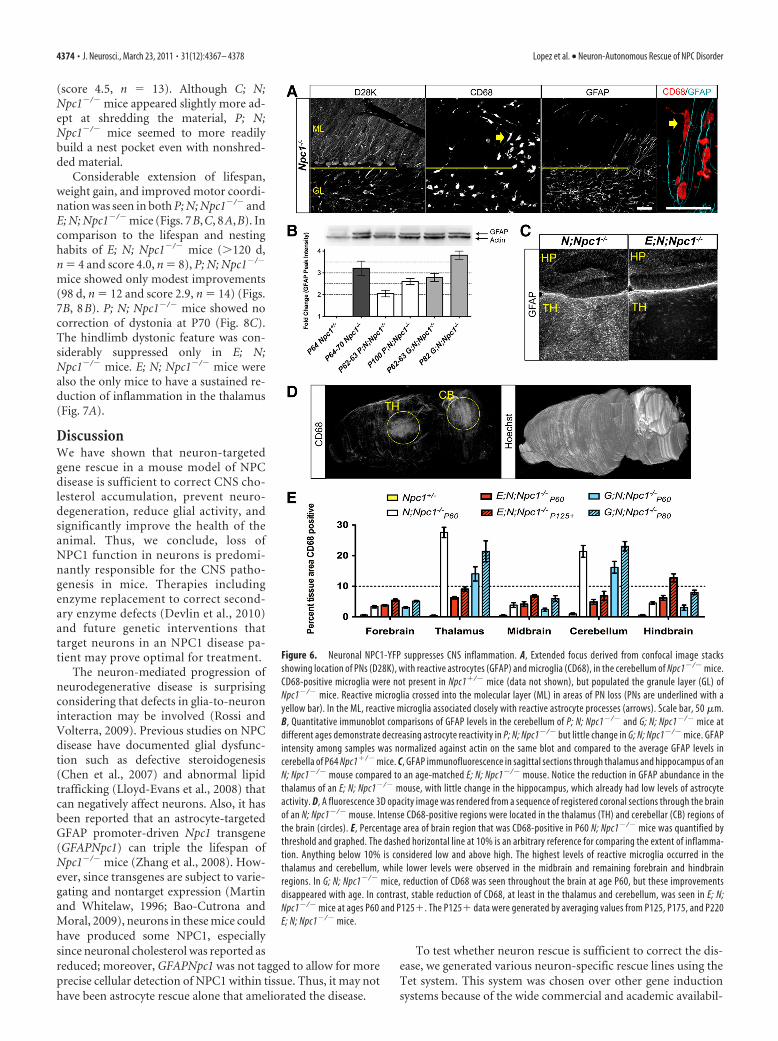
Autonomous neuron rescue paralleledby nonautonomous glial activityIn Npc1�/� mice but not wild-type mice,reactive astrocytes and microglial are per-vasive throughout the CNS during devel-opment and before neuron degenerationoccurs (Baudry et al., 2003). It has pre-viously been suggested that proinflam-matory signals that can trigger glialinflammatory responses originate fromastrocytes as a consequence of NPC1 lossin these cells (Suzuki et al., 2007). How-ever, in the cerebella of Npc1�/� mice, gli-osis was not seen in areas where PNs werestill present (Fig. 6A). In the molecularlayer, reactive astrocytes and microgliamarked by intense GFAP and CD68 im-munoreactivity (Ritz and Hausmann,2008) were concentrated only in or nearsites of PN loss, despite the abundance ofreactive astrocytes and microglia in thegranule layer, the layer containing PNaxon projections. This suggests that glial
cell activity remains responsive to neurons and does not occursolely because of NPC1 loss in glia or in response to inflammatoryprocesses nearby.
Neuron-specific rescue in Npc1�/� mice allowed us the op-portunity to verify that gliosis was a result of neuron degenera-tion and not because of loss of NPC1 function from the astrocytesthemselves. Changes in astrocyte activity were analyzed by pro-tein blots of whole cerebellum lysates from mice of various ages(Fig. 6B). In P64 –P70 mice, approximately threefold more GFAPimmunoreactivity was detected in Npc1�/� compared to controlNpc1�/� cerebellum. Cerebella from P62–P63 P; N; Npc1�/�
mice contained less GFAP than age-matched G; N; Npc1�/�
mice. Although there was an increase in GFAP immunoreactivityin the cerebella of P100 P; N; Npc1�/� mice as compared to P62
Figure 3. Neuron-autonomous correction of the CNS cholesterol storage defect in Npc1�/� mice. Immunofluorescence withanti-GFP marks neuron or astrocyte regions of the hippocampus that are positive for NPC1-YFP. Filipin marks hippocampus areasthat are positive for accumulated free cholesterol. A, Sagittal sections of the hippocampus from an adult Npc1�/� mouse hadsubstantial filipin staining in the CA3 neuron region. In an E; N; Npc1�/� mouse, NPC1-YFP produced in the DG or CA1 neurons didnot reduce cholesterol accumulation in CA3 neurons, where NPC1-YFP expression was weak or absent. B, NPC1-YFP present in CA3neurons (bottom) reduced CA3 cholesterol accumulation (top) in a C; N; Npc1�/� mouse. Despite NPC1-YFP production through-out the hippocampus in a G; N; Npc1�/� mouse, cholesterol accumulation was not reduced in CA3 neurons or elsewhere in thesurrounding area.
Lopez et al. • Neuron-Autonomous Rescue of NPC Disorder J. Neurosci., March 23, 2011 • 31(12):4367– 4378 • 4371

mice, the amount of GFAP remained the same as P62–P63 G; N;Npc1�/� mice. The amount of GFAP in P100 P; N; Npc1�/� micealso remained below the level of GFAP in P64 –P70 Npc1�/�
cerebella. These results demonstrate that PN survival alone sig-nificantly delayed gliosis in the cerebellum. Lowered astrocyteactivity in the thalamus was also observed in E; N; Npc1�/� mice(Fig. 6C), which demonstrates that neuron influence on astrocyteactivity is not limited to PNs in the cerebellum.
As we observed with astrocytes, microglia in Npc1�/� miceproliferated and occupied areas of neuronal loss or degeneration(Fig. 6A). Unlike GFAP, which was also present in nonactivatedastrocytes, CD68 staining was limited to reactive microglia,which facilitated quantitation. To identify brain regions most
affected in Npc1�/� mice, we quantified the amount of tissue areaoccupied by CD68-positive microglia throughout the brain. InP65 Npc1�/� mice, the highest concentration of CD68 immuno-reactivity was found in the cerebellum and thalamus as comparedto other regions of the CNS (Fig. 6D,E). In comparison to G; N;Npc1�/� mice, fewer CD68-positive microglia occupied the thal-amus and cerebellum of E; N; Npc1�/� mice at P60. In contrast toG; N; Npc1�/� mice, microglia activity in E; N; Npc1�/� mice inboth the thalamus and cerebellum remained low despite age (Fig.6E). Microglia activity increased mostly in the hindbrain regionof E; N; Npc1�/� mice with age. This is likely a result of thevariable, and sparse NPC1-YFP expression observed in this re-gion (Fig. 4C).
Figure 4. CNS region-specific neuron rescue of NPC disease in Npc1�/� mice. Sagittal brain sections of adult Npc1�/� mice were stained with filipin to reveal cholesterol accumulation in specificregions. The brain regions shown are portions of cortex (CTX), striatum (STR), thalamus (TH), hypothalamus (HT), midbrain (MB), cerebellum lobule (CB), pons (PO), and medulla (MD). Shown on theright of the images is an outline of the sagittal cross section of the mouse brain depicting the range of NPC1-YFP protein expression in green. Light green represents weak or variable expression basedon the average pattern seen. A, In a representative male P70 N; Npc1�/� mouse, speckled and abundant filipin staining was noticeable in all brain areas. B, In a male P80 C; N; Npc1�/� mouse,decreased filipin staining was noticeable in the CTX, STR, and MB areas. Little to no decrease in filipin staining was noted in HT, PO, and MD. Note that there is a difference in expression profile in miceof the opposite sex. Greater NPC1-YFP coverage and more reduced filipin staining in HT, TH, PO, and MD areas can be seen in female C; N; Npc1�/� mice (data not shown). C, In a male P220 E; N;Npc1�/� mouse, filipin staining was largely absent from most brain regions except the HT, MD, and some of the basal forebrain located to the left of the TH.
4372 • J. Neurosci., March 23, 2011 • 31(12):4367– 4378 Lopez et al. • Neuron-Autonomous Rescue of NPC Disorder

Local reduction of inflammation as an indicator ofregion-specific rescueThe activity of microglia in Npc1�/� mice appeared highly com-partmentalized and locally controlled; lowering inflammation inone area of the brain did not alter the level of inflammation inanother. For example, in P; N; Npc1�/� mice, we have shown thatNPC1-YFP expression is restricted to cerebellar PNs (Figs. 2A–C,5A). As a result, substantially fewer CD68-positive microglia oc-cupied the cerebellum, but the percentage of CD68 remainedunchanged in the thalamus (Fig. 7A). Thus, we used the amountof CD68-positive cells present in a tissue as an indicator of whichinjured areas of the brain were rescued with NPC1-YFP. The levelof reactive macrophages in the liver, also marked by CD68, wasused to determine disease rescue in visceral tissue. The liverexpresses the highest amounts of NPC1 in the body and, inNpc1�/� mice liver, dysfunction and accumulation of cholesterolis prominent (Garver et al., 2007).
As expected, in both C; N; Npc1�/� and E; N; Npc1�/� mice,reduced liver inflammation was not observed since expression ofNPC1-YFP was not identified outside of the brain in these ani-mals. Both mouse strains did have reduced inflammation in thethalamus. In E; N; Npc1�/� mice, however, microglia activity waslowered to levels comparable to areas of brain with the lowestCD68 percentage, such as the midbrain (Figs. 6E, 7A). In R; N;Npc1�/� mice, which produced NPC1-YFP in liver but not in thebrain, CD68 levels in the liver were low, but full diseased levelswere still seen in the brain.
Neuron rescue ameliorates NPC diseaseTo determine the effect of neuron rescueon NPC neurological phenotypes, twodisease signs were measured that presentas clinical symptoms of NPC diseasein humans: ataxia and dystonia. Ataxia inmice was assessed by measuring motor in-coordination (Kamens et al., 2005), anddystonia was characterized by limb hyper-extension and rigidity, plus hypotonia ofthe lumbosacral trunk region.
To quantify the overall benefit to thehealth-related quality of life of the animal,mouse weight and nest building wereused. Weight monitoring allowed us toplot disease progression and assess im-provements in lifespan. Retention ofnest-building activity, which requiresrecognition of bedding material, suffi-cient motor agility for shredding of nestmaterial, and the desire to organize thebedding into a nest structure for protec-tion and warmth, would imply the con-tinued use of coordinated higher mentaland motor functions (Deacon, 2006).
First, production of NPC1-YFP in liverand other tissues of the body, excludingneurons of the CNS (Figs. 2E, 7A), did notprevent weight loss or discernibly delaydisease progression of R; N; Npc1�/� mice(Fig. 7B). These mice were fed doxycy-cline in the drinking water continuouslyfrom birth to induce NPC1-YFP produc-tion during their entire postnatal lifespan.A similar tetracycline, minocycline, amore effective antibiotic than doxycy-
cline, has been shown to not alter the course of the disease inNpc1�/� mice (Erickson and Bernard, 2002). Skin biopsies wereused to check for continuous NPC1-YFP induction (data notshown).
Second, producing NPC1-YFP in astrocytes only slightlymodified disease progression, causing a lifespan increase of only 1week for G; N; Npc1�/� mice (85.5 d, n � 6) in comparison withN; Npc1�/� mice (78.5 d, n � 12). The difference was not statis-tically significant ( p � 0.28) (Fig. 7C). However, G; N; Npc1�/�
mice at age P60 did weigh more than N; Npc1�/� mice (21.99 g,n � 8 for G; N; Npc1�/� mice, 17.67 g, n � 10 for N; Npc1�/�
mice; p � 0.0005). Despite improved weight gain, G; N;Npc1�/� mice failed to build nests (score 1.1, n � 8), as did N;Npc1�/� mice (score 1.4, n � 7) at age P60. Dystonic featuressimilar to that of C; N; Npc1�/� mice (Fig. 8 B) were noticeableat age P70 (date not shown) and mice appeared ataxic al-though motor coordination was not measured.
Third, despite production of NPC1-YFP in neurons in variousareas of the brain in C; N; Npc1�/� mice (Figs. 2D, 4B), weightgain was not improved (Fig. 7B) and increase in lifespan wascomparable to P; N; Npc1�/� mice, although it varied betweenmale and female C; N; Npc1�/� mice (Fig. 7C). No improvementin motor coordination and only a slight correction of dystoniawas seen (Fig. 8A,C). Surprisingly, despite motor deficits, thenest-building performance of C; N; Npc1�/� mice at P60 (score3.1, n � 7) was similar to P; N; Npc1�/� mice (score 2.9, n � 14)(Fig. 8B). Both lines approached wild-type nest-building activity
Figure 5. Cell-autonomous survival of Purkinje neurons. A, Sagittal, midline sections of the cerebellum were stainedwith anti-D28K to mark remaining PNs and anti-GFP to locate NPC1-YFP expression. At age P65, in a G; N; Npc1�/� mouse,patterned loss of PNs (loss of D28K stain from lobules) can be seen. At age P80, significant loss of PNs is noticeable. At ageP90, in a P; N; Npc1�/� mouse, PNs remain and NPC1-YFP is localized to the PN layer (boxed region and zoomed in inset).Note that lobule X was used as a landmark for section comparison since PNs in lobule X are unaffected. B, Representativeimmunoblot comparing the levels of D28K and MBP in whole cerebellum samples from P; N; Npc1�/� and G; N; Npc1�/�
mice to Npc1�/� and Npc1�/� mice. MBP isoforms at 21.5, 18, and 17 kDa are shown. Notice the much larger amount ofNPC1-YFP produced in a G; N; Npc1�/� mouse cerebellum compared to an age-matched P; N; Npc1�/� mouse; despitethis, there is no obvious rescue of MBP and D28K level in a G; N; Npc1�/� mouse sample, but there is in a P; N; Npc1�/�
mouse sample.
Lopez et al. • Neuron-Autonomous Rescue of NPC Disorder J. Neurosci., March 23, 2011 • 31(12):4367– 4378 • 4373
(score 4.5, n � 13). Although C; N;Npc1�/� mice appeared slightly more ad-ept at shredding the material, P; N;Npc1�/� mice seemed to more readilybuild a nest pocket even with nonshred-ded material.
Considerable extension of lifespan,weight gain, and improved motor coordi-nation was seen in both P; N; Npc1�/� andE; N; Npc1�/� mice (Figs. 7B,C, 8A,B). Incomparison to the lifespan and nestinghabits of E; N; Npc1�/� mice (�120 d,n � 4 and score 4.0, n � 8), P; N; Npc1�/�
mice showed only modest improvements(98 d, n � 12 and score 2.9, n � 14) (Figs.7B, 8B). P; N; Npc1�/� mice showed nocorrection of dystonia at P70 (Fig. 8C).The hindlimb dystonic feature was con-siderably suppressed only in E; N;Npc1�/� mice. E; N; Npc1�/� mice werealso the only mice to have a sustained re-duction of inflammation in the thalamus(Fig. 7A).
DiscussionWe have shown that neuron-targetedgene rescue in a mouse model of NPCdisease is sufficient to correct CNS cho-lesterol accumulation, prevent neuro-degeneration, reduce glial activity, andsignificantly improve the health of theanimal. Thus, we conclude, loss ofNPC1 function in neurons is predomi-nantly responsible for the CNS patho-genesis in mice. Therapies includingenzyme replacement to correct second-ary enzyme defects (Devlin et al., 2010)and future genetic interventions thattarget neurons in an NPC1 disease pa-tient may prove optimal for treatment.
The neuron-mediated progression ofneurodegenerative disease is surprisingconsidering that defects in glia-to-neuroninteraction may be involved (Rossi andVolterra, 2009). Previous studies on NPCdisease have documented glial dysfunc-tion such as defective steroidogenesis(Chen et al., 2007) and abnormal lipidtrafficking (Lloyd-Evans et al., 2008) thatcan negatively affect neurons. Also, it hasbeen reported that an astrocyte-targetedGFAP promoter-driven Npc1 transgene(GFAPNpc1) can triple the lifespan ofNpc1�/� mice (Zhang et al., 2008). How-ever, since transgenes are subject to varie-gating and nontarget expression (Martinand Whitelaw, 1996; Bao-Cutrona andMoral, 2009), neurons in these mice couldhave produced some NPC1, especiallysince neuronal cholesterol was reported asreduced; moreover, GFAPNpc1 was not tagged to allow for moreprecise cellular detection of NPC1 within tissue. Thus, it may nothave been astrocyte rescue alone that ameliorated the disease.
To test whether neuron rescue is sufficient to correct the dis-ease, we generated various neuron-specific rescue lines using theTet system. This system was chosen over other gene inductionsystems because of the wide commercial and academic availabil-
Figure 6. Neuronal NPC1-YFP suppresses CNS inflammation. A, Extended focus derived from confocal image stacksshowing location of PNs (D28K), with reactive astrocytes (GFAP) and microglia (CD68), in the cerebellum of Npc1�/� mice.CD68-positive microglia were not present in Npc1�/� mice (data not shown), but populated the granule layer (GL) ofNpc1�/� mice. Reactive microglia crossed into the molecular layer (ML) in areas of PN loss (PNs are underlined with ayellow bar). In the ML, reactive microglia associated closely with reactive astrocyte processes (arrows). Scale bar, 50 �m.B, Quantitative immunoblot comparisons of GFAP levels in the cerebellum of P; N; Npc1�/� and G; N; Npc1�/� mice atdifferent ages demonstrate decreasing astrocyte reactivity in P; N; Npc1�/� but little change in G; N; Npc1�/� mice. GFAPintensity among samples was normalized against actin on the same blot and compared to the average GFAP levels incerebella of P64 Npc1�/� mice. C, GFAP immunofluorescence in sagittal sections through thalamus and hippocampus of anN; Npc1�/� mouse compared to an age-matched E; N; Npc1�/� mouse. Notice the reduction in GFAP abundance in thethalamus of an E; N; Npc1�/� mouse, with little change in the hippocampus, which already had low levels of astrocyteactivity. D, A fluorescence 3D opacity image was rendered from a sequence of registered coronal sections through the brainof an N; Npc1�/� mouse. Intense CD68-positive regions were located in the thalamus (TH) and cerebellar (CB) regions ofthe brain (circles). E, Percentage area of brain region that was CD68-positive in P60 N; Npc1�/� mice was quantified bythreshold and graphed. The dashed horizontal line at 10% is an arbitrary reference for comparing the extent of inflamma-tion. Anything below 10% is considered low and above high. The highest levels of reactive microglia occurred in thethalamus and cerebellum, while lower levels were observed in the midbrain and remaining forebrain and hindbrainregions. In G; N; Npc1�/� mice, reduction of CD68 was seen throughout the brain at age P60, but these improvementsdisappeared with age. In contrast, stable reduction of CD68, at least in the thalamus and cerebellum, was seen in E; N;Npc1�/� mice at ages P60 and P125�. The P125� data were generated by averaging values from P125, P175, and P220E; N; Npc1�/� mice.
4374 • J. Neurosci., March 23, 2011 • 31(12):4367– 4378 Lopez et al. • Neuron-Autonomous Rescue of NPC Disorder

ity of driver transgenes for the targeted expression of a singlereporter transgene. We chose drivers whose cell type specificityand expression profiles have been previously reported (Fig. 1B).Although we cannot be absolutely certain that NPC1-YFP wasabsent from all other cell types except neurons, the use of multi-ple mouse lines with different drivers to induce varied NPC1-YFPexpression patterns in the brain allowed the demonstration of fullneuron autonomy. In an Npc1�/� mouse, NPC1-YFP producedin a specific neuron population corrected the cholesterol accu-mulation phenotype only within those neurons (Fig. 3A) and theproduction of NPC1-YFP in neighboring astrocytes did notchange neuronal cholesterol accumulation (Fig. 3B). Along withthe reduction of cholesterol in the specific brain areas (Fig. 4), theanatomical location of neuron rescue in Npc1�/� mice could beidentified by local reduction of inflammation (Fig. 6). In addi-
tion, the lack of improvement in weightand lifespan of the R; N; Npc1�/� mice(Fig. 7), which produced NPC1-YFP invirtually all tissues except the brain (Fig.2), allowed us to exclude potential con-founding effects of non-nervous systemrescue.
Our study does not exclude the possi-bility that glial NPC1 is required for theoverall health of neurons, but it appearsthat the loss of NPC1 does not signifi-cantly affect glial function that is criticalfor neuron survival. Prior work has shownthat the secretion of sterols was not inhib-ited in Npc1�/� astrocytes. Lipoproteinsgenerated by Npc1�/� glia were capable ofsupporting axon elongation in vitro(Mutka et al., 2004; Karten et al., 2005).Prior work has also demonstrated the cell-autonomous death of PNs by using chi-meric mice, mice comprised of a mixtureof wild-type and Npc1�/� cells (Ko et al.,2005), and conditional knock-out mice,wild-type mice with Npc1 gene deletion inPNs (Elrick et al., 2010). However, thesestudies did not address whether PNs cansurvive alone despite the loss of NPC1from all other neurons, glia cells, or othercells of the body. Here we show in vivo thatdespite glial NPC1 deficiency, cerebellarPNs, which are sensitive to many geneticand acquired disorders as well as toxic en-vironmental factors (Sarna and Hawkes,2003), survived as long as they producedNPC1-YFP (Fig. 5).
The targeted rescue of PNs in anNpc1�/� mouse allowed us to observe thebenefit cerebellar improvements alonecould have on NPC disease. PNs are thesole neuronal output of the cerebellar cor-tex, and loss of PNs in an otherwise nor-mal brain has long been known to causemotor abnormalities. Current researchhas begun to suggest that the cerebellumcan regulate nonmotor brain functionsas well (Strick et al., 2009), including theearly development of the whole brain.Thus, rescuing cerebellar function in a
disease that also affects the cerebellum could have broad andsignificantly beneficial therapeutic outcomes. The benefit PNrescue alone had on weight gain, nest-building activity, motorability, and lifespan in P; N; Npc1�/� mice supports this viewand points to an important cerebellar involvement in the se-verity of NPC disease progression.
Despite significant benefits, cerebellar PN survival ultimatelydid not halt disease progression or prevent premature death of P;N; Npc1�/� mice. Improved motor coordination in these micewas temporary as ataxia seemed to eventually increase with age(Fig. 8A), and weight gain was unsustainable (Fig. 7B). E; N;Npc1�/� mice also showed a similar worsening of condition withage, but these mice exhibited an extraordinary increase in lifespanmarked by a more delayed and gradual weight loss (Fig. 7B). Thebroad neuronal rescue in E; N; Npc1�/� mice would suggest that
Figure 7. Lifespan extended with neuron-specific rescue. A, Graphs of the percentage area of thalamus, cerebellum, and liveroccupied by CD68-positive cells are shown for G, P, C, E and R; N; Npc1�/� mice strains at age P60. As in Figure 6 E, the dotted lineon the graph represents an arbitrary cutoff for low (�10% CD68-positive tissue area) and high (�10% CD68-positive tissue area)microglial activity. B, Weight curves and SD for each strain and sex were plotted and compared with the weight progression ofNpc1�/� and Npc1�/� male mice. C, The mean survival age and 95% confidence limits were graphed for each transgenic linewith the exception of E; N; Npc1�/� mice (asterisk). E; N; Npc1�/� mice were killed for tissue analysis before signs of severemorbidity. Age of death for this line is not known, but mice with the expected NPC1-YFP expression profile (Figs. 2 D, E, 4C) andreduced inflammation (Fig. 6 E) readily surpass 100 d (vertical line).
Lopez et al. • Neuron-Autonomous Rescue of NPC Disorder J. Neurosci., March 23, 2011 • 31(12):4367– 4378 • 4375
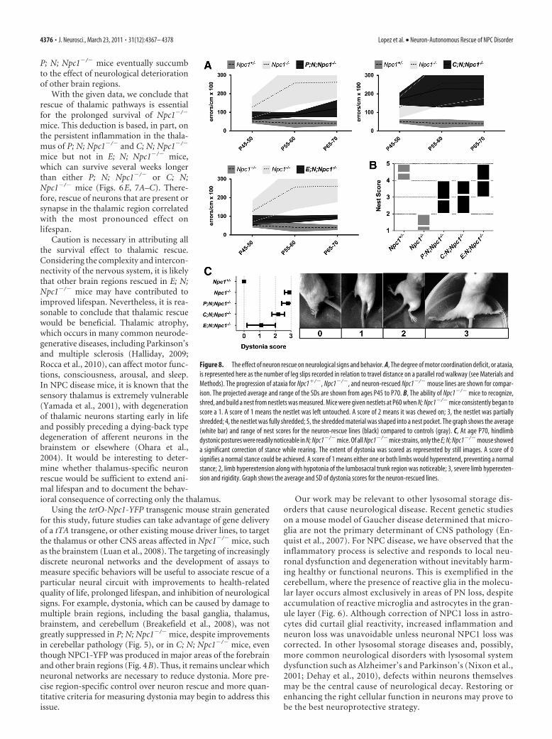
P; N; Npc1�/� mice eventually succumbto the effect of neurological deteriorationof other brain regions.
With the given data, we conclude thatrescue of thalamic pathways is essentialfor the prolonged survival of Npc1�/�
mice. This deduction is based, in part, onthe persistent inflammation in the thala-mus of P; N; Npc1�/� and C; N; Npc1�/�
mice but not in E; N; Npc1�/� mice,which can survive several weeks longerthan either P; N; Npc1�/� or C; N;Npc1�/� mice (Figs. 6E, 7A–C). There-fore, rescue of neurons that are present orsynapse in the thalamic region correlatedwith the most pronounced effect onlifespan.
Caution is necessary in attributing allthe survival effect to thalamic rescue.Considering the complexity and intercon-nectivity of the nervous system, it is likelythat other brain regions rescued in E; N;Npc1�/� mice may have contributed toimproved lifespan. Nevertheless, it is rea-sonable to conclude that thalamic rescuewould be beneficial. Thalamic atrophy,which occurs in many common neurode-generative diseases, including Parkinson’sand multiple sclerosis (Halliday, 2009;Rocca et al., 2010), can affect motor func-tions, consciousness, arousal, and sleep.In NPC disease mice, it is known that thesensory thalamus is extremely vulnerable(Yamada et al., 2001), with degenerationof thalamic neurons starting early in lifeand possibly preceding a dying-back typedegeneration of afferent neurons in thebrainstem or elsewhere (Ohara et al.,2004). It would be interesting to deter-mine whether thalamus-specific neuronrescue would be sufficient to extend ani-mal lifespan and to document the behav-ioral consequence of correcting only the thalamus.
Using the tetO-Npc1-YFP transgenic mouse strain generatedfor this study, future studies can take advantage of gene deliveryof a tTA transgene, or other existing mouse driver lines, to targetthe thalamus or other CNS areas affected in Npc1�/� mice, suchas the brainstem (Luan et al., 2008). The targeting of increasinglydiscrete neuronal networks and the development of assays tomeasure specific behaviors will be useful to associate rescue of aparticular neural circuit with improvements to health-relatedquality of life, prolonged lifespan, and inhibition of neurologicalsigns. For example, dystonia, which can be caused by damage tomultiple brain regions, including the basal ganglia, thalamus,brainstem, and cerebellum (Breakefield et al., 2008), was notgreatly suppressed in P; N; Npc1�/� mice, despite improvementsin cerebellar pathology (Fig. 5), or in C; N; Npc1�/� mice, eventhough NPC1-YFP was produced in major areas of the forebrainand other brain regions (Fig. 4B). Thus, it remains unclear whichneuronal networks are necessary to reduce dystonia. More pre-cise region-specific control over neuron rescue and more quan-titative criteria for measuring dystonia may begin to address thisissue.
Our work may be relevant to other lysosomal storage dis-orders that cause neurological disease. Recent genetic studieson a mouse model of Gaucher disease determined that micro-glia are not the primary determinant of CNS pathology (En-quist et al., 2007). For NPC disease, we have observed that theinflammatory process is selective and responds to local neu-ronal dysfunction and degeneration without inevitably harm-ing healthy or functional neurons. This is exemplified in thecerebellum, where the presence of reactive glia in the molecu-lar layer occurs almost exclusively in areas of PN loss, despiteaccumulation of reactive microglia and astrocytes in the gran-ule layer (Fig. 6). Although correction of NPC1 loss in astro-cytes did curtail glial reactivity, increased inflammation andneuron loss was unavoidable unless neuronal NPC1 loss wascorrected. In other lysosomal storage diseases and, possibly,more common neurological disorders with lysosomal systemdysfunction such as Alzheimer’s and Parkinson’s (Nixon et al.,2001; Dehay et al., 2010), defects within neurons themselvesmay be the central cause of neurological decay. Restoring orenhancing the right cellular function in neurons may prove tobe the best neuroprotective strategy.
Figure 8. The effect of neuron rescue on neurological signs and behavior. A, The degree of motor coordination deficit, or ataxia,is represented here as the number of leg slips recorded in relation to travel distance on a parallel rod walkway (see Materials andMethods). The progression of ataxia for Npc1�/�, Npc1�/�, and neuron-rescued Npc1�/� mouse lines are shown for compar-ison. The projected average and range of the SDs are shown from ages P45 to P70. B, The ability of Npc1�/� mice to recognize,shred, and build a nest from nestlets was measured. Mice were given nestlets at P60 when N; Npc1�/� mice consistently began toscore a 1. A score of 1 means the nestlet was left untouched. A score of 2 means it was chewed on; 3, the nestlet was partiallyshredded; 4, the nestlet was fully shredded; 5, the shredded material was shaped into a nest pocket. The graph shows the average(white bar) and range of nest scores for the neuron-rescue lines (black) compared to controls (gray). C, At age P70, hindlimbdystonic postures were readily noticeable in N; Npc1�/� mice. Of all Npc1�/� mice strains, only the E; N; Npc1�/� mouse showeda significant correction of stance while rearing. The extent of dystonia was scored as represented by still images. A score of 0signifies a normal stance could be achieved. A score of 1 means either one or both limbs would hyperextend, preventing a normalstance; 2, limb hyperextension along with hypotonia of the lumbosacral trunk region was noticeable; 3, severe limb hyperexten-sion and rigidity. Graph shows the average and SD of dystonia scores for the neuron-rescued lines.
4376 • J. Neurosci., March 23, 2011 • 31(12):4367– 4378 Lopez et al. • Neuron-Autonomous Rescue of NPC Disorder

ReferencesBao-Cutrona M, Moral P (2009) Unexpected expression pattern of
tetracycline-regulated transgenes in mice. Genetics 181:1687–1691.Baudry M, Yao Y, Simmons D, Liu J, Bi X (2003) Postnatal development of
inflammation in a murine model of Niemann-Pick type C disease: immu-nohistochemical observations of microglia and astroglia. Exp Neurol184:887–903.
Breakefield XO, Blood AJ, Li Y, Hallett M, Hanson PI, Standaert DG (2008)The pathophysiological basis of dystonias. Nat Rev Neurosci 9:222–234.
Chen G, Li HM, Chen YR, Gu XS, Duan S (2007) Decreased estradiol releasefrom astrocytes contributes to the neurodegeneration in a mouse modelof Niemann-Pick disease type C. Glia 55:1509 –1518.
Chen J, Kelz MB, Zeng G, Sakai N, Steffen C, Shockett PE, Picciotto MR,Duman RS, Nestler EJ (1998) Transgenic animals with inducible, tar-geted gene expression in brain. Mol Pharmacol 54:495–503.
Deacon RM (2006) Assessing nest building in mice. Nat Protoc1:1117–1119.
Dehay B, Bove J, Rodríguez-Muela N, Perier C, Recasens A, Boya P, Vila M(2010) Pathogenic lysosomal depletion in Parkinson’s disease. J Neuro-sci 30:12535–12544.
Devlin C, Pipalia NH, Liao X, Schuchman EH, Maxfield FR, Tabas I (2010)Improvement in lipid and protein trafficking in Niemann-Pick C1 cells bycorrection of a secondary enzyme defect. Traffic 11:601– 615.
Donohue C, Marion S, Erickson RP (2009) Expression of Npc1 in glial cellscorrects sterility in Npc1(�/�) mice. J Appl Genet 50:385–390.
Elrick MJ, Pacheco CD, Yu T, Dadgar N, Shakkottai VG, Ware C, Paulson HL,Lieberman AP (2010) Conditional Niemann-Pick C mice demonstratecell autonomous Purkinje cell neurodegeneration. Hum Mol Genet19:837– 847.
Enquist IB, Lo Bianco C, Ooka A, Nilsson E, Månsson JE, Ehinger M, RichterJ, Brady RO, Kirik D, Karlsson S (2007) Murine models of acute neu-ronopathic Gaucher disease. Proc Natl Acad Sci U S A 104:17483–17488.
Erickson RP, Bernard O (2002) Studies on neuronal death in the mousemodel of Niemann-Pick C disease. J Neurosci Res 68:738 –744.
Fink JK, Filling-Katz MR, Sokol J, Cogan DG, Pikus A, Sonies B, Soong B,Pentchev PG, Comly ME, Brady RO, Barton NW (1989) Clinical spec-trum of Niemann-Pick disease type C. Neurology 39:1040 –1049.
Garver WS, Jelinek D, Oyarzo JN, Flynn J, Zuckerman M, Krishnan K, ChungBH, Heidenreich RA (2007) Characterization of liver disease and lipidmetabolism in the Niemann-Pick C1 mouse. J Cell Biochem101:498 –516.
German DC, Quintero EM, Liang CL, Ng B, Punia S, Xie C, Dietschy JM(2001) Selective neurodegeneration, without neurofibrillary tangles, in amouse model of Niemann-Pick C disease. J Comp Neurol 433:415– 425.
Gimenez E, Montoliu L (2001) A simple polymerase chain reaction assay forgenotyping the retinal degeneration mutation (Pdeb(rd1)) in FVB/N-derived transgenic mice. Lab Anim 35:153–156.
Gossen M, Bujard H (1992) Tight control of gene expression in mammaliancells by tetracycline-responsive promoters. Proc Natl Acad Sci U S A89:5547–5551.
Halliday GM (2009) Thalamic changes in Parkinson’s disease. Parkinson-ism Relat Disord 15 [Suppl 3]:S152–S155.
Hochedlinger K, Yamada Y, Beard C, Jaenisch R (2005) Ectopic expressionof Oct-4 blocks progenitor-cell differentiation and causes dysplasia inepithelial tissues. Cell 121:465– 477.
Hol EM, Roelofs RF, Moraal E, Sonnemans MA, Sluijs JA, Proper EA, deGraan PN, Fischer DF, van Leeuwen FW (2003) Neuronal expression ofGFAP in patients with Alzheimer pathology and identification of novelGFAP splice forms. Mol Psychiatry 8:786 –796.
Imrie J, Dasgupta S, Besley GT, Harris C, Heptinstall L, Knight S, Vanier MT,Fensom AH, Ward C, Jacklin E, Whitehouse C, Wraith JE (2007) Thenatural history of Niemann-Pick disease type C in the UK. J Inherit MetabDis 30:51–59.
Infante RE, Wang ML, Radhakrishnan A, Kwon HJ, Brown MS, Goldstein JL(2008) NPC2 facilitates bidirectional transfer of cholesterol betweenNPC1 and lipid bilayers, a step in cholesterol egress from lysosomes. ProcNatl Acad Sci U S A 105:15287–15292.
Kamens HM, Crabbe JC (2007) The parallel rod floor test: a measure ofataxia in mice. Nat Protoc 2:277–281.
Kamens HM, Phillips TJ, Holstein SE, Crabbe JC (2005) Characterization ofthe parallel rod floor apparatus to test motor incoordination in mice.Genes Brain Behav 4:253–266.
Kapur R, Donohue C, Jelinek D, Erickson RP (2009) Amelioration of en-teric neuropathology in a mouse model of Niemann-Pick C by Npc1expression in enteric glia. J Neurosci Res 87:2994 –3001.
Karten B, Hayashi H, Francis GA, Campenot RB, Vance DE, Vance JE (2005)Generation and function of astroglial lipoproteins from Niemann-Picktype C1-deficient mice. Biochem J 387:779 –788.
Ko DC, Gordon MD, Jin JY, Scott MP (2001) Dynamic movements of or-ganelles containing Niemann-Pick C1 protein: NPC1 involvement in lateendocytic events. Mol Biol Cell 12:601– 614.
Ko DC, Milenkovic L, Beier SM, Manuel H, Buchanan J, Scott MP (2005)Cell-autonomous death of cerebellar Purkinje neurons with autophagy inNiemann-Pick type C disease. PLoS Genet 1:81–95.
Lee Y, Messing A, Su M, Brenner M (2008) GFAP promoter elements re-quired for region-specific and astrocyte-specific expression. Glia56:481– 493.
Lloyd-Evans E, Morgan AJ, He X, Smith DA, Elliot-Smith E, Sillence DJ,Churchill GC, Schuchman EH, Galione A, Platt FM (2008) Niemann-Pick disease type C1 is a sphingosine storage disease that causes deregu-lation of lysosomal calcium. Nat Med 14:1247–1255.
Loftus SK, Morris JA, Carstea ED, Gu JZ, Cummings C, Brown A, Ellison J,Ohno K, Rosenfeld MA, Tagle DA, Pentchev PG, Pavan WJ (1997) Mu-rine model of Niemann-Pick C disease: mutation in a cholesterol homeo-stasis gene. Science 277:232–235.
Loftus SK, Erickson RP, Walkley SU, Bryant MA, Incao A, Heidenreich RA,Pavan WJ (2002) Rescue of neurodegeneration in Niemann-Pick Cmice by a prion-promoter-driven Npc1 cDNA transgene. Hum MolGenet 11:3107–3114.
Luan Z, Saito Y, Miyata H, Ohama E, Ninomiya H, Ohno K (2008) Brains-tem neuropathology in a mouse model of Niemann-Pick disease type C.J Neurol Sci 268:108 –116.
Martin DI, Whitelaw E (1996) The vagaries of variegating transgenes. Bioes-says 18:919 –923.
Mayford M, Bach ME, Huang YY, Wang L, Hawkins RD, Kandel ER (1996)Control of memory formation through regulated expression of a CaMKIItransgene. Science 274:1678 –1683.
Mutka AL, Lusa S, Linder MD, Jokitalo E, Kopra O, Jauhiainen M, Ikonen E(2004) Secretion of sterols and the NPC2 protein from primary astro-cytes. J Biol Chem 279:48654 – 48662.
Nixon RA, Mathews PM, Cataldo AM (2001) The neuronal endosomal-lysosomal system in Alzheimer’s disease. J Alzheimers Dis 3:97–107.
Ohara S, Ukita Y, Ninomiya H, Ohno K (2004) Degeneration ofcholecystokinin-immunoreactive afferents to the VPL thalamus in amouse model of Niemann-Pick disease type C. Brain Res 1022:244 –246.
Park WD, O’Brien JF, Lundquist PA, Kraft DL, Vockley CW, Karnes PS,Patterson MC, Snow K (2003) Identification of 58 novel mutations inNiemann-Pick disease type C: correlation with biochemical phenotypeand importance of PTC1-like domains in NPC1. Hum Mutat 22:313–325.
Patel SC, Suresh S, Kumar U, Hu CY, Cooney A, Blanchette-Mackie EJ,Neufeld EB, Patel RC, Brady RO, Patel YC, Pentchev PG, Ong WY (1999)Localization of Niemann-Pick C1 protein in astrocytes: implications forneuronal degeneration in Niemann-Pick type C disease. Proc Natl AcadSci U S A 96:1657–1662.
Ritz MF, Hausmann ON (2008) Effect of 17beta-estradiol on functionaloutcome, release of cytokines, astrocyte reactivity and inflammatoryspreading after spinal cord injury in male rats. Brain Res 1203:177–188.
Rocca MA, Mesaros S, Pagani E, Sormani MP, Comi G, Filippi M (2010)Thalamic damage and long-term progression of disability in multiplesclerosis. Radiology 257:463– 469.
Rossi D, Volterra A (2009) Astrocytic dysfunction: insights on the role inneurodegeneration. Brain Res Bull 80:224 –232.
Sarna JR, Hawkes R (2003) Patterned Purkinje cell death in the cerebellum.Prog Neurobiol 70:473–507.
Sarna JR, Larouche M, Marzban H, Sillitoe RV, Rancourt DE, Hawkes R(2003) Patterned Purkinje cell degeneration in mouse models ofNiemann-Pick type C disease. J Comp Neurol 456:279 –291.
Strick PL, Dum RP, Fiez JA (2009) Cerebellum and nonmotor function.Annu Rev Neurosci 32:413– 434.
Suzuki M, Sugimoto Y, Ohsaki Y, Ueno M, Kato S, Kitamura Y, Hosokawa H,Davies JP, Ioannou YA, Vanier MT, Ohno K, Ninomiya H (2007) En-dosomal accumulation of Toll-like receptor 4 causes constitutive secre-tion of cytokines and activation of signal transducers and activators oftranscription in Niemann-Pick disease type C (NPC) fibroblasts: a poten-
Lopez et al. • Neuron-Autonomous Rescue of NPC Disorder J. Neurosci., March 23, 2011 • 31(12):4367– 4378 • 4377

tial basis for glial cell activation in the NPC brain. J Neurosci27:1879 –1891.
Takikita S, Fukuda T, Mohri I, Yagi T, Suzuki K (2004) Perturbed myelina-tion process of premyelinating oligodendrocyte in Niemann-Pick type Cmouse. J Neuropathol Exp Neurol 63:660 – 673.
Vanier MT, Rodriguez-Lafrasse C, Rousson R, Duthel S, Harzer K, PentchevPG, Revol A, Louisot P (1991) Type C Niemann-Pick disease: biochem-ical aspects and phenotypic heterogeneity. Dev Neurosci 13:307–314.
Wang J, Lin W, Popko B, Campbell IL (2004) Inducible production ofinterferon-gamma in the developing brain causes cerebellar dysplasiawith activation of the Sonic hedgehog pathway. Mol Cell Neurosci27:489 – 496.
Yamada A, Saji M, Ukita Y, Shinoda Y, Taniguchi M, Higaki K, Ninomiya H,Ohno K (2001) Progressive neuronal loss in the ventral posterior lateral
and medial nuclei of thalamus in Niemann-Pick disease type C mousebrain. Brain Dev 23:288 –297.
Yeckel MF, Berger TW (1990) Feedforward excitation of the hippocampusby afferents from the entorhinal cortex: redefinition of the role of thetrisynaptic pathway. Proc Natl Acad Sci U S A 87:5832–5836.
Zhang M, Strnatka D, Donohue C, Hallows JL, Vincent I, Erickson RP (2008)Astrocyte-only Npc1 reduces neuronal cholesterol and triples life span ofNpc1-/- mice. J Neurosci Res 86:2848 –2856.
Zhu Z, Zheng T, Lee CG, Homer RJ, Elias JA (2002) Tetracycline-controlledtranscriptional regulation systems: advances and application in trans-genic animal modeling. Semin Cell Dev Biol 13:121–128.
Zu T, Duvick LA, Kaytor MD, Berlinger MS, Zoghbi HY, Clark HB, Orr HT(2004) Recovery from polyglutamine-induced neurodegeneration inconditional SCA1 transgenic mice. J Neurosci 24:8853– 8861.
4378 • J. Neurosci., March 23, 2011 • 31(12):4367– 4378 Lopez et al. • Neuron-Autonomous Rescue of NPC Disorder