neurobiologyofdisease ... · neurobiologyofdisease targetingextracellularcyclophilinareduces...
TRANSCRIPT
Neurobiology of Disease
Targeting Extracellular Cyclophilin A ReducesNeuroinflammation and Extends Survival in a Mouse Modelof Amyotrophic Lateral SclerosisLaura Pasetto,1* Silvia Pozzi,1* Mariachiara Castelnovo,1 Manuela Basso,4 Alvaro G. Estevez,5 Stefano Fumagalli,2
Maria Grazia De Simoni,2 Valeria Castellaneta,1 X Paolo Bigini,1 Elena Restelli,2 Roberto Chiesa,2 X Francesca Trojsi,6
Maria Rosaria Monsurro,6 Leonardo Callea,7 Miroslav Malesevic,8 Gunter Fischer,9 XMattia Freschi,3
Massimo Tortarolo,2 Caterina Bendotti,2 and X Valentina Bonetto1
1Department of Molecular Biochemistry and Pharmacology, 2Department of Neurosciences, and 3Italian Foundation for research on ALS (AriSLA) AnimalFacility, IRCCS-Istituto di Ricerche Farmacologiche Mario Negri, 20156 Milano, Italy, 4Centre for Integrative Biology (CIBIO), University of Trento, 38123Trento, Italy, 5Burnett School of Biomedical Sciences, College of Medicine, University of Central Florida, Orlando, Florida 32816, 6Department of Medical,Surgical, Neurological, Metabolic and Aging Sciences, Second University of Naples, 80131 Naples, Italy, 7IRCCS Fondazione “Don Carlo Gnocchi”, 20121Milano, Italy, 8Institute of Biochemistry and Biotechnology, Martin Luther University Halle-Wittenberg, 06099 Halle, Germany, and 9Max-Planck-Institutefor Biophysical Chemistry Gottingen, BO Halle, 06120 Halle, Germany
Neuroinflammation is a major hallmark of amyotrophic lateral sclerosis (ALS), which is currently untreatable. Several anti-inflammatorycompounds have been evaluated in patients and in animal models of ALS, but have been proven disappointing in part because effectivetargets have not yet been identified. Cyclophilin A, also known as peptidylprolyl cis-/trans-isomerase A (PPIA), as a foldase is beneficialintracellularly, but extracellularly has detrimental functions. We found that extracellular PPIA is a mediator of neuroinflammation inALS. It is a major inducer of matrix metalloproteinase 9 and is selectively toxic for motor neurons. High levels of PPIA were found in theCSF of SOD1G93A mice and rats and sporadic ALS patients, suggesting that our findings may be relevant for familial and sporadic cases. Aspecific inhibitor of extracellular PPIA, MM218, given at symptom onset, rescued motor neurons and extended survival in the SOD1G93A
mouse model of familial ALS by 11 d. The treatment resulted in the polarization of glia toward a prohealing phenotype associated withreduced NF-�B activation, proinflammatory markers, endoplasmic reticulum stress, and insoluble phosphorylated TDP-43. Our resultsindicates that extracellular PPIA is a promising druggable target for ALS and support further studies to develop a therapy to arrest or slowthe progression of the disease in patients.
Key words: amyotrophic lateral sclerosis; cyclophilin A; neuroinflammation
IntroductionAmyotrophic lateral sclerosis (ALS) is a devastating, incurableneurodegenerative disease that primarily affects motor neuronsin the brain and spinal cord. Most cases are sporadic, with un-known etiology. Approximately 10% have a family history of a
genetically dominant disorder in which mutations in Cu/Zn su-peroxide dismutase (SOD1) and C9ORF72 genes are the mostcommon cause. Independently from the etiology, it is now estab-lished that ALS is a multifactorial disease involving differentpathogenic mechanisms that require multiple non-neuronal cells
Received Aug. 3, 2016; revised Oct. 24, 2016; accepted Nov. 15, 2016.Author contributions: L.P., S.P., A.G.E., M.G.D.S., P.B., R.C., M.R.M., G.F., C.B., and V.B. designed research; L.P.,
S.P., M.C., M.B., S.F., V.C., E.R., F.T., L.C., M.M., M.F., and M.T. performed research; L.P., S.P., M.B., S.F., P.B., M.F.,C.B., and V.B. analyzed data; L.P., S.P., M.B., A.G.E., C.B., and V.B. wrote the paper.
This work was supported by Telethon Italy (Grant TCR08002 to V.B.), Italian Foundation for research on ALS(AriSLA) (Grant CypALS to V.B.), and the European Community’s Health Seventh Framework Programme (FP7/2007–2013 under Grant 259867 to C.B). We thank Dr. Marco Marzo for contribution to the in vitro experiments, Dr.Giorgia Spano for help in collecting animal tissues, and Judith Baggott for editorial assistance.
Significance Statement
We provide evidence that extracellular cyclophilin A, also known as peptidylprolyl cis-/trans-isomerase A (PPIA), is a mediator of theneuroinflammatory reaction in amyotrophic lateral sclerosis (ALS) and is toxic for motor neurons. Supporting this, a specific extracel-lular PPIA inhibitor reduced neuroinflammation, rescued motor neurons, and extended survival in the SOD1G93A mouse model offamilial ALS. Our findings suggest selective pharmacological inhibition of extracellular PPIA as a novel therapeutic strategy, not only forSOD1-linked ALS, but possibly also for sporadic ALS. This approach aims to address the neuroinflammatory reaction that is a majorhallmark of ALS. However, given the complexity of the disease, a combination of therapeutic approaches may be necessary.
The Journal of Neuroscience, February 8, 2017 • 37(6):1413–1427 • 1413

for rapid disease progression and motor neuron death (Rob-berecht and Philips, 2013). In particular, astrocytes and micro-glia, in association with the neurodegenerative process, acquire aneuroinflammatory phenotype and elicit neuroinflammatoryprocesses that contribute actively to motor neuron degenerationthrough a non-cell-autonomous mechanism (Boillee et al., 2006;Yamanaka et al., 2008).
Several compounds with anti-inflammatory properties havebeen evaluated in the mutant SOD1 mouse model of familialALS. Many of those that had a positive effect on astrocytosis andmicrogliosis delayed the disease onset, but had only a mild or noeffect on its progression (Drachman et al., 2002; Kriz et al., 2002;Van Den Bosch et al., 2002; Schutz et al., 2005; Kiaei et al., 2006;Neymotin et al., 2009). More promising results have been ob-tained in SOD1G93A mice by transgenic inhibition of microglialNF-kB (Frakes et al., 2014), the master regulator of the inflam-matory response, and by knocking out matrix metalloproteinase9 (MMP-9) (Kaplan et al., 2014), a NF-�B transcriptionally acti-vated gene that contributes to the neuroinflammatory responsein many neurological diseases (Yong et al., 2001).
Cyclophilin A, also known as peptidylprolyl cis-/trans-isomerase A (PPIA), is an enzyme acting as an acceleration factorin protein folding and assembly and is the main target of theimmunosuppressive drug cyclosporine A (CsA) (Fischer et al.,1984; Handschumacher et al., 1984). It is expressed ubiquitouslyand abundantly, with the highest expression in the CNS (Goldnerand Patrick, 1996). It is mainly cytoplasmic, but is also secretedextracellularly by different cell types, including neurons (Faure etal., 2006). Its secretion in several cases is constitutive and in-creases under stress and/or pathological conditions (Hoffmannand Schiene-Fischer, 2014). Intracellular PPIA is beneficial: itprotects cells from oxidative stress in various ways (Jaschke et al.,1998; Lee et al., 2001) and mitigates toxicity induced by mutantSOD1 protein aggregates (Lee et al., 1999). We reported recentlythat PPIA regulates key TAR DNA-binding protein 43 (TDP-43)functions, including the regulation of genes involved in the clear-ance of protein aggregates (Lauranzano et al., 2015). Moreover,knocking out PPIA exacerbated aggregation and accelerateddisease progression in the SOD1G93A mouse model. However,extracellular PPIA has some detrimental functions, which aremediated by the extracellular matrix metalloproteinase inducer(EMMPRIN) receptor, also known as CD147/basigin, and de-pends on its peptidylprolyl cis-/trans-isomerase (PPIase) activity(Yurchenko et al., 2002; Malesevic et al., 2013). It shows proin-flammatory cytokine-like behavior, is a potent leukocyte che-moattractant and elicits inflammatory responses in vivo (Sherryet al., 1992; Xu et al., 1992). It induces the expression of matrixmetalloproteinases (MMPs) and proinflammatory cytokines(Kim et al., 2005; Satoh et al., 2009; Seizer et al., 2010). Accord-ingly, PPIA has been linked to a number of human diseases(Nigro et al., 2013). Selective extracellular PPIA inhibitors havebeen introduced recently (Malesevic et al., 2010; Malesevic et al.,2013). These inhibitors are CsA derivatives, cell impermeable,and non-immunosuppressive and reduced the EMMPRIN-mediated effects of extracellular PPIA in a number of mouse
models of chronic and acute inflammatory conditions by in-hibiting its PPIase activity (Hoffmann and Schiene-Fischer,2014).
We identified PPIA as an hallmark of disease in peripheral bloodmononuclear cells and spinal cord of sporadic ALS patients andmutant SOD1 animal models (Massignan et al., 2007; Basso et al.,2009; Nardo et al., 2011). Here, we report that, under ALS condi-tions, high levels of extracellular PPIA exert a toxic effect specificallytoward motor neurons through an EMMPRIN-dependent pathway.We have therefore developed a therapeutic strategy that, by inhibit-ing exclusively PPIA extracellularly, protects motor neurons and re-duces the neuroinflammatory response.
Materials and MethodsAntibodies. Antibodies for immunoblot (Western/dot blot) (IB), immu-nohistochemistry (IH), and immunocytochemistry (IC) were as follows:rabbit polyclonal anti-choline acetyltransferase (ChAT) antibody (1:1000 for IH; Immunological Science); mouse monoclonal anti-glialfibrillary acidic protein (GFAP) antibody (1:1000 for IH; Millipore,RRID:AB_94844); rat polyclonal anti-CD11b antibody (1:800 for IH;Millipore); rat monoclonal anti-CD68 (1:200 for IH; Serotec, RRID:AB_322219); mouse monoclonal anti-lamin A/C antibody (1:500 for IB;Millipore, RRID:AB_94752); mouse monoclonal anti-glyceraldehyde3-phosphate dehydrogenase (GAPDH) antibody (1:10000 for IB; Milli-pore, RRID:AB_10615768); rabbit polyclonal anti-mitochondrial im-port receptor subunit TOM20 (1:1000 for IB; Santa Cruz Biotechnology,RRID:AB_2207533); mouse monoclonal anti-cytochrome C (1:500 forIB; BD Biosciences, RRID:AB_396417); rabbit polyclonal anti-PPIA an-tibody (1:2500 for IB; 1:1000 for IH; Millipore, RRID:AB_2252847);mouse monoclonal anti-SMI32 antibody (1:2500 for IC; Covance, RRID:AB_509997); mouse monoclonal anti-NeuN antibody (1:250 for IC; Mil-lipore, RRID:AB_2298772); goat polyclonal anti-mouse EMMPRINantibody (1:1000 for IB; 1:500 for IC; Santa Cruz Biotechnology, RRID:AB_2066959); rabbit polyclonal anti-apoptosis-inducing factor (AIF)antibody (1:1000 IB; Cell Signaling Technology, RRID:AB_2224542);rabbit polyclonal anti-tumor necrosis factor � (TNF�) antibody(1:500 for IB; Abcam, RRID:AB_778525); mouse monoclonal anti-nitrotyrosine antibody (1:1000 for IB; Hycult Biotechnology, RRID:AB_533156); rabbit polyclonal anti-NF-�B p65 subunit antibody (1:1000 for IB; Cell Signaling Technology, RRID:AB_330561); rabbitpolyclonal anti-phospo-NF-�B p65 (Ser536) antibody (1:1000 for IB;Cell Signaling Technology); rabbit polyclonal anti-78 kDa glucose-regulated protein (BiP) (1:500 for IB; Santa Cruz Biotechnology);mouse monoclonal anti-phospho Ser409/410 TDP-43 antibody (1:2000for IB; Cosmo Bio, RRID:AB_1961900); goat anti-mouse or anti-rabbitperoxidase-conjugated secondary antibodies (1:5000 for IB; Santa CruzBiotechnology); goat anti-rat, anti-mouse, or anti-rabbit biotinylatedsecondary antibodies (1:200 for IH; 1:500 for IC; Vector Laboratories);goat anti-rat biotinylated antibody followed by fluorescent signal cou-pling with streptavidine TSA amplification kit (cyanine 5, PerkinElmer);and goat Alexa Fluor 647 or 597 or 488 anti-mouse or anti-rabbitfluorophore-conjugated secondary antibodies (1:500 for IH and IC;Invitrogen).
Human samples. The study with human samples was approved by theethical committees of the Second University of Naples, Naples, Italy, andwritten informed consent was obtained from all participating subjects. CSFsamples were from 28 sporadic ALS patients (13 males and 15 females) withdefinite ALS according to revised El Escorial criteria, aged 26–79 years(mean � SD: 56 � 12), with a duration of the disease of 11–48 months(mean � SD: 20 � 10) at the time of the lumbar puncture. Control CSFsamples were from 28 patients (13 males and 15 females), aged 28–84 years(mean � SD: 56 � 14) with neurological conditions that do not result inneurodegeneration: multiple sclerosis (n � 16), chronic inflammatory de-myelinating polyneuropathy (n � 4), hydrocephalus (n � 3), pseudotumorcerebri (n � 1), neoplasia (n � 1), vasculitis (n � 1), neurosyphilis (n � 1),and encephalitis (n � 1). CSF samples were collected, centrifuged at 450 � gfor 10 min, and stored at �80°C.
The authors declare no competing financial interests.*L.P. and S.P. contributed equally to this work.S. Pozzi’s present address: Centre de recherche de l’Institut Universitaire en sante mentale de Quebec (IUSMQ),
Laval University, Quebec, Quebec, Canada.Correspondence should be addressed to Valentina Bonetto, IRCCS-Istituto di Ricerche Farmacologiche Mario
Negri, Via La Masa 19, 20156 Milano, Italy. E-mail: [email protected]:10.1523/JNEUROSCI.2462-16.2016
Copyright © 2017 the authors 0270-6474/17/371414-15$15.00/0
1414 • J. Neurosci., February 8, 2017 • 37(6):1413–1427 Pasetto, Pozzi et al. • Extracellular Cyclophilin A in ALS

Animal models. Procedures involving animals and their care were con-ducted in conformity with the following laws, regulations, and policiesgoverning the care and use of laboratory animals: Italian Governing Law(D.lgs 26/2014; Authorization 19/2008-A issued March 6, 2008 by Min-istry of Health); Mario Negri Institutional Regulations and Policiesproviding internal authorization for persons conducting animal experi-ments (Quality Management System Certificate, UNI EN ISO 9001:2008,Reg. No. 6121); the National Institutes of Health’s Guide for the Care andUse of Laboratory Animals (2011 edition), and European Union direc-tives and guidelines (EEC Council Directive, 2010/63/UE). The State-ment of Compliance (Assurance) with the Public Health Service (PHS)Policy on Human Care and Use of Laboratory Animals has been reviewedrecently (9/9/2014) and will expire on September 30, 2019 (Animal Wel-fare Assurance #A5023-01). Animals were bred and maintained at theIRCCS–Istituto di Ricerche Farmacologiche Mario Negri, Milano, Italy,under standard conditions: temperature 21 � 1°C, relative humidity55 � 10%, 12 h light schedule, and food and water ad libitum. Beforeevery analysis, animals were deeply anesthetized with ketamine hydro-chloride (IMALGENE, 100 mg/kg; Alcyon Italia) and medetomidine hy-drochloride (DOMITOR, 1 mg/kg; Alcyon Italia) by intraperitonealinjection and killed by decapitation. Mice of the B6.Cg-Tg(SOD1-G93A)1Gur/J strain (RRID:IMSR_JAX:004435) obtained from TheJackson Laboratory, which express �20 copies of mutant humanSOD1G93A, were used in the preclinical study. Female mice have diseaseonset at 111 � 7 d of age and survive up to 163 � 3 d of age. SOD1G93A
mice and corresponding nontransgenic littermates on a homogeneousC57BL/6JOlaHsd genetic background were used for in vitro studies asdescribed previously (Basso et al., 2013; Tortarolo et al., 2015).SOD1G93A transgenic mice were identified by PCR on DNA tail biopsies.PPIA�/� mice (strain 129S6/SvEvTac Ppia tm1Lubn/Ppiatm1Lbn; stockno. 005320) were obtained from The Jackson Laboratory and crossbredwith SOD1G93A mice on a homogeneous 129S2/SvHsd genetic back-ground (Marino et al., 2015). To generate the double-transgenicSOD1G93APPIA�/� mice, SOD1G93APPIA�/� male mice were crossbredwith female nontransgenic PPIA�/� mice and F2 progeny were used forthe study; SOD1G93APPIA�/� mice have an onset at 101 � 3 d of age andsurvive up to 130 � 10 d, whereas SOD1G93APPIA�/� mice have an onsetat 100 � 3 d of age and survive up to 122 � 8 d (Lauranzano et al., 2015).Genotyping for PPIA and SOD1G93A was done by standard PCR usingprimer sets designed by The Jackson Laboratory. Transgenic rats expressing�64 copies of mutant human SOD1G93A originally generated by Howland etal. (2002) were obtained from Taconic. Nontransgenic and SOD1G93A ani-mals were bred and maintained on a Sprague Dawley rat strain. SOD1G93A
rats were killed at 14–15 weeks of age (presymptomatic), 18–20 weeks of age(onset, when first symptoms of muscular dysfunction appeared), and 21–23weeks of age (end stage). CSF was collected from the cisterna magna of miceand rats, centrifuged at 13.500 � g for 5 min at 4°C, and the supernatant wasstored at �80°C until analysis.
Motor neurons. Motor neurons were prepared as described previously(Sahawneh et al., 2010). Briefly, isolated ventral spinal cords from 15-d-old rat embryos were trypsinized and the tissue was disaggregated. Thecell suspension was centrifuged on top of a 6% OptiPrep cushion (Sigma-Aldrich) and the motor neurons fraction was removed from the inter-face. Motor neurons were further purified by magnet-assisted cellseparation (Miltenyi Biotec) using IgG 192 antibody against p75low-affinity neurotrophin receptor (Millipore Bioscience Research Re-agents). Motor neurons were plated on 96-well plates coated with poly-L-ornithine (Sigma-Aldrich) and laminin (Sigma-Aldrich) at a density of500 cells/well. Motor neurons were maintained in Neurobasal mediumsupplemented with B27, heat-inactivated horse serum, glutamine, gluta-mate, 2-mercaptoethanol (all from Invitrogen), and trophic factors(1 ng/ml brain-derived neurotrophic factor, 0.1 ng/ml glial-derived neu-rotrophic factor, 10 ng/ml cardiotrophin-1) and incubated in a 5% CO2
humidified atmosphere at 37°C.Cortical neurons. Cortical neurons were prepared as described previ-
ously (Restelli et al., 2010). Briefly, cortices from 2-d-old animals weresliced into �1 mm pieces and incubated in cortical neuron dissociationmedium (5.8 mM MgCl2, 0.5 mM CaCl2, 3.2 mM HEPES, 0.2 mM NaOH,30 mM K2SO4, 0.5 �g/ml phenol red, pH 7.4; 292 mOsmol) containing 20
U/ml papain (Sigma-Aldrich) at 34°C for 30 min. Trypsin inhibitor(Sigma-Aldrich) was added to a final concentration of 0.5 mg/ml andthe tissue was dissociated mechanically by passing through a flame-polished Pasteur pipette. Cells were plated at 150 –250,000 cells/cm 2 onpoly-D-lysine-coated (25 �g/ml) plates and maintained in Neurobasalmedium (Invitrogen) supplemented with B27 (Invitrogen), penicillin/streptomycin, and glutamine 2 mM. To reduce the number of non-neuronal cells, aphidicolin (3.3 �g/ml; Sigma-Aldrich) was added to themedium 48 h after plating.
Primary astrocyte cultures. Primary SOD1G93A and nontransgenic as-trocytes were prepared as described previously (Basso et al., 2013).Briefly, cortices from 14-d-old transgenic mouse embryos were dissectedand mechanically dissociated in HBSS containing 33 mM glucose. Aftercentrifugation, the pellet was resuspended in culture medium preparedwith DMEM/F12 containing 2 mM L-glutamine, 33 mM glucose, 5 �g/mlgentamycin, and 10% heat-inactivated horse serum and seeded (500,000cells/ml) on 48-well plates (for coculture preparation) or 6-well plates(for conditioned medium analysis) coated with 1.5 �g/ml poly-L-ornithine and then treated with 10 �M AraC once they reached conflu-ence. Collection of conditioned media was done essentially as describedpreviously (Basso et al., 2013).
Primary microglial cultures. Primary microglial cultures were derivedfrom the cell preparation for primary astrocyte cultures. Once conflu-ence was reached, mixed glial cells were washed with HBSS and shakedovernight at 200 rpm. Supernatant was collected and centrifuged to ob-tain microglia. The pellet was resuspended in culture medium preparedwith 50% astrocyte conditioned medium, previously filtrated, and 50%of DMEM/F12 containing 2 mM L-glutamine, 33 mM glucose, 5 �g/mlgentamycin, and 10% heat-inactivated horse serum and seeded (500,000cells/ml) onto 6-well plates coated with 1.5 �g/ml poly-L-ornithine. After1 week in culture, fresh medium was conditioned for 24 h and collected.Conditioned media were centrifuged at 12,000 � g for 5 min at 4°C, aspreclearing, and analyzed by dot blot.
Primary astrocyte–spinal neuron cocultures. SOD1G93A and nontrans-genic astrocyte–spinal neuron cocultures were prepared as describedpreviously (Basso et al., 2013; Tortarolo et al., 2015). Briefly, spinal cordsfrom 14-d-old embryos were dissected and dissociated mechanically inHBSS with 33 mM glucose. The cells were centrifuged onto a 4% BSAcushion and the pellet was resuspended in neuron culture medium: Neu-robasal (Invitrogen), 2 mM L-glutamine, 33 mM glucose, 5 �g/ml genta-mycin, 1 ng/ml brain-derived neurotrophic factor, 25 �g/ml insulin, 10�g/ml putrescine, 30 nM sodium selenite, 2 �M progesterone, 100 �g/mlapo-transferrin, 10% heat-inactivated horse serum, and 10 �M AraC.Cells were seeded (1,000,000 cells/ml) on a preestablished astrocyte con-fluent layer. Cells were generally fixed with 4% paraformaldehyde(Merck) solution and after 3 washes in PBS 1 � were stored a 4°C.Conditioned medium was collected after 24 h or 6 d in culture.
Immunohistochemistry. Mice were anesthetized and perfused transcar-dially with 50 ml of PBS followed by 100 ml of 4% paraformaldehyde(Sigma-Aldrich) solution in PBS. Spinal cords were rapidly removed,postfixed for 3 h, transferred to 20% sucrose in PBS overnight and then to30% sucrose solution until they sank, frozen in N-pentane at �45°C andstored at �80°C. Before freezing, spinal cord was divided into cervical,thoracic, and lumbar segments and included in Tissue-tec OCT com-pound (Sakura). Coronal sections (30 �m) of lumbar spinal cord tractwere subsequently sliced from L1 to L2 and analyzed. Immunohisto-chemistry for PPIA, ChAT, GFAP, and CD11b and CD68 was done asdescribed previously (Tortarolo et al., 2003; Tortarolo et al., 2006). Sec-tions were also stained with 0.5% cresyl violet to detect the Nisslsubstance of neuronal cells. Sections were examined under an OlympusBX61 light microscope. Images were collected with a camera usingAnalySIS software (Soft Imaging Systems version 3.2). ChAT-immunopositive and Nissl-stained cells were counted in 20 slices, oneevery five sections (segments L1–L2), using TissueQuest analysis soft-ware (TissueGnostic) (Bigini et al., 2010). The number of motor neuronswas calculated for each hemisection and the means used for statisticalanalysis. GFAP, CD11b, and CD68 signals were analyzed in six slices, oneevery 10 sections (segments L1–L2), using ImageJ software (Perego et al.,2011). CD11b- and CD68-stained sections were collected at 20 � by an
Pasetto, Pozzi et al. • Extracellular Cyclophilin A in ALS J. Neurosci., February 8, 2017 • 37(6):1413–1427 • 1415

Olympus BX-61 Virtual Stage microscope so tohave complete stitching of the whole section,with a pixel size of 0.346 �m. Acquisition wasdone over 6-�m-thick stacks with a step size of2 �m. The different focal planes were mergedinto a single stack by mean intensity projectionto ensure consistent focus throughout the sam-ple. CD11b and CD68 signals were analyzed foreach hemisection over the whole gray or whitematter by ImageJ software. Morphology ofCD11b cells was analyzed based on circularity,solidity, and grid crossing (Zanier et al., 2015).Briefly, once segmented, the objects meetingthe minimum size to be analyzed (35 �m 2)were measured for circularity and solidity,both ranging from 0 (linear polygon) to 1 (per-fect circular object). Mean single cell values foreach parameter were used for statistics. To cal-culate grid crossings, segmented objects weresuperimposed on a grid image with horizontaland vertical lines distanced by 9 �m. The totalnumber of object crossings on the grid wasquantified and normalized for the totalnumber of segmented objects; therefore, morecrossings indicate more ramifications per cell.
Western/dot blot. For WB, samples (30 �g)were separated in 12% SDS-polyacrylamidegels and transferred to polyvinylidene difluo-ride membranes (Millipore) as described pre-viously (Basso et al., 2009). For dot blot,proteins (3 �g) were loaded directly onto ni-trocellulose Trans-blot transfer membranes(0.2– 0.45 �m; Bio-Rad) were done by depos-iting each sample on the membrane by vacuumfiltration, as described previously (Massignanet al., 2007; Basso et al., 2009; Nardo et al.,2011). Dot blot was used when several sampleswere analyzed in parallel and exclusively afterverification that the antibody used detectedspecific bands in WB. WB and dot blot mem-branes were blocked with 3% (w/v) BSA(Sigma-Aldrich) and 0.1% (v/v) Tween 20 inTris-buffered saline, pH 7.5, and incuba-ted with primary antibodies and then withperoxidase-conjugated secondary antibodies(Santa Cruz Biotechnology). Blots were devel-oped with the Luminata Forte Western Chemi-luminescent HRP Substrate (Millipore) on theChemiDoc XRS system (Bio-Rad). Densitom-etry was done with Progenesis PG240 version 2006 software (NonlinearDynamics). The immunoreactivity of the different proteins was normal-ized to Ponceau Red staining (Fluka).
ELISA of PPIA. The level of PPIA in conditioned culture medium andCSF was measured by ELISA kits for the mouse protein and the humanprotein (Wuhan USCN Business) following manufacturer’s instructions.
AlphaLISA of MMP-9. An AlphaLISA mouse MMP-9 Kit (PerkinElmer) was used to measure MMP-9 in lumbar spinal cord of mice andastrocyte–spinal neuron coculture conditioned medium. Lumbar spinalcord tissues were homogenized in lysis buffer (10 mM Tris-HCl, pH 7.6,250 mM sucrose, 1 mM EDTA and 0.1 mM PMSF) and centrifuged at845 � g for 20 min at 4°C; then, the supernatant was collected andanalyzed according to the manufacturer’s instructions. The signals werenormalized to the total amount of protein as quantified by BCA proteinassay (Pierce). Astrocyte–spinal neuron coculture medium was collectedafter 6 d of conditioning, centrifuged at 200 � g for 10 min to remove celldebris, and analyzed. AlphaLISA signals were measured using an EnsightMultimode Plate Reader (PerkinElmer).
Subcellular fractionation. Mouse lumbar spinal cords were homoge-nized in buffer A (10 mM Tris-HCl, pH 7.4, 5 mM MgCl2, 25 mM KCl, 0.25
M sucrose, 0.5 mM DTT) containing a protease inhibitor mixture (Roche)and centrifuged at 800 � g for 10 min at 4°C. The supernatant wascentrifuged twice at 800 � g for 10 min at 4°C (cytoplasmic fraction). Thepellet was resuspended in 3 volumes of buffer A and centrifuged 3 timesat 800 � g for 10 min at 4°C. The pellet was resuspended in one volumeof buffer A and one volume of buffer B (10 mM Tris-HCl, pH 7.4, 5 mM
MgCl2, 25 mM KCl, 2 M sucrose) containing a protease inhibitor mixture(Roche) and loaded on a layer of one volume of buffer B. Samples wereultracentrifuged at 100,000 � g for 45 min at 4°C. The pellet (nuclearfraction) was resuspended in 100 �l of buffer A, centrifuged at 800 � g for10 min at 4°C, and resuspended in 40 �l of buffer A. GAPDH and laminA/C were used, respectively, as cytoplasmic and nuclear markers. Cyto-chrome C and TOM20 were used as mitochondrial markers.
Cell treatments. Motor neurons were treated at plating with 0.5 and 5nM human recombinant PPIA (Sigma-Aldrich or R&D Systems) or pu-rified from calf thymus (Sigma-Aldrich) dissolved in the culture mediumand viability was assayed 24 h later. Cortical neurons were treated after14 d in culture with 0.5 and 5 nM human recombinant PPIA (Sigma-Aldrich) dissolved in the culture medium and viability was assayed 24and 72 h later. Nontransgenic astrocyte–spinal neuron cocultures were
Figure 1. PPIA is upregulated in ALS. A, Representative confocal image of lumbar spinal cord ventral horns from nontransgenic(Ntg) and SOD1G93A mice (129Sv) at a presymptomatic stage of the disease (10 weeks) costained for ChAT (red staining) and PPIA(green staining). Scale bar, 50 �m. B, WB analysis for PPIA in lumbar spinal cord, ventral and dorsal horns, from Ntg and SOD1G93A
mice (129Sv) at presymptomatic (Pres, 10 weeks of age) and symptomatic (Symp, 16 weeks of age) stages of the disease. Data, asrelative immunoreactivities (RIs), are normalized to protein loading (Red Ponceau) and expressed as mean � SEM (n � 4). *p �0.05 versus Ntg and Symp by one-way ANOVA, Tukey’s multiple-comparisons test. C, D, Colocalization of PPIA with astrocytes andmicroglia in lumbar spinal cord ventral horn from a SOD1G93A mouse at end stage (18 weeks of age). C, Representative confocalimage of a section costained for GFAP (green staining), PPIA (red staining), and DAPI (blu staining). D, Representative confocalimage of a section costained for CD11b (green staining), PPIA (red staining), and DAPI (blu staining). Scale bar, 10 �m. Whitearrows highlight colocalization between activated astrocytes or microglia and PPIA.
1416 • J. Neurosci., February 8, 2017 • 37(6):1413–1427 Pasetto, Pozzi et al. • Extracellular Cyclophilin A in ALS

treated at neuron plating with 0.5 nM human recombinant PPIA (Sigma-Aldrich) or 0.5 nM MM218 (Malesevic et al., 2010), dissolved in theculture medium or 0.5 nM CsA (Sigma-Aldrich), dissolved in ethanol,and then diluted with culture medium and cell viability was assayed 6 dlater. SOD1G93A astrocyte–spinal neuron cocultures were treated at neu-ron plating with 0.5 nM human recombinant PPIA (Sigma-Aldrich) or 5pM, 50 pM, 0.5 nM, 5 nM MM218, dissolved in the culture medium, andcell viability was assayed 6 d later.
Cell viability assays. Rat motor neuron survival was determined by high-throughput image capture and analysis in 96-well plates using a Flash Cy-tometer (Trophos) as described previously (Sahawneh et al., 2010). Briefly,motor neurons plated in black 96-well plates (Greiner Bio-one) coated with
poly-D-ornithine and laminin (Sigma-Aldrich)were treated with calcein-acetoxymethylester(calcein-AM; Invitrogen) in L15 medium (Invit-rogen) for 1 h at 37°C. Fifteen microliters/well of100 mg/ml hemoglobin (Sigma-Aldrich) inPBS 1 � was added to quench extracellularcalcein and the plates were read using a FlashCytometer.
Corticalneuronviabilitywasassessedbymeasur-ingthecellularreductionof3-(4,5-dimethylthiazol-2-yl)-2,5-dipheny ltetrazolium bromide (MTT) toformazan. Cells were incubated for 30 min at 37°Cwith 0.5 mg/ml MTT (Sigma-Aldrich). MTT wasremoved and cells were resuspended in DMSO andanalyzed spectrophotometrically at 540 nm usingautomatic microplate reader (Infinite 200; Tecan).
Motor neuron survival in astrocyte–spinalneuron cocultures was determined by double im-munocytochemistry for SMI32 and NeuN (Bassoet al., 2013). Motor neuron survival was ex-pressed as the ratio of the number of motor neu-rons (SMI32-positive cells) to the total neuronsin the well (NeuN-positive cells). Wells were an-alyzed with an Olympus camera on a motorizedmicroscope (Olympus). A reproducible grid of9�9 frames (10�enlargement) was created and20 frames were acquired at 488 nm for NeuN and648 nm for SMI32. NeuN-positive cells were au-tomatically counted by ImageJ software andSMI32-positive cells were counted manually withCell^P software (Olympus), which identifiesmotor neurons as cells with extensive dendriticarborization and cell bodies �20 �m.
Preclinical study in the SOD1G93A mouse.Starting at 98 d, SOD1G93A female mice re-ceived continuous intracerebroventricular in-fusions of vehicle (PBS) or MM218 inhibitor. Asubcutaneously implanted osmotic minpumpconnected by polyvinylchloride tubing to astainless steel cannula stereotaxically im-planted into the lateral ventricle (Brain Infu-sion Kit 3; Alzet) was used. Mice received asubcutaneous dose (0.15 mg/kg body weight)of buprenorphine as analgesic immediately be-fore and 12 h after the surgery. During the sur-gery, they were anesthesized by inhalational ofisoflurane 3%. The minipump delivered vehi-cle or drug, 1 or 10 �M in the reservoir, for 28 dat a continuous rate of 0.11 �l/h. After 28 d, theminipump was replaced with another one toinfuse the drug for a further 28 d. Pumps wereweighed before implantation and at the end ofthe experiment to check complete delivery oftheir content. The effect of the MM218 inhibi-tor on the progression of the disease was as-sessed twice a week from 13 weeks of age on thebasis of Rotarod performance (Pizzasegola etal., 2009). The Rotarod apparatus (Ugo Basile)
was accelerated at a constant rate (4.2 rpm/min) from 7 to 28 rpm for amaximum of 5 min. The mice were given up to three attempts and thelongest latency to fall was considered in statistical analysis. To observe thetreatment effect at a late stage of the disease, paralysis was evaluated,assessed as the age at which the mice scored 0 in the Rotarod test, whenthe mice were not able to perform the test anymore. The mice werekilled when they were unable to right themselves within 10 s afterbeing placed on either side. The time was considered the end stage ofthe disease and was used to calculate survival. Life expectancy is con-sidered the mean number of days of life remaining after beginningtreatment.
Figure 2. High levels of extracellular PPIA under ALS conditions. A–C, PPIA levels in conditioned media of nontransgenic (Ntg)and SOD1G93A microglia (A), astrocytes (B), and astrocyte–spinal neuron cocultures (C) were analyzed by dot blot and immunore-activity was normalized to total protein level in the cell lysates, assessed by BCA assay (A) or to protein loading (Red Ponceau; B, C).Data (mean � SEM, n � 6) are shown as percentages of Ntg. *p � 0.05 by Student’s t test. D, CSF samples from Ntg and SOD1G93A
mice (B6.Cg) at the onset of symptoms (Onset, 16 weeks of age) and at a symptomatic stage (Symp, 20 weeks of age) of the diseasewere analyzed by ELISA. Each CSF sample is a pool from three mice. Data are shown as mean � SEM (n � 6). *p � 0.05 versus Ntgand Symp by one-way ANOVA, Tukey’s multiple-comparisons test. E, PPIA levels in CSF from Ntg and SOD1G93A rats were measuredby WB at presymptomatic (Pres, 14 –15 weeks of age; n � 8), onset (18 –20 weeks of age; n � 6), and end (End, 21–23 weeks ofage; n � 6) stages of the disease. Equal volumes of CSF samples (20 �l) were analyzed by WB. *p � 0.05 by one-way ANOVA,Fisher’s least significant difference test. F, CSF samples from sporadic ALS patients (n � 28) and age- and sex-matched non-ALSneurological controls (n � 28) were analyzed by ELISA. *p � 0.05 by Student’s t test. G, PPIA levels in lumbar spinal cord ventralhorns from the same Ntg and SOD1G93A rats as in E were measured by dot blot. PPIA immunoreactivity was normalized to the actualamount of protein loaded (relative immunoreactivity, RI), detected after Red Ponceau staining. *p � 0.05 by one-way ANOVA,Fisher’s least significant difference test.
Pasetto, Pozzi et al. • Extracellular Cyclophilin A in ALS J. Neurosci., February 8, 2017 • 37(6):1413–1427 • 1417

Extraction and analysis of detergent-insoluble proteins. Spinal cord tissuesfrom treated and untreated SOD1G93A mice were processed as describedpreviously (Basso et al., 2009). The fraction insoluble in 2% of Triton X-100was resuspended in 50 mM Tris-HCl, pH 6.8, 1 mM DTT, and 2% SDS andanalyzed by dot blot analysis. Immunoreactivity was normalized to proteinloading (Ponceau red staining). The amount of Triton-resistant proteinsisolated from the tissue was normalized to the soluble protein extracted.Proteins were quantified by the BCA protein assay (Pierce).
ResultsPPIA is highly expressed by motor neurons and increasesextracellularly in ALSPPIA is highly concentrated in the brain and mainly localized inneurons (Goldner and Patrick, 1996). Figure 1A shows that PPIAis highly expressed especially by motor neurons, both in non-transgenic and SOD1G93A animals, as indicated by the intense
Figure 3. The absence of PPIA preserves motor neurons in SOD1G93A mice. A, Representative images of Western blots for PPIA and actin from spinal cord of PPIA�/�, PPIA�/�, and PPIA�/�
mice. B, Quantification of ChAT-stained motor neurons (MNs 250 �m 2) in lumbar spinal cord hemisections from SOD1G93APPIA�/� and SOD1G93APPIA�/� (129Sv) mice at symptom onset(Onset, 14 weeks of age) and at a symptomatic (Symp, 16 weeks of age) stage and corresponding nontransgenic controls (PPIA�/� and PPIA�/�). Data are expressed as mean � SEM (n � 4); (ns)nonsignificant. *p � 0.05 by two-way ANOVA, Fisher’s least significant difference test. Motor neuron loss as percentage of relative nontransgenic control is reported on the bar. C, RepresentativeWB for AIF in the nuclear fraction of lumbar spinal cord tissue of SOD1G93APPIA�/�, SOD1G93APPIA�/� mice at symptom onset and symptomatic stages and corresponding nontransgenic controls(PPIA�/� and PPIA�/�). The level of the active form of AIF (57 kDa) did not change at symptom onset (Onset, 14 weeks of age) and decreased in SOD1G93APPIA�/� mice compared withSOD1G93APPIA�/� mice at a symptomatic (Symp, 16 weeks of age) stage. AIF immunoreactivity was normalized to protein loading (Red Ponceau) and to the mean number of ChAT-stained motorneurons (MNs 250 �m 2) in lumbar spinal cord hemisections. Data are shown as mean � SEM (n � 3). *p � 0.05 by two-way ANOVA, Fisher’s least significant difference test. D, Nuclear–cytoplasmic fractionation was evaluated using an array of markers: lamin A-C for the nucleus, GAPDH for the cytoplasm, TOM20 for the mitochondrial membrane component, and cytochrome C (CytC)for mitochondrial matrix. In the nuclear fraction, anti-AIF antibody detected two AIF isoforms, the active 57 kDa form and the 62 kDa mature protein, which are normally linked to the mitochondrialinner membrane, indicating that, in the nuclear fraction, there is a contamination of mitochondrial membrane components, which was confirmed by the presence of TOM20. E, AlphaLISA analysisof MMP-9 in lumbar spinal cord of SOD1G93APPIA�/� mice at presymptomatic (10 weeks of age) and at symptom onset (Onset, 14 weeks of age) stages compared with the correspondingnontransgenic (Ntg) controls showed lower expression of MMP-9 in the absence of PPIA. Data (mean � SEM, n � 3), shown as percentages of the AlphaLISA signal counts in PPIA�/�, werenormalized to protein concentration as quantified by BCA protein assay. *p � 0.05 by two-way ANOVA, Fisher’s least significant difference test.
1418 • J. Neurosci., February 8, 2017 • 37(6):1413–1427 Pasetto, Pozzi et al. • Extracellular Cyclophilin A in ALS

PPIA immunoreactivity and clear-cut colocalization with theChAT motor neuron marker in the mouse ventral horn spinalcord. PPIA was overexpressed in the spinal cord of SOD1G93A
mice and rats compared with controls at a presymptomatic stageof the disease, specifically in lumbar spinal cord ventral horn(Nardo et al., 2011; Fig. 1A,B). As disease progresses, totalPPIA protein level in ventral horns is apparently lower because ofmotor neuron death and colocalization with GFAP- andCD11b-positive cells is observed, especially at end-stage disease(Fig. 1C,D).
Oxidative stress and inflammatory conditions stimulate PPIAsecretion (Sherry et al., 1992; Jin et al., 2000). PPIA levels were high in
the conditioned medium of mouse glialcells, microglia (Fig. 2A), and astrocytes(Fig. 2B) and astrocyte–spinal neuron co-cultures expressing SOD1G93A (Fig. 2C).High levels of PPIA were also found in theCSF of SOD1G93A mice and rats (Fig. 2D,E)compared with nontransgenic controls andof sporadic ALS patients compared withneurological controls (Fig. 2F). PPIA levelsdecrease in CSF at an advance stage of thedisease, both in SOD1G93A mice and rats(Fig. 2D,E), and this correlates with thePPIA levels in the lumbar spinal cord ventralhorn (Figs. 1B, 2G) and motor neurondeath, confirming that PPIA is abundantlyexpressed by motor neurons and thatthese cells under ALS conditions contrib-ute substantially to the high PPIA levelextracellularly.
Absence of PPIA preserves motorneurons in SOD1G93A miceWe crossbred SOD1G93A mice withPPIA�/� mice (Fig. 3A) and found that, inthe absence of PPIA, motor neurons wereprotected, as assessed by counting theChAT-positive neurons with a soma area 250 �m2 in sections of ventral horn lumbarspinal cord (Fig. 3B). In particular, at theonset of symptoms, there was less motorneuron loss in SOD1G93APPIA�/� than inSOD1G93APPIA�/� mice (22% versus28%), with a greater difference at a laterstage (28% vs 57%). In a PPIA�/� mousemodel, neuroprotection has been linked to adecrease in PPIA-dependent apoptosis-inducing factor (AIF) translocation to thenucleus (Zhu et al., 2007). In the nuclearfraction, anti-AIF antibody detected twoAIF isoforms, the active 57 kDa form andthe 62 kDa mature protein, normally linkedto the mitochondrial inner membrane, in-dicating that, in the nuclear fraction, there isa contamination of mitochondrial mem-brane components, confirmed by the pres-ence of TOM20 (Fig. 3C,D). We indeedfound a decrease in the nuclear transloca-tion of the active 57 kDa-AIF form inthe ventral horn lumbar spinal cord ofSOD1G93APPIA�/� mice compared withSOD1G93APPIA�/� mice. However, the ef-
fect was low and only at a symptomatic stage of the disease (Fig. 3C).Extracellularly, PPIA interacting with the EMMPRIN receptor
induces the expression of MMP-9 (Kim et al., 2005; Yuan et al.,2010), which can cause motor neuron death (Kaplan et al., 2014).We therefore measured the protein level of MMP-9 in lumbar spinalcord of SOD1G93APPIA�/� and SOD1G93APPIA�/� mice. At a pre-symptomatic stage of the disease SOD1G93APPIA�/� mice had ahigher level of MMP-9 than nontransgenic controls, whereasSOD1G93APPIA�/� mice had a substantially lower level of MMP-9than controls (Fig. 3E). At the onset of the disease, the level ofMMP-9 was clearly high in ventral horn lumbar spinal cord ofSOD1G93APPIA�/� mice, whereas in the absence of PPIA, it was
Figure 4. Exogenous PPIA is specifically toxic for motor neurons. A, B, Motor neuron survival was investigated in rat purifiedmotor neurons (MNs) nontransgenic (Ntg; 16 –19 wells per condition; A) and expressing SOD1G93A (4 per condition; B) treated for24 h with 0.5 and 5 nM human recombinant PPIA. Data (mean � SEM) are percentages of untreated (Unt). TFD, Trophic factordeprivation. *p � 0.05 compared with untreated (Unt) by one-way ANOVA, Dunnett’s multiple-comparisons test. C, MTT assayperformed in mouse cortical neurons treated for 72 h with 0.5 and 5 nM human recombinant PPIA revealed no change in cellviability compared with untreated (Unt). Results were similar after a 24 h of treatment. Data are expressed as mean � SEM (n �5 wells per condition). D, Motor neuron survival decreased both in nontransgenic (Ntg) and SOD1G93A expressing astrocyte–spinalneuron cocultures treated for 6 d with 0.5 nM human recombinant PPIA. Data (mean � SEM, n � 6 wells per condition) are shownas the percentage of Ntg untreated. *p � 0.05 by one-way ANOVA, Tukey’s multiple-comparisons test. E, Representative confocalimage of primary spinal neuron culture costained for EMMPRIN (red staining) and SMI32 (green staining). Scale bar, 20 �m.F, Expression of EMMPRIN receptor was increased in ventral horn (VH) lumbar spinal cord of both nontransgenic (Ntg) andSOD1G93A mice (129Sv) at a symptomatic stage (16 weeks of age) compared with dorsal horn (DH) lumbar spinal cord. EMMPRINimmunoreactivity was normalized to protein loading (Red Ponceau). Data, shown as relative immunoreactivity (RI), are expressedas mean � SEM (n � 5). *p � 0.05 by one-way ANOVA, Fisher’s least significant difference test.
Pasetto, Pozzi et al. • Extracellular Cyclophilin A in ALS J. Neurosci., February 8, 2017 • 37(6):1413–1427 • 1419

comparable to controls (Fig. 3E). This indicates that the absence ofPPIA downregulates MMP-9 substantially and this could underliethe motor neuron protection in the SOD1G93APPIA�/� mice, asshown in SOD1G93A mice knocked out for MMP-9 (Kiaei et al.,2007; Kaplan et al., 2014). We conclude that the inhibition of specificextracellular functions of PPIA, for example, induction of MMP-9expression through EMMPRIN receptor, may be a potential thera-peutic strategy.
Motor neurons are vulnerable to extracellular PPIA toxicityTo test whether extracellular PPIA is toxic for motor neurons, wetreated motor neuron cultures purified from nontransgenic andSOD1G93A rats with human recombinant PPIA. In both condi-tions, PPIA induced motor neuron death (Fig. 4A,B). Resultswere similar with different batches of recombinant protein andwith the purified protein from calf thymus (data not shown). Incontrast, human recombinant PPIA had no toxic effect on corti-cal (Fig. 4C) and cerebellar granule neurons (data not shown).We also tested the effect of PPIA in a more complex in vitroparadigm, astrocyte–spinal neuron cocultures expressing or notSOD1G93A, that is, spinal cord neurons, including large motorneurons and smaller rounded neurons, seeded on a preestab-lished astrocyte layer (Tortarolo et al., 2015). We found that re-combinant PPIA at 0.5 nM, the concentration detected in the CSFof SOD1G93A mice at the onset of the disease, induced 30% motorneuron death in nontransgenic cocultures and a further loss com-pared with untreated cocultures in those expressing mutantSOD1 (Fig. 4D), whereas we detected no toxicity in NeuN-positive neuronal cells (data not shown), indicating that PPIA isspecifically toxic for motor neurons. EMMPRIN is highly ex-pressed by large motor neurons, as indicated by the intenseEMMPRIN immunoreactivity and colocalization with SMI32motor neuron marker (Fig. 4E). To look for possible mechanismsof the specific toxic effect toward motor neurons, we measuredthe protein level of EMMPRIN receptor in lumbar spinal cord ofSOD1G93A mice and nontransgenic controls, in ventral horns,rich in motor neurons, and dorsal horns (Fig. 4F). The expres-sion of EMMPRIN receptor was substantially higher in ventralhorns compared with dorsal horns in nontransgenic mice, witha remarkable increase in mutant SOD1 mice at a symptomaticstage of the disease. Because, at this stage, there is significantmotor neuron loss and gliosis, upregulation is also associatedwith glial cells, as described previously (Agrawal et al., 2011).Therefore, higher expression of EMMPRIN may underlie thetoxic effect of extracellular PPIA toward motor neurons by anon-cell-autonomous mechanism.
MM218 extracellular PPIA inhibitor protects motor neuronsin an in vitro paradigm of ALSTo further assess whether extracellular PPIA could be a goodtherapeutic target for ALS, we tested the effect of a specific inhib-itor, MM218 (Malesevic et al., 2010), on astrocyte–spinal neuroncocultures expressing SOD1G93A, in which there is �50% spon-taneous loss of motor neurons after 6 d in culture (Basso et al.,2013; Tortarolo et al., 2015). MM218 is a CsA derivative designedto be cell impermeable and therefore not as toxic for motor neu-ronal cultures as standard CsA (Van Den Bosch et al., 2004). Wetreated nontransgenic astrocyte–spinal neuron cocultures for 6 dto verify this. MM218 has an higher affinity for PPIA than CsA,with Ki values of 1.8 � 0.6 and 8.4 � 2.5 nM, respectively(Malesevic et al., 2010). Nevertheless, CsA at 5 nM induced motorneuron death, whereas MM218 at the same concentration did not(Fig. 5A), confirming that general inhibition of PPIA, intracellu-
larly and extracellularly, is not beneficial for motor neurons. Wethen treated SOD1G93A astrocyte–spinal neuron cocultures withthe CsA derivative and determined motor neuron survival after6 d in culture (Fig. 5B). Because the extracellular PPIA concen-tration in the culture medium was in the picomolar range (datanot shown), we used MM218 at 5 pM to 0.5 nM. At 50 pM and 0.5nM concentrations, MM218 fully rescued SOD1G93A motor neu-rons. Neurons were not affected, as detected by NeuN staining(data not shown).
To test whether the protective effect of MM218, through specificinhibition of extracellular PPIA, was associated with less activation ofEMMPRIN and a consequent decrease in MMP-9 induction, we
Figure 5. MM218 rescues motor neurons in SOD1G93A astrocyte–spinal neuron coculturesthrough inhibition of EMMPRIN activation. A, Nontransgenic astrocyte–spinal neuron cocul-tures treated for 6 d with 5 nM MM218 or CsA or ethanol (Vehicle). CsA, but not MM218, was toxicto motor neurons (MNs). Data (mean � SEM, 6 – 8 wells per condition) are percentages ofuntreated (Unt). *p � 0.05 versus Unt by one-way ANOVA, Bonferroni’s multiple-comparisonstest. B, SOD1G93A astrocyte–spinal neuron cocultures treated for 6 d with increased concentra-tions of MM218. Data (mean � SEM, n � 6 – 8 wells per condition) are percentages of non-transgenic untreated cocultures (Ntg Unt). *p � 0.05 by one-way ANOVA, Bonferroni’smultiple-comparisons test. C, Levels of MMP-9 were measured by AlphaLISA in the medium ofSOD1G93A astrocyte–spinal neuron cocultures treated for 6 d with increasing concentrations ofMM218. Data (mean � SEM, 6 – 8 wells per condition) are in picograms per milliliter. *p �0.05 by one-way ANOVA, Fisher’s least significant difference test. Experiments in A–C wererepeated several times, with consistent results.
1420 • J. Neurosci., February 8, 2017 • 37(6):1413–1427 Pasetto, Pozzi et al. • Extracellular Cyclophilin A in ALS
measured MMP-9 protein levels in the coculture medium (Fig. 5C).There was a high level of MMP-9 in untreated cocultures expressingSOD1G93A compared with nontransgenic controls. The most effec-tive MM218 concentrations, 50 pM and 0.5 nM, caused a significantdecrease in MMP-9 in the conditioned medium compared with un-treated transgenic cocultures. No effect on PPIA levels was observed(data not shown). We therefore concluded that MM218 had a pro-tective effect in an in vitro paradigm of ALS through inhibition ofEMMPRIN activation.
MM218 reduces neuroinflammationand prolongs survival in the SOD1G93A
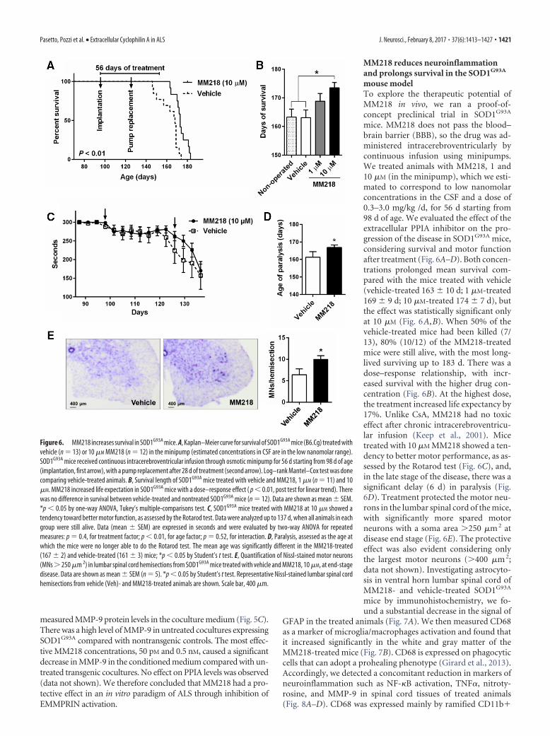
mouse modelTo explore the therapeutic potential ofMM218 in vivo, we ran a proof-of-concept preclinical trial in SOD1G93A
mice. MM218 does not pass the blood–brain barrier (BBB), so the drug was ad-ministered intracerebroventricularly bycontinuous infusion using minipumps.We treated animals with MM218, 1 and10 �M (in the minipump), which we esti-mated to correspond to low nanomolarconcentrations in the CSF and a dose of0.3–3.0 mg/kg /d, for 56 d starting from98 d of age. We evaluated the effect of theextracellular PPIA inhibitor on the pro-gression of the disease in SOD1G93A mice,considering survival and motor functionafter treatment (Fig. 6A–D). Both concen-trations prolonged mean survival com-pared with the mice treated with vehicle(vehicle-treated 163 � 10 d; 1 �M-treated169 � 9 d; 10 �M-treated 174 � 7 d), butthe effect was statistically significant onlyat 10 �M (Fig. 6A,B). When 50% of thevehicle-treated mice had been killed (7/13), 80% (10/12) of the MM218-treatedmice were still alive, with the most long-lived surviving up to 183 d. There was adose–response relationship, with incr-eased survival with the higher drug con-centration (Fig. 6B). At the highest dose,the treatment increased life expectancy by17%. Unlike CsA, MM218 had no toxiceffect after chronic intracerebroventricu-lar infusion (Keep et al., 2001). Micetreated with 10 �M MM218 showed a ten-dency to better motor performance, as as-sessed by the Rotarod test (Fig. 6C), and,in the late stage of the disease, there was asignificant delay (6 d) in paralysis (Fig.6D). Treatment protected the motor neu-rons in the lumbar spinal cord of the mice,with significantly more spared motorneurons with a soma area 250 �m 2 atdisease end stage (Fig. 6E). The protectiveeffect was also evident considering onlythe largest motor neurons (400 �m 2;data not shown). Investigating astrocyto-sis in ventral horn lumbar spinal cord ofMM218- and vehicle-treated SOD1G93A
mice by immunohistochemistry, we fo-und a substantial decrease in the signal of
GFAP in the treated animals (Fig. 7A). We then measured CD68as a marker of microglia/macrophages activation and found thatit increased significantly in the white and gray matter of theMM218-treated mice (Fig. 7B). CD68 is expressed on phagocyticcells that can adopt a prohealing phenotype (Girard et al., 2013).Accordingly, we detected a concomitant reduction in markers ofneuroinflammation such as NF-�B activation, TNF�, nitroty-rosine, and MMP-9 in spinal cord tissues of treated animals(Fig. 8A–D). CD68 was expressed mainly by ramified CD11b�
Figure 6. MM218 increases survival in SOD1G93A mice. A, Kaplan–Meier curve for survival of SOD1G93A mice (B6.Cg) treated withvehicle (n � 13) or 10 �M MM218 (n � 12) in the minipump (estimated concentrations in CSF are in the low nanomolar range).SOD1G93A mice received continuous intracerebroventricular infusion through osmotic minipump for 56 d starting from 98 d of age(implantation, first arrow), with a pump replacement after 28 d of treatment (second arrow). Log–rank Mantel–Cox test was donecomparing vehicle-treated animals. B, Survival length of SOD1G93A mice treated with vehicle and MM218, 1 �M (n � 11) and 10�M. MM218 increased life expectation in SOD1G93A mice with a dose–response effect ( p � 0.01, post test for linear trend). Therewas no difference in survival between vehicle-treated and nontreated SOD1G93A mice (n � 12). Data are shown as mean � SEM.*p � 0.05 by one-way ANOVA, Tukey’s multiple-comparisons test. C, SOD1G93A mice treated with MM218 at 10 �M showed atendency toward better motor function, as assessed by the Rotarod test. Data were analyzed up to 137 d, when all animals in eachgroup were still alive. Data (mean � SEM) are expressed in seconds and were evaluated by two-way ANOVA for repeatedmeasures: p � 0.4, for treatment factor; p � 0.01, for age factor; p � 0.52, for interaction. D, Paralysis, assessed as the age atwhich the mice were no longer able to do the Rotarod test. The mean age was significantly different in the MM218-treated(167 � 2) and vehicle-treated (161 � 3) mice; *p � 0.05 by Student’s t test. E, Quantification of Nissl-stained motor neurons(MNs 250 �m 2) in lumbar spinal cord hemisections from SOD1G93A mice treated with vehicle and MM218, 10 �M, at end-stagedisease. Data are shown as mean � SEM (n � 5). *p � 0.05 by Student’s t test. Representative Nissl-stained lumbar spinal cordhemisections from vehicle (Veh)- and MM218-treated animals are shown. Scale bar, 400 �m.
Pasetto, Pozzi et al. • Extracellular Cyclophilin A in ALS J. Neurosci., February 8, 2017 • 37(6):1413–1427 • 1421

1422 • J. Neurosci., February 8, 2017 • 37(6):1413–1427 Pasetto, Pozzi et al. • Extracellular Cyclophilin A in ALS

cells in the gray and the white matter of the treated animals (Fig.7C,D). Moreover, CD11b� cells showed a decreased ameboid mor-phology (lower circularity and solidity) and more ramifications(higher grid crossing; Fig. 7E) in white and gray matter of the spinalcord of the treated mice, underlining a functional shift of microglia/macrophages upon PPIA inhibition (Fumagalli et al., 2015; Zanier etal., 2015). We also measured 78 kDa glucose-regulated protein(BiP), which is a marker of endoplasmic reticulum (ER) stress andfound that it was lower in treated animals (Fig. 8E). Finally, we in-vestigated whether the treatment interfered with PPIA intracellularfunctions, those linked to protein aggregation and TDP-43 function,such as regulation of histone deacetylase 6 expression (HDAC6),shown in previous works (Basso et al., 2009; Lauranzano et al., 2015).We found no changes in PPIA protein levels, soluble and detergent-
insoluble aggregated SOD1G93A, andHDAC6 expression (data not shown). Weobserved a reduced level of insoluble phos-phorylated TDP-43 in the spinal cord of thetreated mice (Fig. 8F ), indicating thatMM218, by reducing neuroinflammation,may affect TDP-43 pathology.
DiscussionThe key findings of the present work are thatextracellular PPIA is an unexpected media-tor of the neuroinflammatory reaction inALS and is toxic for motor neurons. Sup-porting this, a specific inhibitor of extracel-lular PPIA reduces neuroinflammation,protects motor neurons, and increases sur-vival in SOD1G93A mice.
To decipher the role of PPIA in ALSpathogenesis, we crossbred the PPIA�/�
mouse with the SOD1G93A mouse (Lauran-zano et al., 2015). General depletion of PPIAexacerbated protein aggregation and af-fected key TDP-43 functions, hastening dis-ease progression and shortening thelifespan. We concluded that PPIA has a ben-eficial role in ALS because of its intracellularfunctions in protein folding and complexassembly. Further studies, reported here, in-dicated that SOD1G93APPIA�/� mice havemore spared motor neurons, confirmingthat motor neuron protection is not alwayspredictive of an improved clinical response(Gould et al., 2006). This was consistent
with reports of a possible role of PPIA in cell death pathways, bycooperating to AIF nuclear translocation (Zhu et al., 2007; Tanaka etal., 2011). However, we found a small decrease in AIF nuclear trans-location in SOD1G93APPIA�/� mice only at a symptomatic stage ofthe disease. This suggested that motor neuron protection could notbe explained solely by such a mechanism. One established functionof extracellular PPIA mediated by the EMMPRIN receptor is to pro-mote the induction and release of MMPs (Kim et al., 2005; Satoh etal., 2009; Seizer et al., 2010; Bahmed et al., 2012). MMPs can beneurotoxic through a number of mechanisms, including inductionof ER stress (Kaplan et al., 2014). Most cells express low level ofMMPs, but their expression can be induced by proinflammatorycytokines. High levels of MMPs have been found in tissues and bio-fluids of ALS patients (Lim et al., 1996; Beuche et al., 2000; Fang et al.,2009). Moreover, MMP-9 is a determinant of selective motor neu-ron degeneration (Kaplan et al., 2014). It is strongly expressed byvulnerable motor neurons in SOD1G93A mice and knocking outMMP-9 has led to motor neuron protection (Kiaei et al., 2007; Ka-plan et al., 2014). We found that, in the absence of PPIA, the level ofMMP-9 was substantially lower in SOD1G93A mice already at a pre-symptomatic stage. We therefore deduced that PPIA is a major in-ducer of MMP-9 and low levels of MMP-9 may contribute to motorneuron protection in SOD1G93APPIA�/� mice. Therefore, PPIA hasdistinctive and divergent roles in ALS depending on its interactors/substrates. For example, intracellularly, it is beneficial, acting as afoldase/chaperone for proteins such as TDP-43 and mutant SOD1(Lauranzano et al., 2015), whereas extracellularly, it is detrimental byactivating an EMMPRIN-dependent pathway.
The exact mechanism through which PPIA is secreted by cells hasnot yet been defined. Oxidative stress and inflammation stimulate
4
Figure 7. MM218 modulates glial activation. A, Activation of astrocytes was reduced inanimals treated with 10 �M MM218, as assessed by GFAP immunostaining in lumbar spinal cordhemisections at end-stage disease. Representative GFAP-stained lumbar spinal cord hemisec-tions from vehicle (Veh)- and MM218-treated animals are shown. Scale bar, 20 �m. B, In-creased CD68 staining was detected in the white matter (w.m.) and gray matter (g.m.) of thelumbar spinal cord in the MM218-treated mice at end-stage disease. Representative CD68-stained lumbar spinal cord hemisections from vehicle- and MM218-treated animals are shown.Scale bar, 100 �m. C, D, CD68 staining colocalized exclusively with ramified CD11b� cells inthe gray (C) and the white (D) matter of the treated animals. Representative confocal image oflumbar spinal cord from vehicle- and MM218-treated animals at end-stage disease costainedfor CD11b (green staining) and CD68 (red staining). Scale bar, 20 �m. E, Quantitative analysis ofshape descriptors (circularity, solidity, and grid crossing) indicates that CD11b� cells were lessameboid and had more ramifications in lumbar spinal cord in MM218-treated mice at end-stagedisease, both in the white and the gray matter. For A, B, and E, data are shown as mean � SEM(n � 5). *p � 0.05 by Student’s t test.
Figure 8. MM218 reduces markers of neuroinflammation. A, F, Immnunoblot analysis of lysates from spinal cord of SOD1G93A
mice treated with vehicle (Veh; n � 8) or 10 �M MM218 (n � 10). MM218 reduced NF-�B activation (A), proinflammatorycytokine TNF� (B), nitrotyrosine (C), MMP-9 (D), and ER stress chaperone BiP (E). Immunoreactivity, measured by dot blot (A, C, E)and Western blot (B) with the specific antibodies, was normalized to protein loading (Red Ponceau). NF-�B activation wasquantified by normalization of the phospho (p)-NF-�B p65 signal to total NF-�B p65. Levels of MMP-9 were measured byAlphaLISA and were normalized to protein loading, as quantified by BCA assay, and to the mean number of Nissl-stained motorneurons (MNs 400 �m 2) in lumbar spinal cord hemisections. Data are shown as mean � SEM. *p � 0.05, by Student’s t test.F, Analysis of the Triton-insoluble fraction from spinal cord of SOD1G93A mice treated with vehicle (Veh; n � 8) or 10 �M MM218(n � 10). Insoluble phosphorylated TDP-43 was measured by dot blot with the specific antibody. Immunoreactivity was normal-ized to protein loading (Red Ponceau). Data are shown as mean � SEM. *p � 0.05, by Student’s t test.
Pasetto, Pozzi et al. • Extracellular Cyclophilin A in ALS J. Neurosci., February 8, 2017 • 37(6):1413–1427 • 1423

PPIA secretion possibly by inducing post-translational modifications and through avesicular pathway (Suzuki et al., 2006; Soe etal., 2014). Increased levels of extracellularPPIA have been detected in many inflam-matory conditions and its role in pathogen-esis has been verified in a range of animalmodels (Hoffmann and Schiene-Fischer,2014). We here report that high levels of ex-tracellular PPIA are also associated withALS, as detected in vitro in mouse glial cellsand astrocyte–spinal neuron cocultures ex-pressing SOD1G93A, and in vivo, in the CSFof SOD1G93A mice and rats and sporadicALS patients. Therefore, our findings maybe relevant for both the familial and the spo-radic cases, which account for 90% of all pa-tients. We found that motor neurons aremore vulnerable than other neuronal typesto PPIA toxicity. Extracellular functions ofPPIA are mediated by EMMPRIN (Yurch-enko et al., 2002; Malesevic et al., 2013).EMMPRIN is highly expressed in CNS, es-pecially in spinal cord gray matter (Fan et al.,1998). We found a strong staining forEMMPRIN in large motor neurons and ahigher level of the protein in the ventralhorn compared with the dorsal horn spinalcord, suggesting that EMMPRIN is highlyexpressed in motor neurons. PPIA is a ma-jor inducer of MMP-9, as shown in theSOD1G93APPIA�/� mouse studies. MMP-9is expressed selectively by fast motor neu-rons and accelerates their degeneration byinducing ER stress (Kaplan et al., 2014).Therefore, increased secretion of PPIA byneurons and glial cells under pathologicalconditions could have a selective detrimen-tal effect on vulnerable motor neurons inwhich the toxic EMMPRIN/MMP-9 path-way is upregulated.
Potent PPIA inhibitors, mainly CsAderivatives, have been developed for sev-eral clinical applications; however, all tar-get both intracellular and extracellularPPIA. Our studies suggest that generalPPIA inhibition is not recommended forALS. We and others have shown that stan-dard CsA is toxic for neurons and motorneurons in vitro (Maxwell et al., 2004; VanDen Bosch et al., 2004). Chronic intrathe-cal injection of CsA in mutant SOD1 miceimproved survival, but had severe neurotoxic effects (Keep et al.,2001). Moreover, CsA administered systemically does not reachthe CNS because of the poor BBB permeability and shows nobenefit in ALS patients (Appel et al., 1988). We tested the cell-impermeable CsA derivative MM218 (Balsley et al., 2010;Malesevic et al., 2010) in our in vitro paradigm of ALS, astrocyte–spinal neuron cocultures expressing SOD1G93A, in which thespontaneous loss of motor neurons is associated with increasedextracellular PPIA and MMP-9 levels. In agreement with ourhypothesis, MM218 protected motor neurons and concomi-tantly reduced MMP-9 induction, confirming the causal link be-
tween motor neuron death and activation of EMMPRIN. Wetherefore moved to a proof-of-concept preclinical trial in theSOD1G93A mouse model. The treatment started at approximatelysymptom onset affected disease progression with a dose–re-sponse relationship and the highest dose increased life expec-tancy by 17%. Motor performance, assessed by the Rotarod test,did not improve significantly, even if there was a clear tendency ofamelioration. The test with mice implanted with a brain infusioncannula connected with an osmotic minipump may be challeng-ing and not fully reliable. It is also possible that muscles inSOD1G93A mice are susceptible to extracellular PPIA toxicity.
Figure 9. Model of the non-cell-autonomous mechanism by which extracellular PPIA mediates motor neuron death andMM218 is protective. A, At an early phase of the disease, PPIA is upregulated by motor neurons and neighboring cells as a protectiveresponse against pathogenic pathways induced by mutant SOD1 (protein misfolding, oxidative stress, etc.). As the disease pro-gresses, motor neurons especially, but also glial cells, secrete PPIA, which activates an inflammatory response mediated by theEMMPRIN receptor, leading to NF-�B activation and induction of MMP-9 and proinflammatory cytokines (e.g., TNF�). B, Thecrosstalk between motor neurons and glial cells further exacerbates the proinflammatory response by increasing NF-�B activation,ER stress, and motor neuron death. Treatment with extracellular PPIA inhibitor MM218 at the onset of symptoms blocks theEMMPRIN-initiated pathway upstream, leading to polarization of glia toward a prohealing phenotype associated with reducedNF-�B activation, proinflammatory markers, and ER stress. This resulted in the protection of motor neurons and increased survivalof SOD1G93A mice.
1424 • J. Neurosci., February 8, 2017 • 37(6):1413–1427 Pasetto, Pozzi et al. • Extracellular Cyclophilin A in ALS

Under stress conditions, muscle cells secrete high levels of PPIAthat promote inflammation in vivo (Satoh et al., 2009). It has beenreported that an analog of MM218 reduced myocardial inflam-mation and fibrosis (Heinzmann et al., 2015). Given the tolera-bility of this drug, we would expect the clinical response to begreater with higher doses of MM218, possibly combining intra-cerebroventricular and systemic administration. The exclusiveinhibition of extracellular PPIA by MM218 was sufficient to pro-tect motor neurons in the SOD1G93A mice, as it was in vitro. PPIAactivates through EMMPRIN a signaling pathway that is invari-ably NF-�B dependent (Kim et al., 2005; Satoh et al., 2009; Seizeret al., 2010; Bahmed et al., 2012). Here, we found that EMMPRINis upregulated in ventral horn spinal cord of SOD1G93A mice.NF-�B is activated aberrantly in ALS patients and SOD1G93A
mice and its absence or inhibition was beneficial in differentmouse models of ALS (Swarup et al., 2011; Frakes et al., 2014;Patel et al., 2015). We found a reduced NF-�B activation in thespinal cord of the MM218-treated SOD1G93A mice. Therefore, wepropose that extracellular PPIA, through EMMPRIN, contrib-utes to the aberrant activation of NF-�B, so its pharmacologicaltargeting by MM218 leads to a positive clinical response by in-hibiting NF-�B signaling in motor neurons and neighboring glialcells through a non-cell-autonomous mechanism (Fig. 9A,B).This resulted in the polarization of glia toward a prohealing phe-notype associated with decreased levels of neuroinflammatoryfactors such as TNF� and MMP-9. Extracellular PPIA, in addi-tion to TNF� and MMP-9, induces expression of other proin-flammatory cytokines/chemokines such as IL-8, MCP-1, andIL-1beta, depending on the cell type (Kim et al., 2005). TNF� wasalso reduced in SOD1G93A mice knocked out for MMP-9, whichhas been suggested to regulate the expression and release of sol-uble TNF� (Kiaei et al., 2007). However, our approach was moreeffective than the pharmacological inhibition of MMP-9 (Lorenzlet al., 2006; Kaplan et al., 2014), indicating that inhibiting theEMMPRIN-initiated pathway upstream is a better option, actingat the same time on a number of proinflammatory factors. Extra-cellular PPIA has a potent chemotactic activity toward leukocytesthat is inhibited by MM218 (Yurchenko et al., 2002; Damsker etal., 2007; Malesevic et al., 2010). Therefore, the protective effectof MM218 is unlikely to be linked to a recruitment of immunore-golatory cells to CNS that has been shown with other therapeuticstrategies (Beers et al., 2011; Peviani et al., 2014; Kunis et al.,2015). We can hypothesize that the inhibition of extracellularPPIA limited the infiltration of certain toxic immune cells(Vaknin et al., 2011; Butovsky et al., 2012) and favored a protec-tive activation of resident microglia. Consistent with this,MM218 treatment induced a morphological and phenotypicalswitch of microglia that may be associated with debris clearingand tissue remodeling (Vinet et al., 2012; Hong and Stevens,2016).
We also detected lower ER stress, like in the SOD1G93A miceknocked out for MMP-9 (Kaplan et al., 2014). The molecularmechanisms linking MMP-9 to ER stress have not been clarified.There is ample evidence of an interplay between ER stress andinflammation mediated by NF-�B activation (Hasnain et al.,2012) and pathological conditions in which inflammatory factorsinduce or exacerbate ER stress (Xue et al., 2005; Brozzi et al.,2015). Therefore, hypothetically blocking the EMMPRIN/NF-�Bpathway by inhibiting extracellular PPIA may have a concomi-tant inhibitory effect on ER stress. Finally, the treatment did notaffect PPIA levels and intracellular functions; it only reduced thelevel of detergent-insoluble phosphorylated TDP-43. This effectis probably linked to the reduced neuroinflammation. In fact,
activation of the NF-�B pathway by inflammatory stimuli pro-moted TDP-43 pathology, which is associated with increaseddetergent-insoluble phosphorylated TDP-43 (Correia et al.,2015; Lauranzano et al., 2015).
ReferencesAgrawal SM, Silva C, Tourtellotte WW, Yong VW (2011) EMMPRIN: a
novel regulator of leukocyte transmigration into the CNS in multiplesclerosis and experimental autoimmune encephalomyelitis. J Neurosci31:669 – 677. CrossRef Medline
Appel SH, Stewart SS, Appel V, Harati Y, Mietlowski W, Weiss W, BelendiukGW (1988) A double-blind study of the effectiveness of cyclosporine inamyotrophic lateral sclerosis. Arch Neurol 45:381–386. CrossRef Medline
Bahmed K, Henry C, Holliday M, Redzic J, Ciobanu M, Zhang F, WeekesC, Sclafani R, Degregori J, Eisenmesser E (2012) Extracellularcyclophilin-A stimulates ERK1/2 phosphorylation in a cell-dependentmanner but broadly stimulates nuclear factor kappa B. Cancer Cell Int12:19. CrossRef Medline
Balsley MA, Malesevic M, Stemmy EJ, Gigley J, Jurjus RA, Herzog D, Bukrin-sky MI, Fischer G, Constant SL (2010) A cell-impermeable cyclosporineA derivative reduces pathology in a mouse model of allergic lung inflam-mation. J Immunol 185:7663–7670. CrossRef Medline
Basso M, Samengo G, Nardo G, Massignan T, D’Alessandro G, Tartari S,Cantoni L, Marino M, Cheroni C, De Biasi S, Giordana MT, Strong MJ,Estevez AG, Salmona M, Bendotti C, Bonetto V (2009) Characterizationof detergent-insoluble proteins in ALS indicates a causal link betweennitrative stress and aggregation in pathogenesis. PLoS One 4:e8130.CrossRef Medline
Basso M, Pozzi S, Tortarolo M, Fiordaliso F, Bisighini C, Pasetto L, Spaltro G,Lidonnici D, Gensano F, Battaglia E, Bendotti C, Bonetto V (2013) Mu-tant copper-zinc superoxide dismutase (SOD1) induces protein secretionpathway alterations and exosome release in astrocytes: implications fordisease spreading and motor neuron pathology in amyotrophic lateralsclerosis. J Biol Chem 288:15699 –15711. CrossRef Medline
Beers DR, Henkel JS, Zhao W, Wang J, Huang A, Wen S, Liao B, Appel SH(2011) Endogenous regulatory T lymphocytes ameliorate amyotrophiclateral sclerosis in mice and correlate with disease progression in patientswith amyotrophic lateral sclerosis. Brain 134:1293–1314. CrossRefMedline
Beuche W, Yushchenko M, Mader M, Maliszewska M, Felgenhauer K, WeberF (2000) Matrix metalloproteinase-9 is elevated in serum of patientswith amyotrophic lateral sclerosis. Neuroreport 11:3419 –3422. CrossRefMedline
Bigini P, Steffensen KR, Ferrario A, Diomede L, Ferrara G, Barbera S, SalzanoS, Fumagalli E, Ghezzi P, Mennini T, Gustafsson JA (2010) Neuro-pathologic and biochemical changes during disease progression in liver Xreceptor beta�/� mice, a model of adult neuron disease. J NeuropatholExp Neurol 69:593– 605. CrossRef Medline
Boillee S, Yamanaka K, Lobsiger CS, Copeland NG, Jenkins NA, Kassiotis G,Kollias G, Cleveland DW (2006) Onset and progression in inheritedALS determined by motor neurons and microglia. Science 312:1389 –1392. CrossRef Medline
Brozzi F, Nardelli TR, Lopes M, Millard I, Barthson J, Igoillo-Esteve M,Grieco FA, Villate O, Oliveira JM, Casimir M, Bugliani M, Engin F, Hota-misligil GS, Marchetti P, Eizirik DL (2015) Cytokines induce endoplas-mic reticulum stress in human, rat and mouse beta cells via differentmechanisms. Diabetologia 58:2307–2316. CrossRef Medline
Butovsky O, Siddiqui S, Gabriely G, Lanser AJ, Dake B, Murugaiyan G, Doy-kan CE, Wu PM, Gali RR, Iyer LK, Lawson R, Berry J, Krichevsky AM,Cudkowicz ME, Weiner HL (2012) Modulating inflammatory mono-cytes with a unique microRNA gene signature ameliorates murine ALS.J Clin Invest 122:3063–3087. CrossRef Medline
Correia AS, Patel P, Dutta K, Julien JP (2015) Inflammation InducesTDP-43 Mislocalization and Aggregation. PLoS One 10:e0140248.CrossRef Medline
Damsker JM, Bukrinsky MI, Constant SL (2007) Preferential chemotaxis ofactivated human CD4� T cells by extracellular cyclophilin A. J LeukocBiol 82:613– 618. CrossRef Medline
Drachman DB, Frank K, Dykes-Hoberg M, Teismann P, Almer G, Przedbor-ski S, Rothstein JD (2002) Cyclooxygenase 2 inhibition protects motorneurons and prolongs survival in a transgenic mouse model of ALS. AnnNeurol 52:771–778. CrossRef Medline
Pasetto, Pozzi et al. • Extracellular Cyclophilin A in ALS J. Neurosci., February 8, 2017 • 37(6):1413–1427 • 1425

Fan QW, Yuasa S, Kuno N, Senda T, Kobayashi M, Muramatsu T, KadomatsuK (1998) Expression of basigin, a member of the immunoglobulin su-perfamily, in the mouse central nervous system. Neurosci Res 30:53– 63.CrossRef Medline
Fang L, Huber-Abel F, Teuchert M, Hendrich C, Dorst J, Schattauer D,Zettlmeissel H, Wlaschek M, Scharffetter-Kochanek K, Tumani H, Lu-dolph AC, Brettschneider J (2009) Linking neuron and skin: matrixmetalloproteinases in amyotrophic lateral sclerosis (ALS). J Neurol Sci285:62– 66. CrossRef Medline
Faure J, Lachenal G, Court M, Hirrlinger J, Chatellard-Causse C, Blot B,Grange J, Schoehn G, Goldberg Y, Boyer V, Kirchhoff F, Raposo G, GarinJ, Sadoul R (2006) Exosomes are released by cultured cortical neurones.Mol Cell Neurosci 31:642– 648. CrossRef Medline
Fischer G, Bang H, Mech C (1984) Determination of enzymatic catalysis forthe cis-trans-isomerization of peptide binding in proline-containing pep-tides. Biomed Biochim Acta 43:1101–1111. Medline
Frakes AE, Ferraiuolo L, Haidet-Phillips AM, Schmelzer L, Braun L, MirandaCJ, Ladner KJ, Bevan AK, Foust KD, Godbout JP, Popovich PG, GuttridgeDC, Kaspar BK (2014) Microglia induce motor neuron death via theclassical NF-kappaB pathway in amyotrophic lateral sclerosis. Neuron81:1009 –1023. CrossRef Medline
Fumagalli S, Perego C, Pischiutta F, Zanier ER, De Simoni MG (2015) Theischemic environment drives microglia and macrophage function. FrontNeurol 6:81. CrossRef Medline
Girard S, Brough D, Lopez-Castejon G, Giles J, Rothwell NJ, Allan SM (2013)Microglia and macrophages differentially modulate cell death after braininjury caused by oxygen-glucose deprivation in organotypic brain slices.Glia 61:813– 824. CrossRef Medline
Goldner FM, Patrick JW (1996) Neuronal localization of the cyclophilin Aprotein in the adult rat brain. J Comp Neurol 372:283–293. Medline
Gould TW, Buss RR, Vinsant S, Prevette D, Sun W, Knudson CM, MilliganCE, Oppenheim RW (2006) Complete dissociation of motor neurondeath from motor dysfunction by Bax deletion in a mouse model of ALS.J Neurosci 26:8774 – 8786. CrossRef Medline
Handschumacher RE, Harding MW, Rice J, Drugge RJ, Speicher DW (1984)Cyclophilin: a specific cytosolic binding protein for cyclosporin A. Sci-ence 226:544 –547. CrossRef Medline
Hasnain SZ, Lourie R, Das I, Chen AC, McGuckin MA (2012) The interplaybetween endoplasmic reticulum stress and inflammation. Immunol CellBiol 90:260 –270. CrossRef Medline
Heinzmann D, Bangert A, Muller AM, von Ungern-Sternberg SN, Emscher-mann F, Schonberger T, Chatterjee M, Mack AF, Klingel K, Kandolf R,Malesevic M, Borst O, Gawaz M, Langer HF, Katus H, Fischer G, May AE,Kaya Z, Seizer P (2015) The novel extracellular cyclophilin A (CyPA)inhibitor MM284 reduces myocardial inflammation and remodeling in amouse model of troponin I-induced myocarditis. PLoS One 10:e0124606.CrossRef Medline
Hoffmann H, Schiene-Fischer C (2014) Functional aspects of extracellularcyclophilins. Biol Chem 395:721–735. CrossRef Medline
Hong S, Stevens B (2016) Microglia: phagocytosing to clear, sculpt, andeliminate. Dev Cell 38:126 –128. CrossRef Medline
Howland DS, Liu J, She Y, Goad B, Maragakis NJ, Kim B, Erickson J, Kulik J,DeVito L, Psaltis G, DeGennaro LJ, Cleveland DW, Rothstein JD (2002)Focal loss of the glutamate transporter EAAT2 in a transgenic rat model ofSOD1 mutant-mediated amyotrophic lateral sclerosis (ALS). Proc NatlAcad Sci U S A 99:1604 –1609. CrossRef Medline
Jaschke A, Mi H, Tropschug M (1998) Human T cell cyclophilin18 binds tothiol-specific antioxidant protein Aop1 and stimulates its activity. J MolBiol 277:763–769. CrossRef Medline
Jin ZG, Melaragno MG, Liao DF, Yan C, Haendeler J, Suh YA, Lambeth JD,Berk BC (2000) Cyclophilin A is a secreted growth factor induced byoxidative stress. Circ Res 87:789 –796. CrossRef Medline
Kaplan A, Spiller KJ, Towne C, Kanning KC, Choe GT, Geber A, AkayT, Aebischer P, Henderson CE (2014) Neuronal matrix meta-lloproteinase-9 is a determinant of selective neurodegeneration. Neuron81:333–348. CrossRef Medline
Keep M, Elmer E, Fong KS, Csiszar K (2001) Intrathecal cyclosporin pro-longs survival of late-stage ALS mice. Brain Res 894:327–331. CrossRefMedline
Kiaei M, Petri S, Kipiani K, Gardian G, Choi DK, Chen J, Calingasan NY,Schafer P, Muller GW, Stewart C, Hensley K, Beal MF (2006) Thalido-
mide and lenalidomide extend survival in a transgenic mouse model ofamyotrophic lateral sclerosis. J Neurosci 26:2467–2473. CrossRef Medline
Kiaei M, Kipiani K, Calingasan NY, Wille E, Chen J, Heissig B, Rafii S, LorenzlS, Beal MF (2007) Matrix metalloproteinase-9 regulates TNF-alpha andFasL expression in neuronal, glial cells and its absence extends life in atransgenic mouse model of amyotrophic lateral sclerosis. Exp Neurol205:74 – 81. CrossRef Medline
Kim H, Kim WJ, Jeon ST, Koh EM, Cha HS, Ahn KS, Lee WH (2005) Cy-clophilin A may contribute to the inflammatory processes in rheumatoidarthritis through induction of matrix degrading enzymes and inflamma-tory cytokines from macrophages. Clin Immunol 116:217–224. CrossRefMedline
Kriz J, Nguyen MD, Julien JP (2002) Minocycline slows disease progressionin a mouse model of amyotrophic lateral sclerosis. Neurobiol Dis 10:268 –278. CrossRef Medline
Kunis G, Baruch K, Miller O, Schwartz M (2015) Immunization with amyelin-derived antigen activates the brain’s choroid plexus for recruit-ment of immunoregulatory cells to the CNS and attenuates disease pro-gression in a mouse model of ALS. J Neurosci 35:6381– 6393. CrossRefMedline
Lauranzano E, Pozzi S, Pasetto L, Stucchi R, Massignan T, Paolella K, Mom-brini M, Nardo G, Lunetta C, Corbo M, Mora G, Bendotti C, Bonetto V(2015) Peptidylprolyl isomerase A governs TARDBP function and as-sembly in heterogeneous nuclear ribonucleoprotein complexes. Brain138:974 –991. CrossRef Medline
Lee JP, Palfrey HC, Bindokas VP, Ghadge GD, Ma L, Miller RJ, Roos RP(1999) The role of immunophilins in mutant superoxide dismutase-1linked familial amyotrophic lateral sclerosis. Proc Natl Acad Sci U S A96:3251–3256. CrossRef Medline
Lee SP, Hwang YS, Kim YJ, Kwon KS, Kim HJ, Kim K, Chae HZ (2001)Cyclophilin a binds to peroxiredoxins and activates its peroxidase activity.J Biol Chem 276:29826 –29832. CrossRef Medline
Lim GP, Backstrom JR, Cullen MJ, Miller CA, Atkinson RD, Tokes ZA(1996) Matrix metalloproteinases in the neocortex and spinal cord ofamyotrophic lateral sclerosis patients. J Neurochem 67:251–259. Medline
Lorenzl S, Narr S, Angele B, Krell HW, Gregorio J, Kiaei M, Pfister HW, BealMF (2006) The matrix metalloproteinases inhibitor Ro 28 –2653 [cor-rection of Ro 26 –2853] extends survival in transgenic ALS mice. ExpNeurol 200:166 –171. CrossRef Medline
Malesevic M, Kuhling J, Erdmann F, Balsley MA, Bukrinsky MI, Constant SL,Fischer G (2010) A cyclosporin derivative discriminates between extra-cellular and intracellular cyclophilins. Angew Chem Int Ed Engl 49:213–215. CrossRef Medline
Malesevic M, Gutknecht D, Prell E, Klein C, Schumann M, Nowak RA, SimonJC, Schiene-Fischer C, Saalbach A (2013) Anti-inflammatory effects ofextracellular cyclosporins are exclusively mediated by CD147. J MedChem 56:7302–7311. CrossRef Medline
Marino M, Papa S, Crippa V, Nardo G, Peviani M, Cheroni C, Trolese MC,Lauranzano E, Bonetto V, Poletti A, DeBiasi S, Ferraiuolo L, Shaw PJ,Bendotti C (2015) Differences in protein quality control correlate withphenotype variability in 2 mouse models of familial amyotrophic lateralsclerosis. Neurobiol Aging 36:492–504. CrossRef Medline
Massignan T, Casoni F, Basso M, Stefanazzi P, Biasini E, Tortarolo M, Sal-mona M, Gianazza E, Bendotti C, Bonetto V (2007) Proteomic analysisof spinal cord of presymptomatic amyotrophic lateral sclerosis G93ASOD1 mouse. Biochem Biophys Res Commun 353:719 –725. CrossRefMedline
Maxwell MM, Pasinelli P, Kazantsev AG, Brown RH Jr (2004) RNAinterference-mediated silencing of mutant superoxide dismutase rescuescyclosporin A-induced death in cultured neuroblastoma cells. Proc NatlAcad Sci U S A 101:3178 –3183. CrossRef Medline
Nardo G, Pozzi S, Pignataro M, Lauranzano E, Spano G, Garbelli S, Man-tovani S, Marinou K, Papetti L, Monteforte M, Torri V, Paris L, BazzoniG, Lunetta C, Corbo M, Mora G, Bendotti C, Bonetto V (2011) Amyo-trophic lateral sclerosis multiprotein biomarkers in peripheral bloodmononuclear cells. PLoS One 6:e25545. CrossRef Medline
Neymotin A, Petri S, Calingasan NY, Wille E, Schafer P, Stewart C, Hensley K,Beal MF, Kiaei M (2009) Lenalidomide (Revlimid) administration atsymptom onset is neuroprotective in a mouse model of amyotrophiclateral sclerosis. Exp Neurol 220:191–197. CrossRef Medline
Nigro P, Pompilio G, Capogrossi MC (2013) Cyclophilin A: a key player forhuman disease. Cell Death Dis 4:e888. CrossRef Medline
1426 • J. Neurosci., February 8, 2017 • 37(6):1413–1427 Pasetto, Pozzi et al. • Extracellular Cyclophilin A in ALS
Patel P, Julien JP, Kriz J (2015) Early-stage treatment with Withaferin Areduces levels of misfolded superoxide dismutase 1 and extends lifespanin a mouse model of amyotrophic lateral sclerosis. Neurotherapeutics12:217–233. CrossRef Medline
Perego C, Fumagalli S, De Simoni MG (2011) Temporal pattern of expres-sion and colocalization of microglia/macrophage phenotype markers fol-lowing brain ischemic injury in mice. J Neuroinflammation 8:174.CrossRef Medline
Peviani M, Salvaneschi E, Bontempi L, Petese A, Manzo A, Rossi D, SalmonaM, Collina S, Bigini P, Curti D (2014) Neuroprotective effects of theSigma-1 receptor (S1R) agonist PRE-084, in a mouse model of motorneuron disease not linked to SOD1 mutation. Neurobiol Dis 62:218 –232.CrossRef Medline
Pizzasegola C, Caron I, Daleno C, Ronchi A, Minoia C, Carrì MT, Bendotti C(2009) Treatment with lithium carbonate does not improve disease pro-gression in two different strains of SOD1 mutant mice. Amyotroph Lat-eral Scler 10:221–228. CrossRef Medline
Restelli E, Fioriti L, Mantovani S, Airaghi S, Forloni G, Chiesa R (2010) Celltype-specific neuroprotective activity of untranslocated prion protein.PLoS One 5:e13725. CrossRef Medline
Robberecht W, Philips T (2013) The changing scene of amyotrophic lateralsclerosis. Nat Rev Neurosci 14:248 –264. CrossRef Medline
Sahawneh MA, Ricart KC, Roberts BR, Bomben VC, Basso M, Ye Y, Sa-hawneh J, Franco MC, Beckman JS, Estevez AG (2010) Cu, Zn-superoxide dismutase increases toxicity of mutant and zinc-deficientsuperoxide dismutase by enhancing protein stability. J Biol Chem 285:33885–33897. CrossRef Medline
Satoh K, Nigro P, Matoba T, O’Dell MR, Cui Z, Shi X, Mohan A, Yan C, AbeJ, Illig KA, Berk BC (2009) Cyclophilin A enhances vascular oxidativestress and the development of angiotensin II-induced aortic aneurysms.Nat Med 15:649 – 656. CrossRef Medline
Schutz B, Reimann J, Dumitrescu-Ozimek L, Kappes-Horn K, Landreth GE,Schurmann B, Zimmer A, Heneka MT (2005) The oral antidiabetic pi-oglitazone protects from neurodegeneration and amyotrophic lateralsclerosis-like symptoms in superoxide dismutase-G93A transgenic mice.J Neurosci 25:7805–7812. CrossRef Medline
Seizer P, Schonberger T, Schott M, Lang MR, Langer HF, Bigalke B, KramerBF, Borst O, Daub K, Heidenreich O, Schmidt R, Lindemann S, Herouy Y,Gawaz M, May AE (2010) EMMPRIN and its ligand cyclophilin A reg-ulate MT1-MMP, MMP-9 and M-CSF during foam cell formation. Ath-erosclerosis 209:51–57. CrossRef Medline
Sherry B, Yarlett N, Strupp A, Cerami A (1992) Identification of cyclophilinas a proinflammatory secretory product of lipopolysaccharide-activatedmacrophages. Proc Natl Acad Sci U S A 89:3511–3515. CrossRef Medline
Soe NN, Sowden M, Baskaran P, Kim Y, Nigro P, Smolock EM, Berk BC(2014) Acetylation of cyclophilin A is required for its secretion and vas-cular cell activation. Cardiovasc Res 101:444 – 453. CrossRef Medline
Suzuki J, Jin ZG, Meoli DF, Matoba T, Berk BC (2006) Cyclophilin A issecreted by a vesicular pathway in vascular smooth muscle cells. Circ Res98:811– 817. CrossRef Medline
Swarup V, Phaneuf D, Dupre N, Petri S, Strong M, Kriz J, Julien JP (2011)Deregulation of TDP-43 in amyotrophic lateral sclerosis triggers nuclearfactor kappaB-mediated pathogenic pathways. J Exp Med 208:2429 –2447. CrossRef Medline
Tanaka H, Shimazaki H, Kimura M, Izuta H, Tsuruma K, Shimazawa M, HaraH (2011) Apoptosis-inducing factor and cyclophilin A cotranslocate tothe motor neuronal nuclei in amyotrophic lateral sclerosis model mice.CNS Neurosci Ther 17:294 –304. CrossRef Medline
Tortarolo M, Veglianese P, Calvaresi N, Botturi A, Rossi C, Giorgini A,Migheli A, Bendotti C (2003) Persistent activation of p38 mitogen-activated protein kinase in a mouse model of familial amyotrophic lateral
sclerosis correlates with disease progression. Mol Cell Neurosci 23:180 –192. CrossRef Medline
Tortarolo M, Grignaschi G, Calvaresi N, Zennaro E, Spaltro G, Colovic M,Fracasso C, Guiso G, Elger B, Schneider H, Seilheimer B, Caccia S, Ben-dotti C (2006) Glutamate AMPA receptors change in motor neurons ofSOD1G93A transgenic mice and their inhibition by a noncompetitiveantagonist ameliorates the progression of amytrophic lateral sclerosis-likedisease. J Neurosci Res 83:134 –146. CrossRef Medline
Tortarolo M, Vallarola A, Lidonnici D, Battaglia E, Gensano F, Spaltro G,Fiordaliso F, Corbelli A, Garetto S, Martini E, Pasetto L, Kallikourdis M,Bonetto V, Bendotti C (2015) Lack of TNF-alpha receptor type 2 pro-tects motor neurons in a cellular model of amyotrophic lateral sclerosisand in mutant SOD1 mice but does not affect disease progression. J Neu-rochem 135:109 –124. CrossRef Medline
Vaknin I, Kunis G, Miller O, Butovsky O, Bukshpan S, Beers DR, Henkel JS,Yoles E, Appel SH, Schwartz M (2011) Excess circulating alternativelyactivated myeloid (M2) cells accelerate ALS progression while inhibitingexperimental autoimmune encephalomyelitis. PLoS One 6:e26921.CrossRef Medline
Van Den Bosch L, Tilkin P, Lemmens G, Robberecht W (2002) Minocyclinedelays disease onset and mortality in a transgenic model of ALS. Neurore-port 13:1067–1070. CrossRef Medline
Van Den Bosch L, Storkebaum E, Vleminckx V, Moons L, Vanopdenbosch L,Scheveneels W, Carmeliet P, Robberecht W (2004) Effects of vascularendothelial growth factor (VEGF) on motor neuron degeneration. Neu-robiol Dis 17:21–28. CrossRef Medline
Vinet J, Weering HR, Heinrich A, Kalin RE, Wegner A, Brouwer N, HeppnerFL, Rooijen Nv, Boddeke HW, Biber K (2012) Neuroprotective functionfor ramified microglia in hippocampal excitotoxicity. J Neuroinflamma-tion 9:27. CrossRef Medline
Xu Q, Leiva MC, Fischkoff SA, Handschumacher RE, Lyttle CR (1992) Leu-kocyte chemotactic activity of cyclophilin. J Biol Chem 267:11968 –11971.Medline
Xue X, Piao JH, Nakajima A, Sakon-Komazawa S, Kojima Y, Mori K, YagitaH, Okumura K, Harding H, Nakano H (2005) Tumor necrosis factoralpha (TNFalpha) induces the unfolded protein response (UPR) in areactive oxygen species (ROS)-dependent fashion, and the UPR counter-acts ROS accumulation by TNFalpha. J Biol Chem 280:33917–33925.CrossRef Medline
Yamanaka K, Chun SJ, Boillee S, Fujimori-Tonou N, Yamashita H, GutmannDH, Takahashi R, Misawa H, Cleveland DW (2008) Astrocytes as deter-minants of disease progression in inherited amyotrophic lateral sclerosis.Nat Neurosci 11:251–253. CrossRef Medline
Yong VW, Power C, Forsyth P, Edwards DR (2001) Metalloproteinases inbiology and pathology of the nervous system. Nat Rev Neurosci 2:502–511. CrossRef Medline
Yuan W, Ge H, He B (2010) Pro-inflammatory activities induced byCyPA-EMMPRIN interaction in monocytes. Atherosclerosis 213:415–421. CrossRef Medline
Yurchenko V, Zybarth G, O’Connor M, Dai WW, Franchin G, Hao T, Guo H,Hung HC, Toole B, Gallay P, Sherry B, Bukrinsky M (2002) Active siteresidues of cyclophilin A are crucial for its signaling activity via CD147.J Biol Chem 277:22959 –22965. CrossRef Medline
Zanier ER, Fumagalli S, Perego C, Pischiutta F, De Simoni MG (2015) Shapedescriptors of the “never resting” microglia in three different acute braininjury models in mice. Intensive Care Med Exp 3:39. CrossRef Medline
Zhu C, Wang X, Deinum J, Huang Z, Gao J, Modjtahedi N, Neagu MR,Nilsson M, Eriksson PS, Hagberg H, Luban J, Kroemer G, Blomgren K(2007) Cyclophilin A participates in the nuclear translocation ofapoptosis-inducing factor in neurons after cerebral hypoxia-ischemia.J Exp Med 204:1741–1748. CrossRef Medline
Pasetto, Pozzi et al. • Extracellular Cyclophilin A in ALS J. Neurosci., February 8, 2017 • 37(6):1413–1427 • 1427