neuroblast migration along the anteroposterior axis of c...
TRANSCRIPT

2915RESEARCH ARTICLE
INTRODUCTIONWnt proteins control many aspects of metazoan development, withprominent functions in cell fate determination, cell proliferationand cell migration (van Amerongen and Nusse, 2009). The activityof Wnt proteins is counteracted by a range of inhibitors, includingsecreted proteins such as the secreted Frizzled-related proteins(SFRPs) (Leyns et al., 1997; Bovolenta et al., 2008) and Dickkopf(Niehrs, 2006), and intracellular inhibitors such as the TCF/Leftranscription factor Tcf3 (Kim et al., 2000). During neurectodermdevelopment in vertebrates, these inhibitors are expressed in theanterior and counteract the activity of posteriorly expressed Wntsto enable the formation of anterior brain structures and the eyes(Leyns et al., 1997; Kiecker and Niehrs, 2001; Tendeng andHouart, 2006). Opposing expression of Wnts and Wnt inhibitorshas also been observed in basal metazoan organisms such as thecnidarians Hydra and Nematostella vectensis (Hobmayer et al.,2000; Kusserow et al., 2005; Guder et al., 2006b; Lee et al., 2006),and in the planarian Schmidtea mediterranea (Petersen andReddien, 2008), which led to the hypothesis that a system ofposterior Wnt signaling and anterior Wnt inhibition is an ancientmechanism that may be used across animal phyla to pattern theprimary body axis (Petersen and Reddien, 2009).
The nematode Caenorhabditis elegans expresses five differentWnt proteins that control many aspects of development, includingcell fate specification, cell polarity and the highly stereotypicmigration of neuroblasts along the anteroposterior body axis(Korswagen, 2002; Silhankova and Korswagen, 2007). Neuroblaststhat migrate in a Wnt-dependent manner include: the HSN neurons,which migrate from the posterior to the mid-body region (Sulston etal., 1983; Hedgecock et al., 1987; Pan et al., 2006); the ALM andCAN neurons, which migrate from the anterior to positions in themid-body region (Sulston et al., 1983; Hedgecock et al., 1987;Zinovyeva and Forrester, 2005); and the Q neuroblast descendants,which migrate in opposite directions on the left and right lateral sides(Sulston and Horvitz, 1977; Harris et al., 1996). With the exceptionof the left Q cell descendants, the migration of these neuroblasts iscontrolled through multiple, partially redundantly acting Wntproteins (Zinovyeva et al., 2008). The expression patterns of the C.elegans Wnt genes have been analyzed using transgenic reporterconstructs. These studies revealed a predominantly posteriorexpression for the Wnt genes lin-44, egl-20 and cwn-1 (Herman etal., 1995; Whangbo and Kenyon, 1999; Pan et al., 2006), whereasmom-2 and cwn-2 were reported to be generally expressed along theanteroposterior axis (Gleason et al., 2006), with a more prominentexpression of cwn-2 in the pharynx and anterior muscle cells(Kennerdell et al., 2009; Song et al., 2010). Although there is a largedegree of overlap between the reported expression patterns, there arealso important differences in the extent of expression along theanteroposterior axis and in the specific cell types that are involved,complicating the analysis of Wnt gene function in neuroblastmigration and other aspects of development.
Here, we used single molecule mRNA fluorescent in situhybridization (smFISH) to determine quantitatively the spatio-temporal expression patterns of the five C. elegans Wnt genes. Our
Development 138, 2915-2924 (2011) doi:10.1242/dev.064733© 2011. Published by The Company of Biologists Ltd
1Hubrecht Institute, Royal Academy of Arts and Sciences and University MedicalCenter Utrecht, Uppsalalaan 8, 3584 CT, Utrecht, The Netherlands. 2Department ofPhysics and department of Biology, Massachusetts Institute of Technology, 77 Massachusetts Avenue, Cambridge, MA 02139, USA.
*These authors contributed equally to this work†Present address: Mechanobiology Institute Singapore, National University ofSingapore, Singapore‡Author for correspondence ([email protected])
Accepted 27 April 2011
SUMMARYThe migration of neuroblasts along the anteroposterior body axis of C. elegans is controlled by multiple Wnts that act partiallyredundantly to guide cells to their precisely defined final destinations. How positional information is specified by this system is,however, still largely unknown. Here, we used a novel fluorescent in situ hybridization methods to generate a quantitativespatiotemporal expression map of the C. elegans Wnt genes. We found that the five Wnt genes are expressed in a series ofpartially overlapping domains along the anteroposterior axis, with a predominant expression in the posterior half of the body.Furthermore, we show that a secreted Frizzled-related protein is expressed at the anterior end of the body axis, where it inhibitsWnt signaling to control neuroblast migration. Our findings reveal that a system of regionalized Wnt gene expression andanterior Wnt inhibition guides the highly stereotypic migration of neuroblasts in C. elegans. Opposing expression of Wnts andWnt inhibitors has been observed in basal metazoans and in the vertebrate neurectoderm. Our results in C. elegans support thenotion that a system of posterior Wnt signaling and anterior Wnt inhibition is an evolutionarily conserved principle of primarybody axis specification.
KEY WORDS: C. elegans, Wnt, Neuroblast migration, Secreted Frizzled-related protein
Neuroblast migration along the anteroposterior axis of C. elegans is controlled by opposing gradients of Wnts and a secreted Frizzled-related proteinMartin Harterink1,*, Dong hyun Kim2,*, Teije C. Middelkoop1, Thang Dinh Doan1,†, Alexander van Oudenaarden1,2 and Hendrik C. Korswagen1,‡
DEVELO
PMENT

2916
results show that the different Wnt genes are expressed in a seriesof partially overlapping expression domains, with a predominantexpression in the posterior body half and a single Wnt gene withan anterior expression domain. Furthermore, we show that the C.elegans genome contains a single SFRP ortholog that is specificallyexpressed at the anterior end of the body axis. SFRP-1 functions asan inhibitor of Wnt signaling that represses the most anteriorlyexpressed Wnts to control the migration of neuroblasts in theanterior body region. Our results demonstrate that opposing Wntand Wnt inhibitory activities are also key to anteroposteriorpatterning in C. elegans and provide further support for theevolutionary conservation of this system in primary body axisspecification.
MATERIALS AND METHODSC. elegans strains and culturingGeneral methods for culture, manipulation and genetics of C. elegans wereas described previously (Lewis and Fleming, 1995). Strains were cultured at20°C. Mutations and transgenes used in this study were: LGI, lin-44(n1792)(Herman et al., 1995), mom-5(gk812), pry-1(mu38) (Maloof et al., 1999;Korswagen et al., 2002), ccIs4251[Pmyo-3::gfp] (Fire et al., 1998); LGII,cwn-1(ok546) (Zinovyeva and Forrester, 2005), mab-5(gk670), mig-14(mu71) (Bänziger et al., 2006), vps-35(hu68) (Coudreuse et al., 2006),muIs32[Pmec-7::gfp] (Ch’ng et al., 2003); LGIV, sfrp-1(gk554), cwn-2(ok895) (Zinovyeva and Forrester, 2005), egl-20(hu105) (Coudreuse et al.,2006); otIs33 (Pkal-1::GFP) (Bulow et al., 2002); ayIs7[Phlh-8::gfp] (Harfeet al., 1998); LGV, mom-2(or309) (Zinovyeva and Forrester, 2005) (note thatthe balancer for mom-2, nT1, also complements sfrp-1), muIs35[Pmec-7::gfp] (Ch’ng et al., 2003); heIs63[Pwrt-2::ph::gfp] (M. Wildwater and S.van den Heuvel, unpublished); and unassigned, huIs120[Phsp::sfrp-1].
Single molecule mRNA FISHProbe design and hybridization to perform FISH for single transcriptmeasurement in C. elegans larvae was performed as previously described(Raj et al., 2008) (see also www.singlemoleculefish.com). Animals werecollected by washing plates with M9 and were fixed in 4% formaldehydein 1�PBS for 45 minutes. Fixed animals were permeabilized in 70%ethanol overnight. All probes for hybridization were coupled to either Cy5(GE Amersham), Alexa594 (Invitrogen) or tetramethylrhodamine (TMR)(Invitrogen), depending on the desired gene combinations for imageacquisition. The type of coupled fluorophore did not affect any quantitativeresults in this study. Images were taken in z-stacks using a Nikon TE2000epi-fluorescence microscope with a Princeton Instruments CCD cameraand appropriate optical filters for DAPI, Cy5, Alexa594 and TMR. Allexperiments were performed using either wild-type (N2) or wild-typeanimals expressing cell type specific GFP markers. Three-dimensionalpositions of bright fluorescent spots in each animal were detected with theaid of a custom program written in MATLAB, as described (Raj et al.,2008), which was later manually corrected for further accuracy. The lowbackground of the smFISH staining procedure is illustrated by the absenceof random spots outside the main regions of expression. Examples are thelack of spots for cwn-1, cwn-2, egl-20, lin-44 and sfrp-1 in the earlyembryo (Fig. 2) and the absence of spots for mom-2, egl-20 and lin-44around the anterior region of the L1 pharynx (Fig. 1F). Nuclei werevisualized with DAPI. We used body length to gauge the developmentalage of individual animals. We obtained body lengths of each animal bymeasuring the distance between the nuclei of hyp4 (the anterior-most cell)and hyp10 (the posterior-most cell) along the anteroposterior (AP) axis.The identity of transcript containing cells was determined using specificGFP markers (body wall muscle cells, seam cells and the undifferentiatedM cell descendants), or by determining nuclear positions by DAPI staining(Sulston and Horvitz, 1977; Sulston et al., 1983; Long et al., 2009).
C. elegans phenotypes, expression constructs and transgenesisThe final positions of the CAN, ALM, HSN and Q descendants and thepolarity of the V5 seam cell division were scored in L1 larvae by Nomarskimicroscopy (Harris et al., 1996; Whangbo et al., 2000). Statistical
significance of differences in the final positions of the Q.d, CAN, ALMand HSN neurons was determined using Fisher’s exact test in SPSS(version 19). The polarity of the ALM and PLM neurons, dye filling, mab-5::lacZ reporter transgene activation and P12 to P11 fate transformationwere analyzed as described (Salser and Kenyon, 1992; Herman andHorvitz, 1994; Prasad and Clark, 2006). To generate a heat shock induciblesfpr-1 expression plasmid, the sfrp-1 cDNA was cloned into the pPD49.78vector. The plasmid was injected with a Pmyo-2::Tomato co-injectionmarker and integrated as described (Mello and Fire, 1995).
RESULTSThe five C. elegans Wnt genes are expressed in aseries of partially overlapping domains along theanteroposterior axisTo quantitatively determine the spatio-temporal expressionpatterns of the five C. elegans Wnt genes, we performed singlemolecule mRNA FISH (smFISH) to measure endogenoustranscript levels in staged L1 larvae and during embryonicdevelopment (Raj et al., 2008). Using this technique, we wereable simultaneously to label and visualize individual transcriptsof up to three Wnt genes as bright diffraction-limited fluorescentspots in animals with preserved shape (Fig. 1A). Counting ofthese spots using a custom program written in MATLAB allowedus to measure transcript levels in any three-dimensional regionof the animal (Fig. 1B,C,F). These measurements revealed thatthe expression patterns of the five Wnt genes are strikinglyreproducible in wild-type animals of the same developmentalstage (Fig. 1A,F). In general, the overall transcript expressionprofile of the different Wnt genes agreed with previousexpression patterns obtained with transgenes expressingtranscriptional or translational reporter constructs; but, asdetailed below, there were a number of important differences.
We found that of the five Wnt transcripts, three (lin-44, egl-20and cwn-1) were mostly localized to the posterior half of L1 larvae(Fig. 1A,F), in a pattern that was already present at the commastage of embryonic development (Fig. 2A,B). lin-44 transcriptswere present in the tail hypodermal cells hyp8, hyp9, hyp10 andhyp11, and at later larval stages in the phasmid socket cells PHso1and PHso2 (see Fig. S1A in the supplementary material), aspreviously reported (Herman et al., 1995). In addition, we foundthat lin-44 is expressed in the rectal epithelial cells B and Y,demonstrating that lin-44 has a more anterior expression domainthan has been observed using reporter transgenes. egl-20 wasexpressed in the rectal epithelial cells K, F, U and B, in the analdepressor muscle and in P11/12, which is in agreement withprevious reporter studies (Whangbo and Kenyon, 1999). However,we found that in L1 larvae, egl-20 was also expressed in theposterior ventral body wall muscle quadrants VL23 and VR24 andthe rectal epithelial cell Y. cwn-1 was mainly expressed in posteriorbody wall muscle cells (Fig. 1A,B) and in the M cell descendantsthat give rise to body wall muscle cells and the vulva and uterinemuscle cells (Fig. 1F; see Fig. S1A in the supplementary material).In addition, several cells were found to co-express cwn-1 and egl-20, including the anal depressor muscle, the body wall musclequadrants VL23 and VR24 and P11/12. Interestingly, we observedthat the two lateral canal associated neurons (CANs)simultaneously induce cwn-1 expression during late L1 (see Fig.S1A,B in the supplementary material), an expression that persiststhroughout larval development.
mom-2 has previously been reported to be widely expressedalong the anteroposterior axis of developing larvae, with expressionin body wall muscle cells, ventral cord neurons, intestinal cells and
RESEARCH ARTICLE Development 138 (14)
DEVELO
PMENT

seam cells (Gleason et al., 2006). By contrast, we found that mom-2 shows a restricted expression pattern, with mom-2 transcriptslocalizing only to the germ cell precursors Z2 and Z3, theirdescendants and a few unidentified cells in the tail (Fig. 1D). mom-
2 expression in the germ cells continued throughout larvaldevelopment, whereas the tail expression reached a maximum atthe mid-L1 stage and disappeared before the L1 to L2 molt (seeFig. S1D in the supplementary material). In addition, one or twomom-2 transcripts were occasionally detected in posterior seamcells in early L1 larvae. Consistent with the early embryonicfunction of mom-2 (Thorpe et al., 1997), we found that mom-2transcripts were already present in the zygote (Fig. 2B). At thefour-cell stage, mom-2 transcripts were enriched in the P2blastomere. During later stages of embryonic development, mom-2 transcripts were restricted to the posterior, with expressionremaining in the tail and in the region of the Z2 and Z3 germ lineprecursors in comma stage embryos.
The larval expression of cwn-2 has been described usingdifferent reporter transgenes, showing either a general expressionin body wall muscle cells and ventral nerve cord neurons along thewhole body axis (Gleason et al., 2006), or a more restrictedexpression in the pharynx, anterior muscle cells and the intestine(Kennerdell et al., 2009; Song et al., 2010). We found that cwn-2transcripts mainly localized to head neurons, anterior body wallmuscle cells, anterior P.n cells and the intestine (Fig. 1E,F; see Fig.S1A in the supplementary material). The highest cwn-2 transcriptcount was observed around the terminal bulb of the pharynx, witha gradual decline in expression levels in more posterior cells. Themostly anterior expression of cwn-2 and posterior expression ofcwn-1 was already observed at the 100-cell stage of embryonicdevelopment (Fig. 2A).
Quantification of Wnt transcripts along the anteroposterioraxis of staged L1 larvae revealed that the five Wnt genes areexpressed in a series of partially overlapping expression domains(Fig. 1F). At the posterior end of the animal, only lin-44 isexpressed. Around the rectum, the most abundantly expressedWnt gene is egl-20. In the posterior region between the gonadpremordium and the rectum, cwn-1 is the dominant Wnt,whereas the anterior half of the animal is the domain of cwn-2expression. This overall anteroposterior expression profile wasalready present at the comma stage of embryonic development(Fig. 2A,B) and remained essentially unchanged during theremainder of L1 larval development, although quantification oftotal Wnt transcript numbers revealed changes in the expressionlevels of the five Wnt genes (see Fig. S1A,C in thesupplementary material). Thus, whereas mom-2 expressionremained mostly unchanged during early larval development andthere was only a gradual increase in the expression of lin-44 andegl-20, there was a sharp increase in the expression of cwn-1 andcwn-2.
The anteriorly expressed secreted Frizzled-relatedprotein gene sfrp-1 controls neuronal migrationalong the anteroposterior axisThe activity of Wnt proteins is modulated by secreted Wnt-bindingproteins such as members of the secreted Frizzled-related proteins(SFRPs), an ancient family of Wnt regulators that are present inorganism ranging from sponges to vertebrates (Bovolenta et al.,2008) (see Fig. S2A in the supplementary material). Sequencesimilarity searches revealed that the C. elegans genome contains asingle SFRP ortholog encoded by the predicted gene Y73B6BL.21,which we renamed sfrp-1. Similar to other SFRP family members,SFRP-1 contains a cysteine-rich Frizzled-related domain (CRD)and a netrin-related (NTR) domain (see Fig. S2B in thesupplementary material), which is characterized by positivelycharged residues and six conserved cysteines (Chong et al., 2002).
2917RESEARCH ARTICLEOpposing Wnt and SFRP gradients control neuroblast migration in C. elegans
Fig. 1. Single molecule mRNA FISH analyses of the C. elegansWnt genes and sfrp-1 in staged L1 animals. Images are maximumintensity projections of lateral z-stacks. (A)Detection of cwn-1, egl-20and lin-44 transcripts in the L1 larval tail of six individual animals.Broken circles indicate: top, the anal depressor muscle; from bottomleft to right, P11/P12, Y, B and the body wall muscle cells VL23/VL24.The rectum is indicated by a solid line. Scale bar: 10m. (B)Transcriptidentification using a custom program written in MATLAB. A sagittalview of cwn-1 smFISH spots shows predominant expression in the fourbody wall muscle quadrants. (C)Magnification of the boxed area in A.smFISH spot counts of cwn-1, egl-20 and mom-2 are indicated.(D)Expression of cwn-2, egl-20 and mom-2 in the posterior half of theanimal. The asterisk indicates the Z2 and Z3 germ line precursor cells,the arrow indicates the position of the tail cells that transiently expressmom-2. Scale bar: 10m. (E)Expression of sfrp-1 and cwn-2 in an L1larva. The nuclei of body wall muscle cells are highlighted by nuclearGFP. The posterior ventral nerve cord neuron expressing sfrp-1 wasidentified as DA7 (indicated by an arrow). Scale bar: 10m.(F)Quantification of Wnt and sfrp-1 smFISH spots along theanteroposterior axis of early L1 stage larvae. A DAPI stained animal isincluded for orientation. For all images, anterior is towards the left andposterior is towards the right. Error bars indicate s.e.m. (cwn-1, n10;cwn-2, egl-20 and lin-44, n13; mom-2, n6 and sfrp-1, n9).
DEVELO
PMENT

2918
To determine the expression pattern of sfrp-1, we analyzed sfrp-1mRNA localization using smFISH. As shown in Fig. 1E, sfrp-1 isexpressed in four stripes of cells in the head region, an anteriorspecific expression that is already present at the 100-cell stage ofembryonic development (Fig. 2A). Using a muscle-specific marker,these cells were identified as head body wall muscle cells. Inaddition, we found that sfrp-1 is expressed at low levels in a singleposterior ventral nerve cord neuron and occasionally in one or morecells around the rectum (Fig. 1E; see Fig. S1E in the supplementarymaterial). The predominantly anterior expression of sfrp-1 indicatesthat SFRP-1 and the posteriorly expressed Wnts form opposinggradients. Although we have not been able to visualize directly suchan SFRP-1 concentration gradient, the genetic analysis of sfrp-1function described below demonstrates that SFRP-1 has both short-and long-range functions in modulating Wnt activity.
To investigate the function of sfrp-1, we used the deletion allelegk554, which truncates the sfrp-1 gene upstream of the CRD andNTR domains and probably represents the null phenotype (see Fig.S2B in the supplementary material). sfrp-1(gk554) is viable anddoes not induce obvious morphological defects. However, sfrp-1mutants show clear alterations in the Wnt-dependentanteroposterior positioning of migrating neuroblasts.
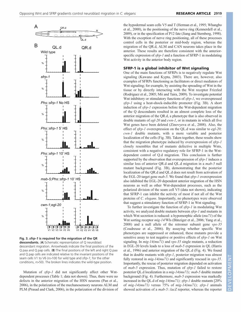
One group of neuroblasts that migrates along the anteroposterioraxis are the Q neuroblast descendants (Sulston and Horvitz, 1977;Hedgecock et al., 1987). At the end of embryogenesis, two Qneuroblasts are generated at equivalent positions on the left (QL) andright (QR) lateral side of the animal (Fig. 3A). During the first stageof larval development, the two Q neuroblasts each generate threedescendants that migrate in opposite directions: on the left side, theQL descendants (QL.d) migrate towards the posterior, whereas onthe right side, the QR.d migrate towards the anterior. Both anteriorand posterior migration is controlled by Wnt signaling. The posteriormigration of the QL.d is mediated by EGL-20, which triggers acanonical Wnt/b-catenin pathway to induce expression of the targetgene mab-5 and to direct migration towards the posterior (Harris etal., 1996; Maloof et al., 1999; Whangbo and Kenyon, 1999). Theanterior migration of the QR.d is also dependent on EGL-20, buthere EGL-20 functions together with CWN-1 to activate a b-catenin
independent Wnt signaling pathway that is required for anteriordirected migration (Zinovyeva et al., 2008). Although the mechanismremains to be established, current models suggest that a difference inresponse threshold to EGL-20 determines which pathway is activated(Whangbo and Kenyon, 1999). Thus, QL is primed to activatecanonical Wnt/b-catenin signaling in response to EGL-20, whereasQR will only activate this pathway when EGL-20 is overexpressed.At intermediate levels, overexpression of EGL-20 inducesovermigration of the QR.d, indicating that Wnt signaling activity notonly specifies the direction of migration, but also influences theposition at which the cells terminate their migration (Whangbo andKenyon, 1999). To investigate whether sfrp-1 regulates the Wntdependent migration of the Q descendants, we determined the finalpositions of the Q descendants Q.paa and Q.pap relative to thehypodermal seam cells V1 to V6. We found that the QL.d localizedaround their normal positions in sfrp-1 mutants (Fig. 3B). There was,however, a clear change in the final position of the QR.d, with theQR.d migrating significantly further into the anterior than in wild-type animals (P<0.001, Fisher’s exact test). As this phenotype issimilar to the extended migration induced by EGL-20overexpression, these data are consistent with a negative regulatoryrole for sfrp-1 in QR.d migration. Loss of this negative regulatoryactivity is, however, insufficient to trigger canonical Wnt/b-cateninsignaling and mab-5 expression in QR.
In addition to the defect in the anterior migration of the QR.d,we found that sfrp-1 mutants show misplacement of the ALM andCAN neurons (Fig. 6A; see Fig. S5A in the supplementarymaterial). Both neurons migrate during the end of embryogenesisfrom the anterior to final positions in the mid-body region (Sulstonet al., 1983; Hedgecock et al., 1987). In both cases, posteriormigration depends on the combined activity of CWN-1 and CWN-2 (Zinovyeva et al., 2008). In sfrp-1 mutants, the posteriormigration of the ALM neurons was significantly truncated(P<0.001) (Fig. 6B). Also, in the case of the CAN neurons,mutation of sfrp-1 induced undermigration (P<0.001), although thiseffect was less pronounced than observed with the ALM neurons(see Fig. S5B in the supplementary material).
RESEARCH ARTICLE Development 138 (14)
Fig. 2. Single molecule mRNA FISH analysesof the C. elegans Wnt genes and sfrp-1during embryonic development. Images aremaximum intensity projections of lateral z-stacks.Detection of (A) sfrp-1, cwn-1 and cwn-2, and (B) mom-2, lin-44 and egl-20 transcripts. Embryoswere staged using DIC microscopy and DAPIstaining of nuclei. Scale bar: 10m.
Table 1. Wnt phenotypes in sfrp-1 and hypomorphic Wnt secretion mutantsWild type sfrp-1 vps-35 vps-35; sfrp-1 mig-14(mu71) mig-14(mu71); sfrp-1
ALM polarity 0 1 21 16 0 N.D.PLM polarity 0 0 29 31 15 N.D.P12 to P11 0 0 6 6 4 2T cell polarity 1 0 12 0 11 4V5 polarity 0 0 32 2 0 0Nerve ring placement 1 0 4 1 0 1
Numbers indicate percentage defective (n>100). The polarity of the ALM and PLM mechanosensory neurons was scored using a mec-7::gfp (muIs32 or muIs35)-expressingtransgene (Ch’ng et al., 2003). The polarity of the V5 division and P12 to P11 fate transformation were scored using Nomarski microscopy at the appropriate developmentalstage. Effects on T-cell polarity were determined by DiO staining of the T-derived phasmid structure in young adults (Herman and Horvitz, 1994). Nerve ring placement wasdetermined by DiO staining of the amphid neurons. N.D., not determined. D
EVELO
PMENT
Mutation of sfrp-1 did not significantly affect other Wnt-dependent processes (Table 1; data not shown). Thus, there were nodefects in the anterior migration of the HSN neurons (Pan et al.,2006), in the polarization of the mechanosensory neurons ALM andPLM (Prasad and Clark, 2006), in the polarization of the division of
the hypodermal seam cells V5 and T (Herman et al., 1995; Whangboet al., 2000), in the positioning of the nerve ring (Kennerdell et al.,2009), or in the specification of P12 fate (Jiang and Sternberg, 1998).With the exception of nerve ring positioning, all of these processescontrol cells in the posterior or mid-body region, whereas themigration of the QR.d, ALM and CAN neurons takes place in theanterior. These results are therefore consistent with the anterior-specific expression of sfrp-1 and a function of SFRP-1 in modulatingWnt activity in the anterior body region.
SFRP-1 is a global inhibitor of Wnt signalingOne of the main functions of SFRPs is to negatively regulate Wntsignaling (Kawano and Kypta, 2003). There are, however, alsoexamples of SFRPs functioning as facilitators or direct mediators ofWnt signaling; for example, by assisting the spreading of Wnt in thetissue or by directly interacting with the Wnt receptor Frizzled(Rodriguez et al., 2005; Mii and Taira, 2009). To investigate potentialWnt inhibitory or stimulatory functions of sfrp-1, we overexpressedsfrp-1 using a heat-shock-inducible promoter (Fig. 3B). A shortinduction of sfrp-1 expression before the Wnt-dependent migrationof the Q descendants resulted in an almost complete loss of theanterior migration of the QR.d, a phenotype that is also observed indouble mutants of egl-20 and cwn-1, or in mutants in which all fiveWnt genes have been deleted (Zinovyeva et al., 2008). Also, theeffect of sfrp-1 overexpression on the QL.d was similar to egl-20;cwn-1 double mutants, with a more variable and posteriorlocalization of the cells (Fig. 3B). Taken together, these results showthat the migration phenotype induced by overexpression of sfrp-1closely resembles that of mutants defective in multiple Wnts,consistent with a negative regulatory role for SFRP-1 in the Wnt-dependent control of Q.d migration. This conclusion is furthersupported by the observation that overexpression of sfrp-1 induces asimilar loss of anterior QR.d and QL.d migration in a mab-5 nullmutant background (Fig. 3B), demonstrating that the posteriorlocalization of the QR.d and QL.d does not result from activation ofthe EGL-20 target gene mab-5. We found that sfrp-1 overexpressionalso inhibited the EGL-20 dependent anterior migration of the HSNneurons as well as other Wnt-dependent processes, such as thepolarized division of the seam cell V5 (data not shown), indicatingthat SFRP-1 can inhibit the activity of most if not all of the Wntproteins of C. elegans. Importantly, no phenotypes were observedthat suggest a stimulatory function of SFRP-1 in Wnt signaling.
To further investigate the function of sfrp-1 in modulating Wntactivity, we analyzed double mutants between sfrp-1 and mutants inwhich Wnt secretion is reduced: a hypomorphic allele (mu71) of theWnt sorting receptor mig-14/Wls (Bänziger et al., 2006; Yang et al.,2008) and a null allele of the retromer subunit gene vps-35(Coudreuse et al., 2006). By assaying whether specific Wntphenotypes are suppressed or enhanced, these mutants provide asensitive assay to test negative or positive effects of sfrp-1 on Wntsignaling. In mig-14(mu71) and vps-35 single mutants, a reductionin EGL-20 levels leads to a loss of mab-5 expression in QL (Harriset al., 1996) and anterior migration of the QL.d (Fig. 4). We foundthat in double mutants with sfrp-1, posterior migration was almostfully restored in mig-14(mu71) and significantly rescued in vps-35.Importantly, the rescue of posterior migration depended on activationof mab-5 expression. Thus, mutation of sfrp-1 failed to restoreposterior QL.d localization in a mig-14(mu71); mab-5 double mutantbackground (Fig. 4). Furthermore, mab-5 expression was markedlyincreased in the QL.d of mig-14(mu71); sfrp-1 double mutants [20%of mig-14(mu71) versus 75% of mig-14(mu71); sfrp-1 animalsshowed activation of a mab-5::lacZ reporter, whereas the reporter
2919RESEARCH ARTICLEOpposing Wnt and SFRP gradients control neuroblast migration in C. elegans
Fig. 3. sfrp-1 is required for the migration of the QRdescendants. (A)Schematic representation of Q neuroblastdescendant migration. Arrowheads indicate the final positions of theQ.paa and Q.pap cells. (B)The final positions of the left and right Q.paaand Q.pap cells are indicated relative to the invariant positions of theseam cells V1 to V6 (n>100 for wild type and sfrp-1; for the otherconditions, n>50). The broken lines indicates the wild-type position.
DEVELO
PMENT

2920
was expressed in 86% and 83% of wild type and sfrp-1 singlemutants, respectively; in each case n>50]. These results suggest thatthe reduction in EGL-20 signaling can be overcome by removal ofSFRP-1, consistent with a negative regulatory role of SFRP-1 in theEGL-20 dependent activation of canonical Wnt/b-catenin signalingin QL. We observed a similar inhibitory role for sfrp-1 in QR.dmigration. Thus, whereas the QR.d showed reduced anteriormigration in mig-14(mu71) and vps-35 mutants, the QR.d localizedat their correct positions in double mutants with sfrp-1 (see Fig. S3in the supplementary material). A comprehensive analysis of otherWnt phenotypes in mig-14(mu71) and vps-35 mutants showed thatloss of sfrp-1 also suppressed defects in the migration of the ALMneurons and the polarity of the seam cells V5 and T (Fig. 6B andTable 1). Importantly, there were no instances in which the vps-35 ormig-14(mu71) phenotype was enhanced by loss of sfrp-1. Takentogether with the strong Wnt inhibitory activity of sfrp-1overexpression, these results support the conclusion that SFRP-1functions as a global inhibitor of Wnt signaling in C. elegans.
The sfrp-1-induced overmigration of the QRdescendants is suppressed by mutation of cwn-2The overmigration of the QR.d in sfrp-1 mutants is similar to theextended migration induced by ubiquitous EGL-20 expression(Whangbo and Kenyon, 1999), indicating that the overmigration is
a result of a gain in Wnt signaling activity. To investigate whichWnts mediate the sfrp-1-induced overmigration, we constructeddouble mutants between sfrp-1 and null mutants of the differentWnt genes. We found that mutation of lin-44 or mom-2 did notsuppress the sfrp-1-induced overmigration (Fig. 5). In doublemutants between sfrp-1 and egl-20, there was a partial suppressionof the overmigration, but also a clear undermigration of the QR.d,an effect that was even more pronounced in double mutants withcwn-1. EGL-20 and CWN-1 function partially redundantly inspecifying anterior QR.d migration and loss of either egl-20 orcwn-1 results in a distinct undermigration of the QR.d (Zinovyeva
RESEARCH ARTICLE Development 138 (14)
Fig. 4. sfrp-1 rescues QL.d migration in hypomorphic Wntsecretion mutants. The final positions of QL.paa and QL.pap cells areindicated relative to the seam cells V1 to V6 (n>50). The broken linesindicate the wild-type position.
Fig. 5. The sfrp-1-induced overmigration of the QR.d issuppressed by mutation of cwn-2. The final positions of QR.paa andQR.pap cells are indicated relative to the seam cells V1 to V6 (n>50).The broken lines indicate the wild-type position.
DEVELO
PMENT

et al., 2008). The intermediate phenotype of the egl-20; sfrp-1 andcwn-1; sfrp-1 double mutants therefore suggests that egl-20 andcwn-1 either function in parallel to sfrp-1 or play only a minor rolein the sfrp-1-induced overmigration of the QR.d. By contrast, wefound that the sfrp-1-induced overmigration was fully rescued bya null mutation in cwn-2 (Fig. 5). Thus, whereas cwn-2 has nosignificant effect on QR.d positioning on its own, the QR.dlocalized at their wild-type position in sfrp-1; cwn-2 doublemutants. Taken together, these results indicate that derepression ofCWN-2 signaling is primarily responsible for the QR.dovermigration phenotype of sfrp-1 mutants.
The sfrp-1-induced undermigration of the ALMand CAN neurons is suppressed by mutation ofcwn-1 or cwn-2The posterior migration of the ALM neurons is dependent on theredundant activity of CWN-1 and CWN-2, with a strong inhibitionof migration in cwn-1; cwn-2 double mutants (Zinovyeva et al.,2008). A similar undermigration is observed in sfrp-1 mutants,indicating that Wnt overactivity also interferes with the correctposterior migration of the ALM neurons. This conclusion issupported by the observation that the ALM undermigrationphenotype of sfrp-1 is suppressed by reducing Wnt secretionthrough mutation of the Wnt sorting receptor mig-14/Wls (Fig. 6B).To investigate which Wnts are required for the sfrp-1-inducedundermigration of the ALM neurons, we analyzed double mutantswith null alleles of each of the five different Wnt genes and testedwhich combination could suppress the ALM undermigrationphenotype. Whereas lin-44, egl-20 and mom-2 did not affect thesfrp-1-induced ALM undermigration, mutation of cwn-1 or cwn-2fully restored the migration of the ALM neurons to their wild-typepositions (Fig. 6B; see Fig. S4B in the supplementary material).These results suggest that in the absence of SFRP-1, overactivityof CWN-1 and CWN-2 interferes with the correct positioning ofthe ALM neurons, and that normal migration can be restored byremoving either of the two Wnt genes. Furthermore, the sfrp-1-induced undermigration of the ALM neurons was fully suppressedin double mutants between sfrp-1 and the Frizzled mom-5 (Fig.6B), indicating that CWN-1 and CWN-2 control ALM positioningthrough the MOM-5/Frizzled receptor.
Similar results were obtained for the sfrp-1-inducedundermigration of the CAN neurons, which was also dependent onCWN-1 and CWN-2. Thus, CAN undermigration was notsuppressed in lin-44, egl-20 or mom-2 mutants, but was rescued ineither cwn-1 or cwn-2 mutants (see Fig. S5B in the supplementarymaterial).
The function of SFRP-1 in suppressing CWN-1 and CWN-2activity is consistent with the anterior expression of sfrp-1 and isin agreement with a role for SFRP-1 in modulating Wnt activity inthe anterior body region. Our results show that this inhibitoryactivity is particularly important for controlling the Wnt-dependentmigration of neuroblasts along the primary body axis of C. elegans.
DISCUSSIONDuring C. elegans development, the migration of neuroblasts alongthe anteroposterior axis is controlled through a complex networkof partially redundantly acting Wnt proteins. Here, we usedsmFISH to map quantitatively the spatio-temporal expressionpattern of the C. elegans Wnt genes. We show that the five Wntgenes are expressed in partially overlapping expression domainsalong the anteroposterior axis, with the most prominent Wntexpression in the posterior body region. Furthermore, we
demonstrate that Wnt signaling in the anterior body region isrepressed by the secreted Frizzled-related protein SFRP-1. Theseresults show that the anteroposterior positioning of neuroblasts iscontrolled by opposing Wnt and Wnt inhibitory activities andprovide further evidence for the evolutionary conservation of thissystem in patterning of the primary body axis.
A quantitative gene expression map of the C. elegans Wnt familyConventional methods for gene expression analysis in C. elegansare mostly based on transgenic reporter constructs (Mello et al.,1991). In this study we used for the first time smFISH (Raj et al.,2008) to determine the spatio-temporal expression pattern of a genefamily in C. elegans. By visualizing single transcripts as brightdiffraction-limited spots, we could directly measure endogenousgene expression in vivo by counting the number of spots in a three-dimensional area of interest, such as a specific cell or tissue.Importantly, this method also allowed us to quantify dynamicchanges in gene expression, such as the transient expression of
2921RESEARCH ARTICLEOpposing Wnt and SFRP gradients control neuroblast migration in C. elegans
Fig. 6. The sfrp-1-induced undermigration of the ALM neurons issuppressed by mutation of cwn-1 or cwn-2. (A)Schematicrepresentation of the ALM migration. Note that the migration takesplace at the end of embryogenesis. (B)The final positions of the ALMLand ALMR neurons are indicated relative to the seam cells V1 to V6(n>50). The broken lines indicate the wild-type position.
DEVELO
PMENT

2922
mom-2 in cells of the tail and the activation of cwn-1 expression inthe CAN neurons during the first stage of larval development. Wefound that the expression level and spatial distribution of transcriptsdetected by smFISH was highly reproducible between stagedanimals, indicating that this method accurately visualizesendogenous gene expression patterns. We conclude that smFISHcan be used to produce quantitative spatio-temporal maps ofendogenous gene expression patterns. It should be noted, however,that the expression pattern of the protein product may be influencedby post-transcriptional regulation.
Our smFISH analysis of the Wnt gene family showed similarities,but also important differences with expression patterns based ontransgenic reporter constructs. We found that cwn-2 is mainlyexpressed in head neurons and anterior body wall muscle cells,resolving a conflict in the previously reported expression patterns forcwn-2 (Gleason et al., 2006; Kennerdell et al., 2009; Song et al.,2010). Furthermore, we observed that mom-2 is not generallyexpressed along the anteroposterior body axis (Gleason et al., 2006),but is restricted to the germ line precursor cells and transiently to agroup of cells in the tail. Another important difference is theexpression of lin-44 outside of the tail hypodermal cells (Herman etal., 1995). The more anterior expression of lin-44 in the B and Yrectal epithelial cells is particularly interesting for the function ofLIN-44 as a directional signal in T-cell polarity (Goldstein et al.,2006) and for the inhibition of presynaptic assemblies in the DA9neuron (Klassen and Shen, 2007).
The smFISH analysis revealed that the five Wnt genes areexpressed in a series of partially overlapping expression domains,with expression of three of the five Wnt genes in the posterior andone in the anterior half of the body. The expression of the differentWnt genes in serial domains correlates with their function incontrolling the migration of neuroblasts along the anteroposterioraxis. Thus, the posteriorly expressed Wnt EGL-20 controls migrationin the posterior and mid-body region, while CWN-1 and CWN-2 areparticularly important for the migration of the QR descendants andthe CAN and ALM neurons in the anterior half of the animal (Harriset al., 1996; Pan et al., 2006; Zinovyeva et al., 2008). We proposethat the staggered series of Wnt expression domains provides asystem for positional information along the anteroposterior body axisof C. elegans.
The secreted Frizzled-related protein SFRP-1 is aninhibitor of Wnt signalingSFRP proteins are characterized by an N-terminal cysteine-richdomain (CRD) that is similar to the Wnt binding CRD domainof Frizzled (Bovolenta et al., 2008). SFRPs are secreted proteinsthat have been shown to act as inhibitors of Wnt signaling, mostprobably by competing with Wnt receptors for Wnt binding.However, SFRPs have also been reported to promote Wntsignaling, for example, by facilitating the spreading of Wnt inthe tissue (Mii and Taira, 2009) or by directly interacting withFrizzleds to stimulate signaling in a Wnt-independent manner(Rodriguez et al., 2005). Phylogenetic analysis has shown thatthe SFRP family appeared very early in metazoan evolution, asclear SFRP orthologs are already present in the cnidarians Hydraand Nematostella vectensis (Guder et al., 2006a). Their functionhas, however, not been studied in any of the genetically tractableinvertebrate model systems. The Drosophila genome does notcontain SFRP orthologs, indicating that this gene family mayhave been lost in insects (but not in all arthropods, as the genomeof the tick Ixodes scapularis contains an SFRP ortholog). In thisstudy, we show that the C. elegans genome contains a single
SFRP ortholog, sfrp-1, which has enabled us to study potentialWnt inhibitory or stimulatory functions of SFRPs in a well-defined model system. We found that SFRP-1 functionsexclusively as an inhibitor of Wnt signaling: first,overexpression of sfrp-1 induced a strong defect in Wntsignaling, similar to the phenotype observed in mutants in whichall five Wnt genes have been mutated (Zinovyeva et al., 2008).Second, loss of sfrp-1 suppressed the Wnt signaling defect ofmutants that induce a reduction in Wnt secretion and, finally, allthe phenotypes observed in sfrp-1 mutants could be suppressedby removing specific Wnts, indicating that mutation of sfrp-1leads to derepression of Wnt signaling. These results suggest thatthe stimulatory function of SFRPs in Wnt signaling has eitherbeen lost in the nematode lineage, or is a more recent inventionof organisms of higher complexity. Studies on the cnidarianSFRPs may shed light on this issue.
An anterior SFRP-1 inhibitory gradient controlsthe positioning of neuroblasts in the anteriorbody regionThe predominant anterior expression of sfrp-1 suggests that itcounteracts the more posteriorly expressed Wnts. Loss of thisinhibitory activity leads to defects in the migration of neuroblasts inthe anterior body region. Thus, the QR.d migrate too far into theanterior, whereas the extent of the posterior migration of the CANand ALM neurons is reduced. In each of these cases, the finalposition of the cells is shifted anteriorly, indicating that SFRP-1counteracts a Wnt activity that promotes anterior localization. Wefound that the sfrp-1-induced anterior displacement of the QR.dcould be suppressed by mutation of cwn-2, whereas ALM and CANmigration could be restored by deletion of either cwn-2 or cwn-1.These results are consistent with a local inhibitory function of SFRP-1 in controlling the activity of the two most anteriorly expressedWnts.
In addition to this short-range function in the anterior bodyregion, our experiments in Wnt secretion mutants showed thatSFRP-1 also has a long-range inhibitory activity. Thus, mutation ofsfrp-1 rescued the posterior migration of the QL.d and the polarityof the V5 and T cell divisions in hypomorphic Wnt secretionmutants, consistent with a function of SFRP-1 in modulating Wntactivity in the mid to posterior body region. This long-rangeinhibition may fine-tune the activity gradients of the posteriorlyexpressed Wnt genes.
An evolutionarily conserved function of Wnts andWnt inhibitors in patterning the primary body axisWe found that four out of the five C. elegans Wnt genes areexpressed in a series of partially overlapping domains along theanteroposterior axis. This staggered expression is remarkably similarto the expression of Wnt genes in the cnidarian Nematostellavectensis (Kusserow et al., 2005) and in the planarian Schmidteamediterranea (Petersen and Reddien, 2008). It has been proposedthat the staggered expression of Wnt genes provides an ancestralmechanism for positional information along the primary body axis(Guder et al., 2006a) and our results suggest that C. elegans hasretained such a system.
Another important similarity is the anterior-specific expression ofsfrp-1 and the mostly posterior expression of the Wnt genes. Thisopposite expression of Wnts and Wnt inhibitors is already present incnidarians, where Wnt inhibitors are expressed at the aboral side andWnts at the oral side of the primary body axis (Hobmayer et al.,2000; Kusserow et al., 2005; Guder et al., 2006b; Lee et al., 2006).
RESEARCH ARTICLE Development 138 (14)
DEVELO
PMENT

Posterior Wnt signaling and anterior Wnt inhibition is also a centralfeature of vertebrate neurectodermal patterning, with the formationof the eyes and anterior brain structures depending on the anterioractivity of both intracellular and secreted Wnt inhibitory factors (Kimet al., 2000; Kiecker and Niehrs, 2001; Niehrs, 2006). Inprotostomes, anterior specific expression of an SFRP has beenobserved in Schmidtea mediterranea (Petersen and Reddien, 2008),but Wnt inhibitors have not been studied in any of the otherprotostome model organisms. Our studies in C. elegans show thatthe opposite expression of Wnts and Wnt inhibitors is also animportant feature of nematode development, supporting the notionthat a system of posterior Wnt activity and anterior Wnt inhibition isa unifying principle of primary body axis specification in animals(Petersen and Reddien, 2009).
AcknowledgementsWe thank Dr Bert Hobmayer for critically reading the manuscript, RemcoMentink for assistance with statistical analysis, Dr Andrew Fire for expressionvectors and the Caenorhabditis Genetic Center (University of Minnesota,Minneapolis) for strains. This work was funded by a NWO VIDI fellowship(016.076.317) to H.C.K., a Boehringer Ingelheim Foundation fellowship toM.H. and a NIH Pioneer award (1DP1OD003936) to A.v.O. Deposited in PMCfor release after 12 months.
Competing interests statementThe authors declare no competing financial interests.
Supplementary materialSupplementary material for this article is available athttp://dev.biologists.org/lookup/suppl/doi:10.1242/dev.064733/-/DC1
ReferencesBänziger, C., Soldini, D., Schütt, C., Zipperlen, P., Hausmann, G. and Basler, K.
(2006). Wntless, a conserved membrane protein dedicated to the secretion of Wntproteins from signaling cells. Cell 125, 509-522.
Bovolenta, P., Esteve, P., Ruiz, J. M., Cisneros, E. and Lopez-Rios, J. (2008).Beyond Wnt inhibition: new functions of secreted Frizzled-related proteins indevelopment and disease. J. Cell Sci. 121, 737-746.
Bulow, H. E., Berry, K. L., Topper, L. H., Peles, E. and Hobert, O. (2002). Heparansulfate proteoglycan-dependent induction of axon branching and axon misroutingby the Kallmann syndrome gene kal-1. Proc. Natl. Acad. Sci. USA 99, 6346-6351.
Ch’ng, Q., Williams, L., Lie, Y. S., Sym, M., Whangbo, J. and Kenyon, C. (2003).Identification of genes that regulate a left-right asymmetric neuronal migration inCaenorhabditis elegans. Genetics 164, 1355-1367.
Chong, J. M., Uren, A., Rubin, J. S. and Speicher, D. W. (2002). Disulfide bondassignments of secreted Frizzled-related protein-1 provide insights about Frizzledhomology and netrin modules. J. Biol. Chem. 277, 5134-5144.
Coudreuse, D. Y. M., Roël, G., Betist, M. C., Destrée, O. and Korswagen, H. C.(2006). Wnt gradient formation requires retromer function in Wnt-producing cells.Science 312, 921-924.
Fire, A., Xu, S., Montgomery, M. K., Kostas, S. A., Driver, S. E. and Mello, C. C.(1998). Potent and specific genetic interference by double-stranded RNA inCaenorhabditis elegans. Nature 391, 806-811.
Gleason, J. E., Szyleyko, E. A. and Eisenmann, D. M. (2006). Multiple redundantWnt signaling components function in two processes during C. elegans vulvaldevelopment. Dev. Biol. 298, 442-457.
Goldstein, B., Takeshita, H., Mizumoto, K. and Sawa, H. (2006). Wnt signals canfunction as positional cues in establishing cell polarity. Dev. Cell 10, 391-396.
Guder, C., Philipp, I., Lengfeld, T., Watanabe, H., Hobmayer, B. and Holstein, T.W. (2006a). The Wnt code: cnidarians signal the way. Oncogene 25, 7450-7460.
Guder, C., Pinho, S., Nacak, T. G., Schmidt, H. A., Hobmayer, B., Niehrs, C. andHolstein, T. W. (2006b). An ancient Wnt-Dickkopf antagonism in Hydra.Development 133, 901-911.
Harfe, B. D., Vaz Gomes, A., Kenyon, C., Liu, J., Krause, M. and Fire, A. (1998).Analysis of a Caenorhabditis elegans Twist homolog identifies conserved anddivergent aspects of mesodermal patterning. Genes Dev. 12, 2623-2635.
Harris, J., Honigberg, L., Robinson, N. and Kenyon, C. (1996). Neuronal cellmigration in C. elegans: regulation of Hox gene expression and cell position.Development 122, 3117-3131.
Hedgecock, E. M., Culotti, J. G., Hall, D. H. and Stern, B. D. (1987). Genetics ofcell and axon migrations in Caenorhabditis elegans. Development 100, 365-382.
Herman, M. A. and Horvitz, H. R. (1994). The Caenorhabditis elegans gene lin-44controls the polarity of asymmetric cell divisions. Development 120, 1035-1047.
Herman, M. A., Vassilieva, L. L., Horvitz, H. R., Shaw, J. E. and Herman, R. K.(1995). The C. elegans gene lin-44, which controls the polarity of certain
asymmetric cell divisions, encodes a Wnt protein and acts cell nonautonomously.Cell 83, 101-110.
Hobmayer, B., Rentzsch, F., Kuhn, K., Happel, C. M., von Laue, C. C., Snyder, P.,Rothbacher, U. and Holstein, T. W. (2000). WNT signalling molecules act in axisformation in the diploblastic metazoan Hydra. Nature 407, 186-189.
Jiang, L. I. and Sternberg, P. W. (1998). Interactions of EGF, Wnt and HOM-Cgenes specify the P12 neuroectoblast fate in C. elegans. Development 125, 2337-2347.
Kawano, Y. and Kypta, R. (2003). Secreted antagonists of the Wnt signallingpathway. J. Cell Sci. 116, 2627-2634.
Kennerdell, J. R., Fetter, R. D. and Bargmann, C. I. (2009). Wnt-Ror signaling toSIA and SIB neurons directs anterior axon guidance and nerve ring placement in C.elegans. Development 136, 3801-3810.
Kiecker, C. and Niehrs, C. (2001). A morphogen gradient of Wnt/b-cateninsignalling regulates anteroposterior neural patterning in Xenopus. Development128, 4189-4201.
Kim, C. H., Oda, T., Itoh, M., Jiang, D., Artinger, K. B., Chandrasekharappa, S.C., Driever, W. and Chitnis, A. B. (2000). Repressor activity of Headless/Tcf3 isessential for vertebrate head formation. Nature 407, 913-916.
Klassen, M. P. and Shen, K. (2007). Wnt signaling positions neuromuscularconnectivity by inhibiting synapse formation in C. elegans. Cell 130, 704-716.
Korswagen, H. C. (2002). Canonical and non-canonical Wnt signaling pathways inCaenorhabditis elegans: variations on a common signaling theme. BioEssays 24,801-810.
Korswagen, H. C., Coudreuse, D. Y. M., Betist, M. C., van de Water, S.,Zivkovic, D. and Clevers, H. C. (2002). The Axin-like protein PRY-1 is a negativeregulator of a canonical Wnt pathway in C. elegans. Genes Dev. 16, 1291-1302.
Kusserow, A., Pang, K., Sturm, C., Hrouda, M., Lentfer, J., Schmidt, H. A.,Technau, U., von Haeseler, A., Hobmayer, B., Martindale, M. Q. et al. (2005).Unexpected complexity of the Wnt gene family in a sea anemone. Nature 433,156-160.
Lee, P. N., Pang, K., Matus, D. Q. and Martindale, M. Q. (2006). A WNT of thingsto come: evolution of Wnt signaling and polarity in cnidarians. Semin. Cell Dev.Biol. 17, 157-167.
Lewis, J. A. and Fleming, J. T. (1995). Basic culture methods. Methods Cell Biol. 48,3-29.
Leyns, L., Bouwmeester, T., Kim, S. H., Piccolo, S. and De Robertis, E. M.(1997). Frzb-1 is a secreted antagonist of Wnt signaling expressed in the Spemannorganizer. Cell 88, 747-756.
Long, F., Peng, H., Liu, X., Kim, S. K. and Myers, E. (2009). A 3D digital atlas of C.elegans and its application to single-cell analyses. Nat. Methods 6, 667-672.
Maloof, J. N., Whangbo, J., Harris, J. M., Jongeward, G. D. and Kenyon, C.(1999). A Wnt signaling pathway controls Hox gene expression and neuroblastmigration in C. elegans. Development 126, 37-49.
Mello, C. and Fire, A. (1995). DNA transformation. Methods Cell Biol. 48, 451-482.Mello, C. C., Kramer, J. M., Stinchcomb, D. and Ambros, V. (1991). Efficient gene
transfer in C. elegans: extrachromosomal maintenance and integration oftransforming sequences. EMBO J. 10, 3959-3970.
Mii, Y. and Taira, M. (2009). Secreted Frizzled-related proteins enhance the diffusionof Wnt ligands and expand their signalling range. Development 136, 4083-4088.
Niehrs, C. (2006). Function and biological roles of the Dickkopf family of Wntmodulators. Oncogene 25, 7469-7481.
Pan, C. L., Howell, J. E., Clark, S. G., Hilliard, M., Cordes, S., Bargmann, C. I.and Garriga, G. (2006). Multiple Wnts and frizzled receptors regulate anteriorlydirected cell and growth cone migrations in Caenorhabditis elegans. Dev. Cell 10,367-377.
Petersen, C. P. and Reddien, P. W. (2008). Smed-b-catenin-1 is required foranteroposterior blastema polarity in planarian regeneration. Science 319, 327-330.
Petersen, C. P. and Reddien, P. W. (2009). Wnt signaling and the polarity of theprimary body axis. Cell 139, 1056-1068.
Prasad, B. C. and Clark, S. G. (2006). Wnt signaling establishes anteroposteriorneuronal polarity and requires retromer in C. elegans. Development 133, 1757-1766.
Raj, A., van den Bogaard, P., Rifkin, S. A., van Oudenaarden, A. and Tyagi, S.(2008). Imaging individual mRNA molecules using multiple singly labeled probes.Nat. Methods 5, 877-879.
Rodriguez, J., Esteve, P., Weinl, C., Ruiz, J. M., Fermin, Y., Trousse, F., Dwivedy,A., Holt, C. and Bovolenta, P. (2005). SFRP1 regulates the growth of retinalganglion cell axons through the Fz2 receptor. Nat. Neurosci. 8, 1301-1309.
Salser, S. J. and Kenyon, C. (1992). Activation of a C. elegans Antennapediahomologue in migrating cells controls their direction of migration. Nature 355,255-258.
Silhankova, M. and Korswagen, H. C. (2007). Migration of neuronal cells alongthe anterior-posterior body axis of C. elegans: Wnts are in control. Curr. Opin.Genet. Dev. 17, 320-325.
Song, S., Zhang, B., Sun, H., Li, X., Xiang, Y., Liu, Z., Huang, X. and Ding, M.(2010). A Wnt-Frz/Ror-Dsh pathway regulates neurite outgrowth in Caenorhabditiselegans. PLoS Genet. 6, e10010056.
Sulston, J. E. and Horvitz, H. R. (1977). Post-embryonic cell lineages of thenematode Caenorhabditis elegans. Dev. Biol. 56, 110-156.
2923RESEARCH ARTICLEOpposing Wnt and SFRP gradients control neuroblast migration in C. elegans
DEVELO
PMENT

2924
Sulston, J. E., Schierenberg, E., White, J. G. and Thomson, J. N. (1983). Theembryonic cell lineage of the nematode Caenorhabditis elegans. Dev. Biol. 100,64-119.
Tendeng, C. and Houart, C. (2006). Cloning and embryonic expression of fivedistinct sfrp genes in the zebrafish Danio rerio. Gene Expr. Patterns 6, 761-771.
Thorpe, C. J., Schlesinger, A., Carter, J. C. and Bowerman, B. (1997). Wntsignaling polarizes an early C. elegans blastomere to distinguish endoderm frommesoderm. Cell 90, 695-705.
van Amerongen, R. and Nusse, R. (2009). Towards an integrated view of Wntsignaling in development. Development 136, 3205-3214.
Whangbo, J. and Kenyon, C. (1999). A Wnt signaling system that specifies twopatterns of cell migration in C. elegans. Mol. Cell 4, 851-858.
Whangbo, J., Harris, J. and Kenyon, C. (2000). Multiple levels of regulation specifythe polarity of an asymmetric cell division in C. elegans. Development 127, 4587-4598.
Yang, P. T., Lorenowicz, M. J., Silhankova, M., Coudreuse, D. Y., Betist, M. C.and Korswagen, H. C. (2008). Wnt signaling requires retromer-dependentrecycling of MIG-14/Wntless in Wnt-producing cells. Dev. Cell 14, 140-147.
Zinovyeva, A. Y. and Forrester, W. C. (2005). The C. elegans Frizzled CFZ-2 isrequired for cell migration and interacts with multiple Wnt signaling pathways.Dev. Biol. 285, 447-461.
Zinovyeva, A. Y., Yamamoto, Y., Sawa, H. and Forrester, W. C. (2008).Complex network of Wnt signaling regulates neuronal migrations duringCaenorhabditis elegans development. Genetics 179, 1357-1371.
RESEARCH ARTICLE Development 138 (14)
DEVELO
PMENT