neurocritical complications of opioid use disorder · 1 neurocritical complications of opioid use...
TRANSCRIPT

1
Neurocritical Complications of Opioid
Use DisorderAlan Velander, MD
Objectives
Recognize signs and symptoms of acute opioid overdose.
Dose naloxone to reverse acute opioid overdose in stupor versus arrest.
Detect, manage, and prognosticate anoxic brain injury after arrest from acute opioid overdose.
Suppress paroxysmal sympathetic hyperactivity after arrest from acute opioid overdose.
Diagnose less common neurocritical complications of opioid use disorder.

2
Acute Opioid Overdose: Synthetics
Contemporary street opioids are a long way from opium.
Increasingly “heroin” is not diamorphine. Instead, it is composed of adulterants and tiny doses of highly potent synthetic opioids (fentanyl, carfentanil, ...).
These synthetic opioids are cheap to synthesize, easy to smuggle, hard to detect, and very, very dangerous.
Acute Opioid Overdose: Synthetics

3
Acute Opioid Overdose: Synthetics
2 mg of fentanyl can cause respiratory depression.
2 mg of ohmefentanyl can kill 12.5 M people.
In 2002 carfentanil implicated in “neutralization” of Chechen terrorists and 128 hostages in Moscow theater.
Cuts of heroin were 3-99% pure. Cuts of “heroin” today are even more heterogeneous.
Hazmat suits are recommended in decontamination of drug sitesby DEA.
IN Narcan is now available OTC in 41-45 states, including LA.
Acute Opioid Overdose: Signs
Respiratory and cardiac arrest
Stupor and coma
Pinpoint pupils
↓ respiratory rate and tidal volume
↓ bowel sounds
↓ temperature
Trauma and exposure
Coingestion (opioid + cocaine) may mask signs.

4
Acute Opioid Overdose: Evaluation
Broad toxicology screen (particularly for acetaminophen)
CT head
ECG
CXR, AXR
Adulterants (baking soda, sucrose, talcum, detergent, caffeine)
In general, no role for charcoal
Acute Opioid Overdose: Naloxone (NIH)
Competitive opioid antagonist
No agonist activity
Faster effect with IV but effective SC, IM, IN.
Rapidly metabolized and ineffective when PO.
Onset of action 1-2 min IV, 2-5 min SC/IM, time to peak 15-30 min, duration of effect 30-120 min, t1/2 30-81 min
First clinical evidence (Evans 1973)
Two dosing scenarios: stupor or arrest

5
Naloxone in Stupor
ABC
If ↓ RR, consider ETCO2 and BVM.
Lower doses of naloxone are in. Start at 0.05 mg IV, not 0.4 mg IV. Escalate q2min until RR >12.
Eg, 0.05 mg→ 0.1 mg→ 0.4 mg→ 1 mg→ 2 mg→ 4 mg. If no effect by 10 mg, diagnosis is probably wrong.
Goal is adequate respiration, not full opioid reversal.
ACLS first, then higher doses of naloxone are in.
Start at 2 mg IN or 0.4 mg IM/IV. Escalate q2min.
Eg, 0.4 mg→ 2 mg→ 5 mg→ 10 mg.
Naloxone in Arrest (Lavonas 2015)

6
Continuous Naloxone Infusion
As naloxone is short-acting, may need to redose q30min, or start infusion if long acting opioids are at play (methadone).
Eg, if naloxone 0.6 mg IVP was initial effective dose, start naloxone 0.4 mg/h IV with naloxone 0.3 mg IV at 30 min and prn for RR <12. (Goldfrank 1986)
Complications of Naloxone
Naloxone precipitates acute opioid withdrawal.
Complications include agitation, seizures, nausea/vomiting, arrhythmia (AF, VT/VF, asystole), acute pulmonary edema.
In a prospective study of ED patients with opioid intoxication, 6 of 453 (1.3%) suffered severe adverse effects in 10 min after naloxone (1 asystole, 3 seizures, 1 pulmonary edema, 1 agitation). No adverse effects were observed after 10 min. (Osterwalder 1996)
↑ PaCO2 before naloxone may be a risk factor.
↑ PaCO2→ ↑ central chemoreceptors→ ↑ sympathetics→ + naloxone→ ↑↑↑ sympathetics↑ parasympathetics
BVM or PPV may counteract.

7
Opioid Receptor Pathways (Benarroch 2012)
µ, δ, κ opioid receptors exist throughout neuraxis.
Opioid receptors detect pain and stimulate reward.
Opioid receptors modulate somatic and autonomic sensation.
Acute opioid withdrawal induces sympathetic hyperactivity, particularly through effects in hypothalamus, insula, amygdala, and periaqueductal gray/reticular formation.
Non-painful sensation becomes painful.
Excitation ↑, inhibition ↓.
A Tale of Two Overdoses
HM was a 22 y man who presented with asystolic arrest. Known heroin abuse. While driving started to swerve then had loss of consciousness. Friend attempted CPR. EMS found
asystole-> ACLS including naloxone-> ROSC. UTOX + opiates. Cooled to 33 °C.
ER was a 38 y man who presented with asystolic arrest. At a party on 9/16. Then found down. In ED bradycardic, bradypneic-> asystole-> ACLS-> ROSC at 11 min-> intubation. Cooled to 36 °C. UTOX + opiates, benzodiazepines. Family was unaware of any drug abuse. After investigation they learned he was smoking heroin.

8
A Tale of Two Overdoses
HMER
Acute Opioid Overdose: Arrest
66% of overdose deaths involve opioids. 115 Americans die every day from opioid overdoses. (CDC)
In retrospective study of 3.8M inpatient hospitalizations for overdose from 2000-2013, 16.4% were due to prescription opioids and 2.3% from illicit heroin. Cardiac arrest occurred in 3.8% of illicit heroin overdose, 1.4% of prescription opioid overdose versus 0.6% of non opioid overdose (p <0.001). (Sakhuja 2017)

9
Anoxic Brain Injury (ABI)
Most feared complication of arrest
Often young
Often prolonged hypoxia, hypotension before arrest
ABI is global in distribution with some selective vulnerability. As ABI is diffuse patient often looks worse than imaging.Neurons >glia (gray >white)Hippocampus, thalamus, putamen >brainstem
Evolves on imaging over time, and brain death or brainstem injury is relatively rare.
Anoxic Brain Injury: Management
Suspect ABI if GCS ≤8 after resuscitation.
Perform targeted temperature management (TTM) to maximize the chances of meaningful recovery. (Nielsen 2013)
Manage opioid withdrawal symptoms and paroxysmal sympathetic hyperactivity (storming).
Provide extra time before prognostication or brain death testing.
I do not stress mass effect/brain edema management. If brain herniation is imminent from ABI, it may be more of a mercy than complication. Brain herniation implies ABI is so severe and diffuse that there is no chance for meaningful recovery.

10
Anoxic Brain Injury: Prognosis (Rossetti 2016)
Anoxic Brain Injury: Exam
Exam is still the gold standard for prognosis in anoxic brain injury.
“Meaningful” recovery is mRS ≤3, independence in daily living, discharge to home or rehabilitation.
Best articlesLevy 1981 pre-coolingGreer 2013Rossetti 2010Fugate 2010
Evidence is complex.

11
Anoxic Brain Injury: Exam (Rossetti 2016)
Value pupils/corneals >myoclonus >>motor.
Anoxic Brain Injury: Seizures/Myoclonus
Seizures were detected in 10-47% of cooled post-cardiac arrest patients (Rossetti 2010, Legriel
209, Abend 2009)
(Lybeck 2017)
Best to separate EEG from seizures/myoclonus.
In other words, goal of EEG in ABI is not to diagnose seizures, it is to prognosticate potential for recovery.

12
Anoxic Brain Injury: Reactivity (Rossetti 2016)
Anoxic Brain Injury: EEG (Rossetti 2016)
EEG background and reactivity are more valuable than presence or absence of seizures/myoclonus.
It may be reasonable to treat seizures/myoclonus aggressively if EEG is encouraging.

13
In general CT head is best first test.
CT is quick. Rules out large stroke, ICH, masses, edema. If brain is bad on CT, it will be worse on MR.
At 24-48 h, CT head gray (caudate nucleus) to white (internal capsule) ratio predicts outcomes:<1.22 vegetative state,<1.18 brain death
Anoxic Brain Injury: Imaging (Gutierrez 2010)
Anoxic Brain Injury: Imaging (Gutierrez 2010)
MR brain illustrates extent of injury.
a acute neuronal damage
b late subacute glial degeneration
c chronic atrophy

14
Anoxic Brain Injury: Imaging (Rossetti 2016)
Use CT head to evaluate cause/complications of arrest.
Use MR brain to guide prognosis.
However, realize PPV of imaging is lower and FPR is higher than other modalities.
Anoxic Brain Injury: Summary (Rossetti 2016)
Trust exam >EEG >imaging.
If EEG background or reactivity are encouraging, it may be reasonable to treat seizures/myoclonus aggressively.
If imaging is the only poor prognostic factor, it may be best to wait before prognosis.
After opioid overdose, it might be worthwhile to wait 7-14 d before prognostication, unless conclusive indicators of poor outcome.

15
A Tale of Two Overdoses
HM and ER were paralyzed while cooled. On rewarming they developed myoclonus and severe autonomic instability. They were hypoxic with P:F <200. When sedated BP and HR
were marginal. With stimulation BP and HR skyrocketed. Both required large doses of sedatives, including propofol and fentanyl to control episodes.
A Tale of Two Overdoses
HMER
E1V1TM1 E1V1TM1
EEG SE, no reactivity EEG slowing, + reactivity

16
Paroxysmal Sympathetic Hyperactivity (PSH) (aka dysautonomia, sympathetic storms, diencephalic seizures, mesencephalic seizures, hypothalamic storms, and 25 others)
(Meyfroydt 2017)
First described by Penfield and recently by Rabinstein.
Definition: “A syndrome, recognized in a subgroup of survivors of severe acquired brain injury, of simultaneous, paroxysmal transient increases in sympathetic (elevated heart rate, blood pressure, respiratory rate, temperature, sweating) and motor (posturing) activity.”
Causes: TBI 80%, ABI 10%, stroke 5%, other 5% (meningitis, encephalitis, overdose, hypoglycemia)
Incidence: Largely depends on recognition
Onset: Days to weeks
Duration: Weeks to months
Paroxysmal Sympathetic Hyperactivity (Meyfroydt 2017)
PSH-AM codifies syndrome to improve diagnosis and
research.
Emphasizes dysautonomia >dystonia/posture
Arrhythmia, stress-induced cardiomyopathy, and neurogenic pulmonary edema (aka ARDS) may coexist with PSH.

17
Paroxysmal Sympathetic Hyperactivity (Meyfroydt 2017)
Massive deafferentation of limbic
system→ ↑ excitation, ↓ inhibition→ ↑ sympathetics
Non-painful sensation becomes painful
Note analogy to acute opioid withdrawal
Paroxysmal Sympathetic Hyperactivity (Meyfroydt 2017)
Hormone levels are higher in TBI + PSH than TBI - PSH.
Sympathetic, not parasympathetic

18
Paroxysmal Sympathetic Hyperactivity: Treatment (Meyfroydt 2017)
Opioids (fentanyl, morphine)
GABAA agonists (propofol, benzodiazepines)
β blockers (labetalol, propranolol >metoprolol)
α2 agonists (dexmedetomidine, clonidine)
D2 agonists (bromocriptine)
Gabapentin, baclofen, dantrolene
Acutely fentanyl, dexmedetomidine v propofol drips. Later fentanyl patch, labetalol, clonazepam. Add bromocriptine for fevers, gabapentin or baclofen for spasticity.
A Tale of Two Overdoses
For HM, I discussed his prognosis at length with his family. Given his severe brain injury, particularly SE, his family elected for withdrawal. He was an organ donor, and his family
consented to donation after cardiac death. He saved 3 lives with his organs.
For ER, I urged his family to continue aggressive medical management. He stayed in the hospital for 3 mos. He required a complex regimen of sedatives as well as prolonged surface and intravascular cooling to control his PSH. He required bilateral chest tubes for pneumothoraces, RRT for AKI, tracheostomy, and gastrostomy.
Why did my advice differ?
19
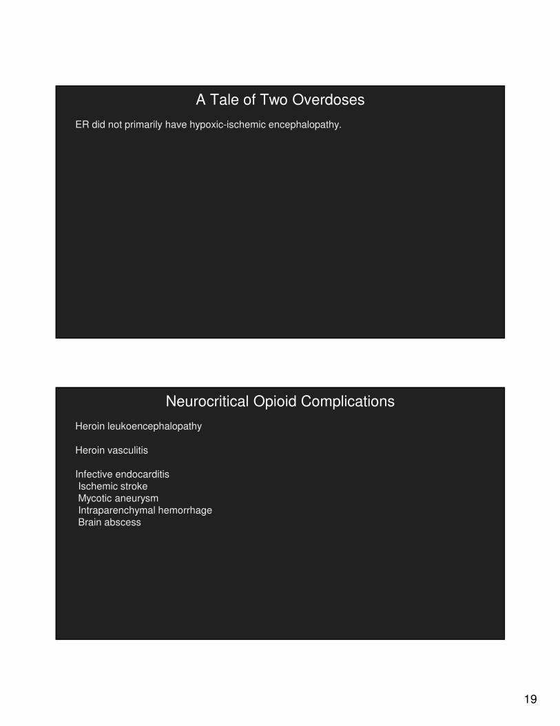
A Tale of Two Overdoses
ER did not primarily have hypoxic-ischemic encephalopathy.
Neurocritical Opioid Complications
Heroin leukoencephalopathy
Heroin vasculitis
Infective endocarditisIschemic strokeMycotic aneurysmIntraparenchymal hemorrhageBrain abscess

20
Heroin Leukoencephalopathy (aka Chasing the Dragon)
Acute-subacute onset of encephalopathy, ataxia, dysarthria, spasticity, choreoathetosis, myoclonus, posturing, and fever that may progress to hypotonia and quadriplegia.
Associated with heroin vapor inhalation (not injection, smoking, or snorting)
Heroin pyrosolate (created by heating heroin) has been implicated but exposure does not reliably replicate condition. Chased heroin is of lower purity than other forms. Adulterants, particularly vapor toluene inhalation, may be the root cause.
Heroin Leukoencephalopathy (aka Chasing the Dragon)
Heroin leukoencephalopathy is likely a rare subset of toxic leukoencephalopathy (toluene, alcohol, MDMA, cocaine), reversible posterior leukoencephalopathy syndrome (RPLS), or
delayed post-hypoxic leukoencephalopathy (DPHL).
Diffuse, symmetric leukoencephalopathy with posterior-anterior gradient and particular involvement of occipital lobes, inferior cerebellum. DWI restriction may occur with ADC hypointensity acutely and hyperintensity later.

21
Heroin Leukoencephalopathy (aka Chasing the Dragon)
Heroin Leukoencephalopathy (aka Chasing the Dragon)
Heroin leukoencephalopathy is likely a rare subset of toxic leukoencephalopathy (toluene, alcohol, MDMA, cocaine), reversible posterior leukoencephalopathy syndrome (RPLS), or
delayed post-hypoxic leukoencephalopathy (DPHL).
Diffuse, symmetric leukoencephalopathy with posterior-anterior gradient and particular involvement of occipital lobes, inferior cerebellum. DWI restriction may occur with ADC hypointensity acutely and hyperintensity later.

22
Heroin Leukoencephalopathy (aka Chasing the Dragon)
Relative arylsulfatase A deficiency, causing delayed myelinotoxicity v delayed apoptosis of oligodendrocytes, implicated in other toxic leukoencephalopathy.
Elevated MRS lactate and possible response to antioxidants in case report of 2 patients (Kriegstein 1999). Consider coenzyme Q10 300 mg q6h +/- vitamin E 2000 mg daily, vitamin C 2000 mg daily given low side effect profile.
Complications include dysautonomia, acute respiratory failure.
Mortality was 23-48% in largest series of 47 patients in Netherlands (Wolter 1982) and 27 patients in Canada (Buxton 2011).
Prognosis, however, is variable and does not necessarily imply irreversible cell death. Over weeks to months some may have a meaningful recovery.
A Tale of One Overdose
3 d1 mo
9 mos later ER is living at home. Speech, vision, and strength are intact. Tubes are removed. Has persistent cognitive and movement disorders but “remarkable recovery”.

23
A Tale of Two Overdoses
Although HM and ER shared cardiac arrests from opioid overdoses with severe PSH, etiology of acute brain injury differed.
HM suffered classic ABI with severe cortical ischemia that manifested as myoclonic status epilepticus and dysautonomia. Prognosis was poor based on large series of post-arrest coma, and he died after withdrawal of life support based on his known wishes.
ER developed heroin leukoencephalopathy. This is a rare diagnosis on the spectrum of toxic leukoencephalopathy, RPLS, and DPHL. Prognosis was uncertain and management was expectant. Rehabilitation continues.
Heroin Vasculitis (Geibprasert
2010)
Even rarer
Possibly from µ opioid receptor induced vasospasm or autoimmune response to adulterant >opioid

24
Infective Endocarditis (IE) (Morris 2014)
IE from IVDA Increased from 6% of hospitalizations in 2006 to 12% in 2013 (SAMHSA).
HIV, HBV, HCV are comorbid. 1/2 from Staphylococcus aureus with frequent metastatic infections.
R >L valvular infection. More pneumonia, septic pulmonary emboli, less murmurs, CHF, splinter hemorrhages. However, if L valvular infection develops, embolization to brain is feared.
In a recent large series of 1345 patients with L valvular IE, 340 (25%) had a neurologic complication, 192 (14%) had ischemic stroke, 86 (6%) had meningitis, 60 (4%) had intraparenchymal hemorrhages, and 20 (1%) had brain abscesses (Garcia-Cabrera 2013).
Ischemic Stroke from IE (Morris 2014)
From septic cardioembolism, often multifocal
If endocarditis is highly suspected, avoid thrombolysis, antiplatelets, or anticoagulants (Walker
2012, Chan 2003, Tornos 1999).
If large stroke occurs, delay valve surgery up to 4 wks for concern of hemorrhagic conversion from inoperative heparinization

25
Mycotic Aneurysm from IE (Morris 2014)
From septic cardioembolism→ intravascular infection→ mycotic aneurysm
Aneurysm is often small, distal, and fragile. Better visualized on angiography than MRA/CTA.
Small aneurysms may resolve with observation and antibiotics
Ruptured, large, or enlarging aneurysms require clip/coil.
Delay valve surgery until aneurysms are stable or 1-2 wks after repair.
Intraparenchymal Hemorrhage from IE (Morris 2012)
From hemorrhagic conversion of ischemic stroke or rupture of mycotic aneurysm
Postpone valve surgery 4 wks.

26
Brain Abscess from Infective Endocarditis (Morris 2014)
Again from septic cardioembolism
If <3 cm, may attempt antibiotics alone. If inadequate response or >3 cm, perform surgery.
In general does not postpone valve surgery.
Conclusions
Suspect acute opioid overdose in any unexplained stupor or arrest, particularly if a patient presents with pinpoint pupils, bradypnea, and bradycardia.
Use naloxone at small escalating doses in stupor and large escalating doses in arrest. With today’s illicit synthetic narcotics, an addict may suffer an unexpected mega-overdose that requires large doses of naloxone before reversal.
Acute opioid overdose frequently causes anoxic brain injury. Resuscitate and initiate targeted temperature management. Use exam then EEG and imaging to guide prognosis at 5-7 d after arrest. If prognosis is uncertain, error on side of time.

27
Conclusions
Paroxysmal sympathetic hyperactivity (PSH) is caused by disinhibited sympathetic reactions to non painful stimuli (analogous to acute opioid withdrawal) after any severe, diffuse brain
injury. PSH is marked by episodes of severe tachycardia, hypertension, fever, and dyskinesias. Opiates, benzodiazepines, and other assorted sympatholytics may be needed to control PSH.
Less common neurocritical complications of opioid use disorder include heroin leukoencephalopathy, heroin vasculitis, and intracranial sequelae of infective endocarditis.

28
Anoxic Brain Injury: Exam
Cranial nerves: fixed pupils, absent corneals, absent oculocephalics
Arousal: no eye opening, no spontaneous eye movements
Motor: extensor or no movement to pain
+/- Myoclonus or seizures
Cranial nerves >arousal >>motor
Anoxic Brain Injury: EEG (Gutierrez 2010)
Classic EEG after cardiac arrest: intermittent cortical activity then delta, theta, and alpha activity
Seizures were detected in 10-47% of cooled post-cardiac arrest patients (Rossetti 2010, Legriel
209, Abend 2009)
Malignant EEG after cardiac arrest:Burst suppression, generalized suppression, status epilepticus, nonreactiveNonreactive FPR 6% v 0% (Crepeau 2013 v Rossetti 2010)
SE FPR 11% (Rossetti 2009)
Myoclonus FPR 7% (Rossetti 2010)
Myoclonic status epilepticus FPR 2% (Greer 2013)

29
Anoxic Brain Injury: SSEP N20s (Leithner 2010)
SSEP N20s more resistant to sedative and metabolic effects than any other
diagnostic.
Hypothermia might delay peaks on SSEP but should not affect amplitude.