neurofisiologia della deglutizione enrico alfonsi …€¦ · 20/10/2015 1 neurofisiologia della...
TRANSCRIPT

20/10/2015
1
NEUROFISIOLOGIA DELLA DEGLUTIZIONE
Enrico Alfonsi
IRCCS Fondazione Istituto Neurologico “C. Mondino”
Pavia
Tongue Larynx Esophagus
Epiglottis
Pharynx
Voluntary phaseOral phase (preparatory)Oral phase (propulsive)
Authomatic phasesPharyngeal phase
Oesophageal phase
SWALLOWINGSWALLOWINGSWALLOWINGSWALLOWING
‘Swallowing is known to be a complex but stereotyped motor sequence, with the
implication that it involves a fixed behavioral pattern’ (Jean, 2001)

20/10/2015
2
20/10/2015
3
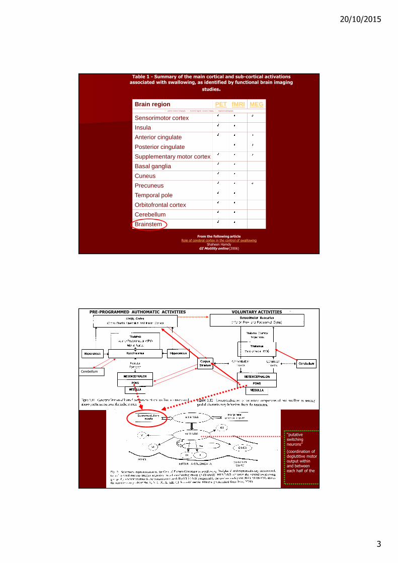
Table 1 - Summary of the main cortical and sub-cortical activations associated with swallowing, as identified by functional brain imaging
studies.
From the following articleRole of cerebral cortex in the control of swallowing
Shaheen HamdyGI Motility online (2006)
Brain region PET fMRI MEGPET, positron emission tomography; fMRI, functional magnetic resonance imaging; MEG, magnetoencephalography.
Sensorimotor cortex
Insula
Anterior cingulate
Posterior cingulate
Supplementary motor cortex
Basal ganglia
Cuneus
Precuneus
Temporal pole
Orbitofrontal cortex
Cerebellum
Brainstem
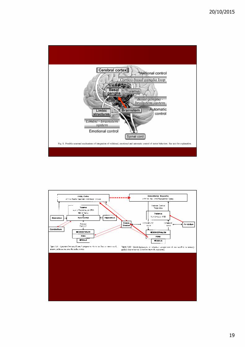
PRE-PROGRAMMED AUTHOMATIC ACTIVITIES VOLUNTARY ACTIVITIES
Cerebellum
“putative switching neurons”
(coordination of deglutitive motor output within and between each half of the medulla)

20/10/2015
4
Subnuclear divisions of rat nucleus tractus solitarii (boxed insert) and distribution of central terminals of afferents coursing in the superior
laryngeal nerve (SLN).
Besides various afferents unrelated to deglutitive function, the SLN contains the majority of swallowing reflex afferents. The intrasolitarial distribution of
anterogradely labeled terminals illustrated in this set of transverse sections overlaps with that revealed after tracer injections into the pharynx, larynx or
upper esophagus. Location of NTS subnucleus centralis (core portion) is marked by dotted outline. Numbers indicate anterior-posterior distance
(micrometers, m) from rostral edge of area postrema. Fascicles of the solitary tract are shown in color. AP, area postrema; DMV, dorsal motor nucleus
of the vagus nerve; D, NTS, nucleus tractus solitarii; cen, central; dl, dorsolateral; gel, gelatinous; int, intermediate; is, interstitial; v, ventral, vl, ventrolateral
subnucleus; D, dorsal; L, lateral; TS, tractus solitarius; IV, fourth ventricle; XII, hypoglossal motor nucleus. (Source : Adapted from Altschuler et al.,30 with
permission of J. Wiley & Sons).
Subnuclear division of NTS:
-NTSis:interstitial
-NTSint:intermediate
-NTSv:ventral
-NTSvl:ventrolateral
-NTSdl:dorsolateral
-NTScen:central
ESOPHAGUS
OROPHARYNX
AND LARYNX
GI Motility online (May 2006) | doi:10.1038/gimo74
Figure 1 Cytodendroarchitecture of the rat ambiguus complex.
Immunohistochemical staining for heat shock protein 27 (HSP27, panel a) reveals contiguous columns of motoneurons that line up in an oblique sagittal plane spanning the length of the ventral rhombencephalon (photomicrograph courtesy of Dr. D.A. Hopkins). The serial reconstruction (b) shows the same neuronal
groupings after retrograde labeling with horse radish peroxidase HRP. Three main divisions are recognizable: a rostral compact formation of esophagomotor neurons (AMB-c), a caudal loose formation(AMB-l) of laryngomotor neurons, and an interposed semicompact formation of chiefly pharyngomotor neurons. The AMB-sc column continues into the rostral tip of the ambiguus complex overlying the facial motor nucleus (VIIm). Note dendritic radiations and bundling. Additional ventral most cell groups (b)
represent the AMB external formation of vagal preganglionic neurons. (Source: Adapted from Bieger and Hopkins,18 with permission of J. Wiley & Sons.)
Esophagomotor neurons
Pharyngomotor neurons
Laryngomotor neurons
External formation of vagal preganglionic
neurons
Facial motor nucleus

20/10/2015
5
GI Motility online (May 2006) | doi:10.1038/gimo74
Proposed network circuit controlling the oral stage of swallowing.
Proposed network circuit controlling the pharyngeal stage of
swallowing.
Proposed network circuit controlling the esophageal stage of swallowing.
PUNTI CHIAVE DEI MECCANiSMI TRONCOENCEFALICI DEL CENTRO DELLA DEGLUTIZIONE
• Le Strutture che formano il generatore centrale della deglutizione o «Central Pattern Generator of swallowing» (SPG) sono intimamente associate al nucleo del tratto solitario (NTS). Circuiti neurali distinti ma interconnessi sono coinvolti nelle fasi orale, faringea ed dell'esofagea della deglutizione.
• Il sottocircuito della fase orale comprende il complesso rostrale del tratto solitario, la formazione reticolare parvicellulare rombencefalica (RFpc), e le sue proiezioni ai nuclei motori dei nervi trigemino (VM), facciale (VIIm) e ipoglosso (XIIm) .
• Il sottocircuito della fase faringea comprende i neuroni premotori delle porzioni intermedia, interstiziale, e ventrali dei subnuclei del Tratto solitario (NTSim, NTSis, NTSV) ed i motoneuroni nel nucleo ambiguo.
• Il sottocircuito della fase esofagea comprende la porzione centrale esterna del subnucleo del tratto solitario (NTSce), neuroni vagali sensoriali primari dell’ esofago ed i motoneuroni della formazione compatta del nucleo ambiguo che innervano la muscolatura striata dell'esofago
• Le connessioni tra i subnuclei del TS e della reticolare rombo-encefalica consentono al SPG di produrre inibizione e eccitazione sequenziale ldei motoneuroni e garantiscono il coordinamento bilaterale dei due «centri unilaterali» della deglutizione. Le connessioni tra NTS e nucleo motore dorsale del nervo vago (DMV) forniscono un meccanismo per la regolazione dell'attività della muscolatura liscia dell'esofago e dello sfintere esofageo inferiore. Questi collegamenti provvedono anche all’l'accoppiamento funzionale delle fasi orofaringea ed esofagea della deglutizione.
• Il trasferimento rapido di informazioni nelle reti SPG utilizza la trasmissione eccitatoria amino-acidica attraverso diversi sottotipi di recettori per il glutammato e il rilascio tonico di acido gamma-aminobutirrico (GABA), inibizione- GABAergica, fornisce il meccanismo necessario per innescare l'attività fasica del SPG.

20/10/2015
6
I processi della respirazione e della deglutizione sono strettamente collegati nel loro controllo
centrale e sono altamente coordinati
� Molti muscoli e le strutture hanno un doppio ruolo nella respirazione e deglutizione:
� I centri di controllo neurali responsabili del coordinamento di respirazione e deglutizione sono contenuti nelle regioni dorsomediale e ventrolaterale del bulbo
� Le strutture corticali svolgono anche un ruolo importante nel facilitare e modulare il coordinamento della respirazione e della deglutizione
� Esistono "neuroni multifunzionali" comuni ad entrambi i controlli: respirazione e deglutizione (Central pattern generator o CPG) possono avere rilevanza anche per una migliore comprensione della neuroplasticità del SNC:
a) E’ possibile che nelle lesioni del CPG della deglutizione, i neuroni comuni sia alla respirazione che alla deglutizione riducano l'impatto complessivo del danno in funzione della deglutizione o viceversa.b) d'altra parte, se un CPG della deglutizione contiene neuroni comuni ad entrambe le funzioni, deglutitoria e respiratoria, una singola lesione del CPG può porre l'individuo in svantaggio funzionale ed a maggior rischio per entrambi i processi funzionali.
� I due processi fisiologici fondamentali sono altamente integrati a livello anatomo-funzionale e non si escludono a vicenda.

20/10/2015
7
PRESBIFAGIA ( PRESBIDEGLUTIZIONE?)
MODIFICAZIONI ANATO-FISIOLOGICHE
1. Edentulia
2. Allungamento del tratto vocale
3. Debolezza muscolare laringea
4. Riduzione dell’olfatto
5. Ridotta sensibilità orale
6. Ridotta sensibilità laringea

20/10/2015
8
MODIFICAZIONI BIOMECCANICHE
1. Diminuita forza linguale ISOMETRICA (con invariata forza deglutitoria) � ridotta riserva funzionale
2. Fase orale prolungata
3. Ritardo di innesco del riflesso
4. Ridotti tempi di apertura dell’UES
5. Ridotta peristalsi faringea
6. Ridotta elevazione laringea
(Schindler et al; in Deglutologia II ed, 2011)
AUMENTO DEI RISTAGNI
AUMENTO DEI FENOMENI DI PENETRAZIONE/ASPIRAZIONE
PRESBIFAGIA
COORDINAZIONE RESPIRAZIONE DEGLUTIZIONE
Hirst et al; Dysphagia 2002; 17: 152-161
Nel soggetto anziano aumenta la durata dell’apnea deglutitoria, la frequenza respiratoria aumenta durante la deglutizione, mentre la saturazione di ossigeno diminuisce
PRESBIFAGIA
20/10/2015
9
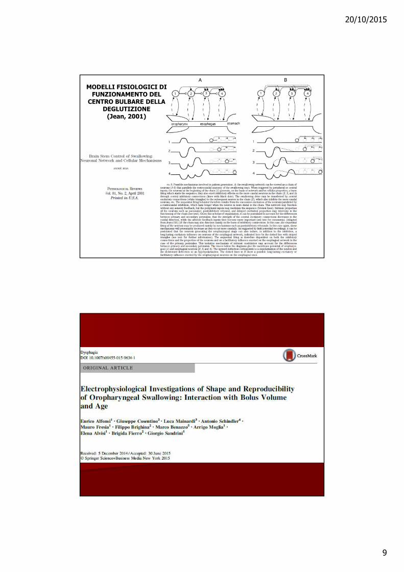
MODELLI FISIOLOGICI DI FUNZIONAMENTO DEL
CENTRO BULBARE DELLA DEGLUTIZIONE
(Jean, 2001)

20/10/2015
10

20/10/2015
11
20/10/2015
12
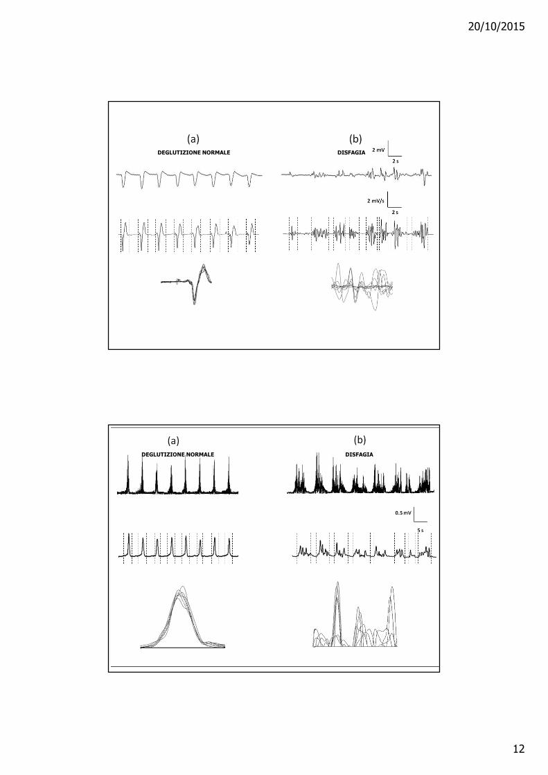
DISFAGIADEGLUTIZIONE NORMALE
DEGLUTIZIONE NORMALE DISFAGIA

20/10/2015
13
CALCULATE THE DISTANCE IN EACH POINT BETWEEN A COUPLE OF CURVES
MEDIAN DISTANCE:
Median value of ‘distance distribution’ for all couples of curves examined
SIMILARITY INDEX (SI):
Percentage of couples of curves having a reciprocal ‘distance‘ lower than a prefixed, threshold (ɛ)ɛ)ɛ)ɛ)value
curve I
curve II
.
‘Unda non est materia sed forma materiae progredientis’(Titus Lucretius Carus, De Rerum Natura, V Libro)
SEGNALE EMG RETTIFICATO «GREZZO»
SEGNALE EMG RETTIFICATO «GREZZO»
«ENVELOPE» DEL SEGNALE EMG
«ENVELOPE» DEL SEGNALE EMG
DEGLUTIZIONE NORMALE DISFAGIA

20/10/2015
14
Meaning of the parameter:SHEMG-E/T-shape

20/10/2015
15
ELECTROPHYSIOLOGICAL INVESTIGATION OF ORAL-PHARYNGEAL SWALLOWING
NORMAL PATTERN
20/10/2015
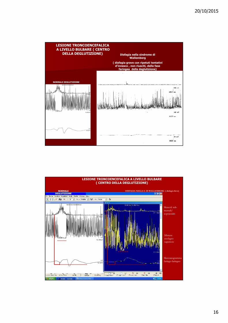
16
Disfagia nella sindrome di Wallemberg
( disfagia grave con ripetuti tentativi d’innesco , non riusciti, della fase
faringea della deglutizione)
MUSCOLO GENERALE
5s 100µV
6.3
5s 200µV
6.4
5s 1mV
6.5
NORMALE DEGLUTIZIONE
LESIONE TRONCOENCEFALICAA LIVELLO BULBARE ( CENTRO
DELLA DEGLUTIZIONE)
DISFAGIA NELLA S. DI WALLEMBERG ( disfagia lieve)
Muscoli sub-
mentali/
sopraioidei
Sfintere
esofageo
superiore
Meccanogramma
faringo-laringeo
MUSCOLO GENERALE
5s 100µV
6.3
5s 200µV
6.4
5s 1mV
6.5
LESIONE TRONCOENCEFALICA A LIVELLO BULBARE ( CENTRO DELLA DEGLUTIZIONE)
NORMALE DEGLUTIZIONE

20/10/2015
17
Dysphagia at ALS onset can be observed in up to 30 % of patients

20/10/2015
18
Brain, Vol. 123, No. 1, 125-140, January 2000© 2000 Oxford University Press
Pathophysiological mechanisms of oropharyngeal dysphagia in amyotrophic lateral sclerosis
Cumhur Ertekin1,2, Ibrahim Aydogdu1,2, Nur Yüceyar2, Nefati
Kiylioglu2, Sultan Tarlaci2 and Burhanettin Uludag1,2
Brain, Vol. 123, No. 1, 125-140, January 2000© 2000 Oxford University Press
Pathophysiological mechanisms of oropharyngeal dysphagia in amyotrophic lateral sclerosis Cumhur Ertekin1,2, Ibrahim Aydogdu1,2, Nur Yüceyar2, Nefati Kiylioglu2, Sultan Tarlaci2 and Burhanettin Uludag1,2
20/10/2015
19
Cerebellum

20/10/2015
20
Bradykinesia of the orale phase of swallowing in PD
MUSCOLO GENERALE
5s 100µV
6.3
5s 200µV
6.4
5s 1mV
6.5
1
“Start hesitation” of swallowing in PD patients with L-Dopa
“Long-term syndrome ”
SHEMG-D increase, with several EMG bursts can be
labeled as ‘start hesitation’ of swallowing
20/10/2015
21
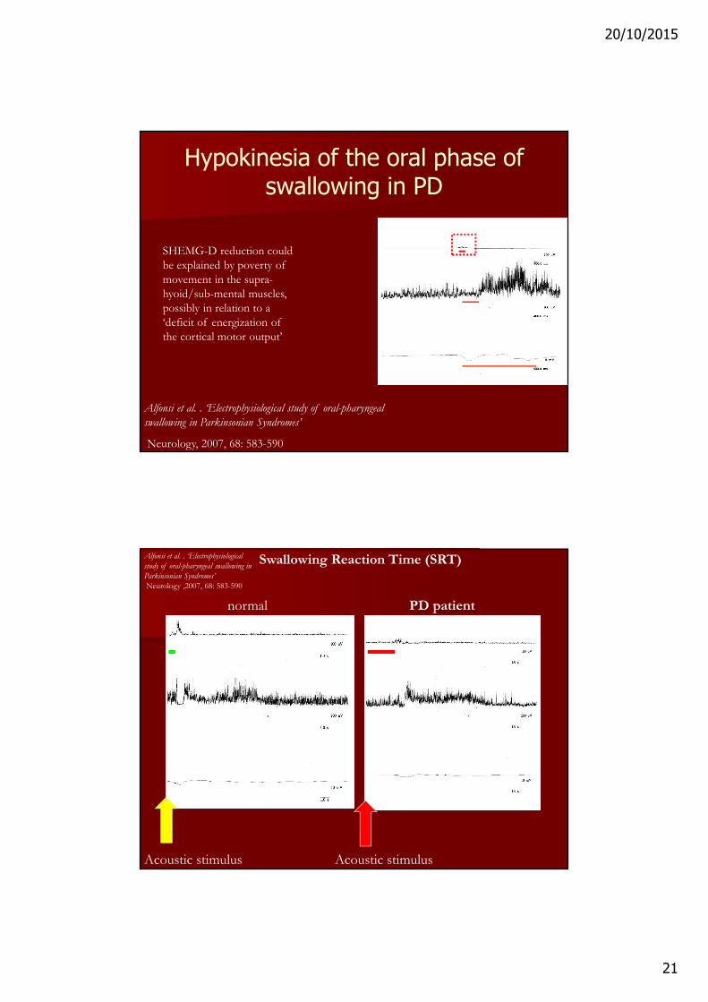
Hypokinesia of the oral phase of swallowing in PD
SHEMG-D reduction could
be explained by poverty of
movement in the supra-
hyoid/sub-mental muscles,
possibly in relation to a
‘deficit of energization of
the cortical motor output’
Alfonsi et al. . ‘Electrophysiological study of oral-pharyngeal
swallowing in Parkinsonian Syndromes’
Neurology, 2007, 68: 583-590
Acoustic stimulus Acoustic stimulus
Swallowing Reaction Time (SRT)
normal PD patient
Alfonsi et al. . ‘Electrophysiological
study of oral-pharyngeal swallowing in
Parkinsonian Syndromes’
Neurology ,2007, 68: 583-590
20/10/2015
22
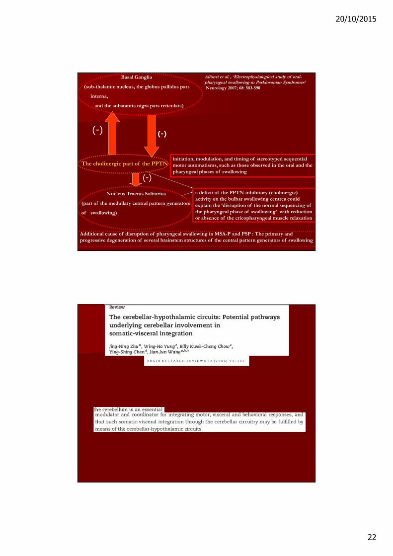
Basal Ganglia
(sub-thalamic nucleus, the globus pallidus pars
interna,
and the substantia nigra pars reticulata)
The cholinergic part of the PPTN
Nucleus Tractus Solitarius
(part of the medullary central pattern generators
of swallowing)
initiation, modulation, and timing of stereotyped sequential
motor automatisms, such as those observed in the oral and the
pharyngeal phases of swallowing
a deficit of the PPTN inhibitory (cholinergic)
activity on the bulbar swallowing centres could
explain the ‘disruption of the normal sequencing of
the pharyngeal phase of swallowing’ with reduction
or absence of the cricopharyngeal muscle relaxation
Alfonsi et al. , ‘Electrophysiological study of oral-pharyngeal swallowing in Parkinsonian Syndromes’Neurology 2007; 68: 583-590
Additional cause of disruption of pharyngeal swallowing in MSA-P and PSP : The primary and
progressive degeneration of several brainstem structures of the central pattern generators of swallowing
(-)
(-)
(-)

20/10/2015
23
(histaminergic fibers)
MUSCOLO GENERALE
5s 100µV
6.3
5s 200µV
6.4
5s 1mV
6.5
200 µV
200 µV
5 mV
PATTERNS OF OROPHARYNGEAL SWALLOWING IN MS
(a): Normal (b): cerebellar-pyramidal sydrome (c): cerebellar syndrome
20/10/2015
24
Grazie per la vostra attenzione