neuroglial activation and neuroinflammation in the...
TRANSCRIPT
Neuroglial Activation andNeuroinflammation in the Brain of Patients
with AutismDiana L. Vargas, MD,1,2 Caterina Nascimbene, MD,1–3 Chitra Krishnan, MHS,1
Andrew W. Zimmerman, MD,1,4 and Carlos A. Pardo, MD1,2,5
Autism is a neurodevelopmental disorder characterized by impaired communication and social interaction and may beaccompanied by mental retardation and epilepsy. Its cause remains unknown, despite evidence that genetic, environ-mental, and immunological factors may play a role in its pathogenesis. To investigate whether immune-mediated mech-anisms are involved in the pathogenesis of autism, we used immunocytochemistry, cytokine protein arrays, and enzyme-linked immunosorbent assays to study brain tissues and cerebrospinal fluid (CSF) from autistic patients and determinedthe magnitude of neuroglial and inflammatory reactions and their cytokine expression profiles. Brain tissues from cer-ebellum, midfrontal, and cingulate gyrus obtained at autopsy from 11 patients with autism were used for morphologicalstudies. Fresh-frozen tissues available from seven patients and CSF from six living autistic patients were used for cytokineprotein profiling. We demonstrate an active neuroinflammatory process in the cerebral cortex, white matter, and notablyin cerebellum of autistic patients. Immunocytochemical studies showed marked activation of microglia and astroglia, andcytokine profiling indicated that macrophage chemoattractant protein (MCP)–1 and tumor growth factor–�1, derivedfrom neuroglia, were the most prevalent cytokines in brain tissues. CSF showed a unique proinflammatory profile ofcytokines, including a marked increase in MCP-1. Our findings indicate that innate neuroimmune reactions play apathogenic role in an undefined proportion of autistic patients, suggesting that future therapies might involve modifyingneuroglial responses in the brain.
Ann Neurol 2005;57:67–81
Autism is a common neurodevelopmental disordercharacterized by impairments in social, behavioral, andcommunicative functions.1,2 Symptoms appear before36 months of age, and regression or loss of skills occursin 30% of affected children, usually between 18 and 24months.1 The syndrome is clinically heterogeneous andcan be associated in up to 10% of patients with well-described neurological and genetic disorders, such astuberous sclerosis, fragile X, and Rett’s and Down’ssyndromes, although in most patients the causes arestill unknown.3,4 Recent epidemiological studies sug-gest that the prevalence of the autistic syndromes hasincreased in recent years to 1 in 250 to 500 chil-dren, perhaps as a result of improved diagnostic ap-proaches.5–7
Although the neurobiological basis for autism re-
mains poorly understood, several lines of research nowsupport the view that genetic, environmental, neuro-logical, and immunological factors contribute to its de-velopment.3,4,8–10 Neuropathological studies haveshown that abnormalities in cytoarchitectural organiza-tion of the cerebral cortex and subcortical structures, aswell as reduced numbers of Purkinje cells in the cere-bellum, are the most consistent findings in postmortembrain tissues from autistic patients. They suggest thatdefects in neuronal maturation and cortical organiza-tion may be responsible for some of the neurologicalproblems seen in autism.11–13
Immune dysfunction has been proposed as a poten-tial mechanism for the pathogenesis of autism.14 Sev-eral studies in peripheral blood have shown various ab-normalities such as T-cell dysfunction, autoantibody
From the 1Department of Neurology and 2Division of Neuroimmu-nology and Infectious Disorders, Johns Hopkins University Schoolof Medicine, Baltimore, MD; 3Department of Neurology, Univer-sity of Milan, Italy; 4Kennedy Krieger Institute; and 5Department ofPathology, Johns Hopkins University School of Medicine, Balti-more, MD.
Received Jul 8, 2004, and in revised form Sep 14. Accepted forpublication Sep 14, 2004.
Published online Nov 15, 2004 in Wiley InterScience(www.interscience.wiley.com). DOI: 10.1002/ana.20315
Address correspondence to Dr Pardo, Department of Neurology,Johns Hopkins University School of Medicine, Pathology 627,600 North Wolfe Street, Baltimore, MD 21287.E-mail: [email protected]
© 2004 American Neurological Association 67Published by Wiley-Liss, Inc., through Wiley Subscription Services
production, and increased proinflammatory cyto-kines.9,15–18 The potential role for maternal antibodiesas a pathogenic factor also has been proposed.19 Cere-brospinal fluid (CSF) studies demonstrated no evidenceof inflammation by standard cell counts, protein elec-trophoresis, or measurements of quinolinic acid andneopterin.20 Despite the growing interest in possibleimmune mechanisms in its pathogenesis, there hasbeen no direct evidence linking findings in the periph-eral blood to immune activity in the brain of autisticpatients.21 Neuropathological studies have given littleattention to immune and neuroglial activity in autism,and the most comprehensive postmortem study re-ported no inflammatory changes or astroglial reac-tions.11 Only a few reports have described gliosis andinflammatory changes.12,22 Such neuroinflammation, ifpresent in the brain, might both participate in and re-sult from dysfunctional CNS development and activityin autism. To investigate whether immune-mediatedmechanisms are involved in the pathogenesis of autismwith respect to the central nervous system (CNS), we
studied brain tissues and CSF from autistic patientsand determined the magnitude of neuroglial and in-flammatory reactions and their cytokine expressionprofiles.
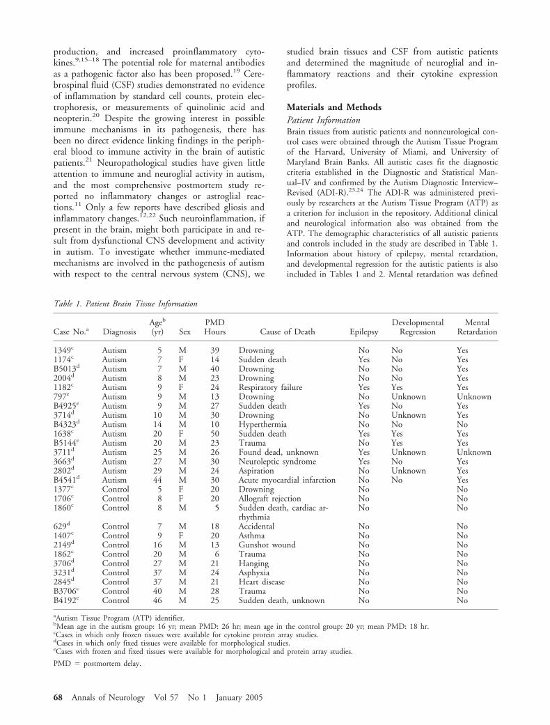
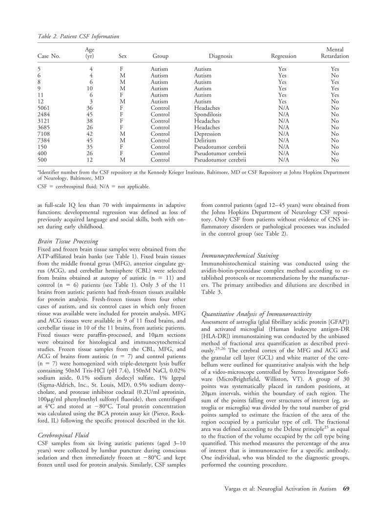
Materials and MethodsPatient InformationBrain tissues from autistic patients and nonneurological con-trol cases were obtained through the Autism Tissue Programof the Harvard, University of Miami, and University ofMaryland Brain Banks. All autistic cases fit the diagnosticcriteria established in the Diagnostic and Statistical Man-ual–IV and confirmed by the Autism Diagnostic Interview–Revised (ADI-R).23,24 The ADI-R was administered previ-ously by researchers at the Autism Tissue Program (ATP) asa criterion for inclusion in the repository. Additional clinicaland neurological information also was obtained from theATP. The demographic characteristics of all autistic patientsand controls included in the study are described in Table 1.Information about history of epilepsy, mental retardation,and developmental regression for the autistic patients is alsoincluded in Tables 1 and 2. Mental retardation was defined
Table 1. Patient Brain Tissue Information
Case No.a DiagnosisAgeb
(yr) SexPMDHours Cause of Death Epilepsy
DevelopmentalRegression
MentalRetardation
1349c Autism 5 M 39 Drowning No No Yes1174c Autism 7 F 14 Sudden death Yes No YesB5013d Autism 7 M 40 Drowning No No Yes2004d Autism 8 M 23 Drowning No No Yes1182c Autism 9 F 24 Respiratory failure Yes Yes Yes797e Autism 9 M 13 Drowning No Unknown UnknownB4925e Autism 9 M 27 Sudden death Yes No Yes3714d Autism 10 M 30 Drowning No Unknown YesB4323d Autism 14 M 10 Hyperthermia No No No1638c Autism 20 F 50 Sudden death Yes Yes YesB5144e Autism 20 M 23 Trauma No Yes Yes3711d Autism 25 M 26 Found dead, unknown Yes Unknown Unknown3663d Autism 27 M 30 Neuroleptic syndrome Yes No Yes2802d Autism 29 M 24 Aspiration No Unknown YesB4541d Autism 44 M 30 Acute myocardial infarction No No Yes1377c Control 5 F 20 Drowning No No1706c Control 8 F 20 Allograft rejection No No1860c Control 8 M 5 Sudden death, cardiac ar-
rhythmiaNo No
629d Control 7 M 18 Accidental No No1407c Control 9 F 20 Asthma No No2149d Control 16 M 13 Gunshot wound No No1862c Control 20 M 6 Trauma No No3706d Control 27 M 21 Hanging No No3231d Control 37 M 24 Asphyxia No No2845d Control 37 M 21 Heart disease No NoB3706c Control 40 M 28 Trauma No NoB4192e Control 46 M 25 Sudden death, unknown No No
aAutism Tissue Program (ATP) identifier.bMean age in the autism group: 16 yr; mean PMD: 26 hr; mean age in the control group: 20 yr; mean PMD: 18 hr.cCases in which only frozen tissues were available for cytokine protein array studies.dCases in which only fixed tissues were available for morphological studies.eCases with frozen and fixed tissues were available for morphological and protein array studies.
PMD � postmortem delay.
68 Annals of Neurology Vol 57 No 1 January 2005
as full-scale IQ less than 70 with impairments in adaptivefunctions; developmental regression was defined as loss ofpreviously acquired language and social skills, both with on-set during early childhood.
Brain Tissue ProcessingFixed and frozen brain tissue samples were obtained from theATP-affiliated brain banks (see Table 1). Fixed brain tissuesfrom the middle frontal gyrus (MFG), anterior cingulate gy-rus (ACG), and cerebellar hemisphere (CBL) were selectedfrom brains obtained at autopsy of autistic (n � 11) andcontrol (n � 6) patients (see Table 1). Only 3 of the 11brains from autistic patients had fresh-frozen tissues availablefor protein analysis. Fresh-frozen tissues from four othercases of autism, and six control cases in which only frozentissue was available were included for protein analysis. MFGand ACG tissues were available in 9 of 11 fixed brains, andcerebellar tissue in 10 of the 11 brains, from autistic patients.Fixed tissues were paraffin-processed, and 10�m sectionswere obtained for histological and immunocytochemicalstudies. Frozen tissue samples from the CBL, MFG, andACG of brains from autistic (n � 7) and control patients(n � 7) were homogenized with triple-detergent lysis buffercontaining 50nM Tris-HCl (pH 7.4), 150nM NaCl, 0.02%sodium azide, 0.1% sodium dodecyl sulfate, 1% Igepal(Sigma-Aldrich, Inc., St. Louis, MD), 0.5% sodium deoxy-cholate, and protease inhibitor cocktail (0.2U/ml aprotinin,100�g/ml phenylmethyl sulfonyl fluoride), then centrifugedat 4°C and stored at �80°C. Total protein concentrationwas calculated using the BCA protein assay kit (Pierce, Rock-ford, IL) following the specific protocol described in the kit.
Cerebrospinal FluidCSF samples from six living autistic patients (aged 3–10years) were collected by lumbar puncture during conscioussedation and then immediately frozen at �80°C and keptfrozen until used for protein analysis. Similarly, CSF samples
from control patients (aged 12–45 years) were obtained fromthe Johns Hopkins Department of Neurology CSF reposi-tory. Only CSF from patients without evidence of CNS in-flammatory disorders or pathological processes was includedin the control group (see Table 2).
Immunocytochemical StainingImmunohistochemical staining was conducted using theavidin-biotin-peroxidase complex method according to es-tablished protocols or recommendations by the manufactur-ers. The primary antibodies and dilutions are described inTable 3.
Quantitative Analysis of ImmunoreactivityAssessment of astroglia (glial fibrillary acidic protein [GFAP])and activated microglial (Human leukocyte antigen-DR[HLA-DR]) immunostaining was conducted by the unbiasedmethod of fractional area quantification as described previ-ously.25,26 The cerebral cortex of the MFG and ACG andthe granular cell layer (GCL) and white matter of the cere-bellum were outlined for quantitative analysis with the helpof a video-microscope controlled by Stereo Investigator Soft-ware (MicroBrightfield, Williston, VT). A group of 30points was systematically placed in random positions, at20�m intervals, within the boundary of each region. Thesum of the points falling over structures of interest (eg, as-troglia or microglia) was divided by the total number of gridpoints sampled to estimate the fraction of the area of theregion occupied by a particular type of cell. The fractionalarea was defined according to the Delesse principle25 as equalto the fraction of the volume occupied by the cell type beingquantified. This method measures the percentage of the areaof interest that is immunoreactive for a specific antibody.One individual, who was blinded to the diagnostic groups,performed the counting procedure.
Table 2. Patient CSF Information
Case No.Age(yr) Sex Group Diagnosis Regression
MentalRetardation
5 4 F Autism Autism Yes Yes6 4 M Autism Autism Yes No8 6 M Autism Autism Yes Yes9 10 M Autism Autism Yes Yes11 6 F Autism Autism Yes Yes12 3 M Autism Autism Yes No5061 36 F Control Headaches N/A No2484 45 F Control Spondilosis N/A No3121 38 F Control Headaches N/A No3685 26 F Control Headaches N/A No7108 42 M Control Depression N/A No7384 45 M Control Delirium N/A No150 35 F Control Pseudotumor cerebrii N/A No400 26 F Control Pseudotumor cerebrii N/A No500 12 M Control Pseudotumor cerebrii N/A No
aIdentifier number from the CSF repository at the Kennedy Krieger Institute, Baltimore, MD or CSF Repository at Johns Hopkins Departmentof Neurology, Baltimore, MD
CSF � cerebrospinal fluid; N/A � not applicable.
Vargas et al: Neuroglial Activation in Autism 69

Confocal MicroscopyFormalin-fixed brain tissues were cryoprotected with sucrosesolutions and then cut with a sliding microtome to yield40�m sections. The sections were incubated with primaryantibodies (GFAP�HLADR) and incubated with the appro-priate fluorogen-tagged secondary antibody (Cy3 or Alexa).Specimens were examined in a Zeiss LSM 5.0 confocal lasermicroscope (Zeiss, Thornwood, NY).
Protein Tissue ArraysTo further characterize the nature of the inflammatory re-sponses in autistic brains, we studied the relative expressionof 79 proteins: cytokines associated with innate and adaptiveimmunity, chemokines, and growth and differentiation fac-tors by human cytokine protein array methods.27,28 Humancytokine array kits (5.1 and V; Raybiotech, Norcros, GA)were used, consisting of 79 different cytokines, chemokines,and growth factors (Table 4) imprinted on a nitrocellulosemembrane. The protocol for analysis followed the manufac-turer’s instructions: Membranes were blocked for 1 hour andincubated with 500�g of human tissue homogenate or 1mlof CSF for 2 hours at room temperature and then washedfor 30 minutes and incubated with a 1 to 250 dilution ofbiotin-conjugated antibody mix for 2 hours. After consecu-tive washes, a 1 to 1,000 dilution of streptavidin-conjugatedperoxidase was added and incubated for 1 hour at room tem-perature. The membranes were washed thoroughly and ex-posed to peroxidase substrate (ECL chemiluminescence; Am-ersham, Arlington Heights, IL), followed by apposition ofthe membranes with autoradiographic film (Hyperfilm ECL;Amersham) for a standard exposure of 1 minutes. The filmwas scanned, and spots were digitized into pixel densities us-ing the NIH imaging software (Image J). The ratio of rela-tive expression was established after subtraction of the back-ground intensity and comparison with the positive spotsavailable in the membrane.
Enzyme-Linked Immunosorbent Assay TechniquesTumor growth factor (TGF)–�1, macrophage chemoattrac-tant protein (MCP)–1, interleukin (IL)–6 (R&D Systems,Minneapolis, MN), and insulin-like growth factor bindingprotein (IGFBP)–1 (Alpha Diagnostics, San Antonio, TX)
were quantified in tissue homogenates by sandwich enzymeimmunoassay using commercially available kits according tothe manufacturers’ protocols. Values were calculated from a
Table 4. Proteins Included in the Cytokine ProteinArray Study
Cytokines ChemokinesGrowth and
Differentiation Factors
IL-2 ENA-78 GCSFIL-4 GRO GM-CSFIL-5 GRO-� IL-3IL-13 I-309 IL-7IFN-� IL-8 MCSFTGF-�1 MCP-1 SCFIL-16 MCP-2 EGFTGF-�2 MCP-3 IGF-ITGF-�3 MDC AngIL-1� MIG OSMIL-1� MIP-1� TpoIL-6 MIP-1� VEGFIL-10 RANTES PDGF-BIL-12 SDF-1 LeptinIL-15 TARC BDNFTNF-� BLC FGF-4TNF-� Ck� 8-1 FGF-6
Eotaxin FGF-7Eotaxin-2 FGF-9Eotaxin-3 Flt-3 ligandFractalkine GDNFGCP-2 HGFIP-10 IGFBP-1MCP-4 IGFBP-2MIF IGFBP-3MIP-3� IGFBP-4NAP-2 LIF
LIGHTNT-3NT-4OsteoprotegerinPARCPIGFTIMP-1TIMP-2
Table 3. Antibody Information
Antibody Type Epitope/Specificity Dilution Source
GFAP Polyclonal Astrocytes 1:100 DakoHLA-DR Monoclonal MHC class II, activated microglia 1:100 DakoCD68 Monoclonal Macrophages, monocytes 1:100 DakoMRP-8, calgranulin A Monoclonal Macrophages in late or chronic infiltrates 1:100 BACHEMCD3 Polyclonal T cells 1:50 DakoCD20 Monoclonal B cells 1:200 DakoC9neo (B7) Monoclonal Complement, membrane attack complex 1:20 Dr P. Morgan, UKIL-6 Polyclonal IL-6 1:750 NovusMCP-1 Polyclonal MCP-1 1:200 PeprotechTGF-�1 Polyclonal TGF-�1 1:200 Santa CruzIGFBP1 Polyclonal IGFBP-1 1:200 Santa Cruz
GFAP � glial fibrillary acidic protein; MRP � migration inhibitory factor (MIF)-realted protein; IL � interleukin; MCP � macrophagechemoattractant protein; TGF � tumor growth factor; IGFBP � insulin-like growth factor binding protein.
70 Annals of Neurology Vol 57 No 1 January 2005
standard curve generated for each enzyme-linked immu-nosorbent assay (ELISA). Samples were diluted 1 to 10, andresults were standardized according to previously establishedprotein concentrations, with the final concentration ex-pressed as picograms per micrograms protein.
Statistical AnalysisSPSS 11.0 was used for all statistical analyses. Because of thenonparametric nature of the data (as determined by tests ofnormality), nonparametric tests were used to increase the ro-bustness of the results. Group differences between autisticcases and controls in the fractional area of immunoreactivityfor astroglia and activated microglia in the various brain re-gions were compared using the Mann–Whitney U testbecause of the non-Gaussian appearance of the data. TheMann–Whitney U test also was used to compare group dif-ferences in protein tissue arrays and ELISA quantification.Significance was assessed at the 0.05 level. For multiple testcomparisons, a Bonferroni correction was performed, andcorrelations were assessed by Spearman’s rank correlation co-efficient because of the ordinal nature of the data. These testswere used because they make no assumptions about the dis-tribution of the data (eg, normality).
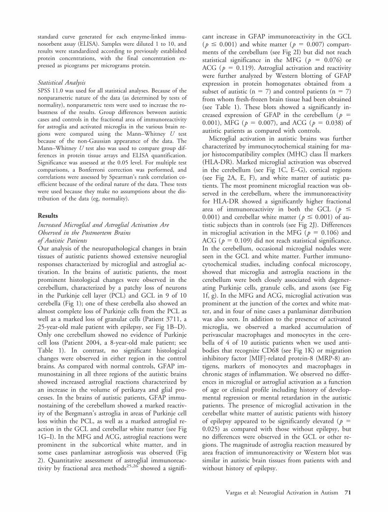
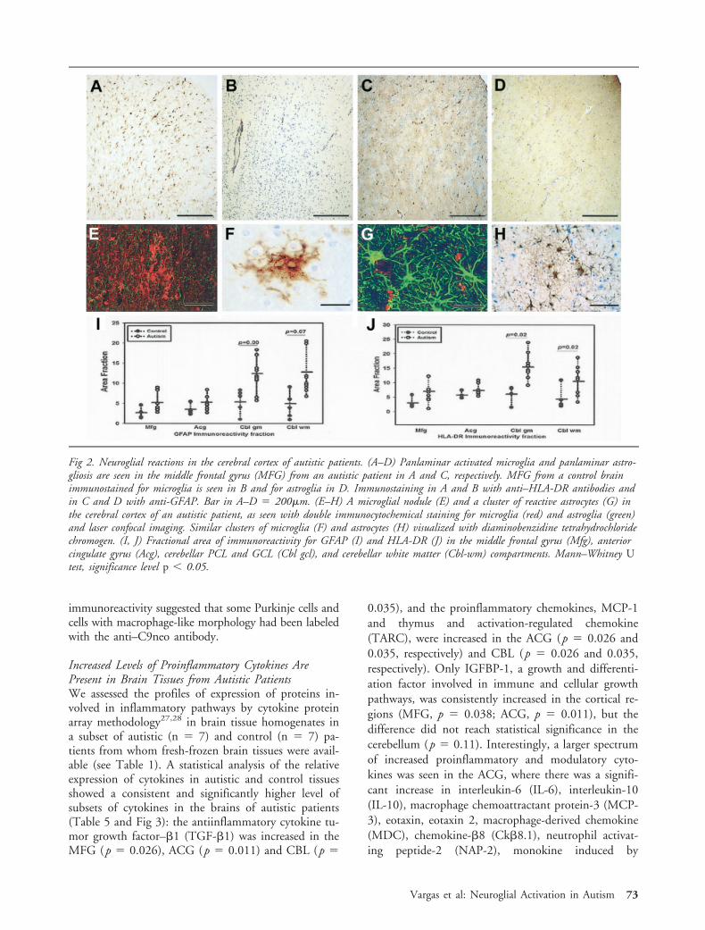
ResultsIncreased Microglial and Astroglial Activation AreObserved in the Postmortem Brainsof Autistic PatientsOur analysis of the neuropathological changes in braintissues of autistic patients showed extensive neuroglialresponses characterized by microglial and astroglial ac-tivation. In the brains of autistic patients, the mostprominent histological changes were observed in thecerebellum, characterized by a patchy loss of neuronsin the Purkinje cell layer (PCL) and GCL in 9 of 10cerebella (Fig 1); one of these cerebella also showed analmost complete loss of Purkinje cells from the PCL aswell as a marked loss of granular cells (Patient 3711, a25-year-old male patient with epilepsy, see Fig 1B–D).Only one cerebellum showed no evidence of Purkinjecell loss (Patient 2004, a 8-year-old male patient; seeTable 1). In contrast, no significant histologicalchanges were observed in either region in the controlbrains. As compared with normal controls, GFAP im-munostaining in all three regions of the autistic brainsshowed increased astroglial reactions characterized byan increase in the volume of perikarya and glial pro-cesses. In the brains of autistic patients, GFAP immu-nostaining of the cerebellum showed a marked reactiv-ity of the Bergmann’s astroglia in areas of Purkinje cellloss within the PCL, as well as a marked astroglial re-action in the GCL and cerebellar white matter (see Fig1G–I). In the MFG and ACG, astroglial reactions wereprominent in the subcortical white matter, and insome cases panlaminar astrogliosis was observed (Fig2). Quantitative assessment of astroglial immunoreac-tivity by fractional area methods25,26 showed a signifi-
cant increase in GFAP immunoreactivity in the GCL(p � 0.001) and white matter (p � 0.007) compart-ments of the cerebellum (see Fig 2I) but did not reachstatistical significance in the MFG (p � 0.076) orACG (p � 0.119). Astroglial activation and reactivitywere further analyzed by Western blotting of GFAPexpression in protein homogenates obtained from asubset of autistic (n � 7) and control patients (n � 7)from whom fresh-frozen brain tissue had been obtained(see Table 1). These blots showed a significantly in-creased expression of GFAP in the cerebellum (p �0.001), MFG (p � 0.007), and ACG (p � 0.038) ofautistic patients as compared with controls.
Microglial activation in autistic brains was furthercharacterized by immunocytochemical staining for ma-jor histocompatibility complex (MHC) class II markers(HLA-DR). Marked microglial activation was observedin the cerebellum (see Fig 1C, E–G), cortical regions(see Fig 2A, E, F), and white matter of autistic pa-tients. The most prominent microglial reaction was ob-served in the cerebellum, where the immunoreactivityfor HLA-DR showed a significantly higher fractionalarea of immunoreactivity in both the GCL (p �0.001) and cerebellar white matter (p � 0.001) of au-tistic subjects than in controls (see Fig 2J). Differencesin microglial activation in the MFG (p � 0.106) andACG (p � 0.109) did not reach statistical significance.In the cerebellum, occasional microglial nodules wereseen in the GCL and white matter. Further immuno-cytochemical studies, including confocal microscopy,showed that microglia and astroglia reactions in thecerebellum were both closely associated with degener-ating Purkinje cells, granule cells, and axons (see Fig1f, g). In the MFG and ACG, microglial activation wasprominent at the junction of the cortex and white mat-ter, and in four of nine cases a panlaminar distributionwas also seen. In addition to the presence of activatedmicroglia, we observed a marked accumulation ofperivascular macrophages and monocytes in the cere-bella of 4 of 10 autistic patients when we used anti-bodies that recognize CD68 (see Fig 1K) or migrationinhibitory factor [MIF]-related protein-8 (MRP-8) an-tigens, markers of monocytes and macrophages inchronic stages of inflammation. We observed no differ-ences in microglial or astroglial activation as a functionof age or clinical profile including history of develop-mental regression or mental retardation in the autisticpatients. The presence of microglial activation in thecerebellar white matter of autistic patients with historyof epilepsy appeared to be significantly elevated (p �0.025) as compared with those without epilepsy, butno differences were observed in the GCL or other re-gions. The magnitude of astroglia reaction measured byarea fraction of immunoreactivity or Western blot wassimilar in autistic brain tissues from patients with andwithout history of epilepsy.
Vargas et al: Neuroglial Activation in Autism 71
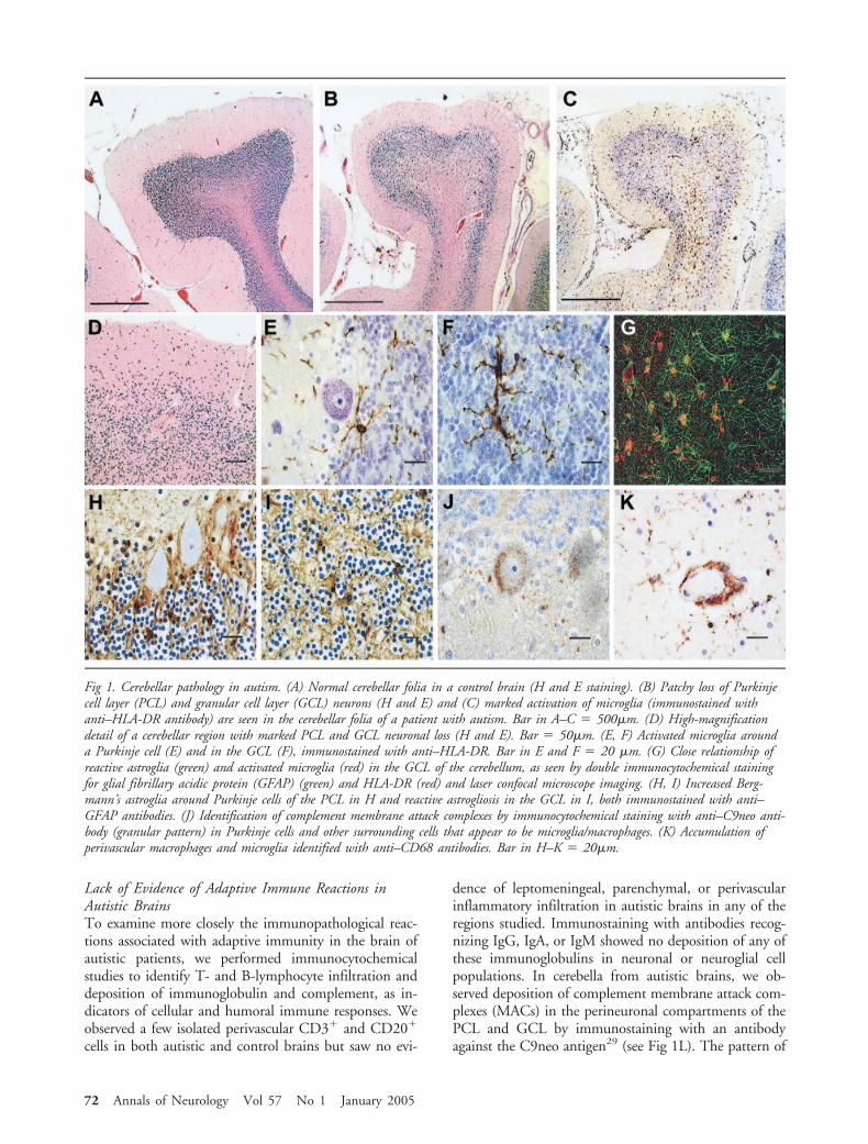
Lack of Evidence of Adaptive Immune Reactions inAutistic BrainsTo examine more closely the immunopathological reac-tions associated with adaptive immunity in the brain ofautistic patients, we performed immunocytochemicalstudies to identify T- and B-lymphocyte infiltration anddeposition of immunoglobulin and complement, as in-dicators of cellular and humoral immune responses. Weobserved a few isolated perivascular CD3� and CD20�
cells in both autistic and control brains but saw no evi-
dence of leptomeningeal, parenchymal, or perivascularinflammatory infiltration in autistic brains in any of theregions studied. Immunostaining with antibodies recog-nizing IgG, IgA, or IgM showed no deposition of any ofthese immunoglobulins in neuronal or neuroglial cellpopulations. In cerebella from autistic brains, we ob-served deposition of complement membrane attack com-plexes (MACs) in the perineuronal compartments of thePCL and GCL by immunostaining with an antibodyagainst the C9neo antigen29 (see Fig 1L). The pattern of
Fig 1. Cerebellar pathology in autism. (A) Normal cerebellar folia in a control brain (H and E staining). (B) Patchy loss of Purkinjecell layer (PCL) and granular cell layer (GCL) neurons (H and E) and (C) marked activation of microglia (immunostained withanti–HLA-DR antibody) are seen in the cerebellar folia of a patient with autism. Bar in A–C � 500�m. (D) High-magnificationdetail of a cerebellar region with marked PCL and GCL neuronal loss (H and E). Bar � 50�m. (E, F) Activated microglia arounda Purkinje cell (E) and in the GCL (F), immunostained with anti–HLA-DR. Bar in E and F � 20 �m. (G) Close relationship ofreactive astroglia (green) and activated microglia (red) in the GCL of the cerebellum, as seen by double immunocytochemical stainingfor glial fibrillary acidic protein (GFAP) (green) and HLA-DR (red) and laser confocal microscope imaging. (H, I) Increased Berg-mann’s astroglia around Purkinje cells of the PCL in H and reactive astrogliosis in the GCL in I, both immunostained with anti–GFAP antibodies. (J) Identification of complement membrane attack complexes by immunocytochemical staining with anti–C9neo anti-body (granular pattern) in Purkinje cells and other surrounding cells that appear to be microglia/macrophages. (K) Accumulation ofperivascular macrophages and microglia identified with anti–CD68 antibodies. Bar in H–K � 20�m.
72 Annals of Neurology Vol 57 No 1 January 2005
immunoreactivity suggested that some Purkinje cells andcells with macrophage-like morphology had been labeledwith the anti–C9neo antibody.
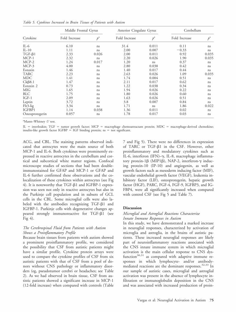
Increased Levels of Proinflammatory Cytokines ArePresent in Brain Tissues from Autistic PatientsWe assessed the profiles of expression of proteins in-volved in inflammatory pathways by cytokine proteinarray methodology27,28 in brain tissue homogenates ina subset of autistic (n � 7) and control (n � 7) pa-tients from whom fresh-frozen brain tissues were avail-able (see Table 1). A statistical analysis of the relativeexpression of cytokines in autistic and control tissuesshowed a consistent and significantly higher level ofsubsets of cytokines in the brains of autistic patients(Table 5 and Fig 3): the antiinflammatory cytokine tu-mor growth factor–�1 (TGF-�1) was increased in theMFG (p � 0.026), ACG (p � 0.011) and CBL (p �
0.035), and the proinflammatory chemokines, MCP-1and thymus and activation-regulated chemokine(TARC), were increased in the ACG (p � 0.026 and0.035, respectively) and CBL (p � 0.026 and 0.035,respectively). Only IGFBP-1, a growth and differenti-ation factor involved in immune and cellular growthpathways, was consistently increased in the cortical re-gions (MFG, p � 0.038; ACG, p � 0.011), but thedifference did not reach statistical significance in thecerebellum (p � 0.11). Interestingly, a larger spectrumof increased proinflammatory and modulatory cyto-kines was seen in the ACG, where there was a signifi-cant increase in interleukin-6 (IL-6), interleukin-10(IL-10), macrophage chemoattractant protein-3 (MCP-3), eotaxin, eotaxin 2, macrophage-derived chemokine(MDC), chemokine-�8 (Ck�8.1), neutrophil activat-ing peptide-2 (NAP-2), monokine induced by
Fig 2. Neuroglial reactions in the cerebral cortex of autistic patients. (A–D) Panlaminar activated microglia and panlaminar astro-gliosis are seen in the middle frontal gyrus (MFG) from an autistic patient in A and C, respectively. MFG from a control brainimmunostained for microglia is seen in B and for astroglia in D. Immunostaining in A and B with anti–HLA-DR antibodies andin C and D with anti-GFAP. Bar in A–D � 200�m. (E–H) A microglial nodule (E) and a cluster of reactive astrocytes (G) inthe cerebral cortex of an autistic patient, as seen with double immunocytochemical staining for microglia (red) and astroglia (green)and laser confocal imaging. Similar clusters of microglia (F) and astrocytes (H) visualized with diaminobenzidine tetrahydrochloridechromogen. (I, J) Fractional area of immunoreactivity for GFAP (I) and HLA-DR (J) in the middle frontal gyrus (Mfg), anteriorcingulate gyrus (Acg), cerebellar PCL and GCL (Cbl gcl), and cerebellar white matter (Cbl-wm) compartments. Mann–Whitney Utest, significance level p 0.05.
Vargas et al: Neuroglial Activation in Autism 73
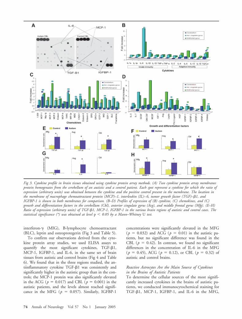
interferon-� (MIG), B-lymphocyte chemoattractant(BLC), leptin and osteoprotegerin (Fig 3 and Table 5).
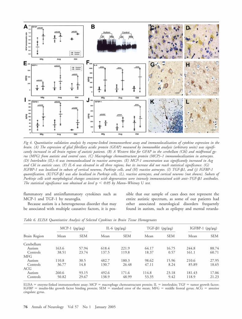
To confirm our observations derived from the cyto-kine protein array studies, we used ELISA assays toquantify the most significant cytokines, TGF-�1,MCP-1, IGFBP-1, and IL-6, in the same set of braintissues from autistic and control brains (Fig 4 and Table6). We found that in the three regions studied, the an-tiinflammatory cytokine TGF-�1 was consistently andsignificantly higher in the autistic group than in the con-trols; the MCP-1 protein was also significantly elevatedin the ACG (p � 0.017) and CBL (p � 0.001) in theautistic patients, and the levels almost reached signifi-cance in the MFG (p � 0.057). Similarly, IGFBP-1
concentrations were significantly elevated in the MFG(p � 0.032) and ACG (p � 0.01) in the autistic pa-tients, but no significant difference was found in theCBL (p � 0.42). In contrast, we found no significantdifferences in the concentration of IL-6 in the MFG(p � 0.45), ACG (p � 0.12), or CBL (p � 0.32) ofautistic and control brains.
Reactive Astrocytes Are the Main Source of Cytokinesin the Brains of Autistic PatientsTo determine the cellular sources of the most signifi-cantly increased cytokines in the brains of autistic pa-tients, we conducted immunocytochemical staining forTGF-�1, MCP-1, IGFBP-1, and IL-6 in the MFG,
Fig 3. Cytokine profile in brain tissues obtained using cytokine protein array methods. (A) Two cytokine protein array membranesprotein homogenates from the cerebellum of an autistic and a control patient. Each spot represent a cytokine for which the ratio ofexpression (arbitrary units) was obtained between the cytokine and the positive control present in the membrane. The location inthe membrane of macrophage chemoattractant protein (MCP)–1, interleukin (IL)–6, tumor growth factor (TGF)–�1, andIGFBP-1 is shown in both membranes for comparison. (B–D) Profiles of expression of (B) cytokine, (C) chemokines, and (C)growth and differentiation factors in the cerebellum (Cbl), anterior cingulate gyrus (Acg), and middle frontal gyrus (Mfg). (E–H)Ratio of expression (arbitrary units) of TGF-�1, MCP-1, IGFBP-1 in the various brain regions of autistic and control cases. Thestatistical significance (*) was obtained at level p 0.05 by a Mann–Whitney U test.
74 Annals of Neurology Vol 57 No 1 January 2005
ACG, and CBL. The staining patterns observed indi-cated that astrocytes were the main source of bothMCP-1 and IL-6. Both cytokines were prominently ex-pressed in reactive astrocytes in the cerebellum and cor-tical and subcortical white matter regions. Confocalmicroscope studies of sections that had been double-immunostained for GFAP and MCP-1 or GFAP andIL-6 further confirmed these observations and the co-localization of these cytokines within astrocytes (see Fig4). It is noteworthy that TGF-�1 and IGFBP-1 expres-sion was seen not only in reactive astrocytes but also inthe Purkinje cell population and in subsets of GCLcells in the CBL. Some microglial cells were also la-beled with the antibodies recognizing TGF-�1 andIGFBP-1. Purkinje cells with degenerative changes ap-peared strongly immunoreactive for TGF-�1 (seeFig 4).
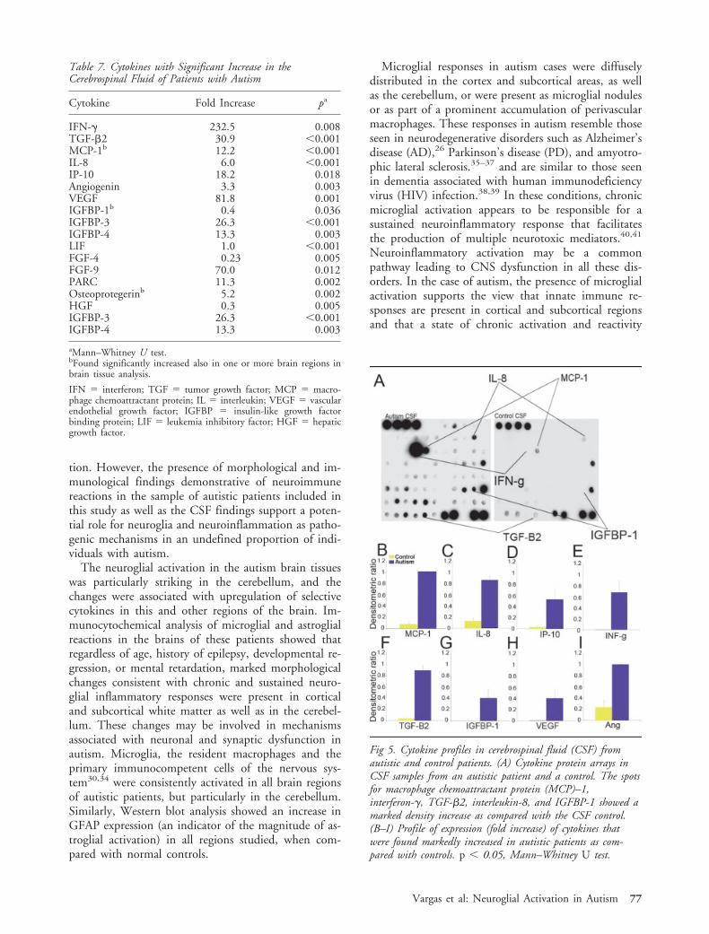
The Cerebrospinal Fluid from Patients with AutismShows a Proinflammatory ProfileBecause brain tissues from patients with autism showeda prominent proinflammatory profile, we consideredthe possibility that CSF from autistic patients mighthave a similar profile. Cytokine protein arrays wereused to compare the cytokine profiles of CSF from sixautistic patients with that of CSF from a pool of do-nors without CNS pathology or inflammatory disor-ders (eg, pseudotumor cerebri or headaches; see Table2). As we had observed in brain tissue, CSF from au-tistic patients showed a significant increase in MCP-1(12-fold increase) when compared with controls (Table
7 and Fig 5). There were no differences in expressionof TARC or TGF-�1 in the CSF. However, otherproinflammatory and modulatory cytokines such asIL-6, interferon (IFN)–�, IL-8, macrophage inflamma-tory protein-1� (MIP1�), NAP-2, interferon-� induc-ing protein-10 (IP-10) and angiogenin, as well asgrowth factors such as mesoderm inducing factor (MIF),vascular endothelial growth factor (VEGF), leukemia in-hibitory factor (LIF), osteoprotegerin, hepatic growthfactor (HGF), PARC, FGF-4, FGF-9, IGFBP3, and IG-FBP4, were all significantly increased when comparedwith control CSF (see Fig 5 and Table 7).
DiscussionMicroglial and Astroglial Reactions CharacterizeInnate Immune Responses in AutismIn this study, we have demonstrated a marked increasein neuroglial responses, characterized by activation ofmicroglia and astroglia, in the brains of autistic pa-tients. These increased neuroglial responses are likelypart of neuroinflammatory reactions associated withthe CNS innate immune system in which microglialactivation is the main cellular response to CNS dys-function30,31 as compared with adaptive immune re-sponses in which lymphocyte- and/or antibody-mediated reactions are the dominant responses.32,33 Inour sample of autistic cases, microglial and astroglialactivation was present in the absence of lymphocyte in-filtration or immunoglobulin deposition in the CNSand was associated with increased production of proin-
Table 5. Cytokines Increased in Brain Tissues of Patients with Autism
Cytokine
Middle Frontal Gyrus Anterior Cingulate Gyrus Cerebellum
Fold Increase pa Fold Increase pa Fold Increase pa
IL-6 6.10 ns 31.4 0.011 0.11 nsIL-10 1.11 ns 2.00 0.007 �0.33 nsTGF-�1 2.33 0.026 2.00 0.011 0.92 0.035MCP-1 2.32 ns 2.20 0.026 1.90 0.035MCP-2 1.24 0.017 1.20 ns 0.37 nsMCP-3 4.00 ns 2.80 0.038 0.42 nsEotaxin 1.46 ns 1.40 0.017 0.44 nsTARC 2.23 ns 2.63 0.026 1.09 0.035MDC 1.41 ns 1.74 0.004 0.51 nsCk�8.1 1.47 ns 2.11 0.017 0.62 nsEotaxin 2 0.73 ns 1.22 0.038 0.34 nsMIG 1.65 ns 1.94 0.026 0.22 nsBLC 1.75 ns 1.80 0.026 0.60 nsIGF-1 2.09 ns 1.63 0.026 0.51 nsLeptin 3.72 ns 3.8 0.007 0.84 nsFlt3-lig 3.34 ns 1.71 ns 1.86 0.022IGFBP1 1.14 0.038 1.36 0.011 0.02 nsOsteoprotegerin 0.057 ns 1.78 0.017 0.03 ns
aMann–Whitney U test.
IL � interleukin; TGF � tumor growth factor; MCP � macrophage chemoattractant protein; MDC � macrophage-derived chemokine;insulin-like growth factor IGFBP � IGF binding protein; ns � not significant.
Vargas et al: Neuroglial Activation in Autism 75
flammatory and antiinflammatory cytokines such asMCP-1 and TGF-1 by neuroglia.
Because autism is a heterogeneous disorder that maybe associated with multiple causative factors, it is pos-
sible that our sample of cases does not represent theentire autistic spectrum, as some of our patients hadother associated neurological disorders frequentlyfound in autism, such as epilepsy and mental retarda-
Table 6. ELISA Quantitative Analysis of Selected Cytokines in Brain Tissue Homogenates
Brain Region
MCP-1 (pg/�g) IL-6 (pg/�g) TGF-�1 (pg/�g) IGFBP-1 (pg/�g)
Mean SEM Mean SEM Mean SEM Mean SEM
CerebellumAutism 163.6 57.94 618.4 221.9 64.17 16.75 244.8 88.74Controls 38.51 23.74 137.5 119.8 18.37 0.57 161.1 60.71
MFGAutism 110.8 30.5 482.7 180.3 98.62 15.96 210.6 27.95Controls 36.77 14.8 130.7 26.48 47.11 8.24 85.89 18.65
ACGAutism 260.6 93.15 492.6 171.6 114.8 23.18 181.43 17.06Controls 50.82 29.67 138.9 48.99 53.35 9.42 118.9 21.23
ELISA � enzyme-linked immunosorbent assay; MCP � macrophage chemoattractant protein; IL � interleukin; TGF � tumor growth factor;IGFBP � insulin-like growth factor binding protein; SEM � standard error of the mean; MFG � middle frontal gyrus; ACG � anteriorcingulate gyrus.
Fig 4. Quantitative validation analysis by enzyme-linked immunosorbent assay and immunolocalization of cytokine expression in thebrain. (A) The expression of glial fibrillary acidic protein (GFAP) measured by immunoblot analysis (arbitrary units) was signifi-cantly increased in all brain regions of autistic patients. (B) A Western blot for GFAP in the cerebellum (Cbl) and midfrontal gy-rus (MFG) from autistic and control cases. (C) Macrophage chemoattractant protein (MCP)–1 immunolocalization in astrocytes.(D) Interleukin (IL)–6 was immunolocalized in reactive astrocytes. (E) MCP-1 concentration was significantly increased in Acgand Cbl in autistic cases. (F) IL-6 was elevated in all three regions, but its increase did not reach statistical significance. (G)IGFBP-1 was localized in subsets of cortical neurons, Purkinje cells, and (H) reactive astrocytes. (I) TGF-�1, and (j) IGFBP-1quantification. (K)TGF-�1 was also localized in Purkinje cells, (L), reactive astrocytes, and cortical neurons (not shown). Subsets ofPurkinje cells with morphological changes consistent with degeneration were intensely immunostained with anti–TGF-�1 antibodies.The statistical significance was obtained at level p 0.05 by Mann–Whitney U test.
76 Annals of Neurology Vol 57 No 1 January 2005
tion. However, the presence of morphological and im-munological findings demonstrative of neuroimmunereactions in the sample of autistic patients included inthis study as well as the CSF findings support a poten-tial role for neuroglia and neuroinflammation as patho-genic mechanisms in an undefined proportion of indi-viduals with autism.
The neuroglial activation in the autism brain tissueswas particularly striking in the cerebellum, and thechanges were associated with upregulation of selectivecytokines in this and other regions of the brain. Im-munocytochemical analysis of microglial and astroglialreactions in the brains of these patients showed thatregardless of age, history of epilepsy, developmental re-gression, or mental retardation, marked morphologicalchanges consistent with chronic and sustained neuro-glial inflammatory responses were present in corticaland subcortical white matter as well as in the cerebel-lum. These changes may be involved in mechanismsassociated with neuronal and synaptic dysfunction inautism. Microglia, the resident macrophages and theprimary immunocompetent cells of the nervous sys-tem30,34 were consistently activated in all brain regionsof autistic patients, but particularly in the cerebellum.Similarly, Western blot analysis showed an increase inGFAP expression (an indicator of the magnitude of as-troglial activation) in all regions studied, when com-pared with normal controls.
Microglial responses in autism cases were diffuselydistributed in the cortex and subcortical areas, as wellas the cerebellum, or were present as microglial nodulesor as part of a prominent accumulation of perivascularmacrophages. These responses in autism resemble thoseseen in neurodegenerative disorders such as Alzheimer’sdisease (AD),26 Parkinson’s disease (PD), and amyotro-phic lateral sclerosis.35–37 and are similar to those seenin dementia associated with human immunodeficiencyvirus (HIV) infection.38,39 In these conditions, chronicmicroglial activation appears to be responsible for asustained neuroinflammatory response that facilitatesthe production of multiple neurotoxic mediators.40,41
Neuroinflammatory activation may be a commonpathway leading to CNS dysfunction in all these dis-orders. In the case of autism, the presence of microglialactivation supports the view that innate immune re-sponses are present in cortical and subcortical regionsand that a state of chronic activation and reactivity
Fig 5. Cytokine profiles in cerebrospinal fluid (CSF) fromautistic and control patients. (A) Cytokine protein arrays inCSF samples from an autistic patient and a control. The spotsfor macrophage chemoattractant protein (MCP)–1,interferon-�, TGF-�2, interleukin-8, and IGFBP-1 showed amarked density increase as compared with the CSF control.(B–I) Profile of expression (fold increase) of cytokines thatwere found markedly increased in autistic patients as com-pared with controls. p 0.05, Mann–Whitney U test.
Table 7. Cytokines with Significant Increase in theCerebrospinal Fluid of Patients with Autism
Cytokine Fold Increase pa
IFN-� 232.5 0.008TGF-�2 30.9 0.001MCP-1b 12.2 0.001IL-8 6.0 0.001IP-10 18.2 0.018Angiogenin 3.3 0.003VEGF 81.8 0.001IGFBP-1b 0.4 0.036IGFBP-3 26.3 0.001IGFBP-4 13.3 0.003LIF 1.0 0.001FGF-4 0.23 0.005FGF-9 70.0 0.012PARC 11.3 0.002Osteoprotegerinb 5.2 0.002HGF 0.3 0.005IGFBP-3 26.3 0.001IGFBP-4 13.3 0.003
aMann–Whitney U test.bFound significantly increased also in one or more brain regions inbrain tissue analysis.
IFN � interferon; TGF � tumor growth factor; MCP � macro-phage chemoattractant protein; IL � interleukin; VEGF � vascularendothelial growth factor; IGFBP � insulin-like growth factorbinding protein; LIF � leukemia inhibitory factor; HGF � hepaticgrowth factor.
Vargas et al: Neuroglial Activation in Autism 77
may be involved in the mechanisms of neuronal andsynaptic dysfunction.
The presence of increased neuroglial responses is rel-evant to the neurobiological mechanisms involved inautism, because both microglia and astroglia are essen-tial for neuronal activity and synaptic function,42 neu-ronal–glial interactions,43 as well as for cortical model-ing, organization, and remodeling during braindevelopment.44 Furthermore, microglial and astroglialactivation seems to play a major role in the neuroim-mune mechanisms of disease in the CNS,34 becausethese cells are part of the first-line response of the in-nate immune system of the CNS30 and contribute tothe modulation of immune responses by producingboth proinflammatory and antiinflammatory cytokinesas well as growth and differentiation factors.45 The mi-croglial and astroglial responses in the CNS may thenhave a dichotomous role in the inflammatory responsesof the brain: as a direct effector of injury and on theother hand as neuroprotectant.46 An issue that remainsunclear is how and when microglia and astroglia be-come activated in the brain of autistic patients. Neu-roglial responses in autism may be part of both primary(intrinsic) neuroglial responses that result from distur-bances of neuroglial function or neuronal–neuroglialinteractions during brain development and secondary(extrinsic), resulting from unknown factors that disturbprenatal or postnatal CNS development. Both astro-cytes and microglia are critical for brain developmentand MHC class II (HLA-DR antigen)–positive micro-glia colonize the developing CNS during the secondtrimester.47,48 It is possible that the presence of acti-vated microglia in the brain in autism may reflect ab-normal persistence of fetal patterns of development inresponse to genetic or environmental (eg, intrauterine,maternal) factors. Even though our studies did notshow any difference in neuroglial activation among au-tistic cases with history of developmental regression ormental retardation, further studies that include largerseries of cases are needed to clarify these issues.
Previous neuropathological studies in autism showedabnormalities in cortical organization and neuronalpacking and reduced cerebellar Purkinje cell num-bers.11 Our findings may indicate that at some pointduring cortical and neuronal organization, unknownfactors influence both neuronal and neuroglial cellpopulations, disturbing neurodevelopment and produc-ing the neurocytoarchitectural changes seen in autismas well as inducing CNS dysfunction that results inneuroinflammation. An alternative explanation is thatextrinsic causative factors (eg, nongenetic, neurotoxic,or environmental) involved in the pathogenesis of au-tism may produce neuronal and cortical abnormalities,to which neuroglial reactions are only secondary re-sponses. Although the meaning of the neuroinflamma-tion in our sample is unknown at this time, these pri-
mary or secondary responses may be valuable clinicalbiomarkers and targets for therapy, if it can be dem-onstrated that they are causing injury to the developingCNS.
Lack of Adaptive Immune Reactions in Brain ofPatients with AutismIn contrast with the prominent presence of activatedmicroglia and astrocytes, features that characterize in-nate immune responses within the CNS, an importantfinding of our study was the lack of specific T-cell re-sponses and the absence of antibody-mediated reac-tions in any of the brain regions studied in autistic sub-jects. These observations suggest that the adaptiveimmune system does not play a significant pathogenicrole in this disorder, at least not during its chronicphase, and that the main immune mechanism involvespredominantly innate immune reactions. Because ourstudy focused on autopsy tissues, we cannot excludethe possibility that specific immune reactions, mediatedby T-cell and/or antibody responses, occurred at theonset of disease, during prenatal or postnatal stages ofdevelopment. An interesting finding in our immuno-cytochemical studies was the observation of comple-ment membrane attack complex in cerebella. The lo-calization and immunoreactivity of C9neo, a markerfor the membrane attack complex,29 in the perineuro-nal Purkinje cell compartment and focal areas of theGCL, suggest that microglial activation may triggercomplement activation, and the complement systemmay play a role in the destructive process that occurs inthe cerebellum of autistic patients. The lack of immu-noglobulin deposition, however, suggests that comple-ment activation may occur in the absence of antibody-mediated pathways and may resemble theimmunopathogenic mechanisms observed in AD, PD,and other neurodegenerative disorders, in which auto-toxic phenomena play a role in neuronal injury andneurodegeneration.49 Further clarification of the role ofthese autotoxic reactions (mediated by complementand associated with other innate immune reactions inthe cerebellum) is required, because degeneration ofthe PCL and GCL seems to occur in the absence ofadaptive immune responses.
Cerebellum Is a Main Focus of Neuroinflammationin AutismOur quantitative analysis of neuroglial reactions showedthat among the brain regions studied, the cerebellumshowed the most prominent neuroglial responses. Thismarked neuroglial activity in the cerebellum is consistentwith previous observations that the cerebellum is one ofthe foci of pathological abnormalities in morphologi-cal11,12 and neuroimaging50–52 studies of autistic pa-tients. Based on our observations, a selective process ofneuronal degeneration and neuroglial activation appear
78 Annals of Neurology Vol 57 No 1 January 2005
to occur predominantly in the PCL and GCL of cere-bellum in autistic subjects, findings that are consistentwith an active and ongoing postnatal process of neuro-degeneration and neuroinflammation. These observa-tions do not support the previously proposed hypothesisthat the changes in the cerebellum in autism result solelyfrom developmental abnormalities in olivary-cerebellarcircuits and a reduced number of Purkinje cells.11 In-stead, our observations suggest that the pathologicalchanges observed in the cerebellum in autistic patientsdo not occur exclusively during prenatal developmentbut appear to involve an ongoing chronic neuroinflam-matory process that involves both microglia and astro-glia. Furthermore, this process continues beyond earlyneurodevelopment and is present even at very late stagesin the life of patients with autism. These findings alsosupport the hypothesis that selective vulnerability ofPurkinje cells plays a role in the etiopathogenesis of au-tism.53
Macrophage Chemoattractant Protein–1 and TumorGrowth Factor–�1 Are the Most ProminentCytokines in the Brain of Autistic PatientsOur study has also demonstrated the presence ofunique profiles of cytokine expression in the brain andCSF of autistic subjects. Two proinflammatory chemo-kines, MCP-1 and TARC, and an antiinflammatoryand modulatory cytokine, TGF-�1, were consistentlyelevated in the brain regions studied. MCP-1, a che-mokine involved in innate immune reactions and im-portant mediator for monocyte and T-cell activationand trafficking into areas of tissue injury,54 appeared tobe one of the most relevant proteins found in cytokineprotein array studies because it was significantly ele-vated in both brain tissues and CSF. The presence ofMCP-1 is of particular interest, because it facilitatesthe infiltration and accumulation of monocytes andmacrophages in inflammatory CNS disease.55 Asshown by our immunocytochemical studies of the ce-rebral cortex and cerebellum, MCP-1 is produced byactivated and reactive astrocytes, a finding that demon-strate the effector role of these cells in the disease pro-cess in autism. The increase expression of MCP-1 hasrelevance to the pathogenesis of autism because we be-lieve its elevation in the brain is linked to microglialactivation and perhaps to the recruitment of mono-cytes/macrophages to areas of neurodegeneration, suchas those we observed in the cerebellum. Our observa-tions resemble findings in other neurological disordersin which elevation of MCP-1 is associated with thepathogenesis of neuroinflammation and neuronal in-jury such as HIV dementia,56 amyotrophic lateral scle-rosis,37 stroke,57 and multiple sclerosis.55 It remainsunclear whether MCP-1 plays a more pleotrophic rolein the CNS or whether its presence is associated onlywith inflammatory conditions. It has been speculated
that MCP-1 may be involved in neuronal survival andneuroprotective mechanisms other than monocyte acti-vation and trafficking58 or even in nonlymphocytic-mediated neuronal injury.59 Expression of MCP-1 inthe CNS appears to be developmentally regulated, andprevious studies have shown its expression in the cere-bellum during prenatal development, a finding thatmay suggest an association with maturation of Purkinjecells.60 Like MHC class II expression in microglia dur-ing CNS modeling,47 MCP-1 elevation in the brain ofautistic patients may reflect persistent fetal patterns ofbrain development.
Our observation that TGF-�1 was increased in thecortex and cerebellum of autistic brains may have im-portant implications for the neurobiology of autism.TGF-�1 is a key antiinflammatory cytokine and is in-volved in tissue remodeling after injury. It can suppressspecific immune responses by inhibiting T-cell prolif-eration and maturation and downregulates MHC classII expression.61 Importantly, cells undergoing celldeath have been shown to secrete TGF-�1, possibly toreduce local inflammation and prevent degeneration ofadditional surrounding cells.62 In our immunocyto-chemical studies, TGF-�1 was localized mostly withinreactive astrocytes and neurons in the cerebellum. Pur-kinje cells that exhibited morphological features of de-generation showed marked immunoreactivity for TGF-�1. These findings suggest that the elevation of thiscytokine in autism may reflect an attempt to modulateneuroinflammation or remodel and repair injured tis-sue. Although TGF-�1, MCP-1, TARC, and IGFBP-1were consistently elevated in at least two of the threeregions examined in the autistic brains, a more remark-able profile of cytokine upregulation was observed inthe ACG, a region in which several cytokines, chemo-kines, and growth factors were markedly elevated whencompared with controls. Both proinflammatory cyto-kines (eg, IL-6) and antiinflammatory cytokines (eg,IL-10) as well as subsets of chemokines were markedlyelevated in the ACG, an important cortical region in-volved in dysfunctional brain activity in autism.63
These findings support the conclusion that an active,ongoing immunological process was present in multi-ple areas of the brain but at different levels of expres-sion in each area.
Marked Expression of Proinflammatory Cytokines inCerebrospinal Fluid of Autistic PatientsCSF studies also confirmed a prominent inflammatorycytokine profile in patients with autism. The presenceof a marked increase of MCP-1 in CSF supports thehypothesis that proinflammatory pathways are acti-vated in the brain of autistic patients and that its pres-ence may be associated with the mechanisms of mac-rophage/microglia activation observed in the braintissue studies. The elevation of MCP-1 in the CSF re-
Vargas et al: Neuroglial Activation in Autism 79
semble observations in other conditions in which mi-croglia/macrophage activation play an important rolesuch as HIV dementia56 and multiple sclerosis.64 Inaddition to the marked elevation in MCP-1, the pres-ence of elevated levels of IFN-�, IL-8, IP-10, and otherproinflammatory molecules such as angiogenin and LIFstrongly supports the view that active neuroinflamma-tory reactions and a network of multiple cytokines arelikely involved in immune-mediated mechanisms inthe CNS of autistic patients. These cytokines play im-portant roles in immune-mediated processes, and theirpresence in the CSF in autistic patients may reflect anongoing stage of inflammatory reactions likely associ-ated with neuroglial activation and/or neuronal injury.Reasons for the relatively greater increases in these cy-tokines in CSF compared with brain are unknown. Itcould be that cytokines derive from neuroglial andneuronal sources as demonstrated by our immunocyto-chemical assessment. The differences we observed incytokines in CSF compared with brain could resultfrom other sources of production, such the leptome-ninges or choroid plexus or might represent a persistentelevation of cytokines as a result of a stage of neurode-velopmental arrest because some of the cytokines arenormally elevated during phases of neurodevelopment.Because the CSF is easily accessible for clinical studies,CSF cytokine profiling may be useful in the future todiagnose, characterize, and follow the clinical course ofautistic disorders.
ConclusionTaken together, our observations suggest that neuro-glial reactions, in the form of innate immune re-sponses, are important in the mechanisms associatedwith neural dysfunction in autism and that the cere-bellum is the focus of an active and chronic neuroin-flammatory process in autistic patients. The presence ofproinflammatory chemokines such as MCP-1 as well asantiinflammatory cytokines such as TGF-�1 supportsthe idea that a chronic state of specific cytokine acti-vation occurs in autism. This hypothesis is also sup-ported by our finding of marked increase in a larger setof cytokines in the CSF that are usually involved ininflammatory pathways. In view of the heterogeneity ofclinical symptoms and possible causes for autism, thepresence of neuroinflammatory changes among thecases we examined suggests that this may be a commonpathogenic mechanism in some patients with autism.Because neuroimmune responses are influenced by thegenetic background of the host, the role of neuroin-flammation in the context of the genetic and other fac-tors that determine the autism phenotype remains animportant issue to be investigated. Because this neu-roinflammatory process appears to be associated withan ongoing and chronic mechanism of CNS dysfunc-tion, potential therapeutic interventions should focus
on the control of its detrimental effects (while preserv-ing reparative benefits) and thereby eventually modifythe clinical course of autism.
This work was supported by grants from the Cure Autism NowFoundation (C.A.P.), the Autism Research Foundation (A.W.Z.)the NIH (National Institute of Drug Abuse, K08DA016160,C.A.P.), and Dr Barry and Renee Gordon and an anonymous do-nor.
We are grateful to Dr J. Pickett, Autism Tissue Program, and theHarvard University, University of Miami and University of Mary-land brain banks for providing brain tissues. We thank Drs S. L.Connors, C. Eberhart, and G. Pradilla, Jr. for their helpful com-ments, Dr B. Paul Morgan for providing the anti–C9neo antibody,Dr D. Irani for providing CSF control samples and Dr D. McClel-lan for editorial assistance.
References1. Rapin I. Autism. N Engl J Med 1997;337:97–104.2. Lord C, Cook EH, Leventhal BL, et al. Autism spectrum dis-
orders. Neuron 2000;28:355–363.3. Rapin I, Katzman R. Neurobiology of autism. Ann Neurol
1998;43:7–14.4. Newschaffer CJ, Fallin D, Lee NL. Heritable and nonheritable
risk factors for autism spectrum disorders. Epidemiol Rev 2002;24:137–153.
5. Fombonne E. Epidemiological surveys of autism and other per-vasive developmental disorders: an update. J Autism Dev Dis-ord 2003;33:365–382.
6. Bertrand J, Mars A, Boyle C, et al. Prevalence of autism in aUnited States population: the Brick Township, New Jersey, in-vestigation. Pediatrics 2001;108:1155–1161.
7. Yeargin-Allsopp M, Rice C, Karapurkar T, et al. Prevalence ofautism in a US metropolitan area. JAMA 2003;289:49–55.
8. Folstein SE, Rosen-Sheidley B. Genetics of autism: complex ae-tiology for a heterogeneous disorder. Nat Rev Genet 2001;2:943–955.
9. Korvatska E, Van de WJ, Anders TF, et al. Genetic and im-munologic considerations in autism. Neurobiol Dis 2002;9:107–125.
10. Lipkin WI, Hornig M. Microbiology and immunology of au-tism spectrum disorders. Novartis Found Symp 2003;251:129–143.
11. Kemper TL, Bauman M. Neuropathology of infantile autism.J Neuropathol Exp Neurol 1998;57:645–652.
12. Bailey A, Luthert P, Dean A, et al. A clinicopathological studyof autism. Brain 1998;121:889–905.
13. Bauman ML, Kemper TL. The neuropathology of the autismspectrum disorders: what have we learned? Novartis FoundSymp 2003;251:112–122.
14. Licinio J, Alvarado I, Wong ML. Autoimmunity in autism.Mol Psychiatry 2002;7:329.
15. Gupta S, Aggarwal S, Rashanravan B, et al. Th1- and Th2-likecytokines in CD4� and CD8� T cells in autism. J Neuroim-munol 1998;85:106–109.
16. Singh VK, Warren R, Averett R, et al. Circulating autoantibod-ies to neuronal and glial filament proteins in autism. PediatrNeurol 1997;17:88–90.
17. Vojdani A, Campbell AW, Anyanwu E, et al. Antibodies toneuron-specific antigens in children with autism: possible cross-reaction with encephalitogenic proteins from milk, Chlamydiapneumoniae and Streptococcus group A. J Neuroimmunol 2002;129:168–177.
80 Annals of Neurology Vol 57 No 1 January 2005
18. Jyonouchi H, Sun S, Le H. Proinflammatory and regulatorycytokine production associated with innate and adaptive im-mune responses in children with autism spectrum disordersand developmental regression. J Neuroimmunol 2001;120:170–179.
19. Dalton P, Deacon R, Blamire A, et al. Maternal neuronal an-tibodies associated with autism and a language disorder. AnnNeurol 2003;53:533–537.
20. Comi AM, Varsou A, Heyes MP, et al. Quinolinic acid andneopterin in children with autism: an analysis of cerebrospinalfluid. Ann Neurol 1999;46:528–529.
21. Zimmerman AW. Commentary: immunological treatments forautism: in search of reasons for promising approaches. J AutismDev Disord 2000;30:481–484.
22. Guerin P, Lyon G, Barthelemy C, et al. Neuropathologicalstudy of a case of autistic syndrome with severe mental retar-dation. Dev Med Child Neurol 1996;38:203–211.
23. Lord C, Pickles A, McLennan J, et al. Diagnosing autism: anal-yses of data from the Autism Diagnostic Interview. J AutismDev Disord 1997;27:501–517.
24. Scahill L, Lord C. Subject selection and characterization in clin-ical trials in children with autism. CNS Spectr 2004;9:22–32.
25. Gundersen HJ, Bendtsen TF, Korbo L, et al. Some new, simpleand efficient stereological methods and their use in pathologicalresearch and diagnosis. APMIS 1988;96:379–394.
26. Vehmas AK, Kawas CH, Stewart WF, et al. Immune reactivecells in senile plaques and cognitive decline in Alzheimer’s dis-ease. Neurobiol Aging 2003;24:321–331.
27. Huang RP. Cytokine protein arrays. Methods Mol Biol 2004;278:215–232.
28. Lin Y, Huang R, Chen LP, et al. Profiling of cytokine expres-sion by biotin-labeled-based protein arrays. Proteomics 2003;3:1750–1757.
29. Storch MK, Piddlesden S, Haltia M, et al. Multiple sclerosis: insitu evidence for antibody- and complement-mediated demyeli-nation. Ann Neurol 1998;43:465–471.
30. Aloisi F. Immune function of microglia. Glia 2001;36:165–179.
31. Kreutzberg GW. Microglia: a sensor for pathological events inthe CNS. Trends Neurosci 1996;19:312–318.
32. Bauer J, Rauschka H, Lassmann H. Inflammation in the ner-vous system: the human perspective. Glia 2001;36:235–243.
33. Medzhitov R, Janeway CA Jr. Innate immune recognition andcontrol of adaptive immune responses. Semin Immunol 1998;10:351–353.
34. Aloisi F. The role of microglia and astrocytes in CNS immunesurveillance and immunopathology. Adv Exp Med Biol 1999;468:123–133.
35. McGeer PL, Kawamata T, Walker DG, et al. Microglia in de-generative neurological disease. Glia 1993;7:84–92.
36. Teismann P, Tieu K, Cohen O, et al. Pathogenic role of glialcells in Parkinson’s disease. Mov Disord 2003;18:121–129.
37. Henkel JS, Engelhardt JI, Siklos L, et al. Presence of dendriticcells, MCP-1, and activated microglia/macrophages in amyotro-phic lateral sclerosis spinal cord tissue. Ann Neurol 2004;55:221–235.
38. Glass JD, Wesselingh SL. Microglia in HIV-associated neuro-logical diseases. Microsc Res Tech 2001;54:95–105.
39. Gartner S. HIV infection and dementia. Science 2000;287:602–604.
40. Banati RB, Gehrmann J, Schubert P, et al. Cytotoxicity of mi-croglia. Glia 1993;7:111–118.
41. Gebicke-Haerter PJ. Microglia in neurodegeneration: molecularaspects. Microsc Res Tech 2001;54:47–58.
42. Auld DS, Robitaille R. Glial cells and neurotransmission: aninclusive view of synaptic function. Neuron 2003;40:389–400.
43. Fields RD, Stevens-Graham B. New insights into neuron-gliacommunication. Science 2002;298:556–562.
44. Nedergaard M, Ransom B, Goldman SA. New roles forastrocytes: redefining the functional architecture of the brain.Trends Neurosci 2003;26:523–530.
45. Allan SM, Rothwell NJ. Cytokines and acute neurodegenera-tion. Nat Rev Neurosci 2001;2:734–744.
46. Nguyen MD, Julien JP, Rivest S. Innate immunity: the missinglink in neuroprotection and neurodegeneration? Nat Rev Neu-rosci 2002;3:216–227.
47. Rezaie P, Male D. Colonisation of the developing human brainand spinal cord by microglia: a review. Microsc Res Tech 1999;45:359–382.
48. Wierzba-Bobrowicz T, Kosno-Kruszewska E, Gwiazda E, et al.Major histocompatibility complex class II (MHC II) expressionduring the development of human fetal cerebral occipital lobe,cerebellum, and hematopoietic organs. Folia Neuropathol 2000;38:111–118.
49. McGeer PL, McGeer EG. Innate immunity, local inflamma-tion, and degenerative disease. Sci Aging Knowledge Environ2002;2002:re3.
50. Courchesne E, Yeung-Courchesne R, Press GA, et al. Hypopla-sia of cerebellar vermal lobules VI and VII in autism. N EnglJ Med 1988;318:1349–1354.
51. Courchesne E. Neuroanatomic imaging in autism. Pediatrics1991;87:781–790.
52. Carper RA, Courchesne E. Inverse correlation between frontallobe and cerebellum sizes in children with autism. Brain 2000;123:836–844.
53. Kinnear KJ. Purkinje cell vulnerability and autism: a possibleetiological connection. Brain Dev 2003;25:377–382.
54. Leonard EJ, Yoshimura T. Human monocyte chemoattractantprotein-1 (MCP-1). Immunol Today 1990;11:97–101.
55. Mahad DJ, Ransohoff RM. The role of MCP-1 (CCL2) andCCR2 in multiple sclerosis and experimental autoimmune en-cephalomyelitis (EAE). Semin Immunol 2003;15:23–32.
56. Kelder W, McArthur JC, Nance-Sproson T, et al. Beta-chemokines MCP-1 and RANTES are selectively increased incerebrospinal fluid of patients with human immunodeficiencyvirus- associated dementia. Ann Neurol 1998;44:831–835.
57. Losy J, Zaremba J. Monocyte chemoattractant protein-1 is in-creased in the cerebrospinal fluid of patients with ischemicstroke. Stroke 2001;32:2695–2696.
58. Eugenin EA, D’Aversa TG, Lopez L, et al. MCP-1 (CCL2)protects human neurons and astrocytes from NMDA or HIV-tat-induced apoptosis. J Neurochem 2003;85:1299–1311.
59. Peterson KE, Errett JS, Wei T, et al. MCP-1 and CCR2 con-tribute to non-lymphocyte-mediated brain disease induced byFr98 polytropic retrovirus infection in mice: role for astrocytesin retroviral neuropathogenesis. J Virol 2004;78:6449–6458.
60. Meng SZ, Oka A, Takashima S. Developmental expression ofmonocyte chemoattractant protein-1 in the human cerebellumand brainstem. Brain Dev 1999;21:30–35.
61. Letterio JJ, Roberts AB. Regulation of immune responses byTGF-beta. Annu Rev Immunol 1998;16:137–161.
62. Chen W, Frank ME, Jin W, et al. TGF-beta released by apo-ptotic T cells contributes to an immunosuppressive milieu. Im-munity 2001;14:715–725.
63. Mundy P. Annotation: the neural basis of social impairments inautism: the role of the dorsal medial-frontal cortex and anteriorcingulate system. J Child Psychol Psychiatry 2003;44:793–809.
64. Franciotta D, Martino G, Zardini E, et al. Serum and CSFlevels of MCP-1 and IP-10 in multiple sclerosis patients withacute and stable disease and undergoing immunomodulatorytherapies. J Neuroimmunol 2001;115:192–198.
Vargas et al: Neuroglial Activation in Autism 81