neuroligand-evoked release of excitatory neurotransmitters
TRANSCRIPT

Retrospective Theses and Dissertations Iowa State University Capstones, Theses andDissertations
1994
Neuroligand-evoked release of excitatoryneurotransmitters from cultured glial cellsFang LiuIowa State University
Follow this and additional works at: https://lib.dr.iastate.edu/rtd
Part of the Animal Sciences Commons, Cell Biology Commons, Neuroscience and NeurobiologyCommons, Neurosciences Commons, Physiology Commons, and the Veterinary PhysiologyCommons
This Dissertation is brought to you for free and open access by the Iowa State University Capstones, Theses and Dissertations at Iowa State UniversityDigital Repository. It has been accepted for inclusion in Retrospective Theses and Dissertations by an authorized administrator of Iowa State UniversityDigital Repository. For more information, please contact [email protected].
Recommended CitationLiu, Fang, "Neuroligand-evoked release of excitatory neurotransmitters from cultured glial cells " (1994). Retrospective Theses andDissertations. 11286.https://lib.dr.iastate.edu/rtd/11286
INFORMATION TO USERS
This manuscript has been reproduced from the microfilm master. UMI
films the text directly from the original or copy submitted. Thus, some
thesis and dissertation copies are in typewriter face, while others may
be from aity type of computer printer.
The quality of this reproduction is dependent upon the quali^ of the
copy submitted. Broken or indistinct print, colored or poor quality
illustrations and photogr^hs, print bleedthrough, substandard margins,
and improper aligmnent can adversely affect reproduction.
In the unlikely event that the author did not send UMI a complete
manuscript and there are missing pages, these will be noted. Also, if
unauthorized copyright material had to be removed, a note will indicate
the deletion.
Oversize materials (e.g., maps, drawings, charts) are reproduced by
sectioning the original, beginning at the upper left-hand comer and
continuing from left to right in equal sections with small overlaps. Each
original is also photographed in one exposure and is included in
reduced form at the back of the book.
Photographs included in the original manuscript have been reproduced
xerographically in this copy. Higher quality 6" x 9" black and white
photographic prints are available for ai^r photographs or illustrations
appearing in this copy for an additional charge. Contact UMI directiy
to order.
A Bell & Howell Information Company 300 North Zeeb Road. Ann Arbor. Ml 48106-1346 USA
313.'761-4700 800/521-0600

Order Number 9518409
Neuroligand-evoked release of excitatory neurotransmitters from cultured glial cells
Liu, Fang, Ph.D.
Iowa State University, 1994
U M I 300 N. ZeebRd. Ann Arbor, MI 48106


Neuroligand-evoked release of excitatory neurotransmitters
from cultured glial cells
by
Fang Liu
A Dissertation Submitted to the
Graduate Faculty in Partial Fulfillment of the
Requirements for the Degree of
DOCTOR OF PHILOSOPHY
Interdepartmental Program: Neuroscience Department: Veterinary Anatomy
Co-majors: Neuroscience Veterinary Anatomy
Approved:
In Char
For the Interd^^rtmental Program
For the p^or Department
F Graduate College
Iowa State University Ames, Iowa
1994
Signature was redacted for privacy.
Signature was redacted for privacy.
Signature was redacted for privacy.
Signature was redacted for privacy.

DEDICATION
To my husband and my parents.
Without their support this would not be possible.
Specially to my lovely son, David.
You make it all worthwhile.
iii
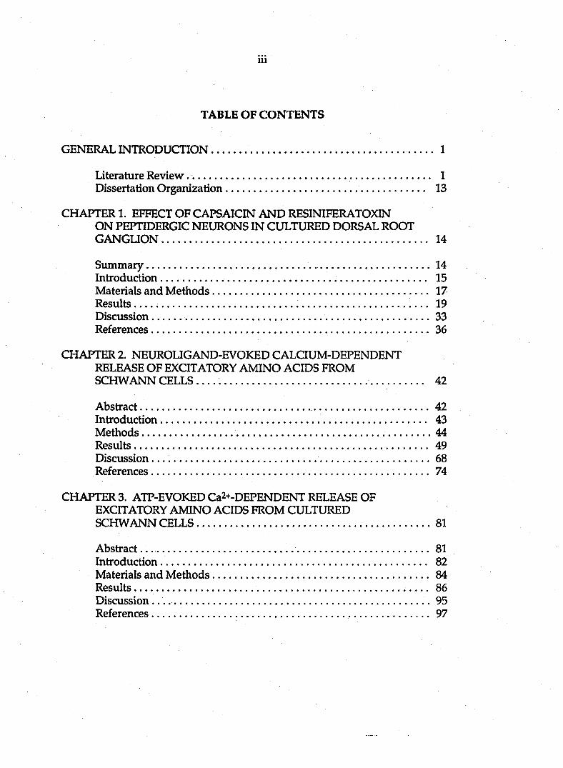
TABLE OF CONTENTS
GENERAL INTRODUCTION 1
Literature Review 1 Dissertation Organization 13
CHAPTER 1. EFFECT OF CAPSAICIN AND RESINIFERATOXIN ON PEPTIDERGIC NEURONS IN CULTURED DORSAL ROOT GANGUON 14
Summary 14 Introduction 15 Materials and Methods 17 Results 19 Discussion 33 References 36
CHAPTER 2. NEUROLIGAND-EVOKED CALCIUM-DEPENDENT RELEASE OF EXCITATORY AMINO ACIDS FROM SCHWANN CELLS 42
Abstract 42 Introduction 43 Methods 44 Results 49 Discussion..... 68 References 74
CHAPTERS. ATP-EVOKEDCa2+-DEPENDENT RELEASE OF EXCITATORY AMINO ACIDS FROM CULTURED SCHWANN CELLS 81
Abstract... 81 Introduction 82 Materials and Methods 84 Results.. 86 Discussion. .95 References 97
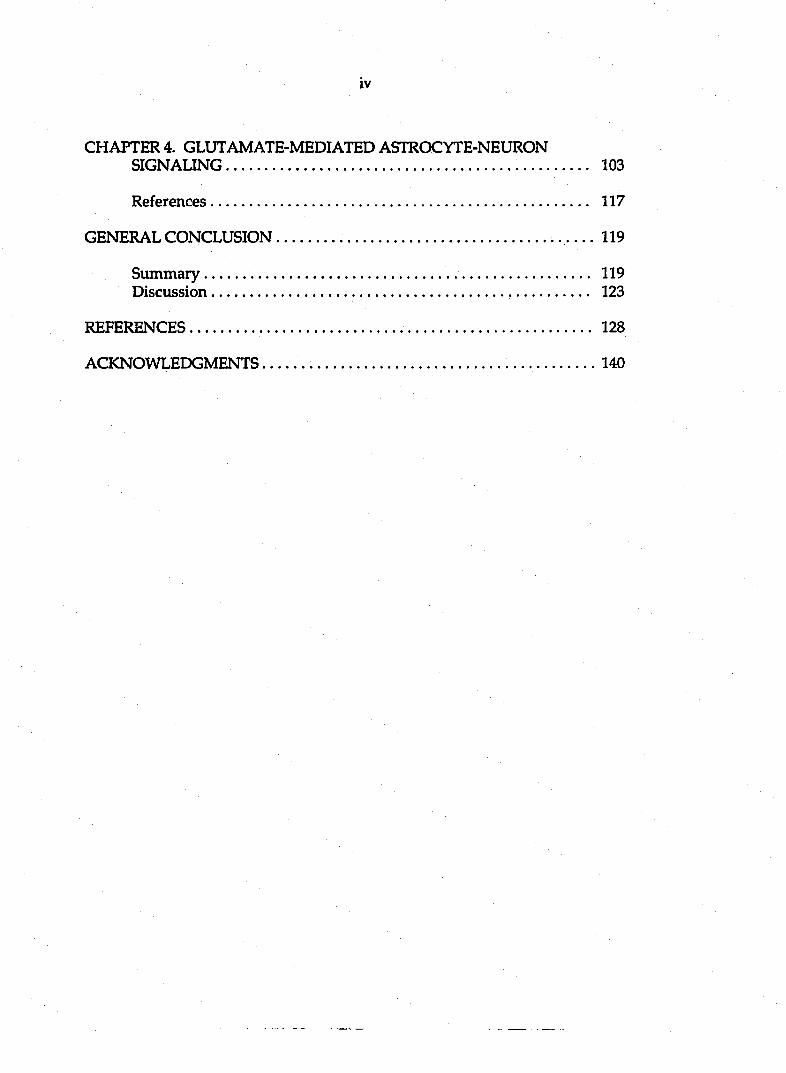
iv
CHAPTER 4. GLUTAMATE-MEDIATED ASTROCYTE-NEURON SIGNALING 103
References 117
GENERAL CONCLUSION 119
Summary 119 Discussion 123
REFERENCES 128
ACKNOWLEDGMENTS 140
1
GENERAL INTRODUCTION
The human brain, which consists of about 10^^ nerve cells and 10^2 gijai cells,
continually receives information, elaborates and perceives it, and makes
decisions. To carry out this task, the nervous system possesses an immense
nim\ber of lines of communication provided by the nerve cells (neurons). Each
neuron makes specific contact with many target cells such as different tj^es of
neurons, gland cells, or muscle cells.
In order for neurons to communicate with each other, they need to have
points of "contact" with each other. These are called synapses, named by the
English physiologist Charles Sherrington in 1897. The term synapse is derived
from the Greek, meaning to clasp, connect or join. Sherrington thought of it,
anatomically as a site of "surfaces of separation", but he always emphasized that
it was first and foremost afunctional connection. In 1911, Ram6n y Cajal foimd
that there were three elements for a s5mapse: a presynaptic terminal, a
postsynaptic target site and the synaptic cleft, the space between the pre- and
post-synaptic plasma membranes. These three elements underline the ability of
neurons to communicate with one another, the process of synaptic transmission.
Literature Review
Synaptic Vesicle and Neurotransmitter Release
It has been widely accepted that there are two major modes of synaptic
transmission: electrical, which depends on current through gap junctions that
2
bridge the cytoplasm of pre- and post-synaptic cells (Furshpan and Potter, 1957;
reviewed by Bennett et al., 1991); and chemical, in which pre- and post-synaptic
cells have no direct continuity (Katz, 1966; Changeux, 1981; Karlin, 1990),
Synaptic transmission across the majority of synapses in the brain is mediated by
the interaction of chemical signals released from the presynaptic terminal with
postsynaptic receptors (reviewed by Jessell and Kandel, 1993).
In the resting state, neurotransmitters such as glutamate, y-aminobutyric acid
(GABA) and acetylcholine (ACh) are stored in uniformly sized small organelles
of 40-50 nm diameter. When an action potential reaches the nerve terminal, the
presynaptic plasma membrane depolarizes and voltage-gated calcium channels
open at the active zone. The ensuing rise in intracellular calcium triggers
exocytosis of synaptic vesicles, resulting in the release of the neurotransmitter
(Katz, 1966). The synaptic vesicle membranes are reclaimed from the plasma
membrane by endocytosis, and the vesicles eventually refill with
neurotransmitter (Siidhof and Reinhard, 1991). To summarize this process, Kelly
(1993) indicates that transmitter release can be considered to involve at least four
steps: the transport of vesicles from the reserve pool to the releasable pool at the
active zones, the docking of vesicles to their release sites at the active zones, the
fusion of the synaptic vesicle membrane with the plasma membrane during
exocytosis in response to an increase in intracellular Ca2+, and the retrieval and
the recycling of vesicle membrane following exocytosis.
Synaptic vesicles have two fundamentally different functions: they take up
and store neurotransmitters, and they fuse with and bud from other membranes,
most notably the presynaptic plasma membrane. These functions must be
performed by the proteins of synaptic vesicles, either alone or in concert with
3
other components of the nerve terminal. A major advance in the analysis of
transmitter release mechanisms has been the cloning and characterization of
several proteins that may participate in different aspects of the release cycle. As
might be expected from the steps involved in release, these proteins fall into four
groups: proteins such as the synapsins that are thought to control the
mobilization of vesicles and thereby to regulate their availability for release (De
Camilli et al, 1990); proteins such as Raba, a member of the p21ras superfamily,
thought to be involved in the intracellular trafficking and docking of sjmaptic
vesicles (Zahraoui et al., 1989); proteins such as synaptotagmin, synaptobrevin,
neurexins, and sjmtaxins, which appear to be involved more specifically in
docking vesicles into release sites on the presynaptic membrane (Bennett et al.,
1991; Trimble et al., 1991; Schiavo et al., 1992); and proteins such as
synaptophysin, thought to be involved in the formation of the vesicle fusion pore
or in recycling (Aimers and Tse, 1990; Siidhof and Jahn, 1991; White, 1992).
The Central Role of in Neurotransmitter Release
As mentioned above, the elevation of the intracellular calcium level
following calcium influx through voltage-gated calcium channels is a critical step
in action potential evoked synaptic transmission (Augustine et al., 1987).
However, in a number of experimental conditions neurotransmitter can also be
released in the absence of extracellular calcium (Adam-Vizi et al., 1984; Hochner
et al., 1989). Neurotransmitter release in the absence of calcium may be derived
from a different intracellular pool than that in the presence of calcium (Adam-
Vizi et al, 1990). The concentration of calcium in the nerve terminal is controlled
by a complex system of soluble buffers, organellar stores and a membrane

4
transport system with varying calcium transport affinities and capacities;
distortion will result in a modulation of transmitter release (Adam-Vizi and
Ashley, 1987). Calcium is sequestered inside the nerve terminal in the smooth
endoplasmic reticulum, synaptic vesicles, mitochondria, and possibly also
caldsomes. Furthermore, there exist intracellular buffering systems like calcium-
binding proteins (Blaustein, 1988; Meldolesi, 1988).
Inositol polyphosphates have been assigned a key role in calcium
intracellular homeostasis. Inositol 1,4,5-trisphosphate (IP3) is formed by receptor-
mediated and G-protein-controlled turnover of phosphatidylinositol 4,5-
bisphosphate together with diacylglycerol (DAG), an activator of protein kinase
C (PKC). The inositol trisphosphate functions via specific receptors in the release
of caldimi from intracellular stores, presumably the endoplasmic reticulum and
the caldsome (Nahorski, 1988). In addition to the IPs-sensitive pool, there is an
IPs-insensitive calcium store, which is located in the peripheral region of the cell
away from the nucleus (Hardie, 1991). Caldum in the IPs-insensitive pool can be
released by the drug caffeine, which also activates the Ca2+-release channels of
sarcoplasmic reticulum (ryanodine receptors). It is believed that, in axon
terminals, receptor-mediated turnover of synaptic phosphoinositides for
mobilizing calcium from internal stores may serve mainly as a modulatory
mechanism.
Mechanisms of Glutamate Release
It is well established that glutamate and aspartate mediate the excitatory
transactions taking place between nerve cells in the vertebrate central nervous
system (QMS). Glutamate is probably the main excitatory amino acid (EAA)
5
neurotransmitter in the mammalian CNS. It works along with other acidic amino
adds, particularly aspartate, homocysteate and cysteine sulfinate (Mayer and
Westbook, 1987). Since the first proposal of the vesicular hypothesis more than 30
years ago (Del Castillo and Katz, 1957), the release of numerous
neurotransmitters has been assumed to be due to the exocytosis of synaptic
vesicles in response to an increase in the cytoplasmic calcium concentration in
nerve terminals. Many arguments favor such a release mechanism for glutamate.
These include the calcium dependency of glutamate release (Nicholls and Sihra,
1986), the presence of glutamate-like compounds in synaptic vesicles (Stom-
Mathisen et al., 1983), and the massive and rapid accumulation of glutamate in
highly purified synaptic vesicles (Naito and Ueda, 1983,1985).
However, glutamate is transported by a reversible, sodium dependent
(Kanner and Sharon, 1978; Kanner and Marva, 1982; Erecinska et al., 1983,1987;
Wilson and Pastuszko, 1986), and electrogenic (Brew and Attwell, 1987;
Szatkowski et al., 1990) uptake mechanism, so it remains possible that the
reversal of the EAA uptake system could account, at least in part, for the release
of EAAs. Such a mechanism may explain the calcium independent EAA release
reported, for example, in the retina (Miller and Schwartz, 1983).
There is the third situation for the accumulation of extracellular glutamate.
Swelling of isolated cells and many vertebrate and invertebrate tissues leads to
release of glutamate, aspartate, taurine, and other amino acids (Kimelberg and
Ransom, 1986). This release is part of the process of regulatory volume decrease
by which swollen cells regain their normal volume (Gilles et al., 1987). Swelling-
induced release of glutamate is inhibited by a number of anion transport blockers
6
such as furosemide and L-644,711, a fluorenyl derivative of ethacrynic add
(Kimelberg et al., 1987).
Types of Glutamate Receptors
Glutamate interacts with at least five classes of membrane receptors, each
with significantly distinct functions (reviewed by Monaghan, 1989). Three have
been defined by the depolarizing actions of selective agonists (N-methyl-D-
Aspartate, NMD A; kainate; quisqualate or a-amino-3-hydroxy-5-
methylisoxazole-4-propionic acid, AMP A) and their blockade by selective
antagonists (Watkins and Olverman, 1987). A fourth, the AP4 receptor (L-2-
amino-4-phosphonobutyrate) appears to represent an inhibitory autoreceptor
(Koemer and Cotman, 1981; Davies and Watkins, 1982; Evans et al., 1982). The
fifth receptor, activated by trans-l-aminocyclopentane-l,3-dicarboxylic acid
(ACPD), modifies inositol phosphate metabolism (Baudry et al., 1986; Nicoletti et
al., 1986; Sladeczek et al, 1985).
The NMDA receptor is best defined. This receptor opens a distinctive
membrane channel characterized by high conductance (main state about 50 pS),
voltage-dependent Mg2+ blockade and permeability to both and Na+ (Nicoll
et al., 1988; Ascher and Nowak, 1988; MacDermott and Dale, 1987; Mayer and
Westbrook, 1987). Voltage-clamp studies of cultured neurons injected with a
calcium-sensitive dye indicate that Ca2+ can enter cells after NMDA application
in a manner independent of the voltage-dependent calcium channels
(MacDermott et al., 1987; Mayer et al., 1987). The increase in intracellular Ca2+
ions is thought to initiate the biochemical processes responsible for both NMDA
receptor-induced plasticity observed in developing and adult animals (Lynch et
7
al., 1983; Collingridge et al., 1987; Cotman et al., 1988) and NMDA receptor-
mediated excitotoxic cell death (Rothman and Olney, 1986; Cotman et al., 1988).
Although NMDA receptors play a critical role in synaptic function, they do
not mediate the excitatory postsynaptic potential resulting from a unitary
synaptic activation. Pharmacological studies indicate that kainate and/or
quisqualate receptors are responsible for the voltage-independent portion of the
synaptic response in many neuronal pathways (reviewed by Mayer and
Westbrook, 1987; Watkins and Evans, 1981), The kainate/quisqualate antagonist,
6-cyano-7-nitroquinoxaline-2,3-done (CNQX), blocks synaptic transmission at
low concentration (Honors et al., 1988; Blake et al., 1988).
Glutamate-induced excitatory transmission appears to involve actions
mediated by one or more combination of glutamate receptors.
Neuroglia
Glial cells are intimately associated with most neurons. The neuroglial cells
used to be called arachnoid cells because of their resemblance to spiders. They
were also described as Deiter's cells in honor of the researcher who discovered
them. The main classes of glial cells in the vertebrate central nervous system are
astrocytes, oligodendrocytes and microglia, whereas Schwann cells are the
predominant glial type in the peripheral nervous system.
Neurobiologists have wondered for 150 years what glial cells do. There were
several hypotheses about the functions of neuroglial cells. One of them was
Golgi's nutritive theory. This theory was based on the fact that the dendrites of
neurons are in contact with the capillaries or with neuroglial cells. The
capillaries, neuroglial cells and dendrites thus form a solid functional unit the
8
role of which is to carry to the cell body of neurons the nutritive elements that
are necessary for it. The second hjqjothesis was Weigert's "filling-out" theory that
the role of neuroglia is simply passive, to fill out the empty spaces left by the
neuronal elements. The third one was the isolation theory favored by Cajal. He
proposed that neuroglial cells form a resistance field of passage for nerve
conduction.
Although there are still different hypotheses about the physiological roles of
neuroglial cells, it has been widely accepted that neuroglia serve as supporting
and protective material to the neurons and to the capillary blood vessels.
However, recent findings suggest that glial cells may be more actively involved
in brain function than had been previously thought. New studies, made possible
with patch-clamp recording, have shown that glial cells in vitro and in situ
possess most of the same kinds of voltage-dependent ion channels that are found
in neurons (reviewed in Barres et al., 1990; Bevan, 1990). Glial cells in culture
respond to a variety of neurotransmitters with changes in membrane potential
(reviewed by Bevan, 1990). Electrophysiological studies show that cultured glial
cells are indeed depolarized by glutamate (Kettenman et al., 1984a; Browman
and Kimelberg, 1984; Kettenmanne et al., 1984b).
The glial membrane is sensitive not only to glutamate but also to some of the
glutamate analogs used to define the subtypes of neuronal glutamate receptors.
Cultured glia are depolarized by kainate which acts on kainate receptors, and
they are also depolarized by quisqualate and AMP A, which are selective agonists
of neuronal quisqualate-AMPA receptors (Teichberg, 1991). As a general rule,
NMDA and other agonists acting at neuronal NMDA receptors do not depolarize
the glial membrane (Backus et al., 1989; Usowicz et al., 1989; Barres et al., 1990b).
9
The presence of glutamate receptors on glia suggests that signaling via
glutamate between neurons and glia might occur. In support of this theory, squid
giant axons have been found to signal their Schwann cells (Lieberman et al., 1989;
Lieberman, 1991). The other example of neuron to glial signaling mediated by
neurotransmitters has been studied in the mammalian brain slice preparation
(Dani et al., 1990). Calcium waves in astrocytes can be elicited by the application
of NMDA in an organotj^ic hippocampal slice preparation. Since the astrocytes
are believed to express only non-NMDA receptors, the calcium waves are most
likely induced by an NMDA-evoked glutamate release from neurons.
Glia-neuron Signaling
Can glial cells signal neurons? Glial cells synthesize and, in some cases,
release neurotransmitters. Schwann cells of the squid giant axon normally
synthesize and release ACh (Heumann et al., 1981). The neurotransmitter GABA
can be detected, using HPLC (high-pressure liquid chromatography), in 02A
glial cells cultured in medium lacking any source of GABA (Barres et al, 1990b).
Astrocytes have been shown to directly modulate the free cytosolic calcium and
signal their neighboring neurons through intercellular connections (Nedergaard,
1994).
Electrogenic glutamate uptake is a commonly occurring system on glia and a
major current carrier. Its likely functions are to terminate the transmitter action of
glutamate and to prevent it from exerting its neurotoxic properties. It may also
play a role in S)maptic plasticity (Barbour et al., 1989). Although it has been
suggested that glutamate release from glia is by "reversed uptake" in special
10
experimental conditions (Schwartz, 1987)., there is not yet convincing evidence to
show the mechanism of glutamate release from glia.
Neuioligands
The research described in this thesis focused on the effects of neurohgand
capsaicin on cultured peptidergic sensory neurons and the mechanism of
neuroligand bradykinin- and adenosine 5'-triphosphate-induced release of
glutamate from glial cultures.
CAPSAICIN Capsaicin (8-methyl-N-vanillyl-6-nonemamide), the pungent
ingredient found in peppers of the capsicum family, has a variety of effects on
the C-type sensory neurons responsible for the transmission of nociceptive
information (Fitzgerald, 1983). The "hot" sensation caused by peppers is due to
excitation of afferent nerve endings in the oral cavity by capsaicin. Similar
excitatory actions occur in the skin, in the airways and in many visceral organs.
This widespread irritancy is due to highly selective excitation of a sub-class of
somatovisceral afferents with unmyelinated axons. Capsaicin selectively
depolarizes dorsal root ganglion (DRG) neurons and excites nociceptive primary
afferents (Heyman and Rang, 1985). The depolarization produced by capsaicin is
due to a non-specific permeability increase for sodium and calcium (Marsh et
al.; 1987, Wood et al., 1988). The C-type sensory neurons activated by capsaicin
contain substance P (SP) and calcitonin gene-related peptide (CGRP) and
participate in pain perception, thermoregulation and in neurogenic inflammation
(Holzer, 1988). In neonatal rats and mice, the systemic injection of capsaicin leads
to large reductions in numbers of C-fibers and DRG neuronal cell bodies (Buck
and Burks, 1986; Holzer et al., 1988). Levels of all peptides found in small afferent
11
neurons supplying somatic and visceral tissues are reduced by capsaicin
treatment (Jessell et al., 1978). Although many experiments have been done with
capsaicin, it is unclear how capsaicin can stimulate the transmission of
nociceptive information. In the first paper of this thesis, we determined the
effects of capsaicin on peptidergic neurons in cultures.
BRADYKININ (BK) The neuroligand bradykinin is a potent inflammatory
nonapeptide (Arg-Pro-Pro-Gly-Phe-Ser-Pro-Phe-Arg) whose generation in
tissues and body fluids elicits numerous responses including vasodilation,
edema, smooth muscle spasm, as well as pain and hyperalgesia via stimulation
of C- and A3-fibers. There is substantial evidence that BK contributes to the
inflammatory response in acute and chronic diseases including allergic reaction,
arthritis, asthma, sepsis, viral rhinitis, and inflammatory bowel diseases. There
are at least two types of BK receptor, Bi and B2. The Bi receptor is not expressed
to any significant extent in normal tissues. It may be of greater significance in
pathophysiological conditions. Most action of BK is mediated through the B2
receptor (Dray, 1993). BK produces an immediate depolarization of sensory
neurons and nociceptive fibers (Dray and Perkins, 1993). It also stimulates
membrane phospholipase C (PLC) to generate IP3 and DAG which elevate
intracellular calcium and activate intracellular PKC, respectively (Burgess et al.,
1989; McGuirk and Dolphin, 1992).
The objectives of the second paper in this thesis were to determine whether
bradykinin can induce release of EAAs from cultured Schwarm cells and the
mechanism of bradykinin-mediated EAA release from Schwann cells.
Furthermore, in the fourth paper, we demonstrated that the release of the
12
excitatory amino add glutamate from astrocytes plays an important role in
astrocyte-neuron signalling.
ADENOSINE 5'-TRIPHOSPHATE (ATP) The significance of intracellular
ATP has been recognized for a long time, but it is only recently that an
extracellular role for this nucleotide has been accepted. It has been demonstrated
that micromolar concentrations of ATP affect several biological processes
including neurotransmission (peripheral and central), cardiac function, muscle
contraction and relaxation, platelet aggregation, vascular tone, secretion of
hormones and other factors, immune responses and cell growth (reviewed by
Gordon, 1986; Bumstock, 1990). Of particular interest is the finding that ATP can
be released from neurons and may be an important chemical mediator of
s)maptic transmission in the central nervous system (White, 1977; Richardson
and Brown, 1987; Wieraszko et al.,1989). Although calcium as a second
messenger plays an important role in a number of signal transduction-regulated
functions in neurons (Miller, 1987), relatively little is known about signaling in
glia and the processes that are influenced by increases in intracellular calcium in
these cells. The purpose of the third study in this thesis was to determine
whether ATP induces the release of EAAs from cultured glia and whether
caldam is involved in the ATP-induced EAA release.
Significance of Research
As we know, the release of excitatory amino acids from nerve terminals plays
an important role in synaptic transmission. Increasing extracellular glutamate
and aspartate are likely to modulate neuronal properties such as learning and
memory, developmental plasticity and epileptogenesis. Excessive glutamate

13
release from glia may also lead to neuroi\ death or i\eurodegeneratory disorders.
Do neuroglial cells play roles in these physiological or pathological states? The
specific effects of excitatory amino acids released from glial cells need to be
elucidated.
Studying the mechanism of neuroligand-induced release of EAAs from glial
cultures will help us to vinderstand the signal transduction pathways of the
neurotransmitter release process in glia. By using this information and
combining it with other research results, we may be able to prevent or attenuate
some brain diseases in the future.
Dissertation Organization
This dissertation is composed of four papers. The papers are preceded by a
general introduction and followed by a general conclusion which include a
summary and discussion of the entire body of work. References cited in the
general introduction and the general conclusion follow the general conclusion.
Most of the experiments were performed in the Department of Veterinary
Anatomy under the guidance of Dr. Srdija Jeftinija.
The experiments involving calcium imaging (chapter 2 and chapter 4) were
performed by Vladimir Parpura in the laboratory of Dr. Philip Haydon.
14
CHAPTER 1. EFFECT OF CAPSAICIN AND RESINIFERATOXIN ON
PEPTIDERGIC NEURONS IN CULTURED DORSAL ROOT GANGLION
A paper published in Regulatory Peptides
Srdija Jeftiiuja, Fang Liu, Ksenija Jeftinija and Laszlo Urban
Summary
The neurotoxic effect of capsaicin has been shown to be selective on a
subpopulation of small dorsal root ganglion neurons in newborn animals. The
aim of this study was to provide evidence of the long lasting effect of capsaicin
and its ultrapotent analog resiniferatoxin (RTX) on sensory peptidergic neurons
maintained in organotypic cultures. The effects of the two irritants were
examined on neurons that contained substance P (SP) and calcitonin gene-related
peptide (CGRP). Exposure of the cultures to 10|iM capsaicin and lOOnM RTX for
periods of 2 days or longer resulted in almost complete elimination of SP-
immunoreactive (IR) neurites and reduction, but not elimination, of CGRP-IR
neurites. In addition, both 10|iM capsaicin and lOOnM RTX significantly reduced
the number of SP- and CGRP-IR cell bodies within ORG explants. Capsaicin in
100|iM concentration produced complete elimination of SP-IR fibers and a
greater decrease in the number of CGRP-IR fibers, but failed to completely
eliminate IR cell bodies. Exposure of the cultures to the irritants in the same
concentrations for 90 min did not produce a measurable effect on SP- or CGRP-
IR in neurites or cell bodies. It is important to establish that the effect of
15
capsaicin and RTX on cultured neurons was of long duration (longer than 4 days)
and is therefore different from depletion of peptides.
These findings demonstrate that processes of cultured sensory neurons are
much more sensitive to capsaicin and RTX than cell bodies. Furthermore, our
results show that SP-IR neuronal elements are more sensitive to capsaicin than
CGRP-IR elements. These data suggest that cultured sensory neurons express
the functional properties of differentiated sensory neurons in vivo.
Key words: Primary sensory neuron. Neurotoxicity, Organotypic culture.
Substance P, Calcitonin gene-related peptide
Introduction
Capsaicin (8-methyl-N-vanillyl-6-nonemamide), the pungent substance found in
plants of the capsicum family, has a variety of effects on the small unmyelinated
and myelinated sensory neurons responsible for the transmission of nociceptive
information [2,8]. Capsaicin selectively depolarizes dorsal root ganglion (DRG)
neurons [18], excites nociceptive primary afferents [9], and inhibits voltage
activated Ca2+ current by evoking intracellular Ca ion accumulation [38]. The
C-type sensory neurons activated by capsaicin contain substance P (SP) and
calcitonin gene-related peptide (CGRP), and participate in nociception,
thermoregulation and in neurogenic inflammation [3,19]. Capsaicin induces
release of CGRP and SP from both peripheral [10,11,20,37] and central
terminals [13,14,26,27]. Capsaicin also evokes release of excitatory amino acids
(glutamate and aspartate) from cultured DRG neurons [25]_
16
Depletion of neuropeptides occurs after administration of higher doses of
capsaicin to intact animals as well as after exposiu-e of isolated tissues to higher
concentrations of capsaicin [30,36]. Depletion of releasable peptide pool or
accumulation of toxic levels of intracellular calcium ions might be involved in the
sensory neuron blocking and neurotoxicity of capsaicin [16,31]. Neurotoxic effect
of capsaicin is restricted to a subpopulation of DRG neurons and is age related;
when administered to neonatal rats, capsaicin causes a large reduction in the
number of C-fibers and "small dark" neurons [23,24,35].
Resiniferatoxin (RTX) is a natural diterpen structurally related to the phorbol
esters that is neither tumor promoting nor does it compete for binding to protein
kinase at concentrations required for the capsaicin-like effects [7,39]. It has been
shown that RTX mimics the effects of capsaicin in stimulating and disensitizing
certain peptidergic unmyelinated primary afferents [6,32]. A common capsaicin-
RTX-binding site discovered by Szallasi and Blumberg [33,34] might correspond
to the capsaicin "receptor" whose presence was predicted [22,32].
The aim of this study was to provide evidence on the effect of capsaicin and
its ultrapotent analog RTX on peptidergic DRG neurons harvested from two day
old animals and maintained in culture. By utilizing cultures we were able to
study the effect of irritant when acting for long time, conditions similar to in vivo
injection. In order to evaluate the irreversible effect of capsaicin and RTX we
chose to monitor changes in SP and CGRP immunoreactivity of DRG somata
and neurite-outgrowth of DRG neurons.

17
Materials and Methods
Preparation of cultures
Organotypic DRG cultures were prepared according to a modification of
methods described by Gahwiler [12] and Delfs et al. [5]. Two day old Sprague-
Dawley rats were decapitated and following laminectomy DRGs were aseptically
dissected out and transferred into chill Gay's balanced salt solution (BSS).
Culturing was carried out on glass coverslips which were specially prepared by
soaking 24 hours in xylene, 4 hours in acetone, 24 hours in absolute alcohol, and
dried for 30 min at SQOC. Explants were placed on glass coverslips which were
coated with chicken plasma. One drop of thrombin solution was added to keep
explants in place. Glass coverslips with explants were placed into Petri dishes
where the culturing medium (25% horse serum, 25% Earls BSS, 50% Basal
Medium Eagles with glucose 6,4 mg/ml) was changed every second day.
Cultures were kept in an incubator at 36 °C in 95% O2 and 5% CO2.
Four exposure schedules were designed in order to study the time course of
capsaicin or RTX's neurotoxic effect: 1) Capsaicin (10, and 100 |iM) or RTX (10
and 100 nM) were present in the growing media for the whole culturing period.
2) Capsaicin or RTX in the concentrations listed above were added to the culture
on day 1 and were removed 48 hours later. The cultures were maintained for
next 4-5 days in normal media. 3) Capsaicin or RTX (in above concentrations)
were added to established 4 day old cultures and were present in the media for 2
days. 4) Capsaicin or RTX (in listed concentrations) were added to newly started
cultures or to 4 day old established cultures for 90 min and then cultures were
kept in normal medial for the rest of culturing.
18
Stock solutions of capsaicin (8-methyl-N-vanillyl-6-noneaneamide; Sigma)
and RTX (resiniferatoxin, Sigma) were made in ethanol or dimethylsulphoxide
(Sigma) and diluted in appropriate growing media.
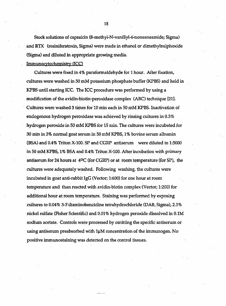
Immunocytochemistry (ICC)
Cultures were fixed in 4% paraformaldehyde for 1 hour. After fixation,
cultures were washed in 50 mM potassium phosphate buffer (KPBS) and held in
KPBS until starting ICC. The ICC procedure was performed by using a
modification of the avidin-biotin-peroxidase complex (ABC) technique [21].
Cultures were washed 3 times for 10 min each in 50 mM KPBS. Inactivation of
endogenous hydrogen peroxidase was achieved by rinsing cultures in 0.3%
hydrogen peroxide in 50 mM KPBS for 15 min. The cultures were incubated for
30 min in 3% normal goat serum in 50 mM KPBS, 1% bovine serum albumin
(BSA) and 0.4% Triton X-100. SP and CGRP antiserum were diluted to 1:5000
in 50 mM KPBS, 1% BSA and 0.4% Triton X-100. After incubation with primary
antiserum for 24 hours at 40C (for CGRP) or at room temperature (for SP), the
cultures were adequately washed. Following washing, the cultures were
incubated in goat anti-rabbit IgG (Vector; 1:600) for one hour at room
temperature and than reacted with avidin-biotin complex (Vector; 1:200) for
additional hour at room temperature. Staining was performed by exposing
cultiures to 0.04% 3-3'diaminobenzidine tetrahydrochloride (DAB, Sigma), 2.5%
nickel sulfate (Fisher Scientific) and 0.01% hydrogen peroxide dissolved in O.IM
sodium acetate. Controls were processed by omitting the specific antiserum or
using antiserum preabsorbed with l|iM concentration of the immunogen. No
positive immunostaining was detected on the control tissues.
19
SP-IR and CGRP-IR neurons were counted in single ganglia under the light
microscope and averaged. Images of IR neurites were quantified by image
analysis system (Zeiss-Kontron; IDAS version 2.00). Samples were viewed with a
Zeiss axiophot microscope at 6.25x magnification (2.5x by 2.5x optivar). Images
were captured with a Sony 3CCD color video camera. The internal scaling
feature of the image analysis software was calibrated to measure in millimeters.
The neurite images imderwent normalization (histogram stretching) and contour
enhancement prior to interactive discrimination. The resulting images were
measured to obtain the area of immunoreactive neurites present in the field.
Four fields, evenly spaced around the DRG explant, were measured from each
culture. The total area measured for each field was 1.15mm2 Data were
analyzed by Student's t-test.
Results
Data are based on the experiments performed on total of 210 DRG cultures.
Of these cultures 90 were used as control and in each experimental group there
were at least 6 DRG cultures.
Dorsal root ganglion explants from 2 day old rats established an extensive
growth of neuronal and non-neuronal elements when cultured for 1 week.
Explants had a tendency to flatten and spread but migration of DRG neurons
into the growing zone surrounding DRG explant was not recorded (Figs 1 A,B
and 4 A,B)- Normal SP- or CGRP-IR in 8 day old cultures is illustrated in Figs. 1
and 4 A,B/C. The number of SP-IR DRG neurons was significantly larger than
number of DRG neurons immunostained for CGRP (P<0.001, Student's t test).
The growth of immunoreactive neurites was radial (Fig. 1A and 4A) and well
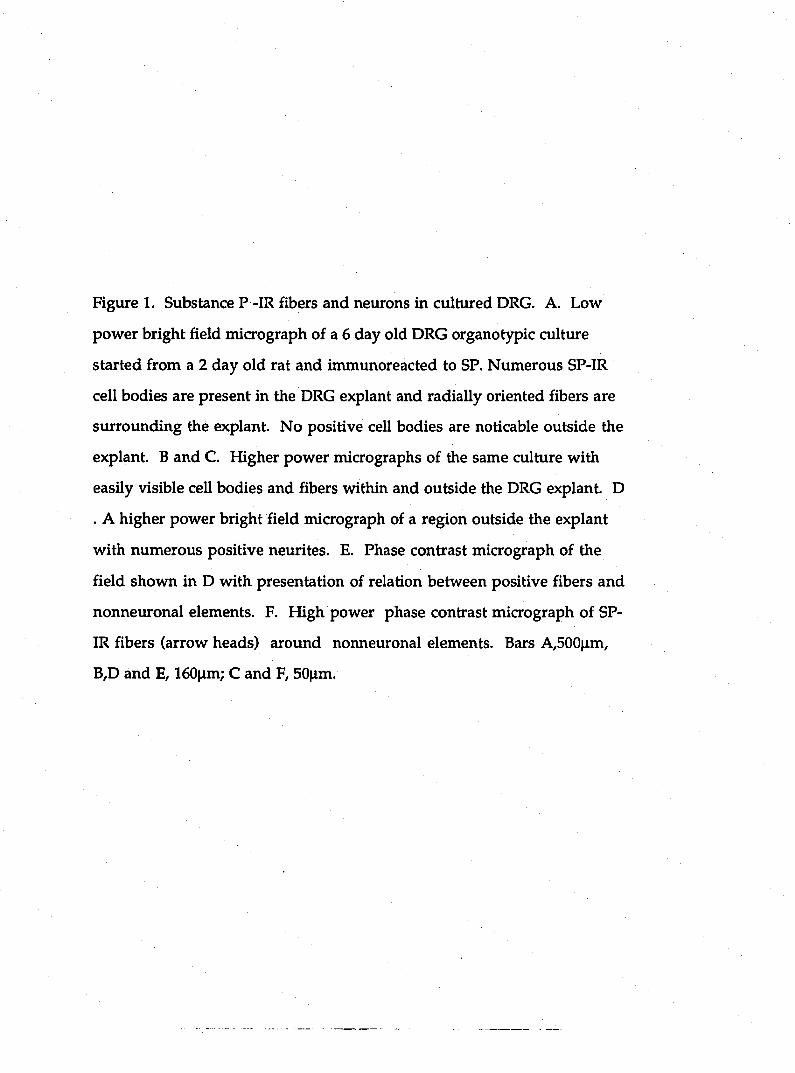
Figure 1. Substance P -IR fibers and neurons in cultured DRG. A. Low
power bright field micrograph of a 6 day old DRG organotypic culture
started from a 2 day old rat and immunoreacted to SP. Numerous SP-IR
cell bodies are present in the DRG explant and radially oriented fibers are
surrounding the explant. No positive cell bodies are noticable outside the
explant. B and C. Higher power micrographs of the same culture with
easily visible cell bodies and fibers within and outside the DRG explant. D
. A higher power bright field micrograph of a region outside the explant
with numerous positive neurites. E. Phase contrast micrograph of the
field shown in D with presentation of relation between positive fibers and
nonneuronal elements. F. High power phase contrast micrograph of SP-
IR fibers (arrow heads) around normeuronal elements. Bars A,500|im,
B,D and E, 160|im; C and F, SO^im.
22
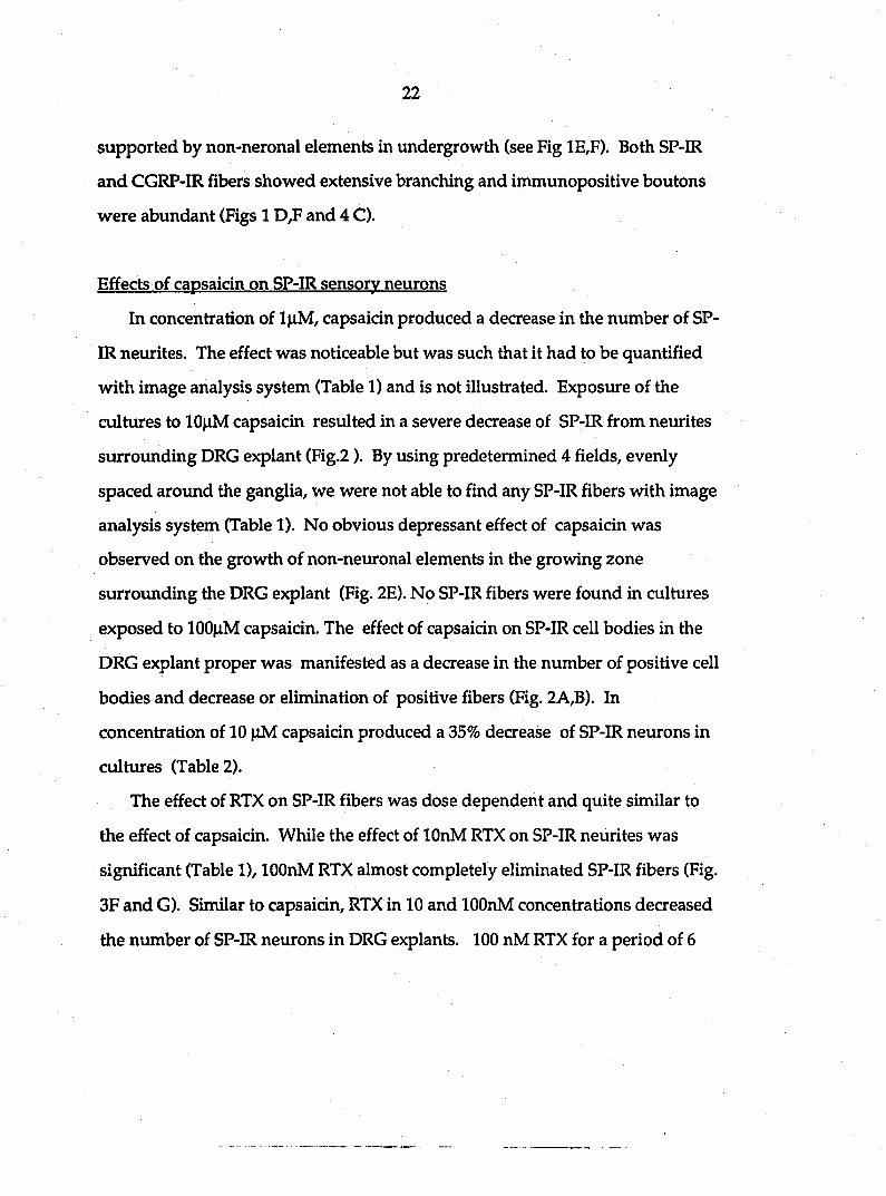
supported by non-neronal elements in undergrowth (see Fig 1E,F). Both SP-IR
and CGRP-IR fibers showed extensive branching and immunopositive boutons
were abundant (Figs 1 D,F and 4 C).
Effects of capsaicin on SP-IR sensory neurons
In concentration of l|xM, capsaicin produced a decrease in the number of SP-
IR neurites. The effect was noticeable but was such that it had to be quantified
with image analysis system (Table 1) and is not illustrated. Exposure of the
cultures to 10|iM capsaicin resulted in a severe decrease of SP-IR from neurites
surrounding DRG explant (Fig.2). By using predetermined 4 fields/evenly
spaced aroimd the ganglia, we were not able to find any SP-IR fibers with image
analysis system (Table 1). No obvious depressant effect of capsaicin was
observed on the growth of non-neuronal elements in the growing zone
surrounding the DRG explant (Fig. 2E). No SP-IR fibers were found in cultures
exposed to 100|aM capsaicin. The effect of capsaicin on SP-IR cell bodies in the
DRG explant proper was manifested as a decrease in the number of positive cell
bodies and decrease or elimination of positive fibers (Fig. 2A,B). In
concentration of 10 |iM capsaicin produced a 35% decrease of SP-IR neurons in
cultures (Table 2).
The effect of RTX on SP-IR fibers was dose dependent and quite similar to
the effect of capsaicin. While the effect of lOnM RTX on SP-IR neurites was
significant (Table 1), lOOnM RTX almost completely eliminated SP-IR fibers (Fig.
3F and G). Similar to capsaicin, RTX in 10 and lOOnM concentrations decreased
the number of SP-IR neurons in DRG explants. 100 nM RTX for a period of 6
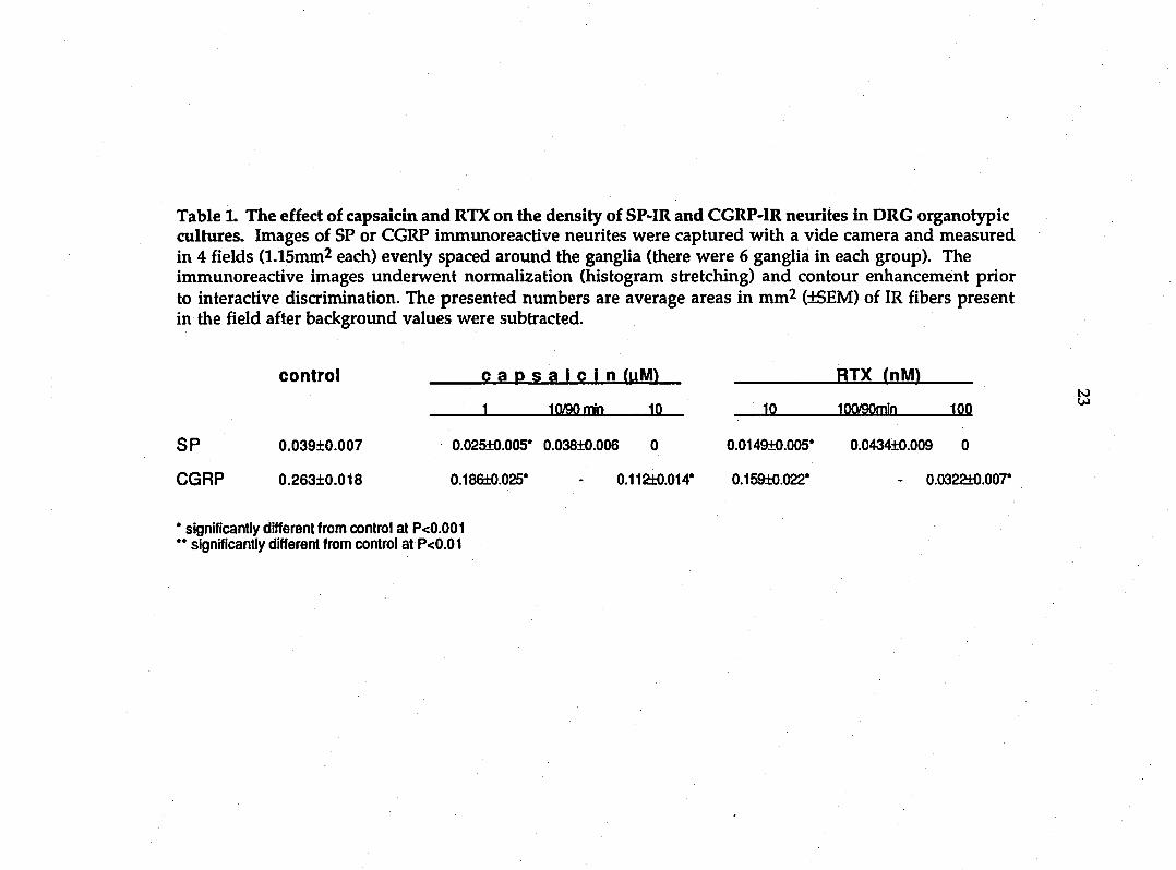
Table 1. The effect of capsaicin and RTX on the density of SP-IR and CGRP-IR neuiites in DRG organotypic cultures. Images of SP or CGRP inununoreactive neurites were captured with a vide camera and measured in 4 fields (l.lSmm^ each) evenly spaced around the ganglia (there were 6 ganglia in each group). The immunoreactive images underwent normalization (histogram stretching) and contour enhancement prior to interactive discrimination. The presented numbers are average areas in mm^ (+SEM) of IR fibers present in the field after backgrotmd values were subtracted.
control c a p s a i c i n ( u M ) RTX ( n m
1 loygorrin Ifl. 10 10(V90min 100
SP 0.039±0.007 0.025±0.005* 0.038±0.006 0 0.0149±0.005* 0.0434±0.009 0
CGRP 0.26310.018 0.18Gi0.025' 0.1121:0.014* 0.159±0.022' 0.0322t0.007*
* significantly different from control at P<0.001 " significantly different from control at P<0.01

Figure 2. Morphological chai\ges produced by capsaicin. 48 hours exposure
to 10|xM capsaicin decreased the number of SP-IR cell bodies (A and B) and
drastically decreased number of SP-IR fibers in the growing zone of the
culture. C. SP-IR soma of sensory neurons in a culture exposed to lOOfiM
capsaicin for 6 days. D and E. Photomicrographs of the field shown in in
bright field (in D) and phase contrast (in E) illustrating the almost
complete elimination of SP-IR fibers in a culture exposed to 10|iM
capsaicin for 6 days and absence of a similar effect on nonneuronal
elements. F. A high power micrograph of a short segment of SP-IR fiber
marked with arrowheads in D and E. Bars; A 160nm, B,C/D and E lOOum:
F SO^im.

26
Table 2. The effect of capsaicin and RTX on the ntunber of SP-IR and CGRP-IR cell bodies in DRG organotypic cultures
control 10 caps lOOnM RTX
n mean±SEM n meantSEM n meartbSEM
SP-IR neurons 21 138±6 20 89±7* 20 64±7*
CGRP-IR neurons 23 97±6** 21 47±4* 20 53±5*
* sigiuficantly different from control at P<0.001
** significantly lower than the number of SP-IR neurons at P<0.001
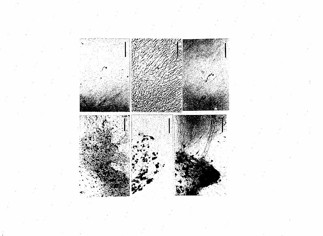
Figure 3. The effects of RTX on SP-IR cultured sensory neurons. A. Low
power micrograph of a culture exposed to lOOnM RTX for 48 hours. B. A
higher power magnification of the same culture with SP-IR cell bodies
inside the explant and several SP-IR fibers. C. lOOnM RTX for 6 days
eliminated all SP-IR fibers in the growing area surrounding the DRG
explant while many SP-IR cells are visible. D and F. Bright field and
phase contrast high power micrographs of SP-IR fibers and nonneuronal
immunonegative elements in a culture exposed to lOOnM RTX for two
days. lOOnM RTX almost completely eliminated all SP-IR fibers (F, arrow
heads are pointing towards structures that may be SP-IR segments of
neurites) without an similarly severe effect on nonneuronal elements in
the same field (G). Bars, A and C, 160|xm; B,D,E,F and G, lOOjim.

29
days produced about a 50% decrease in SP-IR cell bodies in DRG explants (Table
2)-
In order to establish the time dependence of capsaicin and RTX effect, we
exposed tissue to the compounds in different schedules and recorded the effect.
In cultures that were exposed to IGOfiM capsaicin for 2 days a complete
elimination of SP-IR fibers and significant decrease in the number of SP-IR cell
bodies in DRG explant were observed (Table 2). 10^M capsaicin for 48 hours
produced a drastic decrease in the number of SP-IR fibers and 6 day exposures
resulted in almost a complete elimination of SP-IR fibers (Fig,2 D,E and F). l|xM
capsaicin for 7 days capsaicin produced a significant decrease of SP-IR neurites.
However, 10|iM capsaicin (n=18) or lOOnM RTX (n=28) failed to produce an
effect, determined by examining immunoreactivity to peptides 6 days after
exposure, when presented to cultures for 45 to 90 min (Table 1). Exposure of the
cultures to RTX or capsaicin vehicle, in a concentration equal to one used with
the irritants, was without any obvious effect.
Effect of capsaicin and RTX on CGRP-IR neurons
Exposure of the cultures to 10 and 100 capsaicin resulted in an obvious
concentration-dependent decrease of CGRP-IR neurites surrounding DRG
explant (Table 1, Fig. 4 QD,E). lOfxM capsaicin produced a noticeable decrease in
the number of CGRP-IR fibers surrounding explants and 100|iM capsaicin
produced an obvious deaease in the number of CGRP-IR neurites (Table 1, Fig. 4
E,F). It should be noticed, however, that there were many more CGRP-IR fibers
than SP-IR fibers exposed to corresponding concentrations of capsaicin. The
effect of RTX on CGRP-IR fibers was quite similar to the effect of capsaicin.

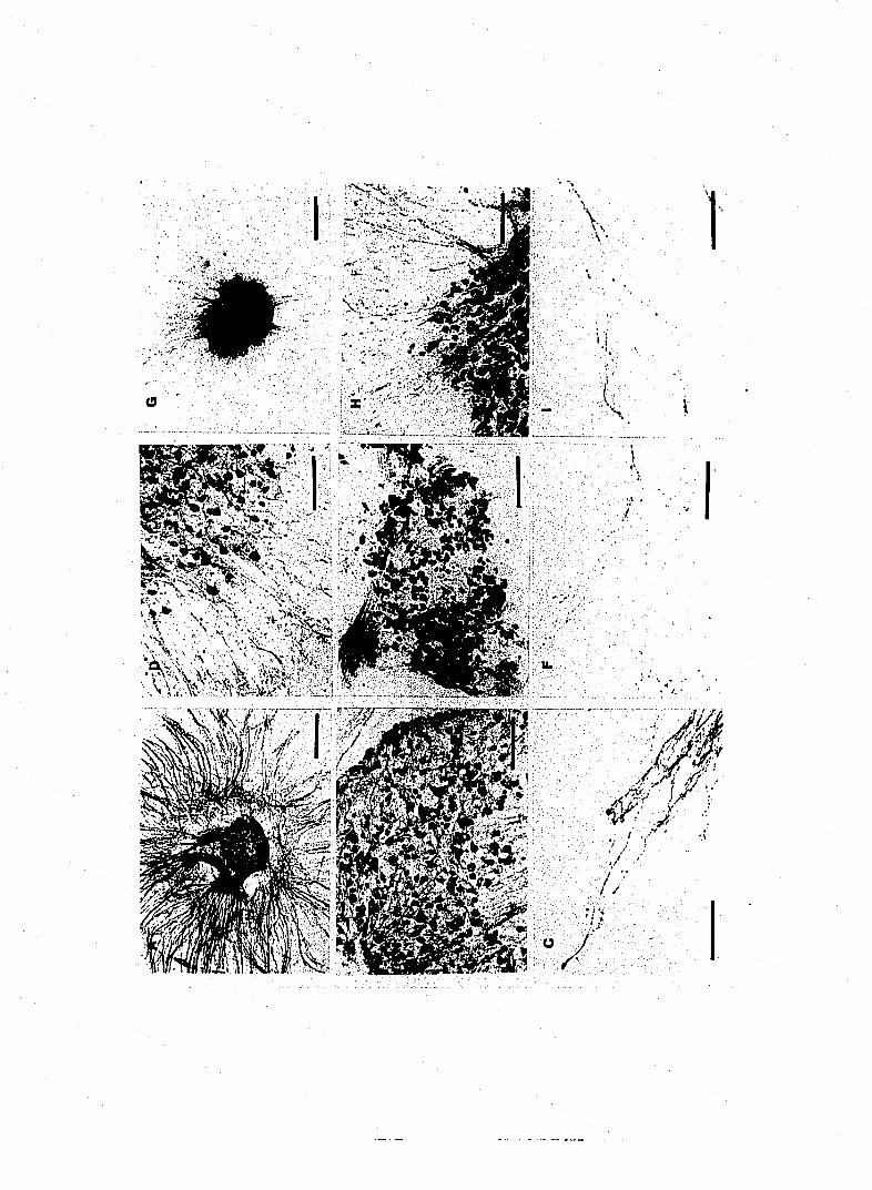
Figure 4. A. A low power bright field micrograph of a 7 day old organotypic culture started from a two day
old rat and immunostained for CGRP. The culture is characterized by numerous CGRP-IR cell bodies in the
DRG explant and CGRP-IR fibers radially oriented in the growing zone of the culture. B. A higher power
nucrograph of CGRP-IR cell bodies and fibers within the DRG explant. C. A bright field micrograph of
CGRP-IR neurites in a 7 day old culture. D. In concentration of 10|iM for 6 days capsaicin decreased ntunber
of CGRP-IR DRG neurons and fibers. E and F. 100|iM capsaicin for 6 days produced an obvious decrease
in the number of CGRP-IR fibers. G, H and I. I concentration of lOOnM for 6 days RTX produced an obvious
decrease in the niunber of CGRP-IR cell bodies and neurites in DRG culture. Bars A and G, 500|im; B,D,E and
H, 160|im, C,F and I, lOOjim.
32
lOnM RTX produced a significant decrease in the CGRP immunoreactivity in
neurites (Table 1), but lOOnM RTX produced a decrease in the number of CGRP-
IR fibers of such magnitude that it could be obvious in a photography (Fig. 4G,H
and I). It is important to notice that in no instance there was complete
elimination of CGRP-IR fibers. The effect of capsaicin and RTX on the number of
CGRP-IR cell bodies in DRG explant proper was similar to that on SP-IR cell
bodies. After counting we established that there was significantly less CGRP-IR
cell bodies in cultures exposed to lOOfiM capsaicin and lOriM RTX in comparison
to controls (Table 2).
The time-dependency of capsaicin and RTX toxic action was examined on
CGRP-IR neurons and is similar to the effect of these toxins on SP-IR neurons
and fibers. The capsaicin and RTX effect on CGRP-IR structures was dependent
on time of exposure as well as on concentration of neurotoxins. In exposures of
the cultures to 10|iM capsaicin or lOOnM RTX for 90 min no obvious effects on
the CGRP-IR in neurites or cell bodies were recorded several days later when the
culture was fixed and ICC procedure performed. The effects on CGRP was not
quantified by using machine because there was no noticeable difference and the
effect on more sensitive SP-IR neurites was absent (Table 1).
On the basis of the data that unique sensory neuron-specific actions of
capsaicin result in an influx of a number of cations including sodium, potassium
and calcium which is specifically antagonized with ruthenium red (RR), we
tested the effect of RR on 22 cultures. In 30 to 60 min exposures to 0.5 |XM RI^
we were unable to demonstrate convincing antagonistic effect on capsaicin or
RTX (n=8). However, culture exposed to 0.5|iM RR for 6 hours or longer (n=8)
failed to show any immunoreactivity to SP or CGRP 4 to 5 days after exposure.

33
In these cultures we were not able to see any immunoreactivity to two peptides
in cell bodies or neurites. In higher concentration (l^M) RR resulted in
disintegration of the cultures (n=6). This "depleting" effect of RR prevented its
use in studying the mechanism of action of capsaicin and RTX.
Discussion
The present series of experiments has examined the effects of capsaicin and
RTX on cultured peptidergic rat sensory neurons. Using immunocytochemistry
for labeling SP- and CGRP-IR we have been able to study the long lasting effect
of these two irritants on growth and survival of peptidergic neurons in
organot)^ic cultures.
It is clear that capsaicin and RTX, in a concentration dependent manner,
eliminate SP -IR fibers and reduce, but do not eliminate, CGRP-IR fibers when
presented to DRG cultures for period of 48 hours or longer. Exposure of the
cultures to the irritants for 90 min did not produce a noticeable effect. It is also
clear that neither capsaicin nor RTX eliminated all of the soma of SP-IR sensory
neurons. These findings are in agreement with previous studies suggesting that
processes of sensory neurons are much more sensitive to capsaicin than cell body
[4,24]. In addition, our findings that SP-IR fibers were much more sensitive to
capsaicin are in agreement with results demonstrating that SP is present in small
DRG neurons and small C-fibers only while CGRP is localized in both small and
large sensory neurons [15,28]. While it seems clear that only small fibers are
affected by capsaicin and RTX in culture, the question of the toxicity of capsaicin
and RTX towards all sensory neurons remains unanswered. Our findings that
SP-IR cell bodies were present in cultures exposed to 100|iM capsaicin or lOOnM

34
RTX for 6 days convincingly demonstrates that these neurons are not killed by
these irritants. Concentration-dependent effect towards peptidergic fibers and
the greater effect on SP-IR fibers is further support that our procedure is sensitive
enough and that these findings are not the result of some nonspecific factors.
These results provide additional support for the data demonstrating that
capsaicin and RTX act with great cellular specificity. Experimental approach,
however, does not allow us to convincingly demonstrate that loss of peptide
immunoreactivity is due to cell death.
It is important to establish that the effect of capsaicin and RTX on cultured
neurons is of long duration and is therefore different from depletion of peptides
from neurites. In cultures exposed to these irritants for two days after which the
cultures were returned to growing media for 4 or 5 days, we were not able to
notice an increase in SP-IR fibers. These cultures were the same as cultures that
were exposed to the irritants for 2 days just prior to fixation and ICC. In
addition, it is important to establish that the sensitivity of fibers is concentration
dependent. In cultures exposed to 10|iM capsaicin or lOnM RTX we were not
able to produce complete elimination of SP-IR fibers, even though the cultures
were exposed to neurotoxins for 7 days. However, 100|iM capsaicin and lOOnM
RTX produced complete elimination of SP-IR fibers in two days. Single exposure
to high concentrations of capsaicin produced a depletion of neuropeptides and
excitatory amino acids from sensory neurons which may account for long lasting
impairment of sensory functions (7,14). A strict correlation was found between
the time-related recovery of neuropeptide levels in sensory terminals and
functional recovery of sensory fibers (9). Present findings demonstrate important

35
role played by the tune of exposure. While singe exposure to capsaicin produced
a reversible impairment of sensory function prolonged exposure in our
experiments resulted in irreversible changes. This may be very important
element in proposed use of capsaicin in chronic pain treatment. Due to strong
effect of ruthenium red on the peptidergic neurons we were not able to study the
mechanism of prolonged capsaicin effect on sensory neurons. However, the
capsaidn-induced excessive Ca accumulation in primary afferents accounts for
"sensory neuron blocking" action produced by high capsaicin and might account
for "irreversible" depletion of SP and CGRP from capsaicin sensitive neurites [30,
36]. Our inability to maintain the cultures for longer than 6 days following
exposure to capsaicin makes it hard to be certain that effects are irreversible.
Previous studies have provided evidence that sensitivity to capsaicin and
content of SP are properties of nociceptive primary sensory neurons but it is not
known whether these markers are expressed in all nociceptive sensory neurons.
It has been documented that in dissociated cultures the fraction of cells sensitive
to capsaicin and fraction of cells that contain SP-IR are larger than in freshly
dissected DRG [1]. In our experiments, the fraction of the cells in the culture that
is stained with antiserum to SP is larger than the fraction of that stained with
CGRP. This result suggests that SP sensory cells survive preferentially in our
culture conditions. Together with finding that capsaicin and RTX eliminate all
SP-IR neurites and only a certain proportion of CGRP-IR neurites, these results
suggest that relatively early in development a large fraction of cultured sensory
neurons are able to express the functional properties of differentiated nociceptive
sensory neurons, and are likely to be useful tool in studying the neuronal
mechanisms of nociception.
36
References
1. Baccaglini, P.I. and Hogan, P.G., Some rat sensory neurones in culture
express characteristics of different pain sensory cells, Proc. Natl. Acad.
Sd., U.S. A., 80 (1983) 594-598.
2. Bevan, S.J., James, I.F., Rang, H.P., Winter, J. and Wood, J.N., The
mechanism of action of capsaicin - a sensory neurotoxin. In P. Jenner (Ed.)
Neurotoxins and their Pharmacological Implications, Raven, New York,
1987, pp. 261-277.
3. Buck, S.H. and Burks, T.F., The neuropharmacology of capsaicin: review
of some recent observations, Pharmacol. Rev., 38 (1986)179-226,
4. Chung, K., Klein, C.M. and Coggeshall, R.E., The receptive part of the
primary afferent axon is most vulnerable to systemic capsaicin in adult
rats. Brain Res., 511(1990) 222-226.
5. Delfs, J., Friend, J., Ishimoto, S. and Saroff, D., Ventral and dorsal horn
acetylcholinesterase neurons are maintained in organotypic cultures of
postnatal rat spinal cord explants. Brain Res., 488 (1989) 31-42.
6. deVries, D.J. and Blumberg, P.M., Thermoregulatory effects of
resiniferatoxin in the mouse: comparison with capsaicin. Life Sd. 44 (1989)
711-715.
7. Driedger, P.E. and Blumberg, P.M., Specific binding of phorbol ester
tumor promoters, Proc. Natl. Acad. Sd. U.S.A. 77 (1980) 567-571.
8. Fitzgerald, M., Capsaicin and sensory neurons - a review. Pain 115
(1983)109-130.

37
9. Foster, R,W. and Ramage, A.G., The action of some chemical irritants on
somatosensory receptors in the cat, Neuropharmacology, 20(1981)191-
198.
10. Franco-Cereceda, A,, Lundberg, J.M., Saria, A., Schreibmayer, W. and
Tritthart, H.A., Calcitonin gene-related peptide: release by capsaicin and
prolongation of the action potential in the guinea-pig heart. Acta Physiol.
Scand., 132 (1988) 181-190.
11. Franco-Cereceda, A. and Rudehill, A.R., Capsaicin-induced vasodilatation
of human coronary arteries in vitro is mediated by calcitonin gene-related
peptide rather than substance P or neurokinin A, Acta Physiol. Scand., 136
(1989)575-580.
12. Gahwiler, B.H., Organotypic monolayer cultures of nervous tissue, J.
Neurosci. Methods, 4 (1981) 329-342.
13. Gamse, R., Lackner, D., Gamse, G. and Leeman, S.E., Effect of capsaicin
pretreatment of capsaicin-evoked release of immunoreactive somatostatin
and substance P from primary sensory neurons. Naunyn-Schmied, Arch.
Pharmacol., 316 (1981)19-24.
14. Gamse, R., Molnar, A. and Lembeck, F., Substance P release from spinal
cord slices by capsaicin. Life Sci., 25 (1979) 629-636.
15. Gibson, G.J., Polak, J.M., Bloom, S.R., Sabate, I.M., Mulderry, P.M., Ghatei,
M. A., McGregor, G.P., Morrison, J. F. B., Kelley, J. S., Evans, R. M. and
Rosenfeld, M.G., Calcitonin gene-related peptide immunoreactivity in the
spinal cord of man and of eight other species, J. Neurosci., 4 (1984) 3101-
3111.
38
16. Hakanson, R., Beding, B., Ekman, R., Heiling, M., Wahlestedt, C. and
Sundler, F., Multiple tachikinin pools in sensory nerve fibers in the rabbit
iris, Neurosdence, 21(1987) 943-950.
17 Hayes, A. G., Oxford, A., Reynolds, M., Shinger, A. H., Skingle, M., Smith,
C. and Tyers, M.B., The effect of a series of capsaicin analogs on
nociception and body temperature in the rat. Life. Sci., 34 (1984) 1241-1248.
18. Hejmian, I. and Rang H.P., Depolarising responses to capsaicin in a
subpopulation of rat dorsal root ganglion cells, Neurosci. Lett., 56 (1985)
69-75.
19. Holzer, P., Local effector function of capsaicin-sensitive sensory nerve
endings: involvement of tachykinins, calcitonin gene-related peptide and
other neuropeptides, Neuroscience, 24 (1988) 739-768.
20. Hoover, D.B., Effects of capsaicin on release of substance P-like
immimoreactivity and physiological parameters in isolated guinea-pig
hearts, Eur. J. Pharmacol., 141 (1987) 489-492.
21. Hsu, S-M, Raine, L. and Fanger, H., Use of avidin-biotin-peroxidase
complex (ABC) in immunoperoxidase techniques: a comparison between
ABC and unlabeled antibody (PAP) procedures, J. Histochem. Cytochem.,
29 (1981) 577-580.
22. James, LF., Walpole, C.S., Hixon, J., Wood, J.D. and Wrigglesworth, R.,
Long-lasting agonist activity produced by a capsaicin-like photoaffinity
probe, Molec. Pharmac., 33 (1988) 643-649.
23. Jancso, G., Kiraly, A. and Jancso-Gabor, A., Pharmacologically induced
selective degeneration of chemosensitive primary neurones. Nature, 270
(1977) 741-743.
39
24. Jancso, G., Kiraly, E., Loo, F., Such, G. and Nagy, A., Selective
degeneration by capsaicin of a subpopulation of primary sensory neurons
in the adult rat, Neurosci. Lett., 59 (1985) 209-214.
25. Jeftinija, S., Jeftinija, K., Liu, R, Skilling, S.R., Smullin, D.H. and Larson,
A.A., Excitatory amino acids are released from rat primary afferent
neurons in vitro. Neurosci Lett, (1991) in press.
26. Jessell, T.M., Iversen, L.L. and Cuello, A.C., Capsaicin-induced depletion
of substance P from primary sensory neurones, Brain Res., 152 (1976)183-
188.
27. Jhamandas, K., Yaksh, T.L., Harty, G., Szolcsanyi, J. and Go, V.L.W.,
Action of intrathecal capsaicin and its structural analogues on the content
and release of spinal substance P: Selectivity of action and relationship to
analgesia. Brain Res., 306 (1984) 215-225.
28. Lee, Y., Takami, K., Kawai, Y., Girgis, S., Hillyard, C.J., Maclntyre, L,
Emson, P. E. and Tohyams, M., Distribution of calcitonin gene-related
peptide in the rat peripheral nervous system with reference to its
coexistence with substance P, Neuroscience, 15 (1985)1227-1237.
29. Marsh, S..J., Stansfeld, C.E., Brown, D.A., Davey, R. and McCarthy, D., The
mechaiusm of action of capsaicin on sensory C-type neurons and their
axons in vitro. Neuroscience, 23 (1987) 275-289.
30. Maggi, C. A. and Meli, A., The sensory-efferent function of capsaicin-
sensitive sensory neurons. Gen. Pharmacol., 19 (1988) 1-43.
31. Maggi, C.A., Santicioli, P., Geppetti, P., Parlani, M., Astolfi, A., Pradelles,
P., Patacchini, R. and Meli, A., The antagonism induced by ruthenium red
40
of the actions of capsaicin on the peripheral terminals of sensory neurons:
further studies^ Eur. J. Pharmac., 154 (1988) 1-10.
32. Maggi, C.A., Patacchhini, R., Tramontana, M., Amann, R., Giuliani, S. and
Santicioli/P./ Similarities and differences in the action of resiniferatoxin
and capsaicin on central and peripheral endings of primary sensory
neurons, Neuroscience, 37(1990)531-539.
33. Szallasi, A. and Blumberg, P.M., Resiniferatoxin, a phorbol-related
diterpene, acts as an ultrapotent analog of capsaicin, the irritant
constituent in rat pepper, Neuroscience, 30 (1989) 515-520.
34. Szallasi, A. and Blumberg, P.M., Specific binding of resiniferatoxin, an
ultrapotent capsaicin analog, by dorsal root ganglion membranes. Brain
Res., 524 (1990) 106-111.
35. Szolcsanyi, J., Capsaicin: hot new pharmacological tool. Trends Neurosci.,
4(1984)495-497.
36. Szolcsanyi, J., Capsaicin-sensitive chemoceptive neural system with dual
sensory-efferent function. In L.A. Chahl, J. Szolcsanyi and F. Lembeck
(Eds.), Antidromic Vasodilation and Neurogenic Inflammation,
Akademiai Kiado, Budapest, 1984, pp 26-52.
37. Theriault, E., Otsuka, M. and Jessell, T., Capsaicin-evoked release of
substance P from primary sensory neurons. Brain Res., 170 (1979) 209-213.
38. Wood, J.N., Winter, J., James, I.F., Rang, H.P., Yeats, J. and Bevan, S.,
Capsaicin-induced ion fluxes in dorsal root ganglion cells in culture, J.
Neurosci. 8 (1988) 3208-3220.
41
Zur Hausen, H., Bomkamm, G. W., Schmidt, R. J. and Hecker, E., Tumor
iiutiators and promoters in the induction of Epstein-Barr virus, Proc. Natl.
Acad. Sci. U.S. A. 76 (1979) 782-785.
42
CHAPTER 2. NEUROLIGAND-EVOKED CALCIUM-DEPENDENT
RELEASE OF EXCITATORY AMINO ACIDS FROM SCHWANN CELLS
A paper submitted to The Journal of Neuroscience
Vladimir Parpura*, Fang Liu*, Ksenija Jeftinija'
Srdija Jeftinija and Philip G. Haydon
Abstract
Bradykinin caused a receptor-mediated increase in release of the excitatory
amino acids (EAAs) glutamate and aspartate from Schwann cell cultures
obtained from dorsal root ganglia (DRG) together with an increase in the
cytoplasmic level of free calcium. Perturbations which inhibited bradykinin-
induced calcium mobilization prevented the release of EAAs from glia. The
addition of ionomycin caused a calcium-dependent release of EAAs. Therefore,
bradykinin causes calcium dependent-release of EAAs from DRG Schwann cells.
Bradykinin did not cause cell swelling and p-chloromercuriphenylsulphonic
acid, an inhibitor of the electrogenic glutamate transporter, did not reduce
bradyknin-induced EAA release. Therefore, bradykinin stimulates EAA release
from Schwann cells through a mechanism that is neither the previously described
volume regulated release mechanism nor due to the reversal of the glutamate
transporter.
* Contributed equally

43
Introduction
It has been well established that glia have multiple roles in the nervous
system which include the regulation of external potassium and the uptake of the
excitatory amino adds glutamate and aspartate which are released from neurons
into the extracellular space (NichoUs and Atwell, 1990; Barres, 1991; Kanai et al.,
1993). Several lines of evidence suggest that glial cells may also play important
roles in releasing neurotransmitters. Schwann cells can release acetylcholine
following axotomy of motoneurons (Dennis and Miledi, 1974). Retinal Miiller
cell depolarization stimulates the release of glutamate through a reversal of an
electrogenic glutamate uptake carrier (Szwatkovski et al., 1990). Depolarization
causes the calcium-dependent release of the amino acid taurine from
hippocampal glia (Philibert et al., 1988). Hyposmotic shock of cortical astrocytes
stimulates cell swelling, an increase in internal calcium and a correlated release
of the amino acids aspartate and taurine (Kimelberg et al., 1990; O'Connor and
Kimelberg, 1993). Since glial cell calcium levels are regulated by
neurotransmitters (Reisner et al., 1989; Cornell-Bell et al., 1990; Glaum et al., 1990;
Charles et al., 1991; Cornell-Bell and Finkbeiner, 1991; Inagaki et al., 1991; Jensen
and Chiu, 1991; McCarthy and Salm, 1991) this has prompted us to ask whether
the neuroligand bradykinin causes a calcium-dependent release of excitatory
amino acids (EAAs) from glial cells.
In the peripheral nervous system, the nonapeptide bradykinin is released
from its precursors, the kininogens, by the action of the enzyme kallikrein in
response to trauma (Dray and Perkins, 1993). Bradykinin is known to mobilize
calcium from inositol 1,4,5-trisphosphate (IP3)-sensitive calcium stores in several

44
cell types (for ref see Miller, 1987; Higashida and Ogura, 1991). Since bradykinin
can mobilize internal calcium, we have asked whether this agent can cause the
release of excitatory amino acids from glial cells derived from the dorsal root
ganglia. This study demonstrates that the neuroligand, bradykinin, mobilizes
internal calcium from Schwann cells which stimulates a calcium-dependent
release of the excitatory anuno acids, glutamate and aspartate.
Methods
Cell Culture
Glial cultures from DRG were obtained by modification of the organotypic
procedure (Gahwiler, 1984). Following anesthesia with ether and decapitation,
DRG from 1- to 2-day-old Sprague-Dawely rats were rapidly removed and
washed in cold (4°C) oxygenated Gey's balanced salt solution (GBSS; Gibco)
modified by the addition of 2% glucose. The capsular sheet was carefully"
removed form the DRG to mirumize fibroblast contamination. DRG were than
embedded onto a glass coverslip inlayed in a 35 mm dish (for release studies) or
into a glass bottomed dish (for calcium imaging) in a plasma-thrombin clot.
Cultures were maintained at 36°C in a humidified 5% C02/air atmosphere. The
culture medium consisted of 25% horse serum (Gibco), 25% Earle's Balanced Salt
Solution (Gibco) and 50% Basal Medium Eagle (Gibco) containing 36 mM
glucose. The central part of explants which contains neuronal cell bodies was
removed from established cultures (4 to 7 days in culture). The residual glial cells
were maintained for additional 72 hours when all experiments were performed.
Neurites degenerated 24 to 48 hours following the removal of the central portion
45
of explants leaving cultures containing only non-neuronal cells. The absence of
neuronal elenients was confirmed by immunocytochemistry.
In some experiments we enriched the number of Schwann cells in cultures
using two methods (see Kleitman et al, 1992). In the Wood method, DRG were
cultured for 48-96 hours in the presence of cytosine arabinoside (Ara-C; 5
mg/mL). They were then transplanted to a new coverslip coated with chicken
plasma. DRG explants were then cultured as described above to obtain DRG
glia. In the Brockes method, DRG glia were incubated with Thy 1.1 antibody
(hybridoma supernatant, American Type Culture, No. TIB103) and addition of
rabbit complement (Sigma). Since Thy 1.1 is expressed on fibroblasts, but not on
Schwann cells, this procedure leads to a complement mediated lysis of
fibroblasts. In both culture methods, immunocytochemistry revealed that
Schwann cells were enriched to >95%.
Immunocytochemistry
Antibody visualization was accomplished by using a Vectastain ABC kit
(Vector) and the nickel-enchanced DAB method (Hsu et al., 1981).
Immunocytochemistry was performed using antibodies directed against the
synaptic proteins synaptophysin (1:1000; clone 7.4a, provided by R. Jahn) and
synaptotagmin (1:5000; clone 41.1, provided by R. Jahn), against glial fibrillary
acidic protein (GFAP; 1:5000, ICN Immimobiologicals) and against the low-
affinity NGF receptor (1:2000; 192-IgG, provided by E.M. Johnson, Jr.). Anti-
synaptophysin, synaptotagmin and anti-GFAP were found to be negative on
DRG glia. Positive controls were performed with these antibodies using

46
hippocampal neurons and glia and using DRG explants in which axons were
maintained in culture.
EAA release
The coverslips with glial cultures were mounted into a 50 |il perfusion
chamber. A modified Ringer's perfusion solution containing (in mM) NaCl 128,
KCl 1.9, KH2PO41.2, CaCl2 2.4, MgS041.3, NaHCOs 26 and glucose 10
(pH=7.4) bubbled with 95% O2/ 5% CO2 was used for constant flow through at a
rate of 200 ml/min and 35-37°C. After an equilibration period of 40 to 60
minutes, samples of perfusate for amino acid determination were collected every
minute. The amino acid content in samples was determined by high-
performance liquid chromatography (HPLC) with fluorescence detection
(Lindroth and Mopper, 1979). Prior to injection, aliquots of samples were
derivatized with o-phthalic aldehyde (OPA) 2-mercaptoethanol reagent (Pierce).
Chromatography was performed on a 15 cm Microsorb-MV HPLC column
(Rairun Instrument Co.) using a sodium acetate methanol gradient (pH=5.9).
Bradykinin (10 nM; Sigma) and its antagonist (D-ArgO, Hyp3, fi-Thi^/^, D-Phe^)-
bradykirun (5 nM; Bachem Bioscience Inc.) were added to a perfusion solution.
In control experiments we co-administered hydroxyproline (180 nM) together
with bradykinin and determined that there is a 1 minute lag time from initiation
of the addition of an agent until it is collected for HPLC analysis due to dead-
volume in the chamber. All figures for HPLC have been correspondingly
corrected. Elevated K+ (50 mM) solution for HPLC was prepared by
modification of a Ringer's solution where NaCl was replaced by KCl.
47
Calcium imaging
Cells were loaded with fura-2 AM (4 ^M; Molecular Probes) for 40-60
minutes at 37°C. of 25%(w/w) of Pluronic F-127 (Molecular Probes) was
mixed with 1ml of 4nM Fura-2 AM ester solution. After washing, cells were kept
for 30-60 minutes at 37°C to permit dye de-esterification . All experiments took
place at 22-24°C. All image processing and analysis were performed using
ratiometric software (QFM, Quantex Corp. or Image-l/Fl, v 1.63g, Universal
Imaging Corp.). Background subtracted, ratio images (340/380 nm or 350/380
nm) were used to calculate the [Ca^+Jj according to Equation 5 of Grynkiewicz et
al. (1985). Calibration was performed in situ (Thomas and Delaville, 1991) using
the Ca2+-ionophore 4-bromo-A23187 (10 |iM, Molecular Probes), An estimate of
autofluorescence at each wavelength was achieved by addition of MnCl2 (20
mM). Cells were included in analysis if the first addition of bradykinin caused a
[Ca2+]i accumulation that was greater than 50% of the resting calcium level.
During calcium imaging, bradykinin (10 nM) and elevated K+ (50 mM) were
applied to glia for 1 minute by pressure ejection from a puffer pipette (opening
diameter ~2-3 [im, 10 psi). All other drugs were uniformly applied to the bath by
flow through. Repeated applications of bradykinin were spaced at 10 minute
intervals. In some experiments the second bradykinin application followed the
first application after 30 minutes. Ryanodine (10 |iM), caffeine (10 mM) and
furosemide (5 n\M) were applied 10 minutes prior to the second application of
bradykinin while thapsigargin was applied to the bath 30 minutes prior to the
second application of bradykinin. Thapsigargin (1 |iM) was dissolved in dry
DMSO. Control experiments with 0.1% (v/v) DMSO showed that DMSO did not
48
affect the action of bradykinin. Normal saline contained (in mM): NaCl 140, KCl
5, MgCl2 2, CaCl2 2 and HEPES10 (pH 7.4). Calcium-depleted solution
contained (in mM): NaCl 128, KCl 1.9, KH2PO41.2, CaCl2 0.2, EGTA 1, MgS04
2.5, NaHC03 26 and glucose 10 (pH=7.4). Elevated K+ saline was prepared by
modification of normal saline where NaCl was replaced by KCl.
Max Chelator (version 5.6), written by Chris Patton at Stanford University,
was used to calculate free extracellular calcium levels. The calculation includes a
correction for extracellular Mg2+ions.
Cell volume measurement
To determine whether bradykinin caused a shrinkage of cells in our studies
we monitored cell volume using BCECF as described by Eriksson et al. (1992).
Cells were loaded by incubation in membrane permeable BCECF AM (10 |iM) for
40-60 minutes, followed by 40 minutes for de-esterification. BCECF was excited
at the pH-insensitive wavelength of 440 run. The emission intensity (510 DF
40nm bandpass) was monitored with a SIT camera. When cells swell, BCECF is
diluted, reducing the fluorescent intensity detected. We confirmed that this
approach effectively detected volume changes in our studies by applying
hyposmotic saline that was made by removing 50 mM of NaCl from our
standard saline (see above).
49
Results
Bradykinin stimulates excitatory amino add release from dorsal root ganglia
Schwann cells
Primary glial cultures of dorsal root ganglia (DRG) were obtained using a
modification of the organotypic procedure (Gahwiler, 1984). Dorsal root ganglia
were cultured on coverslips to permit neurite extension and migration of glial
cells. After cultures were established, the central portion of the ganglionic
explant, which contains neuronal cell bodies, was removed from the culture.
During the next 48 hours the residual neurites degenerated leaving a neuron-free
glial cell culture. The release of EAAs from these cultures was assayed using
HPLC on the superfusate. The basal release of glutamate and aspartate into
superfusate produced levels of 26 + 2 nM (mean + SEM; n= 13) and 6 + 1 nM,
respectively. Addition of bradykinin caused a dose-dependent increase in
release of the EAAs glutamate and aspartate from glial cultures. The threshold
concentration of bradykinin was 1 nM. 10 nM bradykinin caused a greater than
nine-fold inaease in the release of glutamate to 239 ± 42 nM and a greater than
three-fold increase in aspartate to 21 + 3 nM (n=12; Fig. 1 A, B). A second
application of bradykinin (n=12) similarly caused EAA release, although the
magnitude of this response was attenuated (Fig. 1 A, B). This action is receptor-
mediated since the B2 receptor antagonist, (D-ArgO, Hyp3, fi-Thi^rS, D-Phe7>
bradykinin (5 ^M), reversibly blocked the stimulatory action of bradykinin (n=6;
Fig. IC). While bradykinin stimulated the release of glutamate and aspartate
from glial cultures it did not significantly affect the release of serine. The basal
level of serine was 14 + 2 nM compared to 16 + 2 nM in the presence of

Figure 1. Bradykinin causes a receptor-mediated release of the EAAs, glutamate
and aspartate, from DRG glia. Using HPLC, the amounts of glutamate and
aspartate were determined in superfusate from rat DRG glia cultures. Addition
of bradykinin (BK; 10 nM) caused a 9-fold elevation of glutamate release (A) and
a 3-fold elevation of aspartate release (B). The B2 receptor antagonist, (D-ArgO,
Hyp3, fi-Thi5/8^ D-Phe7)-bradykinin, blocked the secretory action of bradykinin
(C). After washout of the antagonist, bradykinin reliably increased glutamate
release. Points represent means + SEM.

n
Glutamate concentration (nM)
in o
en -
H 3
-
in
S. IS CIS ^
Aspartate concentration (nM) Glutamate concentration (nM) h' t>> M h' N' Kl O O O e t n o u i e u i o o o o
H 1'
3. 5'

52
bradykinin (n=13; p>0.5). These data demonstrate that the nonapeptide
bradykinin potently causes the selective release of the EAAs glutamate and
aspartate from DRG glia.
The cell types present in the glial cultures derived from DRG was examined
using inunimocytochemistry. We confirmed that glial cultures were neuron-free
by using antibodies against the synaptic proteins synaptophysin and
synaptotagmin. In these cultures immunoreactivity for these two synaptic
proteins was absent while neurons were immunopositive in separate cultures.
An antibody against glial fibrillary acidic protein was negative in DRG glial
cultures. However, using an antibody against the low-affinity NGF receptor,
characteristically expressed in Schwann cells localized distal to the site of
axotomy, but not in surrounding fibroblasts (Taniuchi et al., 1988), we found that
about 74% of the cultured glial cells possess NGF receptors (Fig. 2). Therefore
the predominant glial cell type in these DRG cultures is Schwann cells.
Since fibroblasts are likely to be a contaminating cell type in these cultures,
we modified our techniques to enrich specifically in Schwann cells. Purified
populations of Schwann cells (>95% of cells) were obtained by the Wood (n=6)
and Brockes (n=6) methods (see Kleitman et al., 1992). These cultures showed
sinular increases of the EAAs glutamate and aspartate in the superfusate after
application of bradykinin. Bradykinin increased the glutamate levels in the
superfusate from a basal level of 127+9 and 35+7 to 465+23 and 123+16 using the
Brockes and Wood methods, respectively. We therefore conclude that the
nonapeptide, bradykinin causes the release of the the amino acids glutamate and
aspartate from DRG Schwann cells.
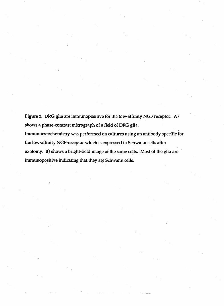
Figure 2. DRG glia are immunopositive for the low-affinity NGF receptor. A)
shows a phase-contrast micrograph of a field of DRG glia.
Immunocytochemistry was performed on cultures using an antibody specific for
the low-affiruty NGF-receptor which is expressed in Schwann cells after
axotomy. B) shows a bright-field image of the same cells. Most of the glia are
immunopositive indicating that they are Schwann cells.
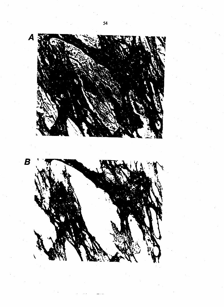
54

55
Bradykinin mobilizes calcium in dorsal root ganglion Schwann cells.
The release of neurotransmitter at neuronal synapses is calcium-dependent.
To ask whether calcium might play a role in bradykinin-induced EAA release
from DRG Schwann cells, we used ratiometric imaging techniques to monitor
glial calcium levels. Cells were loaded with the membrane permeant calcium
indicator fura-2 AM. Glia were imaged at low plating density, where they did
not form a confluent monolayer. In resting conditions, the cytoplasmic calcium
level of glia was 102 + 2 nM (n=136). Bradykinin (10 nM) reliably raised the
cytoplasmic level of free calcium in 72% of cells tested (n=136 of 188). This
increase of intracellular calcium reached the peak level of 747 + 24 nM (n=136;
Fig. 3A) about 20 seconds after the onset of bradykinin application and was
sustained for several minutes. In all subsequent experiments, we have reported
the calcium levels for only those cells that responded to bradykinin. These
bradykinin responsive cells had the morphology of Schwann cells. In some
experiments we performed immunocytochemistry following calcium imaging
using the antibody directed against the low-affinity NGF receptor which is
present in Schwann cell cultures. Immuncytochemistry demonstrated that those
cells that responded to bradykinin with an elevation of internal calcium were
immimopositive with the antibody directed against the low-affinity NGF-
receptor (n=22 of 22 cells). Therefore, bradykinin elevates internal calcium levels
in Schwann cells in a manner temporally coincident with the timing of release of
glutamate and aspartate.
Repeated applications of bradykinin reliably elevated calcium levels in
Schwann cells (Fig. SB). The B2 receptor antagonist, (D-ArgO, Hyp3, C-Thi^/S^ D-
Phe7)-bradykinin (5 |iM), reversibly attenuated the stimulatory action of
Figtue 3. Bradykinin mobilizes Ca2+ from internal stores in DRG glia. In (A) a
focal application of bradykinin (BK; lOnM) elevates DRG glial calcium level to a
peak 20 seconds after the onset of application. During sustained application of
bradykinin, glial calcium is maintained at a stable elevated plateau from basal
calcium level (dashed line). In (B) repeated applications of bradykinin (10 nM)
caused repeated elevations of glial cytoplasmic calcium levels. In (C) the
external calcium v^as reduced by exchange of normal saline for calcium-depleted
saline. In this saline bradykinin was able to elevate calcium from its resting level
indicating that calcium is mobilized from internal stores. However, a second and
third addition of bradykinin had little effect on internal calcium levels (compare
CwithB). Bars represent means + SEM.
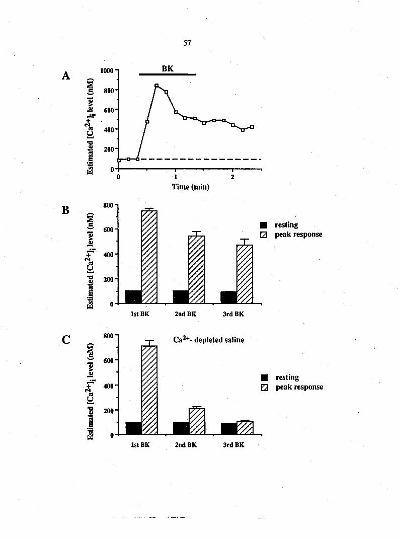
57
10001
Time (min)
B
i :r 600
i i
(2 0
• resting • peak response
IstBK 2ndBK 3rd BK
Ca^+- depleted saline
i rr 600
i
iS 0 1st BK
• resting • peak response
2ndBK 3rdBK

58
bradykinin on calcium mobilization. In the presence of the B2 receptor
antagonist^ the average resting calcium level (107 + 3 nM) was moderately
increased by application of bradykinin (151 + 11 nM; n=32). However, after
washout of the antagorust, bradykinin increased the cytoplasmic calcium level to
480 + 24 nM (n=32), a value significantly greater than in the presence of the B2
receptor antagoiust (Scheffe^'s test, p<0.001).
Removal of external calcium from the bathing medium did not prevent the
initial calcium mobilizing action of bradykinin (Fig. 3C). Cells were bathed in a
caldum-depleted saline containing 0.2 mM calcium with the addition of 1 niM
EGTA to yield an estimated free extracellular calcium level of 24 nM. We found
that it was necessary to have some calcium in the bathing medium otherwise
cells detached from the culture substrate. In this calcium-depleted saline,
bradykinin elevated cytoplasmic free calcium from 98 + 2 nM to 713 + 38 nM
(n=36). However, subsequent additions of bradykinin in calcium-depleted
bathing medium had little calcium mobilizing action (n=36 and n=23 for the
second and third applications, respectively; Fig. 3C). These data suggest that
bradykinin mobilizes calcium from internal stores and that these stores must
refill with calcium from the extracellular medium.
Calcium mobilization from an internal store is further supported by
observations using thapsigargin, an inhibitor of the Ca2+-ATPase of internal
calcium stores. Following the first addition of bradykinin, thapsigargin (1 mM)
was added to the culture to prevent reloading of internal stores with calcium.
Addition of thapsigargin alone caused a transient increase in calcium from 90 + 5
to 320 + 42 nM (n=15; Student t-test, p<0.0001). In the presence of thapsigargin, a
second application of bradykinin failed to mobilize internal calcium as compared

59
to matched control cells which were incubated in the DMSO carrier saline (n=26,
Scheffe^'s test, p<0.001; Fig. 4) supporting the notion that bradykinin mobilizes
calcium from an internal store. To determine the type of internal calcium store
which bradykinin acts on, we added ryanodine, which can block the release of
calcium from the caffeine-sensitive calcium store. Ryanodine (10 |iM) did not
affect the calcitmi mobilizing action of repeated applications of bradykirun (n=21
and n=20 for the second and third applications, respectively; Scheffe^'s test,
p>0.25; Fig. 4C). Furthermore, the sustained presence of caffeine (10 mM) did not
affect the calciimi-mobilizing action of bradykinin (n=23, Scheffe^'s test, p>0.25;
Fig. 4C). Thus, we conclude that bradykinin predominantly mobilizes calcium
from a ryanodine/caffeine-insensitive internal calcium store. This suggests that
bradykinin's actions are mediated through an IP3- sensitive calcium store. This is
consistent with other studies which have shown that bradykinin causes
phosphoinositide hydrolysis in oligodendrocytes and astrocytes (Ritchie et al.,
1987).
Excitatory amino acid release from Schwann cells is calcium-dependent
Calcium-imaging data raise the possibility that bradykinin stimulates EAA
release through mobilizing internal calcium. To test whether calcium is
necessary for glutamate release we determined whether multiple applications of
bradykinin caused glutamate release in calcium-depleted saline. Consistent with
the ability of bradykirun to mobilize calcium in calcium-depleted saline the first
bradykinin application stimulated the release of glutamate from glial cultures
(n=6. Fig. 5A) in calcium-depleted saline. However, a second application of
bradykinin in calcium-depleted saline did not cause significant glutamate release
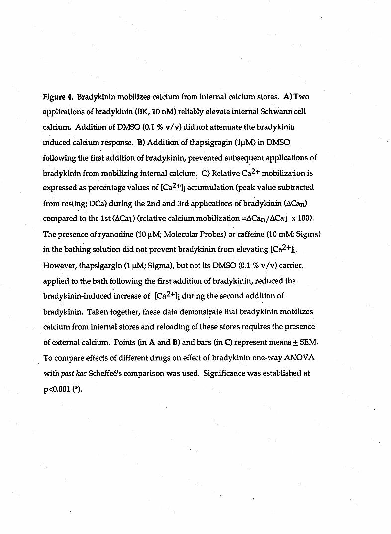
Figure 4. Bradykinin mobilizes calcium from internal calciiun stores. A) Two
applications of bradykinin (BK, 10 nM) reliably elevate internal Schwann cell
calcium. Addition of DMSO (0.1 % v/v) did not attenuate the bradykinin
induced calcivim response. B) Addition of thapsigragin (l|iM) in DMSO
following the first addition of bradykinin, prevented subsequent applications of
bradykinin from mobilizing internal calcium. C) Relative Ca2+ mobilization is
expressed as percentage values of [Ca^+Jj accumulation (peak value subtracted
from resting; DCa) during the 2nd and 3rd applications of bradykinin (ACan)
compared to the 1st (ACai) (relative calcium mobilization =ACan/ACai x 100).
The presence of ryanodine (10 nM; Molecular Probes) or caffeine (10 mM; Sigma)
in the bathing solution did not prevent bradykinin from elevating [Ca2+]i.
However, thapsigargin (1 nM; Sigma), but not its DMSO (0.1 % v/v) carrier,
applied to the bath following the first addition of bradykinin, reduced the
bradykinin-induced increase of [Ca2+]i during the second addition of
bradykinin. Taken together, these data demonstrate that bradykinin mobilizes
calcium from internal stores and reloading of these stores requires the presence
of external calcium. Points (in A and B) and bars (in C) represent means ± SEM.
To compare effects of different drugs on effect of bradykinin one-way ANOVA
with post hoc Scheffe^'s comparison was used. Significance was established at
p<0.001(»).

61
B
C
IstBK 2ndBK + DMSO
40 Time (sec)
40 Time (sec)
S « 20
IstBK 2ndBK + Thapsigargin
• 2ndBK
3rdBK
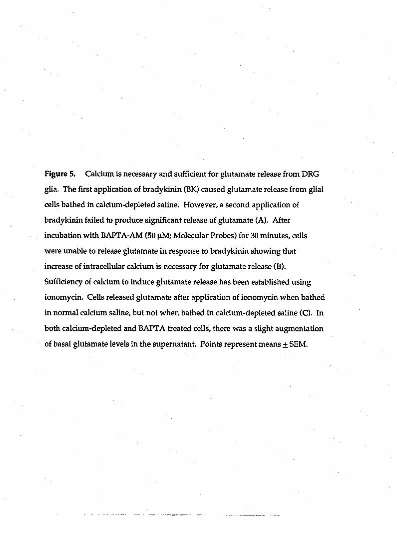
Figure 5. Calcium is necessary and sufficient for glutamate release from DRG
glia. The first application of bradykinin (BK) caused glutamate release from glial
cells bathed in calcium-depleted saline. However, a second application of
bradykinin failed to produce significant release of glutamate (A). After
incubation with BAPTA-AM (50 |iM; Molecular Probes) for 30 minutes, cells
were unable to release glutamate in response to bradykinin showing that
increase of intracellular calcium is necessary for glutamate release (B).
Sufficiency of caldum to induce glutamate release has been established using
ionomycin. Cells released glutamate after application of ionomycin when bathed
in normal calcimn saline, but not when bathed in calcium-depleted saline (C). In
both calcium-depleted and BAPTA treated cells, there was a slight augmentation
of basal glutamate levels in the supernatant. Points represent means ± SEM.
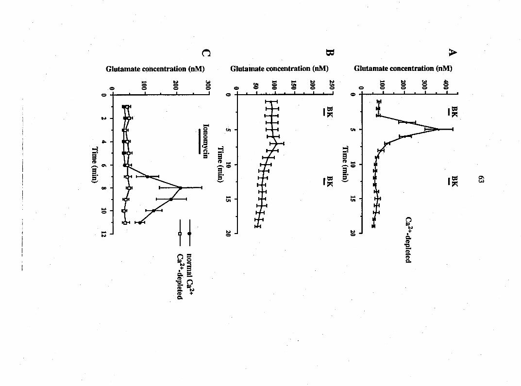
Glutamate concentration (nM)
o s o "J
+ 1 3 D. o
n n fT K> n + o.
Glutamate concentration (nM) Glutamate concentration (nM)
64
(n=6; Student t-test, p>0.5; Fig. 5A). To further test the calcium hypothesis for
glutamate release, we incubated glial cultures for 30 minutes in 50 |iM BAPTA-
AM in caldum-containing saline. This membrane permeant calcium chelator
blocked the stimulatory action of bradykinin on glutamate lelease (n=6; Student
t-test, p>0.3; Fig. 5B).
As a critical test of the calcium hypothesis we exposed cultures to the
caldum-ionophore, ionomycin, in calcium-containing saline. Addition of
ionomydn (5 |iM) stimulated the release of both glutamate (n=6; Student t-test,
p<0.025. Fig. 5C) and aspartate (data not shown). Ionomycin was without
stimulatory effects when glia were bathed in calcium-depleted saline (n=6;
Student t-test, p>0.9; Fig. 5C). Taken together, these data demonstrate that
caldum is mobilized in Schwann cells coincident with the timing of EAA release.
This caldum mobilization is both necessary and sufficient for stimulating the
release of EAAs from Schwann cells.
p-chloromercuriphenylsulphonic add does not inhibit EAA release from
Schwann cells
Glutamate can be released from cells during depolarization through the
reversal of an electrogenic glutamate uptake carrier (Szwatkowski et al., 1990).
To determine whether this mechanism might regulate glutamate release from
DRG Schwann cells, we depolarized them using elevated potassium saline.
Application of elevated K+ (50 mM) did not cause a change in Schwann cells
calcium levels (n=27; Student t-test, p>0.1) nor stimulated EAA release from DRG
glia. In control conditions we detected 48 + 7 nM glutamate in the superfusate.
Following addition of 50 mM K+ saline we detected 46 + 6 nM glutamate (n=6.
65
Student t-test, p>0.7). The lack of glutamate release from DRG glia during the
application of elevated K+ saline is not consistent with a role for the reversal of a
glutamate uptake carrier in mediating the bradykinin-induced release of EAAs
from DRG Schwann cells. Futhermore, addition of the glutamate transporter
inhibitor p-chloromercuriphenylsulphonic acid (p-CMPS; 50 mM) did not reduce
the bradykinin stimulation of glutamate and aspartate release. However
consistent with previous observations using glutamate transporter inhibitors
(Balcar and Johnston, 1971; Isaacson and Nicoll, 1993), p-CMPS did elevate the
basal level of glutamate in the bathing medium.
EAA release from Schwann cells is not mediated by a cell swelling-dependent
mechanism.
Hyposmotic media cause a swelling-dependent release of aspartate and
taurine from cortical astrocytes (Kimelberg et al, 1990). This release is sensitive
to anion transport inhibitors. We determined whether Schwann cells have a
similar sensitivity by incubating them in furosemide (5 mM). Bradykinin-
induced release of glutamate was reduced by 98% (n = 5; p <0.0001, Student t-
test) in the presence of furosemide (Fig. 6A). To ask whether furosemide inhibits
glutamate release by acting at the level of the calcium signal we determined the
effect of furosemide on bradykirun-induced calcium mobilization. Using
repeated applications of bradykinin (see Fig. 4 for protocol) we found that
addition of furosemide (5mM) significantly attenuated (n = 43; p <0.0001,
Student t-test) the ability of the second application of bradykinin to mobilize
calcium (Fig. 63).
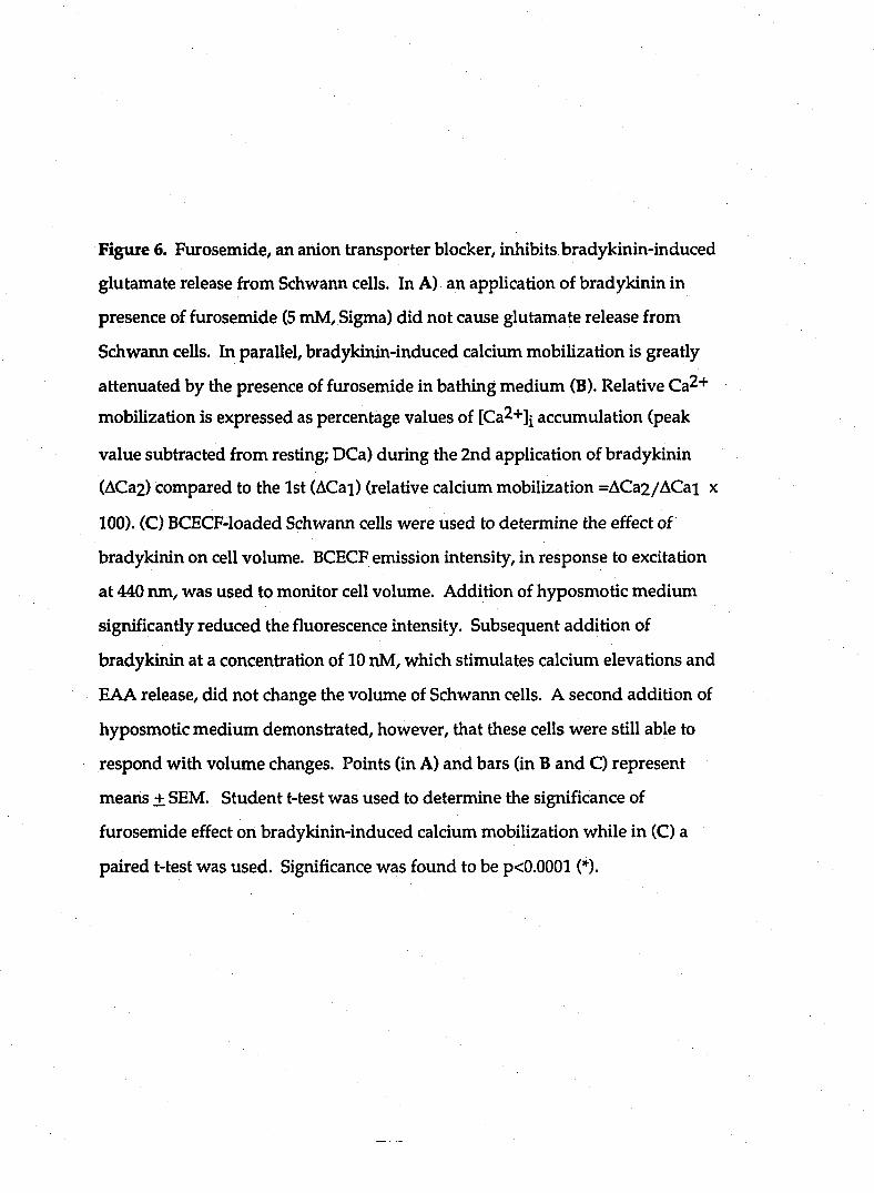
Figtue 6. Furosemide, an anion transporter blocker, inhibits bradykinin-induced
glutamate release from Schwann cells. In A) an application of bradykinin in
presence of furosemide (5 mM, Sigma) did not cause glutamate release from
Schwann cells. In parallel, bradykinin-induced calcium mobilization is greatly
attenuated by the presence of furosemide in bathing medium (B). Relative Ca2+
mobilization is expressed as percentage values of [Ca2+]| accumulation (peak
value subtracted from resting; DCa) during the 2nd application of bradykinin
(ACa2) compared to the 1st (ACai) (relative calcium mobilization =ACa2/ACai x
100). (C) BCECF-loaded Schwann cells were used to determine the effect of
bradykinin on cell volume. BCECF emission intensity, in response to excitation
at 440 run, was used to monitor cell volume. Addition of hyposmotic medium
significantly reduced the fluorescence intensity. Subsequent addition of
bradykinin at a concentration of 10 nM, which stimulates calcium elevations and
EAA release, did not change the volume of Schwann cells. A second addition of
hyposmotic medium demonstrated, however, that these cells were still able to
respond with volume changes. Points (in A) and bars (in B and C) represent
means + SEM. Student t-test was used to determine the significance of
furosemide effect on bradykinin-induced calcium mobilization while in (C) a
paired t-test was used. Significance was found to be p<0.0001 (*).
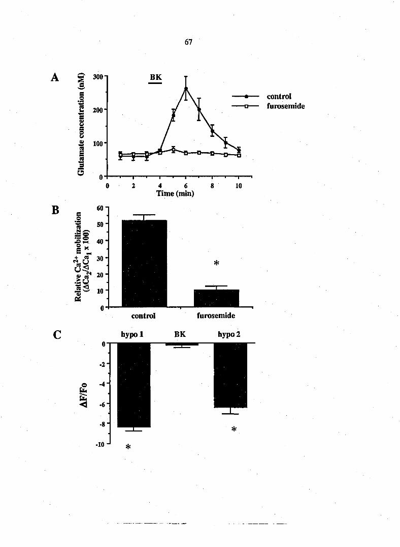
67
3001
B
s s o
2 200-
§ B 8 3 100
E 5 9
5
control furosemide
4 6 Time (min)
10
control furosemide
hypol BK hypo 2

68
Since furosemide reduces bradykinin-induced calcium mobilization, it is not
clear whether the release mechanism per se is sensitive to this anion transport
irUiibitor as has been demonstrated in astrocytes in response to hyposmotic
media (Kimelberg et al, 1990). Therefore, to further test whether bradykinin
stimulates glutamate release from Schwann cells through a cell swelling
mechaiusm we monitored cell volume during bradykinin application. Schwann
cells were loaded with BCECF and the fluorescence emission was monitored in
response to excitation at the pH-insensitive wavelength of 440 nm. Sequential
additions of hyposmotic media repeatedly and reversibly reduced the intensity
of BCECF fluorescence demonstrating that this method can detect swelling of
Schwarm cells (Fig. 6C; n = 36, p < 0.0001; paired t-test). However, application of
bradykinin (10 nM, 1 minute), a stimulus which mobilizes calcium and evokes
EAA release, did not change Schwann cell volume (Fig. 6C; n = 36, p > 0.25;
paired t-test). Since bradykinin does not regulate cell volume, it does not liberate
EAAs from Schwarm cells through a previously described cell swelling-
dependent release mechanism.
Discussion
Our data demonstrate that the release from Schwann cells of the excitatory amino
adds, glutamate and aspartate, is regulated by the neuroligand bradykinin.
Bradykinin causes the mobilization of calcium from internal stores in Schwann
cells within seconds of addition to cultures. This calcium elevation is sustained
for several minutes. In parallel cultures, bradykinin releases glutamate and
aspartate within one minute of delivery of bradykinin to the superfusion
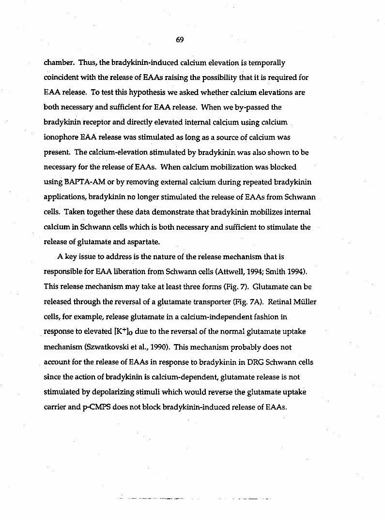
69
chamber. Thus, the bradykinin-induced caldiuii elevation is temporally
coincident with the release of EAAs raising the possibility that it is required for
BAA release. To test this hypothesis we asked whether calcium elevations are
both necessary and sufficient for EAA release. When we by-passed the
bradykinin receptor and directly elevated internal calcium using calcium
ionophore EAA release was stimulated as long as a source of calcium was
present. The calcium-elevation stimulated by bradykinin was also shown to be
necessary for the release of EAAs. When calcium mobilization was blocked
using BAPTA-AM or by removing external calcium during repeated bradykii\in
applications/ bradykirun no longer stimulated the release of EAAs from Schwann
cells. Taken together these data demonstrate that bradykinin mobilizes internal
calcium in Schwann cells which is both necessary and sufficient to stimulate the
release of glutamate and aspartate.
A key issue to address is the nature of the release mechanism that is
responsible for EAA liberation from Schwann cells (Attwell, 1994; Smith 1994).
This release mechanism may take at least three forms (Fig. 7). Glutamate can be
released through the reversal of a glutamate transporter (Fig. 7A). Retinal Miiller
cells, for example, release glutamate in a calcium-independent fashion in
response to elevated [K+]o due to the reversal of the normal glutamate uptake
mechanism (Szwatkovski et al., 1990). This mechanism probably does not
account for the release of EAAs in response to bradykinin in DRG Schwann cells
since the action of bradykinin is calcium-dependent, glutamate release is not
stimulated by depolarizing stimuli which would reverse the glutamate uptake
carrier and p-CMPS does not block bradykinin-induced release of EAAs.

Figure 7. Summary of EAA release mechanisms. Previous studies have demonstrated that EAAs can be released
from cells due to (A) the reversal of an electrogenic transporter during depolarization (Szatkowski et al., 1990).
Alternatively hyposmotic medium causes cell swelling which stimulates EAA release that is sensitive to the anion
transport inhibitor, furosemide (B) (Kimelberg et al., 1990). Since Schwann cell depolarization does not induce
EAA release, the glutamate transport inhibitor p-CMPS does not inhibit bradykinin stimulated EAA release and
since bradykinin does not induce swelling of Schwann cells, neither of these release mechanisms is likely to be
responsible for neuroligand-induced EAA release. The properties of the bradykinin induced EAA release
mechanism of Schwann cells are summarized in (C). Bradykinin acts on a B2 receptor which leads to a furosemide-
sensitive elevation of calcium. Calcium ions, liberated from internal stores, are both necessary and sufficient to
stimulate EAA release from Schwann cells. Further studies are required to determine whether this caldum-
dependent release mechaiusm is similar to the neuronal vesicular release mechanism of synaptic trarismission.
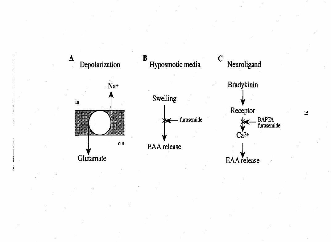
A Depolarization
Na+
A in
out
Glutamate
B Hyposmotic media
Swelling
iurosemide
Y EAA release
Neuroligand
Bradykinin
I Receptor
BAPTA fiirosemide
Ca2+
EAA release
72
In cortical astrocytes hyposmotic medium causes cell swelling and the
consequent release of glutamate, taurine and aspartate (Kimelberg et al., 1990;
O'Connor and Kimelberg, 1993; Fig. 7B). An anion transport blocker,
furosenude, reduces cell swelling dependent release from cortical astrocytes
(Kimelberg et al., 1990). This raises the hypothesis that bradykinin causes cell
swelling which in turn stimulates EAA release from Schwann cells. We
therefore tested the action of furosemide on EAA release. In Schwann cells
furosemide reduced bradykinin-stimulated calcium mobilization and EAA
release. Since furosemide significantly modified bradykinin-induced calcium
signaling this experiment did not allow us to determine whether the cell swelling
dependent release mechanism is utilized. Therefore, we directly measured cell
volume and asked whether bradykinin induces cell swelling. Using BCECF our
data clearly demonstrated that while hyposmotic media induced swelling of
Schwann cells, bradykinin did not. These data are not consistent with
bradykinin inducing the release of EAAs through the previously described
volume regulated mechanism (Kimelberg et al., 1990; O'Connor and Kimelberg,
1993). Furthermore, these previous studies have shown that cell swelling
induced EAA release is calcium-independent, while calcium is both necessary
and sufficient for bradykinin-induced EAA release from Schwann cells.
A third potential mechanism of bradykinin-stimulated EAA release is
through a membrane/vesicle release mechanism similar to that of neurons.
Cultures were free of immunoreactivity for the synaptic proteins sjmaptophysin
and synaptotagmin while axons of DRG explants (separate cultures; data not
shown) were immunopositive for these synaptic proteins. Thus, proteins
characteristic of neuronal transmitter release apparatus are absent in Schwann

73
cells. However, these observatioi\s do not rule out quantal membrane release
mechanisms since muscle (Dan and Poo, 1992) and fibroblasts (Adler et al., 1993)
have been demonstrated competent to release neurotransmitters in a quantal
fashion.
Figure 7C summarizes our current understanding of the bradykinin-induced
EAA release mechanism. Bradykinin, acting through a receptor, mobilizes
internal calcium which is necessary and sufficient to stimulate EAA release from
Schwaim cells independent of cell volume changes. The nature of this calcium-
dependent release apparatus remains to be defined.
The regulated release of neurotransmitters from glia may be a widespread
property. Stimulation of motoneurons leads to the elevation of calcium in the
Schwann cells which surround the presynaptic terminal 0ahromi et al., 1992).
After denervation, Schwann cells have been shown capable of releasing
acetylcholine (Dennis and Miledi, 1974). Depolarization of hippocampal
astrocytes causes the calcium-dependent release of the amino acid taurine, but
not glutamate (Philibert et al., 1988). Recently, we demonstrated that glial cells
from the cerebral cortex release aspartate and glutamate in response to
bradykinin (Parpura et al., 1994). Since many neurotransmitters utilized by the
nervous system can mobilize calcium in glia (Reisner et al., 1989; Cornell-Bell et
al., 1990; Glaum et al., 1990; Charles et al., 1991; Cornell-Bell and Finkbeiner,
1991; Inagaki et al., 1991; Jensen and Chiu, 1991; McCarthy and Salm, 1991), it is
possible that neurons have in addition to their fast neuron-neuron synaptic
actions, a calcium mobilizing action on neighboring glia (Dani et al., 1992) which
in turn leads to the release of other neurotransmitters from glia that may serve
secondary roles in glia-neuronal transmission. In support of this possibility

74
recent studies have demonstrated that glia can signal to neurons (Parpura, 1993;
Nedergaard, 1994; Parpura et al., 1994).
The specific roles for release of excitatory amino acids from glial cells
remains to be elucidated. In the peripheral nervous system, bradykinin is
released from its precursors, the kirunogens, by the action of the enzyme
kallikrein in response to trauma (Dray and Perkins, 1993). Thus, during trauma
and tissue damage, bradykinin is produced where it can initiate the transmission
of nociceptive information. Perhaps the bradykinin induced-release of glutamate
and aspartate from Schwarm cells is involved in mediating certain aspects of
immediate responses to injury. In the central nervous system, elevations of
external glutamate and aspartate are likely to modulate neuronal properties
including excitability and synaptic transmission. Given the important role of
glutamate in the induction of long-term potentiation, it will be important to
determine whether glia regulate synaptic plasticity, learning and memory by
releasing the neurotransmitter glutamate and whether excessive glutamate
release from glia also contributes to neurodegenerative disorders.
References
Adler J, Popov S, Girod R, Poo M-m (1993) Calcium-dependent quantal
secretion of neurotransmitter from fibroblasts. Soc. Neurosd. Abstr. 19: 611.
Attwell D (1994) Glia and neurons in dialogue. Nature 369:707-708.
Barres B (1991) New roles for glia. J. Neurosd. 11: 3685-3694.
75
Balcar VJ, Johnston GAR (1972) The structural specificity of the high affinity
uptake of L-glutamate and L-aspartate by rat brain slices. J. Neurochem. 19:
2657-2666.
Charles AC, Merrill JE, Dirksen ER, Sanderson MJ (1991) Intercellular signaling
in glial cells: calcium waves and oscilations in response to mechanical
stimulation and glutamate. Neuron 6: 983-992.
Cornell-Bell AH, Finkbeiner SM, Cooper MS, Smith SJ (1990) Glutamate induces
calcium waves in cultured astrocytes: long-range glial signaling. Science 247:
470-473.
Cornell-Bell AH, Finkbeiner SM (1991) Ca^^ waves in astrocytes. Cell Calciimi
12:185-204.
Dan Y, Poo M-m (1992) Quantal transmitter secretion from myocytes loaded
with acetylcholine. Nature 359:429-440.
Dani JW, Chemjavsky A, Smith SJ (1992) Neuronal activity triggers calcium
waves in hippocampal astrocyte networks. Neuron 8:429-440.
Dennis MJ, Miledi R (1974) Electrically induced release of acetylcholine from
denervated Schwann cells. J. Physiol. 237:431-452.

76
Dray A, Perkins M (1993) Bradykinin and inflammatory pain. Trends Neurosci.
16:99-104.
Eriksson PS, Nilsson M, WSgberg M, Ronnback L, Hansson E (1992) Volume
regulation of single astroglial cells in primary culture. Neurosci. Lett. 143:195-
199.
Gahwiler BH (1984) Slice cultures of cerebellar, hippocampal and hypothalamic
tissue. Experientia 40:235-243.
Glaim\ SR, Holzwarth JA, Miller RJ (1990) Glutamate receptors activate Ca2+
mobilization and Ca2+ influx into astrocytes. Proc. Natl. Acad. Sci. USA 87:
3454-3458.
Grjmkiewicz G, Poenie M, Tsien RY (1985) A new generations of Ca2+
indicators with greatly improved fluorescence properties. J. Biol. Chem. 260:
3440-3450.
Higashida H, Ogura A (1991) Inositol triphosphate/calcium-dependent
acetylcholine release by bradykinin in NG108-15 rodent hybrid cells. Ann. N. Y.
Acad. Sd. 635:153-166.
Hsu S-M, Raine L, Fanger H (1981) Use of avidin-biotin-peroxidase complex
(ABC) in immunoperoxidase techniques: a comparison between ABC and
unlabelled antibody (PAP) procedures. J. Histochem. Cytochem. 29: 577-580.
77
Inagaki N, Fukui H, Ito S, Wada H (1991) Type-2 astrocytes show intracellular
Ca2+ elevation in response to various neuroactive substances. Neurosci. Lett.
128:257-260.
Isaacson JS, Nicoll RA (1993) The uptake inhibitor L-trans-FDC enhances
responses to glutamate but fails to alter the kinetics of excitatory synaptic
currents in hippocampus. J. Neurophysiol. 70:2187-2191.
Jahromi BS, Robitaille R, Charlton MP (1992) Transmitter release increases
intracellular calcium in perisynaptic Schwann cells in situ. Neuron 8:1069-1077.
Jensen AM, Chiu SY (1991) Differntial intracellular calcium responses to
glutamate in type 1 and type 2 cultured brain astrocytes. J. Neurosci 11:1674-
1684.
Kanai Y, Smith C P, Hediger MA (1993) The elusive transporters with a high
affinity for glutamate. Trends Neurosci. 16:365-370.
Kimelberg HK, Goderie SK, Higman S, Pang S, Waniewski RA (1990) Swelling-
induced release of glutamate, aspartate and taurine from astrocyte culture. J.
Neurosci. 10:1583-1591.

78
Kleitman N, Wood PM, Bunge RP (1992) Tissue culture methods for the study
of myelination. In: Culturing nerve cells (Banker G, Goslin K, eds.), pp 337-377.
Cambridge: MIT press.
Lindroth P, Mopper K (1979) High performance liquid chromatographic
determination of subpicomole amounts of amino acids by precolumn
fluorescence derivatization with o-phthalaldehyde. Anal. Chem. 51:1667-1674.
McCarthy KD, Salm AK (1991) Pharmacologically-distinct subsets of astroglia
can be identified by their calciiun response to neuroligands. Neuroscience 41:
325-333.
Miller RJ (1987) Bradykinin highlights the role of phospholipid methabolism in
the control of nerve excitability. Trends Neurosci. 10:226-228.
Nedergaard M (1994) Direct signaling from astrocytes to neurons in cultures of
mammalian brain cells. Science 263:1768-1771.
Nicholls D. Attwell D (1990) The release and uptake of excitatory amino acids.
Trends Pharm. Sci. 11:462-468.
O'Connor ER, KimelbergHK (1993) Role of calcium in astrocyte volume
regulation and in release of ions and amino acids. J. Neurosci. 13: 2638-2650.
Parpura V (1993) Glia-neuron signaling. Thesis. Iowa State University, Ames.
79
Parpura V, Basarsky TA, Liu F, Jeftinija K, Jeftinija S, Haydon PG (1994)
Glutamate-mediated astrocyte-neuron signaling. Nature 369: 744-747
Philibert RA, Rogers KL, Allen AJ, Dutton GR (1988) Dose-dependent, K+-
stimulated efflux of endogenous taurine form primary astrocyte cultures is Ca2+-
dependent. J.Neurochem. 51; 122-126.
Reiser G, Donie F, Binmoller F-J (1989) Serotonin regulates cytosolic Ca2+
activity and membrane potential in a neuronal and in a glial cell line via 5-HT3
and 5-HT2 receptors by different mechanisms. J.Cell Sci. 93: 545-555.
Ritchie T, Cole R, Him H-S, deVellis J, Noble EP (1987) Inositol phospholipid
hydrolysis in cultured astrocytes and oligodendrocytes. Life Sci. 41: 31-39.
Smith SJ (1994) Neuromodulatory astrocytes. Curr. Biol. 4:807-810.
Szatkowski M, Barbour B, Attwell D (1990) Non-vesicular release of glutamate
from glial cells by reversed eletrogenic glutamate uptake. Nature 348:443-446.
Taniuchi M, Clark BH, Schweitzer JB, Johnson EMJr (1988) Expression of nerve
growth factor receptors by Schwan cells of axotomized peripheral nerves:
ultrastructural location, suppression by axonal contact, and binding properties.
J. Neurosci. 8: 664-681.

80
Thomas AP, Delaville F (1991) The use of fluorescent indicators for
measurements of cytosolic-free calcium concentration in cell populations and
single cells. In: Cellular calcium (McCormackJG, CobboldPH, eds.), ppl-54.
Oxford: Oxford University Press.
81
CHAPTER 3. ATP-EVOKED Ca2+-DEPENDENT RELEASE OF EXCITATORY
AMINO ACIDS FROM CULTURED SCHWANN CELLS
A paper to be submitted to Brain Research
Fang Liu, Ksenija Jeftinija, Srdija Jeftinija
Abstract
The specific objective of this study was to determine the mechanism by which
ATP evokes release of EAAs from cultured Schwann cells. Basal concentrations of
aspartate and glutamate in Schwann cell cultures were 7.6410.74 nM (mean±SEM)
and 30.52±3.89 nM, respectively. Perfusion application of 100|iM ATP for 2
minutes to cultured Schwann glia resulted in an 201±28 % increase of aspartate
and 203±15 % increase of glutamate. A second application of ATP 10 minutes after
the first application similarly caused EAA release, although the magnitude of this
response was attenuated. Application of ATP to glia for 10 minutes resulted in a
peak inaease in the release followed by a decline to a plateau significantly higher
than baseline release. Bath application of adenosine (100|xM) was without effect on
release of EAAs suggesting involvement of P2 receptors. The release of EAAs
evoked by ATP was not abolished in low Ca^+-EGTA saline. Pretreatment of the
glial cultures with 50|aM BAPTA-AM abolished the effect of ATP. Thapsigargin
(l|iM), an inhibitor of Ca2+-ATPase or the Ca2+ pump of internal stores,
significantly reduced the release of EAAs evoked by ATP. Our results show that

82
ATP evokes the release of EAAs from cultured Schwann cells by activating
intracellular calcium stores.
Introduction
Although the fimctional importance of intracellular adenosine 5'-
triphosphate (ATP) levels in cellular metabolism has been recognized for many
years, the observation that extracellular ATP can influence neurotransmission,
cardiac function, muscle contraction and other biological processes is more recent
(reviewed by Gordon, 1986; Bumstock, 1990). ATP acting as a neurotransmitter
or neuromodulator performs important functions in the central and peripheral
nervous system.
It has been shown that ATP can be released or co-released with acetylcholine
or norepinephrine from nerve endings (Bumstock, 1976; 1986a; 1986b; Fyffe and
Perl, 1984). Extracellular ATP excites a suppopulation of rat dorsal horn neurons
(Jahr and Jessel, 1983) and rat sensory neurons (Krishtal et al., 1988). It was also
reported ATP depolarized cultured hippocampal neurons and induced release of
glutamate from the neurons (Inoue et al., 1992).
The actions of ATP are mediated by P2-purinoceptors which are present on
many cell types including neurons and astrocytes (Phillis and Wu, 1981; Salter
and Henry, 1985; Gebicke-Haerter et al, 1988; Pearce et al., 1989). There is
evidence that activation of P2-purinoceptors leads to an increase in intracellular
calcium in a variety of excitable and unexcitable cells (O'Connor et al., 1991; El-
Moatassim et al., 1992). Two mechanisms are involved in ATP-evoked
intracellular calcium increase. First, ATP can activate nonselective cation

83
channels, resulting in depolarization and subsequent opening of voltage-
dependent Ca2+ charmels (Nakazawa and Matsuki, 1987; Friel, 1988). The
increased calcium influx is also the result of direct activation of ATP-gated cation
channels (Benham and Tsien 1987; Charest et al., 1987). Second, the extracellular
ATP can stimulate the breakdown of inositol phospholipids and the resulting
increase in inositol 1,4,5,-triphosphate (IP3) is responsible in part for the elevation
of cytosolic Ca2+ (Boeynaerus et aL, 1988; Danziger et al., 1988).
There is an increasing body of evidence showing that neuroglia may be more
actively involved in brain function than has been previously thought. It has been
demonstrated that neuroglia have multiple roles in the nervous system which
include the regulation of extracellular potassium, the uptake of excitatory amino
adds (EAAs) and the release of neurotransmitter such as glutamate (Glu),
aspartate (Asp) and acetylcholine (ACh) (Nicholls and Atwell, 1990; Barres, 1991;
Kanai et al., 1993; Snuth, 1994). Our recent study shows that the release of the
excitatory neurotransmitter glutamate from glial cells plays a key role in glia-
neuron signalling (Parpura et al., 1994a).
Although it has been determined that extracellular ATP can stimulate Ca2+
influx and increase intracellular calcium in cultured astrocytes (Neary et al., 1991;
Salter and Hicks, 1994), little is known about the effects of extracellular ATP on
Schwann cells, which are the predominate glial cells in the peripheral nervous
system. Our previous study shows that the neuroligand bradykinin can induce
the release of EAAs from cultured Schwann cells in a Ca2+-dependent manner
(Parpura et al.; 1994b). This evidence prompted us to propose that extracellular
ATP might influence the functions of Schwann cells. In this study, we first
84
demonstrate that ATP can evoke release of EAAs from cultured Schwann and the
intracellular Ca2+plays a role in the release of EAAs.
Materials and Methods
Cell culture Organotypic DRG cultures will be prepared according to modification
of methods described by Gahwiler (1984), and previously reported (Jeftinija et al.,
1991). Following decapitation, DRG from one to four day old Sprague-Dawley rats
will be rapidly removed and washed in cold (4oC) oxygenated Gey's balanced salt
solution (GBSS, Gibco) modified by the addition of 2.0% glucose. Culturing will be
carried out on glass coverslips which are specially prepared by soaking 24 hours in
xylene, 4 hours in acetone, 24 hours in absolute alcohol, and dried for 30 min at 80°
C. Isolated explants will be placed on a glass coverslip completely covered with
chicken plasma (Sigma) and coagulated by adding thrombin solution (Sigma). The
coverslips bearing the explants will be placed into 35 mm Petri dishes. Cultures will
be incubated in a humid 5% COz atmosphere at 37° C. The culture medium
contained 25% horse serimi, 25% Earls Balanced Salt Solution, 50% Basal Medium
Eagles with added glucose (6.4 mg/ml). When the culture is incubated for 3-4 days,
it has a lot of processes arising from DRG explant and also Schwann cells in
growing zone. In order to get DRG glial cells, we'll remove the central part of
explant which contains neuronal cell bodies from established cultures (4 to 5 days
old culture). The residual glia cells left behind will be maintained for additional 72
hours. Fibroblasts will be removed by incubating the culture with thy 1.1 antibody
(hybridoma supernatant, American Type Culture, No. TIB103) and addition of rabbit
serum complement (Sigma). This procedure is used to lysogenize the fibroblasts and

85
purified Schwann cells. The DRG glial cells and fibroblasts were identified by
inununocytochemistry using antibodies against low affinity NGF receptor and
fibronectin. Immunocytochemistry revealed that at least 95% of the cell population
were Schwann cells.
Release method and HPLC This procedure has been developed in our
laboratory. The coverslips with cell cultures are mounted in a 50 ml perfusion
chamber. The modified Ringer's perfusion solution (in mM: NaCl 127, KCl 1.9,
KH2PO4I.2, CaCl2 2.4, MgS041.3, NaHCCb 26, glucose 10, pH7.4) bubbled vnth
95% oxygen and 5% carbon dioxygen is pumped into the perfusing system by a
n\inipuls pump at a rate of 200 ml/min. After an equilibration period of half an
hour, samples of perfusate are collected every minute for amino acids
determinations. The amino acid content in the samples is determined by high-
performance liquid chromatography (HPLC) with fluorescence detection using a
Shimadzu system. Prior to injection, aliquots of the samples are derivatized with
o-phthalaldehyde (OPA) 2-mercaptoethanol reagent (Pierce). Chromatography is
performed on a 15 cm Rainin column using a pH 5.9 sodium acetate methanol
gradient.
The concentrations of Glu and Asp are expressed in nMols. Basal
concentrations of EAAs were determined as the mean of the EAA content of
three samples collected just prior to stimulation with ATP, adenosine or
bradykirun. All data are presented as the mean values ± S.E.M.. The data were
analyzed by Student's t-test.
86
Results
ATP evokes EAA release from dorsal root ganglion Schwann cells
The basal concentration of glutamate and aspartate was 30.52±3.89 nM
(meardSEM) and 7.64±0.74 nM, respectively. Application of 100|iM ATP for two
min produced a significant increase in the concentrations of Glu and Asp. The
peak concentrations of Glu and Asp were 203±15 % (n=8, p<0.0005) and 201+28
% (n=8, P<0.005) of the basal concentration, respectively. These data show that
extracellular ATP can stimulate the release of EAAs from cultured Schwann cells.
A second application of ATP similarly caused EAA release, although the
magnitude of this response was attenuated. The second peak concentrations of
glutamate and aspartate were 163± 11% (n=8, P<0.0005) and 176±11% (n=8,
P<0.0005) of the control concentration, respectively (Figure la and Figure lb).
Application of ATP to glial cultures for 10 minutes resulted in a biphasic
response, which involved a increased glutamate peak that was 232±10 % (n=4,
P<0.005) of the basal concentration followed by a declining release to a sustained
release plateau significantly higher than the control release. The increased release
returned to the basal concentration when ATP was removed (Figure Ic). This
data illustrate that prolonged exposure of ATP to Schwann cells could induce
long lasting release of EAAs.
Pi^-purinoceptors are not involved in ATP-evoked release of EAAs
To test receptor specificity of ATP-evoked release of EAAs, we used
adenosine to stimulate cultures. Application of adenosine (100 |aM) for two
minutes failed to induce release of EAAs from cultured Schwann cells
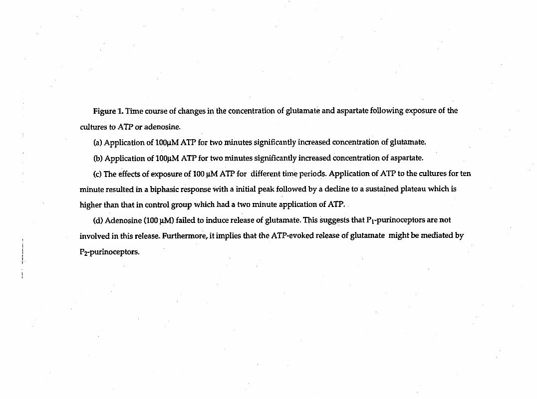
Figure 1. Time covirse of changes in the concentration of glutamate and aspartate following exposure of the
cultures to ATP or adenosine.
(a) Application of 100|iM ATP for two minutes significantly increased concentration of glutamate.
(b) Application of lOO^iM ATP for two minutes significantly inaeased concentration of aspartate.
(c) The effects of exposure of 100 |iM ATP for different time periods. Application of ATP to the cultures for ten
minute resulted in a biphasic response with a initial peak followed by a decline to a sustained plateau which is
higher than that in control group which had a two minute application of ATP.
(d) Adenosine (100 nM) failed to induce release of glutamate. This suggests that Pi-purinoceptors are not
involved in this release. Furthermore, it implies that the ATP-evoked release of glutamate might be mediated by
I P2-purinoceptors. t
I 1

ATP 100(iM
(C)
S c 3
a
60-q
50 H
40-j
30 H
20 H
10-
0
-B- 10 min ATP
2 min ATP
( d )
250
200
S 150 c 3
a 100
I I I I 1 I I I I I I I I I I I I I I
50
0
ATPlOOpM
ATP lOOfiM
adonoafna 100(iM BKIOnM

89
(n=4, P>0.3). Exposure of 10 nM bradykinin to the cultures ten minutes after the
application of adenosine evoked a six fold increase of glutamate release. This
result indicates that these cultures responded to the stimulation of bradykinin
very well, although they were not sensitive to adenosine (Figure Id).
It has been proposed that there are two classes of purinergic receptors
(Burnstock/1978). One is the adenosine-sensitive Pi-purinoceptor, and the other
one is the ATP-sensitive P2-purinoceptor. The latter is insensitive to adenosine.
Because adenosine did not induce the release of EAAs from cultured Schwann
cells, the possibility that the Pi-purinoceptor is involved in this process is
eliminated. Furthermore, it implies that ATP might evoke release of EAAs by
activating P2 -purinoceptors.
ATP-induced release of EAAs is calcium-dependent
ATP has been found to increase intracellular Ca2+ concentration in many
cells (reviewed by Burnstock, 1990). On the basis of this evidence, we have
investigated whether Ca2+ plays a role in ATP-induced release of EAAs.
Cultures were incubated with low Ga2+ (0.2mM), high Mg2+ (2.5 mM), EGTA
(1 mM) saline for 30 minutes. We found that, after removing the extracellular
calcium, ATP still caused a increased release of EAAs similar to that seen in the
control group. The peak concentration of glutamate was 162+25 % of the basal
concentration (n=6, P<0.05). This result demonstrated that extracellular calcium
is not involved in ATP-induced release of EAAs from Schwann cells (Figure 2a).
In order to determine whether Ca2+ from internal stores is involved in
glutamate release, we pretreated cultures with 50 piM BAPTA-AM [l,2bis(2-
aminophenoxy) ethane-N,N,N',N'-tetraceticacid, acetoxymethyl ester] saline for

Figure 2. ATP-evoked release of glutamate is Ca2+-dependent.
(a) Low Ca^+-EGTA saline didn't significantly affect the ATP-evoked release
of glutamate.
(b) BAPTA-AM (50 iM), the membrane permeant calcium chelator, blocked
the ATP-evoked glutamate release.
(c) Thapsigargin (1 |iM), the inhibitor of the Ca2+-ATPase of internal calcium
stores, abolished the stimulatory effect of ATP on release of glutamate.
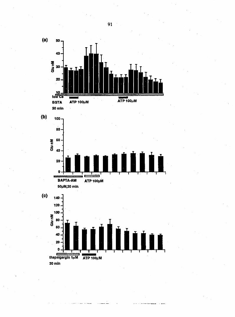
(a) 50-,
(b)
low (;a
EQTA ATP lOO^M 30 min
100-1
ATP100|iM
O 40-
BAPTA-AM ATPIOOMM 50|jM,30 mfn
(C) 140-
120-
100-
thapsigargin IjiM atp lOOjiM
30 min
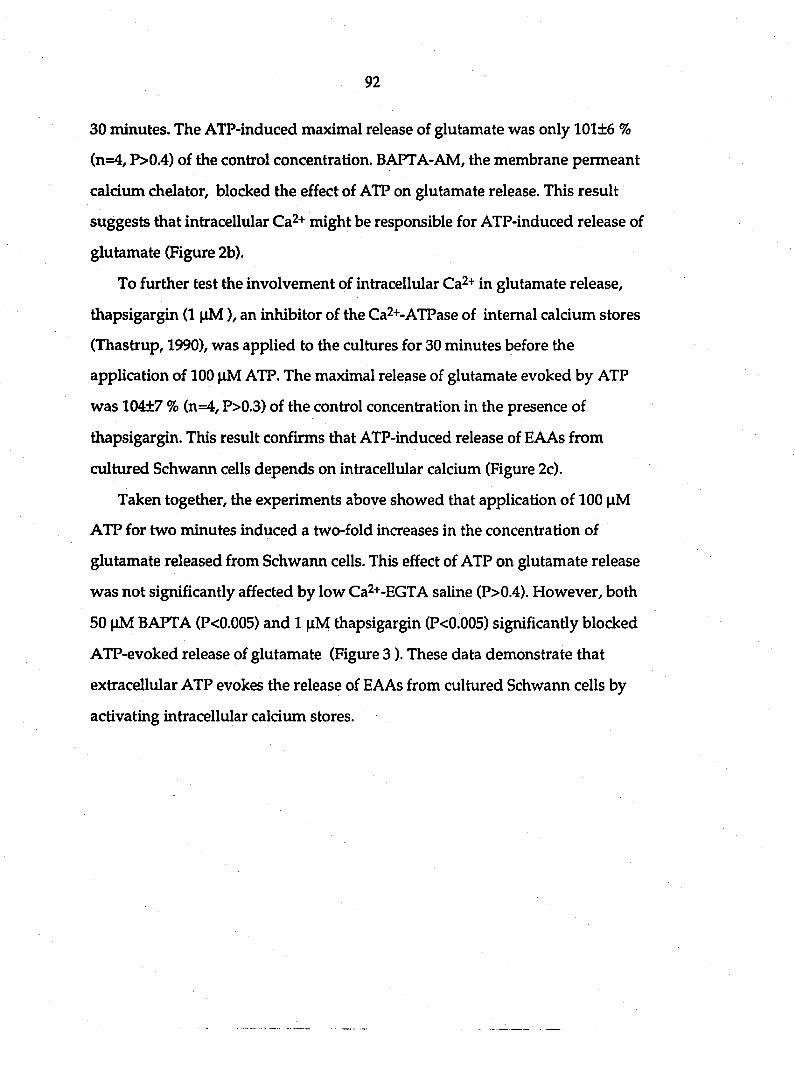
92
30 minutes. The ATP-induced maximal release of glutamate was only 101±6 %
(n=4, P>0.4) of the control concentration. BAPTA-AM, the membrane permeant
calcium chelator, blocked the effect of ATP on glutamate release. This result
suggests that intracellular Ca2+ might be responsible for ATP-induced release of
glutamate (Figure 2b).
To further test the involvement of intracellular Ca2+ in glutamate release,
thapsigargin (1 |iM), an inhibitor of the Ca2+-ATPase of internal calcium stores
(Thastrup, 1990), was applied to the cultures for 30 minutes before the
application of 100 |i,M ATP. The maximal release of glutamate evoked by ATP
was 104±7 % (n=4, P>0.3) of the control concentration in the presence of
thapsigargin. This result confirms that ATP-induced release of EAAs from
cultured Schwann cells depends on intracellular calcium (Figure 2c).
Taken together, the experiments above showed that application of 100 |iM
ATP for two minutes induced a two-fold increases in the concentration of
glutamate released from Schwann cells. This effect of ATP on glutamate release
was not significantly affected by low Ca2+-EGTA saline (P>0.4). However, both
50 |iM BAPTA (P<0.005) and 1 |iM thapsigargin (P<0.005) significantly blocked
ATP-evoked release of glutamate (Figure 3). These data demonstrate that
extracellular ATP evokes the release of EAAs from cultured Schwann cells by
activating intracellular calcium stores.
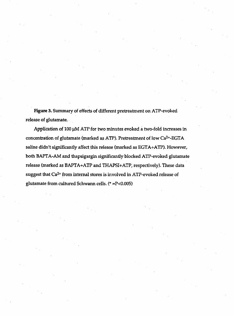
Figiue 3. Summary of effects of different pretreatment on ATP-evoked
release of glutamate.
Application of 100 |4M ATP for two minutes evoked a two-fold increases in
concentration of glutamate (marked as ATP). Pretreatment of low Ca2+-EGTA
saline didn't significantly affect this release (marked as EGTA+ATP). However,
both BAPTA-AM and thapsigargin significantly blocked ATP-evoked glutamate
release (marked as BAPTA+ATP and THAPSI+ATP, respectively). These data
suggest that Ca2+ from internal stores is involved in ATP-evoked release of
glutamate from cultured Schwann cells. (* =P<0.005)

Giu Reiease (% of basai concentration)
-A -1. lO
//.••.'.•//.••.v.'.
!• vO

95
Discussion
In this study, we present data demonstrating that extracellular ATP can
evoke the release of the excitatory amino acids glutamate and aspartate from
cultured Schwarm cells.
It has been shown that neuroglia, including Schwann cells, release
neurotransmitter. Dermis and Miledi (1974) reported that Schwann cells can
release acetylcholine following axotomy of motoneurons. Szwatkouski et al.
(1990) found that depolarization of retinal Miiller cells stimulates the release of
glutamate. Our previous studies show that the nonapeptide bradykinin can
evoke the release of glutamate and aspartate from cultured astrocytes (Parpura et
al., 1994a) and Schwann cells (Parpura et al., 1994b) in a calcium-dependent
maimer. The present study provides additional evidence that Schwann cells can
release glutamate and aspartate in response to stimulation of ATP.
Extracellular ATP can activate specific cell surface receptors, P2-
purinoceptors (Burnstock, 1978). This purinergic receptor is insensitive to
adenosine, which activates Pi-purinoceptors. We demonstrates that ATP
significantly induced release of EAAs from Schwann cells, whereas adenosine
doesn't stimulate the release. Although neither P2-purinoceptors nor Pi-
purinoceptors has ever been identified on the surface of Schwann cells, our data
suggests that purinergic P2-receptors might be involved ATP-induced release of
EAAs.
ATP can cause an increase in intracellular calcium in many cell types. This
process involves calcium entry across the plasma membrane via receptor-
operated chaimels as well as calcium mobilization from internal stores via
96
receptor couplmg to phosphatidylinositol hydrolysis (reviewed by Bumstock,
1990; Neary and Norenberg,1992). We found that ATP-evoked release of EAAs
from cultured Schwann cells might result as a functional consequence of
increases in intracellular calcium. Removing extracellular calcium by using low
Ca2+-EGTA saline did not significantly affect the release of EAAs from Schwann
cells. This result indicated that at least Ca2+ influx is not involved in the release.
In contrast, BAPTA-AM, which can penetrate cell membrane and chelate
intracellvilar Ca2+, abolished the ATP-evoked release of EAAs. This suggests that
calcium from internal stores has to be responsible for this release. Thapsigargin,
which depletes internal calcium stores, also blocked ATP-evoked release of
EAAs. This result provided more evidence that ATP-induced release of EAAs
from cultured Schwann cells is dependent upon intracellular calcium.
It has been shown that extracellular ATP confines its effects to glial cells. ATP
activating its receptors evoked increases in intracellular calcium in astrocytes
(Salter and Hicks, 1994). Furthermore, ATP not only evoked changes in astrocytic
calcium signal, but also in protein phosphorylation and morphology (Neary and
Norenberg, 1992). Our present study that ATP can induce release of EAAs from
cultured Schwann cells first demonstrated that extracellular ATP has effects on
Schwann cells, the glial cells in the peripheral nervous system. There is evidence
that axon-to-Schwann cell signalling was mediated by neurotransmitter
glutamate via quisqualate/kainate receptors in the squids (Liebemman et al.,
1989). Our data provides new evidence that extracellular ATP might also serve as
a mediator in neuron-glia signalling.
The fimctions of ATP-evoked release of EAAs from Schwann cells are not
clear. Our recent study demonstrated that release of glutamate from astrocytes
97
mediates the process of astrocyte-neuron signalling (Parpura et al., 1994a). This
prompts us to propose that the release of EAAs from Schwann cells might play a
role in glia-neuron signalling in peripheral nervous system. Since ATP can be
released under physiological and pathological conditions (Neary and Norenberg,
1992), ATP-evoked release of EAAs from Schwann cells might be involved in
certain functions of Schwann cells in both situations.
References
Barres B. (1991) New roles for glia. J. Neurosci. 11:3685-3694.
Benham C.D. (1990) ATP-gated channels in vascular smooth muscle cells. Ann.
N. Y. Acad. Sci. 603: 275-285.
Benham C.D., Tsien R.W. (1987) A novel receptor-operated Ca2+-permeable
channel activated by ATP in smooth muscle. Nature 328:275-278.
Boeynaems J.M., Pirotton S., Van-Coevorden A., Raspe E., Demolle D., Erneux C.
(1988) P2-purinergic receptors in vascular endothelial cells: from concept to
reality. J. Recept. Res. 8:121-132.
Bumstock G. (1976) Purine nucleotides. Adv. Biochem. Psychopharmacol. 15:
225-235.
98
Bumstock G. (1978) A basis for distinguishing two types of purinergic receptors.
In: L. Bolis and R.W. Straub (Eds.), Cell and Membrane Receptors for Drugs and
Hormones: a Multidisciplinary Approach, Raven Press, New York, pp. 107-118.
Bumstock G. (1990) Overview: purinergic mechanisms. Ann. N.Y. Acad. Sci. 603:
1-18.
Bumstock G. (1986a) In Coexistence of Neuronal Messengers: A New Principle in
Chemical Transmission. Progress in Brain Research. Vol. 68:193-203. Elsevier.
Amsterdam.
Bumatock G. (1986b) The changing face of autonomic neurotransmission. Acta.
Physiol. Scand. 126: 67-91.
Charest R., Prpic V., Exton J.H., Blackmore P.P. (1985) Stimulation of inositol
trisphosphate formation in hepatocytes by vasopressin, adrenaline and
angiotensin II and its relationship to changes in cytosolic free Ca2+. Biochem J.
227 (1): 79-90.
Danziger R.S., Raffaeli S., Moreno-Sanchez R., Sakai M., Capogrossi M.C.,
Spurgeon H.A., Hansford R., Lakatta E.G. (1988) Extracellular ATP has a potent
effect to enhance cytosolic calcium and contractility in single ventricular
myocytes. Cell Calcium 9 (4): 193-199.

99
Dennis MJ., Miledi R. (1974) Electrically induced release of acetylcholine from
denervated Schwann cell. J. Physiol. 237:431-452.
Dubyak G.R. (1986) Extracellular ATP activates polyphosphoinositide
breakdown and Ca2+ mobilization in enrich ascites tumor cells. Arch. Biochem
Biophys. 245 (1): 84-95.
El-Moatassim C., Dornand J., Mani J.C. (1992) Extracellular ATP and cell
signalling. Biochim. Biophys. Acta. Mol. Cell Res 1134:31-45.
Friel D.D. (1988) An ATP-sensititive conductance in single smooth muscle cells
from the rat vas deferens. J. Physiol. Lond. 401:361-380.
Fyffe R.E.W., Perl E.R. (1984) Is ATP a central synaptic mediator for certain
primary afferent fibers from mammalian skin? Proc. Natl. Acad. Sci. USA 81:
6890-6893.
Gahwiler B.H. (1984) Slice cultures of cerebellar, hippocampal and hypothalamic
tissue. Experientia 40: 235-243.
Gebicke-Hearter P.J., Wurster S., Schobert A., Hertting G. (1988) P2-purinoceptor
induced prostaglandin synthesis in primary rat astrocyte cultures. Naunyn.
Schmiedebergs. Arch. Pharmacol. 338: 704-707.

100
Gordon J.L. (1986) Extracellular ATP: effects, sources and fate. Biochem. J., 233:
309-319.
Inoue K., Nakazawa K., Fujimori K., Watano T., Takanaka A. (1992) Extracellular
adenosine 5' -triphosphate-evoked glutamate release in cultured hippocampal
neurons. Neurosci. Lett. 134:215-218.
Jahr C.E., Jessel T.M. (1983) ATP excites a subpopulation of rat dorsal horn
neurons. Nature 304:730-733.
Jeftinija S., Jeftinija K., Liu F., Skilling S.R., Smullim D.H., Larson A,A. (1991)
Excitatory amino acid are released from rat primary afferent neurons in vitro.
Neurosci. Lett. 125:191-194.
Kanai Y., Smith C.P., Hediger M.A. (1993) The elusive transporters with a high
affinity for glutamate. Trends Neurosci. 16:365-370.
Krishtal O.A., Marchenko S.M., Obukhov A.G. (1988) Cationic channels activated
by extracellular ATP in rat sensory neurons. Neuroscience 27:995-1000.
Lieberman E.M., Abbott N.J., Hassan S. (1989) Evidence that glutamate mediates
axon-to-Schwann cell signalling in squid. Glia 2:94-102.
101
Nakazawa K., Matsuki N. (1987) Adenosine triphosphate-activated inward
current in isolated smooth muscle cells from rat vas deferens. pflugers-Arch. 409:
644-646.
Neary J.T., Norenberg M.D. (1992) Signaling by extracellular ATP: physiological
and pathological considerations in neuronal-astrocytic interactions. Pro. Brain
Res. 94:145-151.
Nicholls D., Attwell D. (1990) The release and uptake of excitatory amino acids.
Trends Pharm. Sci. 11:462-468.
O'Connor S.E., Dainty I.A., Leff P. (1991) Further subclassification of ATP
receptors based on agonist studies. Trends Pharmacol. Sci. 12:137-141.
Parpura V., Basarsky T.A., Liu F., Jeftinija K., Jeftinija S., Haydon P.G. (1994a)
Glutamate-mediated astrocyte-neuron signalling. Nature 369: 744-747.
Parpuar V., Liu F., Jeftinija K., Jeftinija S., Haydon P.G. (1994b) Neuroligand-
evoked caldum-dependent release of excitatory amino acids from Schwann cells.
J. Neurosci. in press.
Pearce B., Murphy S., Jeremy J., Morrow C., Dandona P. (1989) ATP-evoked Ca2+
mobilization and prostanoid release from astrocytes: P2-purinergic receptors
lii\ked to phosphoinositide hydrolysis. 52:971-977.

102
Phillis J.W., Wu P.H. (1981) The role of adenosine and its nucleotides in central
synaptic transmission. Prog. Neurobiol. 16:187-239.
Salter M.W., Henry J.L. (1985) Effects of adenosine 5'-monophosphate and
adenosine 5'-triphosphate on functionally identified units in cat spinal dorsal
horn. Evidence for a differential effect of adenosine 5' -triphosphate on
nociceptive vs non-nociceptive units. Neuroscience 15:815-825.
Salter M.W., Hicks J.L. (1994) ATP-evoked increases in intracellular calcium in
neurons and glia from the dorsal spinal cord. J. Neurosci. 14:1563-1575.
Smith S.J. (1994) Neuromodulatory astrocytes. Curr. Biol. 4: 807-810.
Szatkowski M., Barbour B., Attwell D. (1990) Non-vesicular release of glutamate
from glial cells by reversed electrogenic glutamate uptake. Nature 348:443-446.
Thastrup O., Cullen P.J., Drobak B.K., Hanley M.R., Dawson A.P. (1990)
Thapsigargin, a tumor promoter, discharges intracellular Ca2+ stores by specific
inhibition of the endoplasmic reticulum Ca2+-ATPase. Proc. Natl. Acad. Sci. USA
87:2466-2470.

103
CHAPTER 4. GLUTAMATE-MEDIATED ASTROCYTE-NEURON
SIGNALING
A paper published in Nature
Vladimir Parpura, Trent A. Basarsky, Fang Liu, Ksenija Jeftinija,
Srdija Jeftinija and Philip G. Hay don
Neurotransmitter released from neurons is known to signal to neighboring
neurons and glia ^'3. Here we demonstrate an additional signaling pathway in
which glutamate is released from astrocytes and causes an NMDA (N-methyl-D-
aspartate) receptor-mediated increase in netuonal calcium . Internal calciiun was
elevated and glutamate release stimidated by application of the neuroligand
biadykinin to cultured astrocytes. Elevation of astrocyte internal calcium was
also sufficient to induce glutamate release. To determine whether this released
glutamate signals to neurons, we studied astrocyte-neuron co-cultures.
Bradykinin significantly increased calcium levels in neurons co-cultured with
astrocytes, but not in solitary neurons. The glutamate receptor antagonists D-2-
amino-5-phosphonopentanoic acid and D-glutamylglycine prevented
bradykinin-induced neuronal calcium elevations. When single astrocytes were
directly stimulated to increase internal calcium and release glutamate, calcitmi
levels of adjacent neurons were increased; this increase could be blocked by D-
glutamylglycine. Thus, astrocytes regulate neuronal calciiun levels through the
calcium-dependent release of glutamate.
104
The release of glutamate from neuron-free cultures of neocortical astrocytes was
monitored using high-performance liquid chromatography (HPLC). The
neuroligand bradykinin (100 nM) elevated glutamate levels in the superfusate (n
= 16; p <0.02, paired t-test; Fig. la). To ask whether internal calcium might play a
role in glutamate release from astrocytes, we monitored internal calcium levels
using fura-2. Bradykinin stimulated an elevation of astrocyte calcium levels from
99 ± 10 to 568 ± 89 nM (mean ± SEM, n = 20; Fig. 2). To determine whether
elevated internal calcium is sufficient to stimulate glutamate release, we used the
calcium ionophore ionomycin. Addition of ionomycin (5|iM) in the presence of
external calcium, but not in its absence, stimulated the release of glutamate from
astrocytes (Fig. lb). Furthermore, bradykinin did not cause significant glutamate
release when calcium was removed from the external saline (p > 0.1, paired t-
test). These data demonstrate that bradykinin induces the release of glutamate
from neocortical astrocytes and that elevated internal calcium can induce
glutamate release.
The role of glutamate transporters as mediators of bradykinin-induced glutamate
release was investigated by using glutamate transport inhibitors. Consistent
with previous observations p-chloromercuriphenylsulfonic acid (p-CMPS, 50
fiM) and L-trans-pyrrolidine-2,4-dicarboxylate (PDC, 100 |iM -1 mM) raised the
basal level of glutamate in the astrocyte superfusate (p < 0.02, Mann-Whitney U-
test 6). Furthermore, neither p-CMPS (Fig. Ic, n = 6) nor PDC (n = 6) impaired
the ability of bradykinin to stimulate glutamate release from astrocytes. While
glutamate transporter inhibitors block glutamate uptake it is not known whether
they also block reversed uptake. Furosemide, an anion transport inhibitor.
Figure 1. Bradykinin causes calcium-dependent release of glutamate from
astrocytes. The superfusate from astrocyte cultures was collected at one minute
intervals and levels of glutamate were measured using HPLC. a) Bradykinin
(100 i\M) elevates glutamate release from astrocytes (n = 4). b). Addition of
ionomydn (5|iM) to cultures stimulated glutamate release in calcium contairung
saline (+ Ca, n = 6), but not in its absence (0 Ca; n = 7). c) The glutamate
transport inhibitor p-CMPS (50 |iM) raised the basal level of glutamate and
enhanced the bradykinin-induced elevation of glutamate in superfusate (n = 6).
These enhancements are presumably due to the inhibition of glutamate uptake,
d) Furosemide (5 mM) blocks bradykinin-induced release of glutamate (n = 4)
without significantly affecting bradykinin-induced astrocyte calcium
mobilization.
METHODS. Cultures from 1-4 day-old Sprague-Dawely rat cortices were
enriched in type-1 astrocytes according to the methods of Levison and
McCarthy 25 and were maintained in a-MEM. These cultures were neuron-free
as revealed by immunocytochemistry using an antibody directed against the
sjmaptic protein synaptotagmin (1:250; clone 41.1, provided by R. Jahn). The
anuno acid content of samples was determined by HPLC with fluorescence
detection. Prior to injection, aliquots of samples were derivatized with o-
phthalic aldehyde (OPA) 2-mercaptoethanol reagent (Pierce).
Chromatography was performed on a 15 cm Microsorb-MV HPLC column
(Raiiun Instrument Co.) using a sodium acetate (pH 5.9) methanol gradient.
Points represent mean + SEM. Solutions. A modified Ringer's solution used for
perfusion contained (in mM): NaCl 128, KCl 1.9, KH2PO41.2, CaCl2 2.4,
MgS041.3, NaHCOs 26, and glucose 10 (pH = 7.4). In 0 Ca solution calcium
was replaced by 2 mM EGTA.

Glutamate concentration (nM) <3* Glutamate concentration (nM) »-* NJ
O S S i I I I I I I
Glutamate concentration (nM) cx Glutamate concentration (nM)
O s 8 S 8 S
o
K> g» ^ ^ o o o o O o o
to -
S. OV -
to *
I-] •
n s
CD
o o\

Figture 2. Bradykinin causes a glutamate-mediated elevation of neuronal calcium level in neurons co-cultured with astrocytes. Calcium levels in neocortical neurons and astrocytes were morutored using fura-2. a) Puffer application of bradykinin (1 |iM, 75s) elevated calcium levels of neurons (circled) and astrocytes (A). When neurons were cultured alone (b), bradykinin did not significantly alter their caldimi levels. In (c) three neurons are shown on top of astrocytes in the presence of DGG (ImM). Bradykinin elevated the astrocyte calcium levels, but not the level of calcium in adjacent neurons. Color scale is a linear pseudocolor representation of [Ca^+Jj shown in the form of the ratio of fura-2 emission at 510 nm due to sequential excitation at 350 and 380 nm. Scale bar, 10 |im. d-e) Time course of astrocyte and neuronal calcium responses after addition of bradykinin (BK) in control conditions (d) and in the presence of IXjG (1 mM) (e). The astrocyte change in calcium (peak value subtracted from resting) induced by bradykinin (469 + 86 nM, n=20; control) was not sigruficantly affected by DGG (424 + 45 nM, n=22; Student's t-test, p > 0.6). In f) the mean bradykinin-evoked calcium accumulations from experiments reported in (a-e) are shown, g) Calcium elevations in neurons co-cultured with astrocytes were moiutored using fluo-3 and confocal microscopy. Changes in calcium are represented as AF/Fo. Bradykinin-induced elevations of neuronal caldiun were reduced by D-AP5 (50 |iM). Mg2+-deficient medium containing glycine (10 |iM) augmented the neuronal calcium response to bradykinin, which was significantly reduced by D-AP5 (50 |xM). Furosemide (5 mM) reduced neuronal calcium elevations in response to bradykinin. Bars represent means + SEM (n=18 for each group in g). METHODS. Dissociated cultures from visual cortices were prepared using a modification of the Heuttner & Baughman procedure 26-28 and were used after 10-15 days in culture. A monoclonal antibody directed against glial fibrillary acidic protein (GFAP, 1:500; ICN Immunobiologicals), showed that glial cells in this culture were astrocytes (n=159 of 159 tested). Calcium Imaging, (a-f) Cells were loaded with fura-2 AM (2 |iM) and calcium levels were estimated according to Basarsky et al. 28 In (g) cells were loaded with fluo-3AM (20 |ig/mL). 29 Images were acquired using a Noran Odyssey real-time confocal nucroscope (Noran). Imaging saline contained (in mM); NaCl 140, KCl 5, MgCl2 2, CaCl2 2 and HEPES10 (pH 7.35). In magnesium deficient saline Mg2+ was replaced by calcium and glydne (10 ^M) was added. Pharmacological agents were bath applied 10 minutes prior to application of bradykinin. Cocktail contained D-AP5 (50 |iM), CNQX (10 nM) and L-AP3 (1 mM). Statistical analysis. One-way ANOVA followed by Scheffe^'s (f) or Tukey's (g) posf/loc comparisons was used. Significance was established at p < 0.05 (*) and p < 0.01
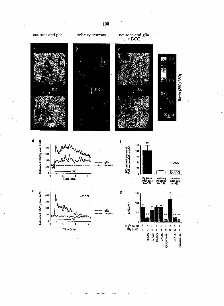
108
neurons and glia solitary neurons
i
neurons and glia + DGG
'i-''i _ v . v • .
- I BK ••
5^'':" 1.0
LO um
"S 400 i ^ "300
a 200
E s eS
100 ap BK
glia neuron
1 2 Time (min)
-I if 8 Q S'S •n'a si
«a
+ DGG
neurons solitaiy withr" with elia neurons with glia (n=2S) (n=26) (n=3f)
S I
400
300
200
100
Oi ca?tV>
+ DGG 150
glia neuron
1 2 3 Time (min)
(ft u: <
I"*!!!''!-„ WWBBMiaiwBiS!!^
Mg2+(mM> 2 2 2 2 2 2 0 0 2 G l y ( h M ) 0 0 0 0 0 0 1 0 1 0 0
2 o S, S u) o 3 § z •J Q u
a. < Q
a >3
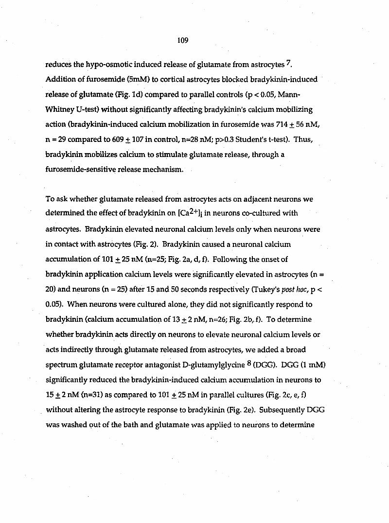
109
reduces the hypo-osmotic induced release of glutamate from astrocytes 7,
Addition of furosemide (5mM) to cortical astrocytes blocked bradykinin-induced
release of glutamate (Fig. Id) compared to parallel controls (p < 0.05, Mann-
Whitney U-test) without significantly affecting bradykinin's calcium mobilizing
action (bradykinin-induced calcium mobilization in furosemide was 714 + 56 nM,
n = 29 compared to 609 + 107 in control, n=28 nM; p>0.3 Student's t-test). Thus,
bradykinin mobilizes calcium to stimulate glutamate release, through a
fiirosemide-sensitive release mechanism.
To ask whether glutamate released from astrocytes acts on adjacent neurons we
determined the effect of bradykinin on [Ca^"*"]! in neurons co-cultured with
astrocytes. Bradykinin elevated neuronal calcium levels only when neurons were
in contact with astrocytes (Fig. 2). Bradykinin caused a neuronal calcium
accumulation of 101 + 25 nM (n=25; Fig. 2a, d, f). Following the onset of
bradykinin application calcium levels were significantly elevated in astrocytes (n =
20) and neurons (n = 25) after 15 and 50 seconds respectively (Tukey's post hoc, p <
0.05). When neurons were cultured alone, they did not significantly respond to
bradykiiun (calcium accumulation of 13 + 2 nM, n=26; Fig. 2b, f). To determine
whether bradykinin acts directly on neurons to elevate neuronal calcium levels or
acts indirectly through glutamate released from astrocytes, we added a broad
spectrum glutamate receptor antagonist D-glutamylglycine 8 (DGG). DGG (1 mM)
significantly reduced the bradykinin-induced calcium accumulation in neurons to
15 + 2 nM (n=31) as compared to 101 + 25 nM in parallel cultures (Fig. 2c, e, f)
without altering the astrocyte response to bradykinin (Fig. 2e). Subsequently DGG
was washed out of the bath and glutamate was applied to neurons to determine

110
whether glutamate receptors were present. Direct application of glutamate (100
laM) elevated internal calcium in each category of neurons shown in figure 2a, f
(neurons and glia, solitary neurons and neurons and glia that had previously been
exposed to DGG). These responses were not significantly different from one
another (Scheffe^'s post hoc comparison 9), These data suggest that bradykinin
elevates neuronal calcium by the action of glutamate, which is released from
astrocytes in response to bradykinin.
To critically determine whether bradykinin causes a neuronal change in calcium
level and to further study receptor pharmacology we used confocal microscopy.
Co-cultures of astrocytes and neurons were loaded with fluo-3 and the fluorescent
emission at the plane of neuronal somata was monitored. Bradykinin reliably
elevated neuronal calcium in astrocyte-neuron co-cultures (Fig. 2g). Addition of
the N-methyl-D-aspartate (NMDA) receptor antagonist D-2-amino-5-
phosphonopentanoic acid (D-AP5,50 M), but not 6-cyano-7-nitroquinoxaline-2,3
dione (CNQX, 10 ^M) or L-2-amino-3-phosphonopropionic acid (L-AP3, ImM),
significantly reduced bradykinin-induced neuronal calcium elevation (Fig. 2g).
Consistent with a role for NMDA receptors in mediating the action of released
glutamate, removal of external Mg2+ and addition of glycine (10 |iM) significantly
augmented the bradykinin-induced neuronal calcium elevation (Fig. 2g).
HPLC data demonstrated that furosemide blocks bradykinin-induced glutamate
release from astrocytes (Fig. 1). If glutamate released from astrocytes signals to
adjacent neurons then furosemide should block this intercellular signaling
pathway. Furosemide (5mM) significantly reduced the ability of bradykinin to

I l l
elevate neuronal calcium levels by 84 % (Fig. 2g) and reduced the neuronal calcium
elevation in response to focally applied glutamate (100 mM) by 43% (n = 25; p<
0.001, Student's t-test). Since furosemide blocks bradykiiun-induced glutamate
release from astrocytes and reduces glutamate-induced neuronal calcium
mobilization, the sensitivity of the bradykinin-induced neuronal calcium elevation
to furosemide is consistent vwth glutamate mediating astrocyte-neuron signaling.
Since calcium elevations in astrocytes stimulate glutamate release we used two
methods which directly raise calcium in astrocytes and asked whether there was a
consequent elevation in neuronal calcium. First, tapping astrocytes with
micropipettes elevated astrocyte calcium levels and significantly raised neuronal
calcium levels (AF/Fo = 62 ± 25 %, n = 6; p < 0.005, Mann-Whitney U-test).
Second, we photo-stimulated astrocytes to elevate their calcium levels A
portion of a single astrocyte was exposed to focal UV light. Photo-stimulation
reliably elevated astrocyte calcium levels (AF/Fo = 202 + 19 %, n=7), which then
spread to adjacent astrocytes (Fig. 3). When astrocytes were photo-stimulated, the
calcium levels of neurons in contact with astrocytes also increased (Fig. 4). Photo-
stimulation of astrocytes caused a maximal increase in neuronal AF/Fo by 81 + 20
% (n= 15) which was not significantly altered by addition of tetrodotoxin (1 |iM;
AF/Fo = 57 + 6 %, n=15; p>0.25, Scheffee's test) to the medium. However, addition
of the glutamate receptor antagonist DGG significantly reduced the astrocyte-
induced elevation of calcium in adjacent neurons (AF/Fo = 14 + 4 %, n=15; p<0.01,
Scheffee's test; Fig. 4).
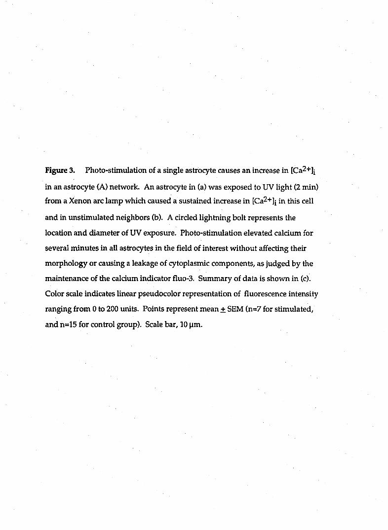
Figtire 3. Photo-stimulation of a single astrocyte causes an increase in [Ca2+]i
in an astrocyte (A) network. An astrocyte in (a) was exposed to UV light (2 min)
from a Xenon arc lamp which caused a sustained increase in [Ca2+]j in this cell
and in unstimulated neighbors (b). A circled lightning bolt represents the
location and diameter of UV exposure. Photo-stimulation elevated calciimi for
several minutes in all astrocytes in the field of interest without affecting their
morphology or causing a leakage of cytoplasmic components, as judged by the
maintenance of the calcium indicator fluo-3. Summary of data is shown in (c).
Color scale indicates linear pseudocolor representation of fluorescence intensity
ranging from 0 to 200 units. Points represent mean + SEM (n=7 for stimulated,
and n=15 for control group). Scale bar, 10 jim.
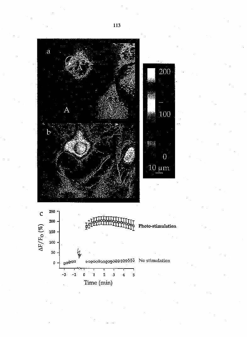
113
20G
100
PI •
0
10 Lim
Fhoto-stiiiiulation
No StilHulaiioil
1 I I I , l
- 9
Time (min)

Figure 4. Photo-slimulation of astrocytes causes an increase of [Ca2+]j in adjacent unstimulated neurons due to
the action of glutamate. Images represent calcium levels in the confocal plane of the neuronal body before (a) and
after (b) UV stimulation of a single astrocyte. After stimulation, [Ca2+]j increased both in astrocytes (A) and in the
neuron (N). The increase in neuronal calcium is not due to a direct action of UV exposure, because neuronal
processes (arrov^s, c) were not in the path of UV excitation (circled lightning bolt), c) Microinjection of
sulforhodamine 101 (10 mM) into individual neurons (n = 8) confirmed that fluo-3 revealed the full extent of
neuronal processes. In control experiments (n=6) we gradually moved the photo-stimulus closer to an isolated cell
and determined that calcium levels were only elevated when the stimulus was within less than 5 |im from the cell
border. Neurites were always greater than 11 |im from the edge of the photo-stimulus and thus were not directly
activated by this stimulus. The image in (c) was acquired at the end of the experiment at the level of astrocytes
and neuronal processes. The time course of [Ca^+Jj changes in neurons due to photo-stimulation (PS) of astrocytes
is shown in (d). The presence of DGG (1 mM), but not tetrodotoxin (TTX, 1 (iM) greatly reduced the neuronal
response. Color scale for (a) and (b) indicates linear pseudocolor representation of fluorescence intensity ranging
from 0 to 160 units. Points represent means ± SEM (n=15 for each group). Scale bar, 10 ^m.
METHODS. TTX (1 |iM) and DGG (1 mM) were uniformly applied to the bath by saline exchange 10-20 minutes
prior to UV stimulation.
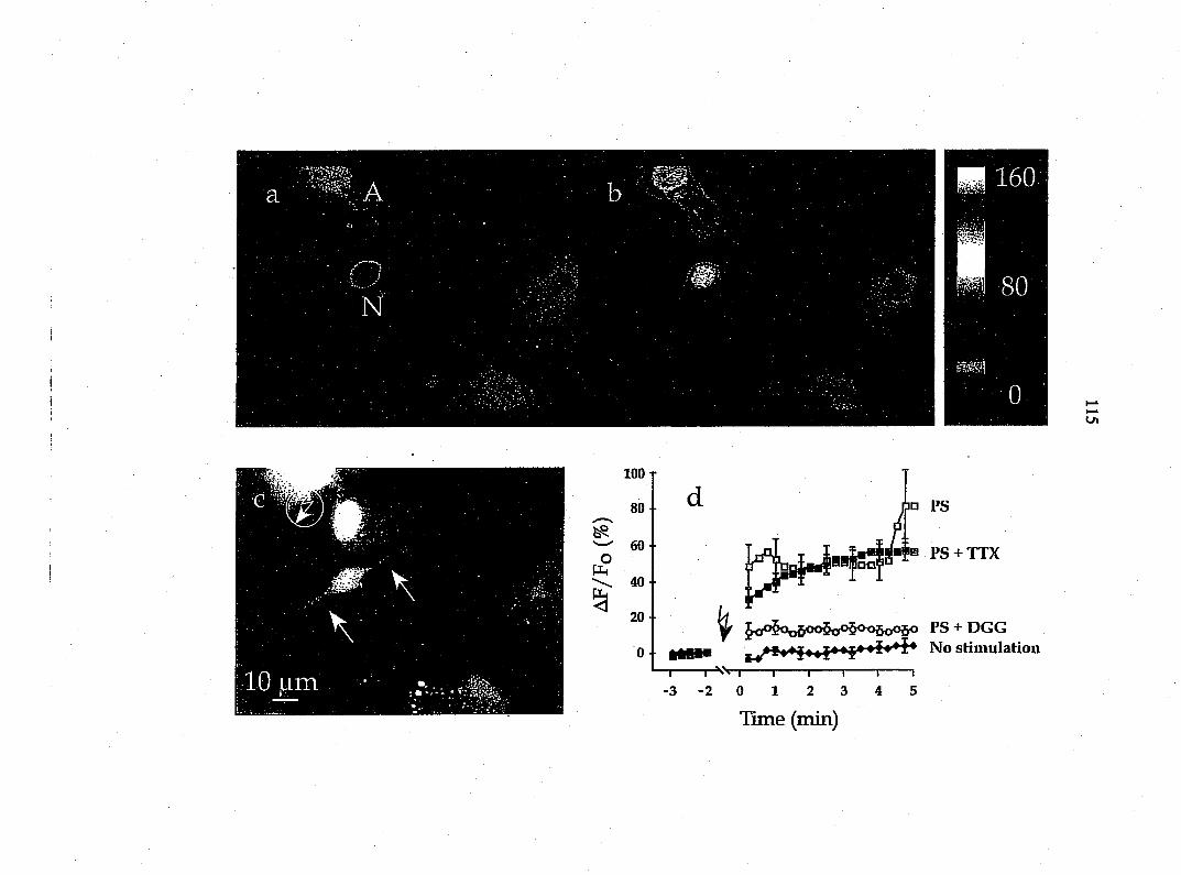
Ul
100
80 y—»s
60 o
til 40-
20 •
0 •
• PS
'a PS + TTX
^ i^oo5oo6®®5o®5o«6o05o PS + DGG ^ No stimulation
—I 1 Vv I 1 1 1 1 1 -3 -2 0 1 2 3 4 5
Time (min)
116
This study demonstrates that a neuroligand, bradykinin, causes the calcium-
dependent release of the neurotransmitter glutamate from neocortical astrocytes.
The calcium-dependent glutamate release mechanism of astrocytes is likely to be
distinct from neuronal release mechanisms since we found that astrocytes are not
immimoreactive for the calcium-sensitive vesicle protein synaptotagmin. Using
three distinct stimuli which each elevate astrocyte calcium levels ODradykinin,
tapping astrocytes with micropipettes and direct astrocyte photo-stimulation) we
demonstrate that astrocytes can cause a glutamate-dependent elevation of
neuronal calcium. These data point to a novel form of transmitter signaling in the
nervous system in which stimuli that mobilize calcium in astrocytes cause a
caldum-dependent release of the excitatory amino acid glutamate, that can then
act on adjacent neurons. It is possible that other pathways, such as gap junctions
^2, also contribute to astrocyte-neuron signaling. However, since gap-junction
uncoupling agents such as octanol have many additional actions 13-15 which
include a reduction of calcium influx through the NMDA receptor ^3 we have not
tested this possibility. Glutamate released at synaptic terminals can trigger
calcium waves in an astrocyte network 1. Since many neurotransmitters can
mobilize glial calcium ^0/16-22^ jg possible that neurons stimulate neighboring
astrocytes to release glutamate, which in turn signals back to the neuron. In this
case astrocytes could be considered as an integral computational element within
the brain. In addition to mediating fast sjoiaptic transmission, glutamate serves a
role in synaptic plasticity 23^ and under conditions of excessive release, can cause
neuronal damage and degeneration 24. jhis raises the possibility that astrocyte-

117
mediated glutamate release plays an important role in regulating neuronal
physiology and pathology.
References
1. Dani, J. W., Chernjavsky, A. & Smith, S. J. Neuron 8,429-440 (1992).
2. Murphy, T. H., Blatter, L.A., Wier, W.G. & Baraban, J.M. /. Neurosci. 13
2672-2679 (1993).
3. Mennerick, S. & Zorumski, C.F. Nature 368,59-62 (1994).
4. Balcar, V. J. & Johnston, G. A. R. /. Neurochem. 19,2657-2666 (1972).
5. Isaacson, J. S. & NicoU, R. A. /. Neurophysiol. 70,2187-2191(1993).
6. Sokal, R. R. & Rohlf, F. J. In: Biometry (ed. Wilson, J.) pp 400-453 (W. H.
Freeman & Co., New York, 1981).
7. Kimelberg, H. K., Goderire, S. K., Higman, S. Pang, S. & Waniewski, R. A. /.
Neurosci. 10,1583-1591 (1990).
8. Jones, A. W., Smith, D. A. S. & Watkins, J. C. Neuroscience 13,573-581
(1984).
9. Scheffee, H. Biometrika 40,87-104 (1953).
10. Charles, A. C., Merrill, J. E., Dirksen, E. R. & Sanderson, M. J. Neuron 6,
983-992(1991).
11. Guthrie, P. B., Segal, M. & Kater, S. B. Nature 354,76-80 (1991).
12. Nedergaard, M. Science 263,1768-1771 (1994).
13. McLamon, J. G., Wong, J. H. & Sawyer, D. Can. J. Physiol. Pharmacol. 69,
1422-1427(1991).
14. Triggle, D. J. et. al Ann. N. Y. Acad. Sci. 635,123-138 (1991).

118
15. Ma, J., Fill, M., Knudson, M. C., Campbell, K. P. & Coronado, R. Science
242,99-102(1988).
16. Reiser, G., Doni6, F. & BinmoUer, F.-J. /. Cell Sci. 93,545-555 (1989).
17. Cornell-Bell, A. H., Finkbeiner, S. M., Cooper, M. S. & Smith, S. J. Science
247,470-473(1990).
18. Glaum, S. R., Holzwarth, J. A. & Miller, R. J. Proc. Natl. Acad. Sci. USA
87,3454-3458(1990).
19. Cornell-Bell, A. H.& Finkbeiner, S. M. Ce//Cfl/ciMm 12,185-204(1991).
20. Inagaki, N., Fukui, H., Ito, S. & Wada, H. Neurosci.Lett. 128,257-260 (1991).
21. Jensen, A. M. & Chiu, S. Y. J. Neurosci. 11,1674-1684 (1991).
22. McCarthy, K. D. & Salm, A. K. Neuroscience 41,325-333 (1991).
23. Malgaroli, A. & Tsien, R. W. Nafure 357,134-139(1992).
24. Choi, D. W. Neuron 1,623-634 (1988).
25. Levison, S.W., & McCarthy, K.D. In: Culturing Nerve Cells (eds. Banker, G.
& Goslin, K.) pp 309 - 336 (The MIT Press, Cambridge Mass, USA, 1991).
26. Heuttner, J. E. & Baughman, R. W. /. Neurosci. 6,3044-3060 (1986).
27. Parpura, V., Haydon, P. G. & Henderson, E. /. Cell Sci. 104,427-433 (1993).
28. Basarsky, T.A., Parpura, V. & Haydon, P. G. J. Neurosci. In press
29. Kao, J. P. Y., Hartootunian, A. T. & Tsien, R. Y. /. Biol. Chem. 14,8179-8184
(1989).
119
GENERAL CONCLUSION
It has become dear that the excitatory amii\o acids glutamate and aspartate
do not confine their excitatory effects to nerve cells, but also interact with glial
cells. The objectives of this dissertation are to elucidate effects of neuroligand
capsaicin on cultured peptidergic sensory neurons and to determine whether
neuroligands such as bradykinin (BK) and adenosine 5'-triphosphate (ATP) can
stimulate release of EAAs from cultured glial ceils as well as to identify the
possible signal transduction pathways for the neuroligand-induced release of
EAAs.
Summary
Effects of capsaicin and resiniferatoxin on peptidergic neurons in cultured
dorsal root ganglion
The purpose of the first study was to provide evidence of the long lasting
effect of capsaicin and its ultrapotent analog resiniferatoxin (RTX) on sensory
peptidergic neurons maintained in organotypic cultures. The effects of the two
irritants were examined on neurons that contained substance P (SP) and
calcitonin gene-related peptide (CGRP). Exposure of the cultures to 10|iM
capsaicin and lOOnM RTX for periods of 2 days or longer resulted in almost
complete elimination of SP-immunoreactive (IR) neurites and reduction, but not
elimination, of CGRP-IR neurites. In addition, both lO^iM capsaicin and lOOnM
RTX significantly reduced the number of SP- and CGRP-IR cell bodies within

120
DRG explants. Capsaicin in lOO^M concentration produced complete
elimination of SP-IR fibers and a great decrease in the number of CGRP-IR fibers,
but failed to completely eliminate IR cell bodies. Exposure of the cultures to the
irritants in the same concentrations for 90 min did not produce a measurable
effect on SP- or CGRP-IR in neurites or cell bodies. It is important to establish
that the effect of capsaicin and RTX on cultured neurons was of long duration
(longer than 4 days) and is therefore different from depletion of peptides.
The neurotoxic effect of capsaicin has been shown to be selective on a
subpopulation of small dorsal root ganglion neurons in newborn animals. Our
findings demonstrate that processes of cultured sensory neurons are much more
sensitive to capsaicin and RTX than cell bodies. Furthermore, these results show
that SP-IR neuronal elements are more sensitive to capsaicin than CGRP-IR
elements. Our data suggest that cultured sensory neurons express the functional
properties of differentiated sensory neurons in vivo.
Neuroligand-evoked calcium-dependent release of excitatory amino acids from
Schwann cells
This study deals with release of neurotransmitter from Schwann cells
obtained from cultured rat DRGs. We found that neither capsaicin nor high
potassium can stimulate the release of EAAs from Schwann cells. However, BK
(lOnM) caused significant release of glutamate and aspartate from cultured
Schwann cells together with an increase in the cytoplasmic level of free calcium.
BK-induced release of glutamate and the increase of the cytoplasmic calcium are
receptor-mediated. Pretreatment with the BK receptor antagonist.

121
(D-Arg®, Hyp3, b-Thi^'®, D-Phe'')-bradykinin (5|iM), blocked glutamate release
ar\d calcium inaease evoked by BK. Although BK still stimulated release of
glutamate without extracellular calcium, this effect was abolished by
pretreatment with the membrane permeant calcium chelator BAPTA-AM
[l,2bis(2-aminophenoxy) ethane-N,N,N',N'-tetraceticacid, acetoxymethyl ester,
50|iM]. Perturbations which inhibited BK-induced calcium mobilization
prevented the release of EAAs from Schwarm cells. The calcium ionophore
ionomycin (5|iM) significantly increased the concentration of glutamate in glial
cultures. The increase of glutamate induced by ionomycin was abolished by low
Ca2+-EGTA saline. These results prove that calcium mobilization is both
necessary and sufficient for stimulating the release of EAAs from Schwann cells.
To answer the question whether BK-induced increase of glutamate is due to the
reversal uptake of glutamate, we used the glutamate transport blocker p-
chloromercuriphenylsulphonic acid (p-CMPS, SO^iM). Although p-CMPS
elevated the basal concentration of glutamate, it didn't block the release of
glutamate evoked by BK. The anion inhibitor furosemide (5mM) significantly
attenuated the ability of the second application of BK to mobilize calcium.
Furthermore, furosemide abolished the BK-induced release of glutamate from
Schwann cells. To test whether the release of EAAs is through the mechanism of
BK-induced cell swelling, we measured the volume of the Schwann cell and
found that BK didn't cause cell swelling. Taken together, these data prove that
the neuroligand BK can induce release of EAAs from Schwann cells. The
mechanism of BK-induced release of EAAs is neither due to the reversal of the
glutamate transporter nor volume regulated release mechanism. BK evokes EAA
release by mobilizing calcium from intracellular calcium stores.
122
ATP-evoked Ca2+-dependent release of excitatory amino acids from cultiired
Schwann cells
The specific objective of this study was to determine the mechanism by which
ATP evokes release of EAAs from cultured Schwann cells. Basal concentrations of
aspartate and glutamate in Schwann cell cultures were 7.64±0.74 nM (mean+SEM)
and 30.52±3.89 r\M, respectively. Perfusion application of lOOjiM ATP for 2
minutes to cultured Schwann glia resulted in an 201±28 % increase of aspartate
and 203±15 % increase of glutamate. A second applications of ATP 10 minutes
after the first application similarly caused EAA release, although the magnitude of
this response was attenuated. Application of ATP to glia for ten minutes resulted
in a peak increase in the release followed by a decline to a plateau significantly
higher than baseline release. Bath application of adenosine (100|iM) was without
effect on the release of EAAs suggesting involvement of P2 receptors. The release
of EAAs evoked by ATP was not abolished in low Ca2+-EGTA saline.
Pretreatment of the glial cultures with 50|iM BAPTA-AM abolished the effect of
ATP. Thapsigargin (IjiM), an inhibitor of Ca^+'ATPase or the Ca2+ pump of
internal stores, significantly reduced the release of EAAs evoked by ATP. Our
results show that ATP evokes the release of EAAs from cultured Schwann cells by
activating intracellular calcium stores.
Glutamate-mediated astrocyte-neiuron signalling
The last study focused on glutamate-mediated astrocyte-neuron signaling.
Neurotransmitter released from neurons is known to signal to neighboring
neurons and glia. We demonstrate an additional signaling pathway in which
123
glutamate is released from astrocytes and causes an NMD A receptor-mediated
neuronal calcium elevation. Application of the neuroligand bradykinin to cultured
astrocytes elevated internal calcium and stimulated glutamate release. Elevation
of astrocytic internal calcium was also sufficient to induce glutamate release. To
determine whether this released glutamate signals to neurons, we studied
astrocyte-neuron co-cultures. Bradykinin significantly elevated calcium levels in
the neurons co-cultured with astrocytes, but not in solitary neurons. The
glutamate receptor antagonists, D-2-amino-5-phosphonopentanoic acid and D-
glutamylglycine, prevented bradykinin-induced neuronal calcium elevations.
When single astrocytes were directly stimulated to increase internal calcium, and
release glutamate, calcium levels of adjacent neurons were increased, this increase
could be blocked by D-glutamylglycine. Thus, astrocytes regulate neuronal
calcium levels through the calcium-dependent release of glutamate.
Discussion
The studies in this dissertation demonstrate that the neuroligands can induce
release of EAAs from cultured glial cells.
There are three different mechanisms proposed for increase of extracellular
glutamate. One of them is the calcium dependency of glutamate release (Nicholls
and Sihra, 1986), which appears to be due to the exocytosis of synaptic vesicles;
The second one is reversal of uptake of glutamate (Miller and Schwartz, 1983);
Furosemide-sensitive release of glutamate from swelling cells is the third proposed
124
mechanism (Kimelberg and Ransom, 1986). According to our studies, calcium
plays an important role in the neuroligand-induced release of EAAs.
Release of EAAs evoked by BK or ATP from Schwaim cells depends on
intracellular Ca '*'
Internal calcium appears to be responsible for BK- or ATP-induced release of
EAAs from cultured glial cells. As we know, there are two intracellular stores
(Hardie, 1991). One is IPs-sensitive calcium store and the other one is caffeine-
ser\sitive store. Which Ca2+ store is involved in release of EAAs? On the basis of
the fact that caffeine doesn't induce any release of EAAs at all and ryanodine fails
to block BK-induced release, we infer that IPs-sensitive Ca2+ stores must be
responsible for the EAA release. Furthermore, the involvement of the IPs-sensitive
Ca2+ store is confirmed by the result that pretreatment of phospholipase C
inhibitor (IJ73122,8|iM) significantly blocks the BK-induced release of EAAs (our
unpublished data).
There is no evidence yet that glial cells contain synaptic vesicles. It has been
suggested that glia might release neurotransmitters by Ca2+ independent carrier
mechanisms. A recurrent suggestion is that the release might be mediated by the
reverse operation of electrogenic neurotransmitter carrier protein, which normally
function to take up neurotransmitters (reviewed by Barres, 1991). Although there is
evidence that glia release neurotransmitters by "reversed uptakes" in special
experimental conditions (Schwartz, 1987) and under physiological conditions
(Barbour, 1987; Szatkowski et al., 1990), this "Ca2+-independent release" theory is
challenged now. We demonstrated that elevated intracellular Ca2+ is necessary and
sufficient for release of EAAs from glial cells. Our data suggest that there might be
125
a novel neurotransmitter release mechanism for glial cells. The release of EAAs
from glia probably reflects a Ca2+-dependent vesicle exocytosis process. However,
this process has to be different from the exocytosis which occurs at nerve
terminals, since glial lack typical structure of synapses - the focal active zone
(reviewed by Smith, 1994) and also lack immunoreactivity to synaptic vesicle
proteins such as synaptotagmin, synaptophysin and synaptobrevin (our
unpublished data).
Release of glutamate from astrocytes plays a key role in glia-neuron signalling
Although a glia-neural signalling theory of brain function was proposed thirty
years ago (Galambos, 1961), it has only recently received much attention. Our
studies give new evidence that glial cells (astrocytes) indeed signal to neurons and
the neurotransmitter glutamate released from the glial cells plays a key role in this
process.
Our data show that neuroligand BK induced elevated free Ca2+ in purified
astrocyte cultures, but not in neuron cultures. However, BK caused an intracellular
Ca2+ increase in both astrocytes and neurons when it was applied to co-cultures of
the neurons and the astrocytes. This result strongly suggests signalling from
astrocyte to neuron. The next question is how a astrocyte could signal to a neuron.
We found that BK greatly evoked release of glutamate from cultured neuron-free
astrocytes. Furthermore, D-2-amino-5-phosphonopentanoic acid, the glutamate
receptor antagonist, effectively blocked the astrocyte-neuron signalling. Taken
these evidence together, we conclude that neurotransmitter glutamate plays a key
role in glia-neuron signalling.

126
Nedergaard (1994) provides similar evidence for astrocyte-neuron signalling.
However, he indicates that gap-junctions might mediate the astrocyte-neuron
signalling. The reasons that there are tv/o different mechanisms for astrocyte-
neuron signalling may be due to gap-junctions functioning under some specific co-
culture conditions or with some combination of neuronal or astrocytic sub-types,
but the glutamate-mediated mechanism predominating in other circumstances
(Smith, 1994).
The function of glia-neuron signalling is not clear yet. On the basis of the
available information, one would expect that glia-neuron signalling might
participate modulation of brain functions such as mood, motivational state, arousal
and learning and memory.
Is neuroligand-evoked release of EAAs involved in neurodegenerative
disorders?
Excessive glutamate is toxic to neurons. Exposed to glutamate for even a few
minutes, neurons undergo a degenerative process leading to cell death (Teichberg,
1991). The toxic effect of glutamate may be involved in acute and chronic
neurodegenerative disorders. It has been shown that considerate endogenous
glutamate can be released into the extracellular space after cerebral ischemia and
trauma (Benveniste et al., 1987, Katayama et al., 1988). NMDA antagonist can
protect against hypoxic/ischemic brain damage in vivo (Gill et al., 1987; Park et al.,
1987; Kochhar et al., 1988). There is also some evidence which implicates that EAA
neurotoxity may be a factor in chronic degenerative diseases such as Huntington's
disease, Alzheimer's disease and Parkinsonism (reviewed by Olney, 1990 and
Choi, 1988).
127
The studies in this dissertation deal with neuroligand-induced release of
EAAs. It has been shown that the neuroligand BK is released from its precursors
in response to trauma, anoxia and inflammation (Dray and Perkins, 1993) and
higher extracellular levels of ATP may be released following tissue injury (Neary
and Norenbery, 1992). Although it is not clear what effects that BK- or ATP-
induced EAA release from glia would have on CNS under centain pathological
condition, the present information hints that neuroligand-induced release of EAAs
might potentiate a pathological state associated with brain damage. Furthermore,
utilizing certain neuroligand antagonists, which block the release of EAAs, might
attenuate the pathological state.
Conclusion
This dissertation focuses on elucidating the mechanism of neuroligand-evoked
release of EAAs. Our studies have demonstrated that the EAA release appears to
be in a Ca2+-dependent manner. We have showed, at least in part, the signal
pathways of release of EAAs. However, there is still a gap left. Further
investigation is needed to explore how Ca2+ triggers EAA release from glia, which
lack synaptic vesicles.
As we know, release of EAAs plays an important role in neuron-neuron
signalling. However, there is increasing evidence that the release of EAAs is
actively involved in the signalling processes between neuron and glia. Although it
is not clear what functions these signalling processes serve, they deserve further
study.

128
REFERENCES
Adam-Vizi, V., Ashley, R.H. (1987) Relation of acetylcholine release to Ca2+
concentration in Guinea-pig cortex synaptosomes. J. Neuroche. 49,1013-
1021.
Adam-Vizi, V., Ligeti, E. (1984) Release of acetylcholine from rab brain
synaptosomes by various agents in the absence of external calcium ions. J.
Physiol, (lond.) 353,505-531.
Adam-Vizi, V., Deri, Z., Vizi, E.S., Sershen, H., Lajtha, A. (1991) Ca^+-
independent veratridine-evoked acetylcholine release from striatal slices is
not iiUiibited by vesamicol (AH5183): mobilization of distinct transmitter
pools. J. Neurochem. 56(1), 52-8.
Aimers W., Tse F.W. (1990) Transmitter release from s3mapses: does a
preassembled fusion pore initiate exocytosis? Neuron 4,813-818.
Ascher P., Nowak L. (1988) Quisqualate- and Kainate-activated channels in
mouse cnetral neurones in culture. J. Physiol. 399,277-245.
Augustine G,J., Charlton M.P., Smith, S. (1987) Calcium action in synaptic
transmitter release. Annu. Rev. Neurosci. 10,633-693.
Backus, K.H., Kettenmann, H., Schachner, M. (1989) Pharmacological
characterization of glutamate receptor in cultured astrocytes. J. Neurosci.
Res. 22,274-282.
Barbuor B., Brew H., Attwell D. (1988) Electrogenic glutamate uptake in glial
cells is activated by introcellular potassium. Nature 335,433-435.

129
Barbour, B., Szatkowski, M., Ingledew, N., Attwell, D. (1989) Arachidonic add
induces a prolonged inhibition of glutamate uptake into glial cells. Nature
(Lond) 342,918-920.
Barres, B.A. (1991) New roles for glia. J. Neurosci. 11(12), 3685-3694.
Barres, B.A., Chun, L.L.Y., Corey, D.P. (1990a) Ion channels in vertebrate glia.
Aimu. Rev. Neurosci. 13,441-474.
Barres, B.A., Koroshetz W.J., Swartz K.J, Chun L.L.Y., Corey D.P. (1990b) Ion
channel expression by white matter glia: the 02A glial progenitor cell.
Neuron 4,507-524.
Baudry M, Evans J., Lynch G. (1986) Excitatory amino acids inhibit stimulation o
fphosphatidylinositol metabolism by aminergic agonists in hippocampus.
Nature 319,329-331.
Benveniste H., Drejer J., Schousboe A., Diemer N.M. (1984) Elevation of the
extracellular concentrations of glutamate and aspartate in rat hippocampus
during transient cerebral ischemia monitored by intracerebral microdialysis.
J. Neurochem. 43,1369-1374.
Berrmett M.V.L., Barrio L.C., Bargiello T.A., Spray D.C., Hertzerg E., Saez J.C.
(1991) Gap junctions: new tools, new answers, new questions. Neuron 6,
305-320.
Bevan, S. (1990) Ion channels and neurotransmitter receptor in glia. Semin
Neurosci. 2,467-481.
Blake J.F., Brown M.W., Collingridge G.L. (1988) CNQX blocks acidic amino acid
induced depolarizations and synaptic components mediated by non-NMDA
receptors in rat hippocampal slices. Neurosci. Lett. 89,182-186.

130
Blaustein^ M.P. (1988) Calcium transport and buffering in neurons. Trends
Neurosci. 10,438-443.
Bowman, C.L., Kimelberg, H.K. (1984) Excitatory amino acids directly depolarize
rat brain astrocytes in primary culture. Nature (Lond) 311,656-659.
Brew H., Attwell D. (1987) Electrogenic glutamate uptake is a major current
carrier in the membrane of axolotl retinal glial cells. Nature 327, 707-709.
Buck S.H., Burks, T.F. (1986) The neuropharmacology of capsaicin: review of
some recent observations. Pharmacol. Rev., 38,179-226.
Burgess G.M., Mullaney I., McNeill M.; Coote P.R., Minhas A., Wood J.N. (1989)
Activation of guanylate cyclase by bradykinin in rat sensory neurons is
mediated by calcium influx: possible role of the increase in cyclic GMP. J.
Neurochem. 53,1212-1218.
Bumstock G. (1990) Overview: purinergic mechanisms. Ann. N.Y. Acad. Sci. 603,
1-18.
Changeux J.-P. (1981) The acetylcholine receptor: an "allosteric" membrane
protein. Harvey Lect. 75,85-254.
Choi D.W. (1988) Glutamate neurotoxicity and diseases of the nervous system.
Neuron 1 (8), 623-634.
CoUingridge G.L., Bliss T.V.P. (1987) NMDA receptors-their role in long term
potentiation. Trands Neurosci. 10,288-294.
Cotman C.W., Monaaghan D.T., and Ganong A.H. (1988) Excitatory amino acid
neurotransmission: NMDA receptors and hebb-type synaptic plasticity.
Annu. Rev. Neurosci. 11,61-80.
Cragoe E.J., Woltersdorf O.W., Gould N.P., Pietruszkiewicz A.M. (1986) Agents
for the treatment of brain edema. 2. [(2,3,9,9a-tetrahydro-3-oxo-9a-

131
substituted-lH-fluren-7-yl)oxy] alkanoic acids and some of their analogues.
J. Med. Chem. 29,825-841.
Dani J.W., Chernjavsky A., Smith S.J. (1990) Calcium waves propagate through
astrocyte networks in developing hippocampal brain slices. J. Cell biol., 3,
389a
Davies ]., Watkins J.C. (1982) Actions of D- and L-forms of 2-amino-5-
phosphonobutyrate and 2-amino-4-phosphonobutyrate in the cat spinal-
cord. Brain Res. 235,378-386.
De Camilli P., Benfenati F., Valtorta F., and Greengard P. (1990) The synapsins.
Annu. Rev. CeU Biol. 6,433-460.
Del Castillo J. and Katz B. (1957) La base "quantale" de la transmisson
neuromusculaire, in Microphysiologie Comparee des Elements Excitables.
Vol 67, pp 245-258. Centre National de la Recherche Scientifique, Colloque
International, Paris.
Dray A., Forbes C.A., Burgess G.M. (1990) Ruthenium red blocks the capsaicin-
induced increase in intracellular calcium and activation of membrane
currents in sensory neurons as well as the activation of peripheral
nociceptors in vitro. Neurosci. Lett. 110,52-59.
Dray, A. and Perkins, M. (1993) Bradykinin and inflammatory pain. TINS 16,99-
104.
Erednska M. and Nelson D. (1987) Amino acid neurotransmitters in CNS.
Relationships between net uptake and exchange in rat brain synaptosomes.
FEBS Lett. 213,61-66.
Erednska M., Wantorsky D., and Wilson D. F. (1983) Aspartate transport in
synaptosomes from rat brain. J. Biol. Chem. 258,9069-9077.
132
Evans R.H., Frands A-A., Jones A.W, Smith D.A.S., Watkins, J.C. (1982) The
effects of a series of -phosphonic -carboxylic amino adds on electrically
evoked and amino add induced responses in isolated spinal cord
preparations. Br. J. Pharmacol. 75,65-75.
Faden A.I., Demediuk P., Panter S.S., Vink R. (1989) The role of excitatory amino
acids and NMDA receptors in traumatic brain injury. Science 244,798-800.
Fitzgerald, M. (1983) Capsaidn and sensory neurons. Pain 15, 109-130.
Furshpan E.J., and Potter D.D. (1957) Mechanism of nerve-impulse transmission
at a crayfish synapse. Nature 180,342-343.
Galambos R. (1961) A glia-neural theory of brain function. Proc. Natl. Acad. Sci.
USA 47,129-136.
Gill R., Foster A.C., Woodruff G.N. (1987) Systemic administration of MK-801
protects against ischemia-induced hippocampal neurodegeneration in
gerbil. J. Neurosd. 7,3343-3349.
Gilles R., Kleinzeller A., Bolis L. (1987) Current topics in membranes and
transport. Vol. 30, Academic, New York.
Gordon J.L. (1986) Extracellular ATP: effects, sources and fate. Biochem. J. 233,
309-319.
Hardie D.G. (1991) Biochemical Messengers - hormones, neurotransmitters and
growth factors. Chapman & Hall, Lodon, 175pp.
Hayes R.L., Chapouris R., Lyeth B.G., Jenkins L., Robinson S.E., et al. (1987)
Pretreatment with phencyclidine (PCP) attenuates long term behavioral
deficits following concussive brain injury in the rat. Neurosd. Abstr. 13,
1254.

133
Heumann R., Villegas J., Herzfeld D.W. (1981) Acetylcholine synthesis in the
Schwann cell and axon in the giant nerve fiber of the squid. J. of neurochem.
36,765-768.
Heyman I., Rang H.P. (1985) Depolarizing responses to capsaicin in a sub-
population of rat dorsal root ganglion cells. Neurosci. Lett. 56,69-75.
Hochner, B., Pamas, H. and Pamas, I. (1989) Membrane depolarization evokes
neurotransmitter release in the absence of calcium entry. Nature 342,433-
435.
Holzer P.(1988) Local effector functions of capsaicin-sensitive sensory endings:
involvement of tachykinins, calcitonin gene-related peptide and other
neuropeptides, Neuroscience, 24,739-769.
Holzer P. (1991) Capsaicin: cellular target, mechanism of action, and selectivity
for thin sensory neurons. Pharmaco. Rev. 43,144-190.
Honors T., Davies S.N., Drejer J., Fletcher E.J., Jacobsen P., et al. (1988)
Quinoxalinediones: potent competitive non-NMDA glutamate receptor
antagonists. Science 241,701-703.
Jessell T.M., Iversen L.L. and Cuello A.C. (1978) Capsaicin-induced depletion of
substance P from primary sensory neurons. Brain Res., 152,183-188.
Jessell T.M., Kandel E.R. (1993) Synaptic transmission: a bidirectional and self-
modifiable form of cell-cell communication. Cell 72,1-30.
Katmer B.L and Marva E. (1982) Effux of L-glutamate by membrane vesicles
isolated from rat brain. Biochemistry 21,3143-3147.
Kaimer B.L and Sharon I. (1978) Active transport of L-glutamate by membrane
vesicles isolated from rat brain. Biochemistry 17,3949-3953.

134
Karlin A. (1990) Explorations of the nicotinic acetylcholine receptor. Harvey Lect.
85,71-107.
Katayama Y., Cheung M.K., Gorman L., Tamura T., Becker D.P. (1988) Increase in
extrocellular glutamate and associated massive ionic fluxes following
concussive brain injury. Neurosci. Abstr. 14,1154.
Katz B. (1966) Nerve, Muscle, and Synapse. McGraw-Hill, New York.
Kelly R. B. (1993) Storage and release of neurotransmitters. Cell 72,43-53.
Ketteiunarm, H., Gilbbert, P. and Schachner, M. (1984a) Depolarization of
cultured oligodendrocytes by glutamate and GABA. Neurosci. Lett. 47,271-
276.
Kettenmann, H., Backus, K. H. and Schachner, M. (1984b) Aspartate, Glutamate
and GABA-aminobutyric acid depolarize cultured astrocytes. Neurosci.
Lett. 52,25-29.
Kimelberg H.K., and Ransom B.R. (1986) Physiological and pathological aspects
of astrocytic swelling. Astrocytes 3,129-166.
Kimelberg H.K., Cragoe E.J., Nelson L.R., Popp A.J. (1987) Improved recovery
from a traumatic-hypoxic brain injury in casts by intracisternal injection of
an anion transport inhibitor. CNS Trauma 4,3-14.
Kochhar A., Zivin J.A., Lyden P.D., Mazzarella, V. (1988) Glutamate antagonist
therapy reduces neurologic deficits produced by focal central nervous
system ischemia. Arch. Neurol. 45,148-153.
Koemer J.F., Cotman C.W. (1981) Micromolar L-2-amino-4-phosphonobutyric
acid selectively inhibits perforant path synapses from lateral entorhinal
cortex. Brain Res. 216,192-198.
135
Lieberman E.M. (1991) Role of glutamate in axon-Schwann cell signaling in the
squid. Ann NY Acad. Sci. 633,448-57
Lieberman E.M. Abbott N.J., Hassan S. (1989) Evidence that glutamate mediates
axon-Schwann cell signalling in the squid. Glia 2,94-102.
Lynch G., Larson J., Kelso S., Barionuevo G., and Schottler F. (1983) Intracelluar
injections of EGTA block induction of hippocampal long-term potentiation.
Nature 305,719-721.
MacDermott A.B., and Dale N. (1987) Receptors, ion channels and synaptic
potentials underlying the integrative actions of excitatory amino acids.
Trands Neurosci. 10,280-284.
MacDermott A.B., Mayer M.L., Westbook G.L., Smith S. J., and Barker, J.L. (1987)
NMDA-receptor activation increases cytoplasmic calcium concentration in
cultured spinal cord neurones. Nature 321,519-522.
Marsh S.J., Stansfeld C.E., Brown D.A., Davey R. and McCarthy D. (1987) The
mechanism of action of capsaicin on sensory C-type neurons and their
axons in vitro. Neuroscience 23,275-289.
Mayer M.L., MacDermott A.B., Westbook G.L., Smith S. J., and Barker, J.L. (1987)
Agonist- and voltage-gated calcium entry in cultured mouse spinal cord
neurons under voltage clamp measured using arsenazo-IIL J. Neurosci. 7,
3230-3244.
Mayer M.L., and Westbrook G.L. (1987) The physiology of excitatory amino acids
in the vertebrate central nervous system. Prog. Neurobiol. 28,197-276.
McGuirk S.M., Dolphin A.C. (1992) G-protein mediation in nociceptive signal
transduction: an investigation into the excitatory action of bradykinin in a
subpopulation of cultured rat sensory neuron. Neuroscience 48,117-128.

136
Meldolesi J., Volpe, P. and Pozzan, T. (1988) The intracellular distribution of
calcium. Trends neurosci. 10,449-452.
Miller A.M., Schwartz E.A. (1983) Evidence for the identification of synaptic
transmitters released by photoreceptors of the toad retina. J. Physiol. (Lond.)
334,325-349.
Miller, R. (1987) Multiple calcium channels and neuronal function. Science 235,
46-52.
Monaghan D.T. (1989) The excitatory aminoacid receptors: their calsses,
pharmacology, and distinct properties in the function of central nervous
system. Aimu. Rev. Pharmacol. Toxicol. 29,365-402.
Nahorski, S.R. (1988) Inositol polyphosphates and neuronal calcium homeostasis.
Trands Neurosci. 10,444-448.
Naito S., Ueda T. (1983) Adenosine triphosphate-dependent uptake of glutamate
into protein I associated synaptic vesicles. J. Biol. Chem. 258,696-699.
Naito S., Ueda T. (1985) Characterization of glutamate uptake into synaptic
vesicles. J. Neurochem. 44,99-109.
Neary J.T., Norenberg M.D. (1992) Signaling by extracellular ATP: physiological
and pathological considerations in neuronal-astrocytic interactions. Pro.
Brain Res. 94,143-151.
Nedergaard M. (1994) Direct signaling from astrocytes to neurons in cultures of
mammalian brain cells. Science 263,1768-1771.
NichoUs D.G., Sihra T. (1986) Synaptosomes possess an exocytotic pool of
glutamate. Nature 321,772-773.
Nicoletti F., ladarola M.J., Wroblewski J.T., Costa E (1986) Excitatory amino acid
recognition sites coupled with inositol phospholipid metabolism:
137
developmental changes and interaction with alphai -adrenoceptors. Proc.
Nati. Acad. Sd. USA 83,1931-1935.
NicoU R.A., Kauer J.A., Malenka R.C. (1988) The current excitement in long-term
potentiation. Neuron 1,97-103.
Olney J.W. (1990) Excitotoxic amino acids and neuropsychiatric disorders. Annu.
Rev. Pharmacol. Toxicol. 30,47-71.
Park C., Nehls D.G., Ozyurt E., Graham D.I., McCulloch J. (1987) Ischemin brain
damage is reduced by systemic administration of NMDA antagonist, MK-
801. Neurosci. Abstr. 13,1029.
Richardson, P.J., Brown, S.J. (1987) ATP release from affinity-purified rat
cholinergicv nerve terminals. J. Neurochem. 48,622-630.
Rothman S.M., Olney J.W. (1986) Glutamate and the patho-physiology of
hypoxic/ischemic barin-damage. Ann. Neuol. 19,105-111.
Schiavo G., Benfenati F., Poulain B., Rossetto, O., Polverino de Laureto P,
DasGupta B.R., Montecucco C. (1992) Tetanus and botulinum-B neurotoxins
block neurotransmitter release by proteolytic cleavage of synaptobrevin.
Nature 359,832-835.
Schwartz E.A. (1987) Depolarization without calcium can release GABA from a
retinal neuron. Science 238,350-355.
Storm-Mathisen J., Leknes A. K., Bore A. T., Waaland J. L., Edminson P., Haug F-
M. S., and Ottersen O. P. (1983) First visualization of glutamate and GABA
in neurons by immunocytochemistry. Nature 301,517-520.
Sladeczek F., Pin J.P., Recasens M, Bockaert J., Weiss S. (1985) Glutamate
stimulates inositol phosphate formation in striatal neurones. Nature 317,
717-719.

138
Smith S.J. (1994) Neuromodulatory astrocytes. Current Biology 4 (9), 807-810.
Sudhof T.C., Jahn R. (1991) Proteins of synaptic vesicles involved in exocytosis
and membrane recycling. Neuron 6,665-677.
Szatkowski M., Barbour B., Attwell D. (1990) Non-vesicular release of glutamate
from glial cells by reversed electrogenic glutamate uptake. Nature 348,443-
446.
Teichberg V.I. (1991) Glial glutamate receptors: likely actors in brain signaling.
FASEB 5,3086-3091
Thureson-Klein AK and Klein RL, (1990). Exocytosis from neuronal large dense-
cored vesicles. Int. Rev. Cytol. 121,67-126.
Trimble W.S., Linial M. and Scheller R.H. (1991) Cellular and molecular biology
of the presynaptic nerve terminal. Annu. Rev. Neurosci. 14,93-123.
Usowicz, M.M., Gallo, V., and Cull-Candy, S.G. (1989) Multiple conductance
channels in type-2 cerebellar astrocytes activated by excitatory amino acids.
Nature (Lond.), 339,380-383.
Vizi, E.S. (1978) Na+- K+- activated adenosinetriphosphatase as a trigger in
transmitter release. Neurosci. 3,367-384.
Watkins J.C., Evans R.H. (1981) Excitatory amino acid transmitters. Annu. Rev.
Pharmacol. Toxicol. 21,165-204.
Watkins J.C., Olverman, H.J. (1987) Agonists and antagonists for excitatory
amino acid receptors. Trands Neurosci. 10,265-272.
White J.M. (1992) Membrane fusion. Science 258,917-924.
White, T.D. (1977) Direct detection of depolarization-induced release of ATP
from synaptosomal preparation. Nature 267,67-69.
139
Wieraszko, A., Goldsmith, G., Seyfrid, T.N. (1989) Stimulation-dependent release
of adenosine triphosphate from hippocampal slices. Brain Res. 485,244-250.
Wilson D.F., Pastuszko A. (1986) Transport of cysteate by synaptosomes isolated
from brain: evidence that it utilizes the same transporter as aspartate,
glutamate, and cysteine sulfinate. J. Neurochem. 47,1091-1097.
Winter J., Dray A., Wood J.N., Yeats J.C., Bevan S. (1990) Cellular mechanism of
action of resiniferatoxin: a potent sensory neuron excitotoxin. Brain Res. 520,
131-140.
Wood J.N., Coote P.R., Minhas A., Mullaney I., McNeill M., Burgess G.M. (1989)
Capsaicin-induced ion fluxes increase cyclic GMP but not cyclic AMP levels
in rat sensory neurons in culture. J. Neurochem. 53,1203-1211.
Wood J.N., Winter,J., James,I.F., Rang, H.P., Yeats, J. and Bevan, S. (1988)
Capsaicin-induced ion fluxes in dorsal root ganglion cells in culture, J.
Neurosci., 8,3207-3220.
Zahraouf A., Touchot N., Chardin P., Tavitian A. (1989) The human rab genes
encode a family of GTP-binding proteins related to yeast YPTl and SEC4
products involved in secretion. J. Biol. Chem. 264,12394-12401.
140
ACKNOWLEDGMENTS
I would like to thank my major professor. Dr. Srdija Jeftinija, for his advice,
guidance and support throughout this research.
I also would like to thank the members of my committee: Dr. Mary Helen
Greer, for her understanding and help; Dr. Philip Haydon, for his input and
expertise in this research; Dr. Walter Hsu, for the discussions and suggestions
regarding this study; and Dr. Carol Jacobson for her comments and
encouragement. Special thanks go to Ksenija Jeftinija for her technical support
and much help.
I gratefully acknowledge the following organizations at Iowa State
University for their financial support: Graduate College, Neuroscience Program,
Department of Veterinary Anatomy, and Signal Transduction Training Group.