neurons & the nervous system - part 2 - wikispacesand... · ... spinal cord peripheral nervous...
TRANSCRIPT

BIO 301
Human Physiology
Neurons & the Nervous System - Part 2
The Human Nervous System consists of the Central Nervous System & the Peripheral
Nervous System.
Central Nervous System:
1 - Brain
2 - Spinal cord
Peripheral Nervous System: 1 - Cranial nerves (12 pair) & their branches
2 - Spinal nerves (31 pair) & their branches
Source: http://mail.med.upenn.edu/~hessd/Lesson3.htm

Divisions of Peripheral Nervous System -
1 - Somatic - supplies & receives fibers (neurons) to & from the skin, skeletal muscles,
joints, & tendons
Used with permission of John Kimball
2 - Visceral - supplies & receives fibers to & from smooth muscle, cardiac muscle, and
glands. The visceral motor fibers (those supplying smooth muscle, cardiac muscle, &
glands) make up the Autonomic Nervous System. The ANS has two divisions:
o Parasympathetic division - important for control of 'normal' body functions, e.g.,
normal operation of digestive system
o Sympathetic division - also called the 'fight or flight' division; important in
helping us cope with stress
Source: http://faculty.washington.edu/chudler/nsdivide.html
Divisions of the Human Brain:
1 - Myelencephalon, which includes the medulla
2 - Metencephalon, which includes the pons and cerebellum
3 - Mesencephalon, which includes the midbrain (tectum and tegmentum)
4 - Diencephalon, which includes the thalamus and hypothalamus
5 - Telencephalon, which includes the cerebrum (cerebral cortex, basal ganglia, & medullary
body)
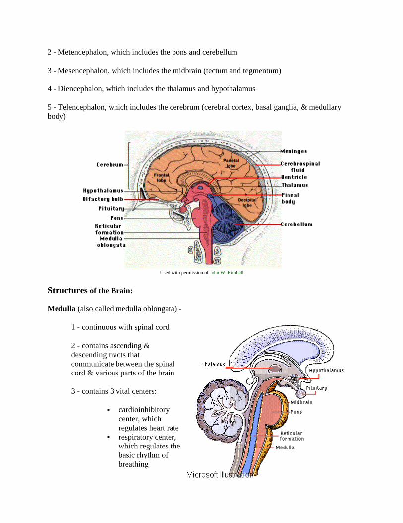
Used with permission of John W. Kimball
Structures of the Brain:
Medulla (also called medulla oblongata) -
1 - continuous with spinal cord
2 - contains ascending &
descending tracts that
communicate between the spinal
cord & various parts of the brain
3 - contains 3 vital centers:
cardioinhibitory
center, which
regulates heart rate
respiratory center,
which regulates the
basic rhythm of
breathing
vasomoter center, which regulates the diameter of blood vessels
4 - origin of five cranial nerves (VIII or vestibulocochlear, IX or glossopharyngeal, X or
vagus, XI or accessory, & XII or hypoglossal)
Pons -
1 - Bridge connecting spinal cord w/ brain & parts of brain w/ each other
2 - Origin of four cranial nerves (V or trigeminal, VI or abducens, VII or facial, & VIII or
vestibulocochlear)
3 - contains pneumotaxic center (a respiratory center)
Midbrain -
1 - Corpora quadrigemina - visual reflexes & relay center for auditory information. Two pairs of rounded knobs on the upper surface of the midbrain mark the location of four nuclei, which
are called collectively the "corpora quadrigemina." These masses contain the centers for certain visual
reflexes, such as those responsible for moving the eyes to view something as the head is turned. They also
contain the hearing reflex centers that operate when it is necessary to move the head so that sounds can be
heard better.
2 - Cerebral peduncles - ascending & descending fiber tracts
3 - Origin of two cranial nerves (III or oculomotor & IV or trochlear)
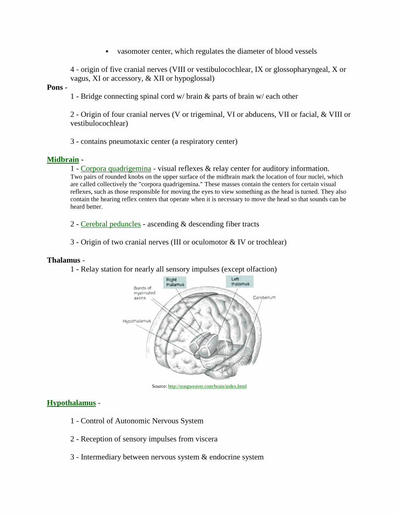
Thalamus -
1 - Relay station for nearly all sensory impulses (except olfaction)
Source: http://songweaver.com/brain/index.html
Hypothalamus -
1 - Control of Autonomic Nervous System
2 - Reception of sensory impulses from viscera
3 - Intermediary between nervous system & endocrine system

4 - Control of body temperature
5 - Regulation of food intake
6 - Thirst center
7 - Part of limbic system (emotions such as rage and
aggression)
8 - Part of reticular formation
Reticular formation -
1 - portions located in the spinal cord, medulla, pons,
midbrain, & hypothalamus
2 - needed for arousal from sleep & to maintain
consciousness
Cerebellum -
1 - functions in coordination, maintenance of posture, & balance
Cerebrum -
1 - largest portion of the human brain
2 - consists of 2 hemispheres divided by a fissure
Source: http://faculty.washington.edu/chudler/split.html
3 - includes cerebral cortex, medullary body, & basal ganglia:

o Cortex:
outer 2 - 4 mm of the cerebrum
consists of gray matter (cell bodies & synapses; no myelin)
'folded', with upfolded areas called gyri & depressions or grooves called
sulci
consists of four primary lobes
Source: http://faculty.washington.edu/chudler/lobe.html
functional areas include motor areas (initiate impulses that will cause
contraction of skeletal muscles) (see A Map of the Motor Cortex), sensory
areas (receive sensory impulses from throughout the body), and
association areas (for analysis)
Source: http://faculty.washington.edu/chudler/functional.html
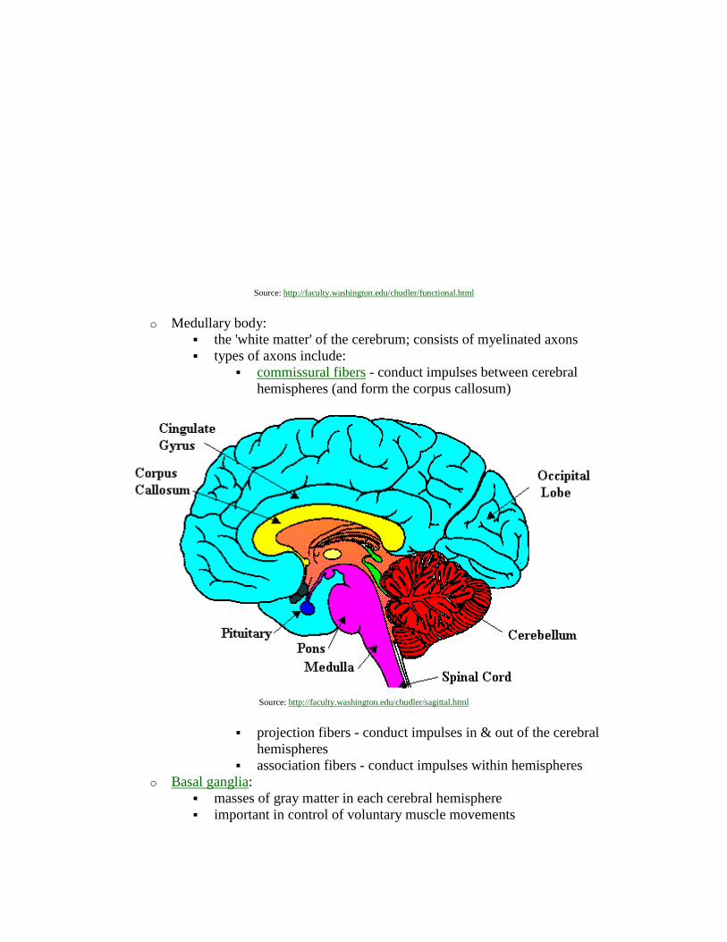
o Medullary body:
the 'white matter' of the cerebrum; consists of myelinated axons
types of axons include:
commissural fibers - conduct impulses between cerebral
hemispheres (and form the corpus callosum)
Source: http://faculty.washington.edu/chudler/sagittal.html
projection fibers - conduct impulses in & out of the cerebral
hemispheres
association fibers - conduct impulses within hemispheres
o Basal ganglia:
masses of gray matter in each cerebral hemisphere
important in control of voluntary muscle movements
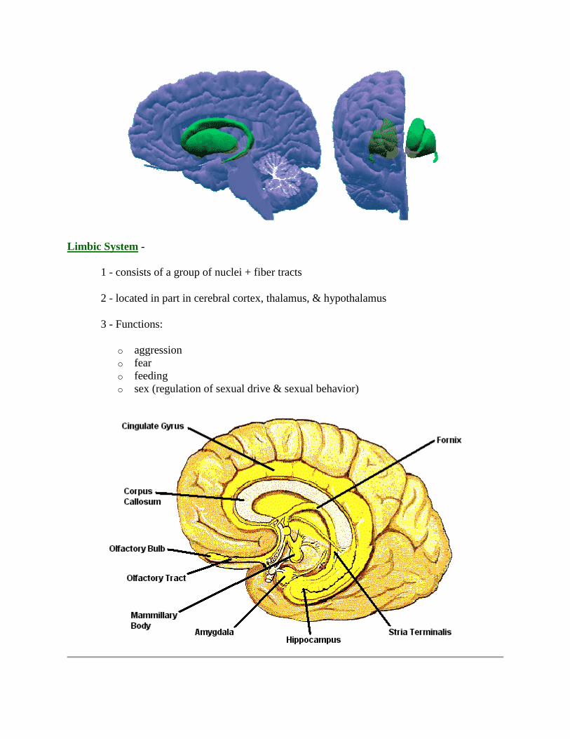
Limbic System -
1 - consists of a group of nuclei + fiber tracts
2 - located in part in cerebral cortex, thalamus, & hypothalamus
3 - Functions:
o aggression
o fear
o feeding
o sex (regulation of sexual drive & sexual behavior)

Spinal cord
Used with permission of John W. Kimball
The spinal cord extends from the skull (foramen magnum) to the first lumbar vertebra. Like the
brain, the spinal cord consists of gray matter and white matter. The gray matter (cell bodies &
synapses) of the cord is located centrally & is surrounded by white matter (myelinated axons).
The white matter of the spinal cord consists of ascending and descending fiber tracts, with the
ascending tracts transmitting sensory information (from receptors in the skin, skeletal muscles,
tendons, joints, & various visceral receptors) and the descending tracts transmitting motor
information (to skeletal muscles, smooth muscle, cardiac muscle, & glands). The spinal cord is
also responsible for spinal reflexes.
Reflex- rapid (and unconscious) response to changes in the internal or external environment
needed to maintain homeostasis
Reflex arc - the neural pathway over which impulses travel during a reflex. The components of a
reflex arc include:
1 - receptor - responds to the stimulus
2 - afferent pathway (sensory neuron) - transmits impulse into the spinal cord
3 - Central Nervous System - the spinal cord processes information
4 - efferent pathway (motor neuron) - transmits impulse out of spinal cord
5- effector - a muscle or gland that receives the impulse from the motor neuron & carries
out the desired response
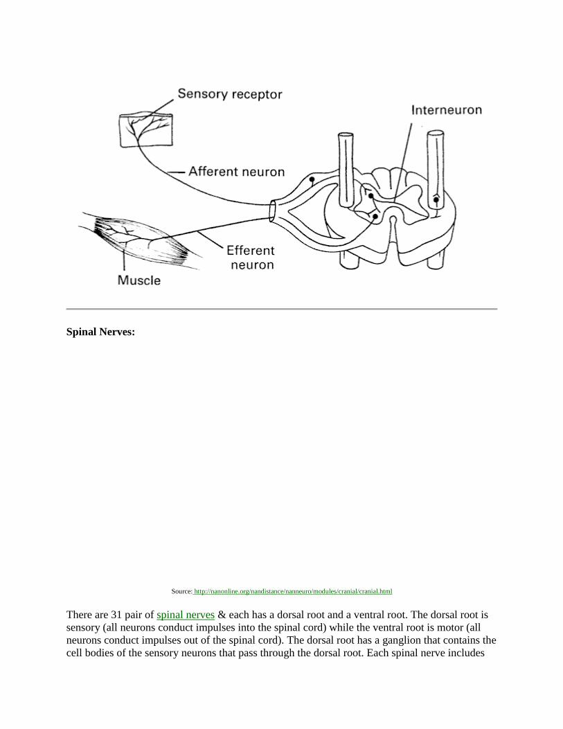
Spinal Nerves:
Source: http://nanonline.org/nandistance/nanneuro/modules/cranial/cranial.html
There are 31 pair of spinal nerves & each has a dorsal root and a ventral root. The dorsal root is
sensory (all neurons conduct impulses into the spinal cord) while the ventral root is motor (all
neurons conduct impulses out of the spinal cord). The dorsal root has a ganglion that contains the
cell bodies of the sensory neurons that pass through the dorsal root. Each spinal nerve includes
numerous sensory, or afferent, & motor, or efferent, neurons. Some of these neurons are
classified as somatic, and these neurons conduct impulses to or from 'somatic' structures (skin,
skeletal muscles, tendons, & joints). Other neurons are 'visceral', and these conduct impulses to
or from 'visceral' structures (smooth muscle, cardiac muscle, and glands). Thus, all neurons in
spinal nerves (& the peripheral nervous system) can be placed in one of four categories:
Somatic afferent
Somatic efferent
Visceral afferent
Visceral efferent
Somatic afferent neurons are sensory neurons that conduct impulses initiated in receptors in the
skin, skeletal muscles, tendons, & joints. Receptors in the skin are responsible for sensing such
things as touch, temperature, pressure, & pain and are called exteroceptors. Receptors in the
skeletal muscles, tendons, & joints provide information about body position & movement and
are called proprioceptors. Somatic afferent neurons are unipolar neurons that enter the spinal
cord through the dorsal root & their cell bodies are located in the dorsal root ganglia.
Somatic efferent neurons are motor neurons that conduct impulses from the spinal cord to
skeletal muscles. These neurons are multipolar neurons, with cell bodies located in the gray
matter of the spinal cord. Somatic efferent neurons leave the spinal cord through the ventral root
of spinal nerves.
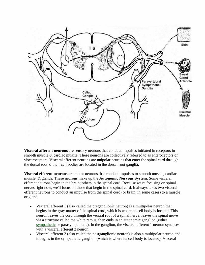
Visceral afferent neurons are sensory neurons that conduct impulses initiated in receptors in
smooth muscle & cardiac muscle. These neurons are collectively referred to as enteroceptors or
visceroceptors. Visceral afferent neurons are unipolar neurons that enter the spinal cord through
the dorsal root & their cell bodies are located in the dorsal root ganglia.
Visceral efferent neurons are motor neurons that conduct impulses to smooth muscle, cardiac
muscle, & glands. These neurons make up the Autonomic Nervous System. Some visceral
efferent neurons begin in the brain; others in the spinal cord. Because we're focusing on spinal
nerves right now, we'll focus on those that begin in the spinal cord. It always takes two visceral
efferent neurons to conduct an impulse from the spinal cord (or brain, in some cases) to a muscle
or gland:
Visceral efferent 1 (also called the preganglionic neuron) is a multipolar neuron that
begins in the gray matter of the spinal cord, which is where its cell body is located. This
neuron leaves the cord through the ventral root of a spinal nerve, leaves the spinal nerve
via a structure called the white ramus, then ends in an autonomic ganglion (either
sympathetic or parasympathetic). In the ganglion, the visceral efferent 1 neuron synapses
with a visceral efferent 2 neuron.
Visceral efferent 2 (also called the postganglionic neuron) is also a multipolar neuron and
it begins in the sympathetic ganglion (which is where its cell body is located). Visceral
efferent 2 neurons may exit the ganglion through the gray ramus, then proceed to some
visceral structure (smooth muscle, cardiac muscle, or gland).

Source: http://www.mmi.mcgill.ca/Unit2/Mandl/lect20autonomicnervoussystem.htm

The 4 types of peripheral neurons: somatic afferent (top right), somatic efferent (bottom right), visceral afferent (top left), and visceral efferent (bottom left).
Autonomic Nervous System:
1 - entirely motor (consisting of the visceral efferent fibers)
2 - has two divisions: sympathetic & parasympathetic
o sympathetic neurons leave the central nervous system through spinal nerves in the
thoracic & lumbar regions of the spinal cord
o parasympathetic neurons leave the central nervous system through cranial nerves
plus spinal nerves in the sacral region of the spinal cord

Used with permission of John W. Kimball
3 - impulses always travel along two neurons: preganglionic & postganglionic
o sympathetic division - preganglionic neurons are relatively short & postganglionic
neurons are relatively long
o parasypathetic division - preganglionic neurons are relatively long &
postganglionic neurons are very short
4 - Chemical transmitters - all autonomic neurons are either cholinergic or adrenergic
o cholinergic neurons - use acetylcholine as a neurotransmitter
includes all preganglionic neurons (both sympathetic & parasympathetic
divisions), all parasympathetic postganglionic neurons, plus the
sympathetic postganglionic neurons that supply the sweat glands
o adrenergic neurons - used norepinephrine (also called noradrenalin) as a
neurotransmitter
includes all postganglionic sympathetic neurons (except those that go to
the sweat glands)
5 - Functions of the Autonomic Nervous System:
o sympathetic division - prepares the body for strenuous physical activity in
stressful situations. This response is often referred to as the 'fight-or-flight'
response because the sympathetic division prepares the body to fight against or
flee from a threat.
o parasympathetic division - regulates important body functions such as digestion &
'slows down' the body after a 'flight-or-flight' response ('rest & digest')
6 - Control of Autonomic Nervous System - primary control center is the hypothalamus
Source: http://www.mmi.mcgill.ca/Unit2/Mandl/lect20autonomicnervoussystem.htm
Back to Neurons & the Nervous System I
Related links:
Development of Transmembrane Resting Potential
The Physical Factors Behind the Action Potential
Nerve Action Potentials
Saltatory Conduction of Action Potentials
Neurons: Our Internal Galaxy
Synaptic Transmission

The Autonomic Nervous System
The Nervous System
Explore the Brain and Spinal Cord
The Animated Brain
Back to BIO 301 syllabus