neuropathologic and clinical features of human medial
TRANSCRIPT
Copyright © 2010 Korean Neurological Association 73
Print ISSN 1738-6586 / On-line ISSN 2005-501310.3988/jcn.2010.6.2.73
ORIGINAL ARTICLEJ Clin Neurol 2010;6:73-80
Neuropathologic and Clinical Features of Human Medial Temporal Lobe Epilepsy
Eun-Kee Bae, MD, PhDa,b,c*; Keun-Hwa Jung, MD, PhDa,b,c*; Kon Chu, MD, PhDa,b,c*; Soon-Tae Lee, MD, PhDa,b; Jin-Hee Kim, MD, PhDa; Kyung-Il Park, MD, PhDd; Manho Kim, MD, PhDa,b; Chun-Ki Chung, MD, PhDc,e; Sang Kun Lee, MD, PhDa,b,c; Jae-Kyu Roh, MD, PhDa,b
aStroke & Stem Cell Laboratory in Clinical Research Institute, Stem Cell Research Center, Department of Neurology, bProgram in Neuroscience, Neuroscience Research Institute of SNUMRC, Seoul National University, Seoul, KoreacComprehensive Epilepsy Center, Seoul National University Hospital, Seoul, KoreaDepartment of dNeurology, Seoul Paik Hospital, Inje University College of Medicine, Seoul, KoreaDepartment of eNeurosurgery, Seoul National University Hospital, Seoul, Korea
Received November 12, 2009Revised March 20, 2010Accepted March 23, 2010
CorrespondencesSang Kun Lee, MD, PhDDepartment of Neurology, Seoul National University Hospital,101 Daehak-ro, Jongno-gu, Seoul 110-744, KoreaTel +82-2-2072-2923Fax +82-2-3672-4949E-mail [email protected]
Jae-Kyu Roh, MD, PhDDepartment of Neurology, Seoul National University Hospital,101 Daehak-ro, Jongno-gu, Seoul 110-744, KoreaTel +82-2-2072-3265Fax +82-2-3672-4949E-mail [email protected]
*The authors contributed equally to this study.
Background and PurposezzThere is recent evidence of various types of morphological chang-es in the hippocampus of a rodent model of medial temporal lobe epilepsy (mTLE). However, little is known about such changes in humans. We examined the histological changes [i.e., neuro-nal loss, cell genesis, and granule cell dispersion (GCD)] in surgical hippocampal specimens tak-en from patients with mTLE.
MethodszzNissl staining, and nestin and Prox1 immunohistochemistry were performed on human hippocampal specimens obtained from patients with medically intractable mTLE, thus allowing the analysis of neuronal loss, cell genesis, and GCD, respectively. We also assessed the correlations between clinical parameters and the histopathologic findings.
ResultszzThe degree of cell genesis in the granule cell layer was significantly correlated with the severity of GCD, history of childhood febrile seizures, and frequent generalized seizures. Cell gen-esis was not correlated with cell death, age at seizure onset, duration of epilepsy, or the mean fre-quency of all seizures.
ConclusionszzOur results indicate that cell genesis in the dentate gyrus of patients with mTLE is associated with GCD and is influenced by the presence of febrile seizures during childhood and the frequency of episodes of generalized seizures. J Clin Neurol 2010;6:73-80
Key Wordszz epilepsy, temporal lobe, dentate gyrus, hippocampus, pathology.
Introduction
Hippocampal sclerosis (HS) is the most common lesional ab-normality identified in patients with temporal lobe epilepsy (TLE).1,2 HS is characterized by severe loss of the principal neurons in areas CA1 and CA3 of the hippocampus, and is fre-quently associated with widening of the granule cell layer of the dentate gyrus, termed granule cell dispersion (GCD), which is observed in about 40-50% of surgical temporal lobe speci-mens.3-6 Despite their importance in TLE, the pathogenic mech-anisms underlying this distinctive hippocampal pathology have
not yet been identified. Whether HS represents the cause or the consequence of chronic seizure activity and pharmacoresis-tant TLE also remains to be established.7
The clinical significance of the morphological changes ob-served in the hippocampi of patients with medial TLE (mTLE) is ambiguous, in spite of the many relevant studies that have been conducted over the last 50 years. Although there have been many suggestions of a positive correlation between the severi-ty of hippocampal neuronal loss and clinical parameters that indicate a greater seizure burden [e.g., younger age of onset, longer duration of epilepsy, greater number of generalized sei-
Neuropathologic Features of TLE
74 J Clin Neurol 2010;6:73-80
zures, and presence of status epilepticus (SE) and initial precipi-tating injuries (IPI)], there is currently no reliable evidence of such a relationship.8-13
In addition to the relationship between clinical and patho-logical parameters, there is no established evidence of a rela-tionship between the pathological parameters themselves. For example, the development of GCD in the hippocampus is th-ought to be affected by excessive neurogenesis as a result of seizures in rodent models of TLE.14 However, there are insuffi-cient and conflicting data about the relationship between GCD and cell proliferation in the human hippocampus.15,16
In the study presented here, we examined the histological ch-anges associated with TLE in terms of neuronal loss, cell gen-esis, and GCD in surgical hippocampal specimens taken from patients with pharmacoresistant mTLE and HS. We also exam-ined both the clinicopathologic relationship and the interrela-tionship between these pathologic changes to better underst-and the clinical significance and pathologic mechanisms underlying the histological changes in mTLE.
Methods
SubjectsCases were selected from the pathology archives at the De-partments of Neurology and Neurosurgery, Seoul National University Hospital, Seoul, Korea. The research was approved by the Hospital Committee on Human Ethics. The hippocam-pal specimens were obtained from 26 patients who had under-gone temporal lobectomy between 2000 and 2006, including hippocampal resection, for the treatment of medically intracta-ble mTLE with HS. Medically intractable patients were defined as those whose seizures were poorly controlled with two or more anticonvulsant drugs prescribed by an epileptologist, including at least one of the following: phenytoin, carbamaze-pine, and valproic acid.17 All patients went through a compre-hensive clinical, electrophysiological, neuropsychological, and imaging evaluation before the epilepsy surgery.
Clinical data collectionClinical data were obtained for each patient by reviewing all records available in the electronic medical records at the hos-pital. These data included the patient’s sex, age at epilepsy on-set, age at surgery, duration of epilepsy, average frequency of preoperative complex partial and generalized seizures, histo-ry of childhood febrile seizure (FS) or other significant IPI,18 age at FS or IPI, history of SE, and surgical outcomes.
Tissue preparation and immunohistochemistryThe brain specimens were cryopreserved as tissue blocks for cryostat sectioning at a thickness of 7 μm. Coronal sections were
taken from the right or left hippocampus, and every seventh se-ction of the hippocampus (6 sections per specimen) was sub-jected to semiquantitative immunohistochemical analysis. All hippocampal specimens were processed identically to mini-mize the effect of differential shrinkage on neuronal density measurements. Histological evaluations were performed as de-scribed previously.19-21 Paraffin-embedded sections from se-lected HS patients were dewaxed, rehydrated through graded alcohols, and taken to water. Sections were microwaved for 15 minutes in 0.05 M EDTA (pH 7.5) and then allowed to cool for 20 minutes.
Sections were stained using the Nissl method and labeled with antinestin (1 : 100, Chemicon, Temecula, CA, USA) and anti-Prox1 antibodies (1 : 60, Abcam, Cambridge, UK). Nestin, which is a protein of the intermediate filament family, is typi-cal of undifferentiated neural stem and progenitor cells.22 Prox1 is expressed in postmitotic dentate granule cells, is spe-cific for this cell type in the adult rat dentate gyrus,23-25 and is used to identify ectopic granule cells following pilocarpine-induced SE.26,27 Biotinylated goat antimouse IgG (ABC, Sig-ma, Poole, UK) was used as the secondary antibody. The bi-otin signal was detected using 3,3’-diaminobenzidine (brown) and Vector VIP (purple). Prox1 expression was detected by nickel-enhanced diaminobenzidine peroxidase immunohisto-chemistry, as described previously.26,27 We also examined the extrahippocampal pathological findings. The surgically ex-cised temporal lobe specimens were fixed in formaldehyde after extirpation and then stained with hematoxylin and eosin.
Grading systemHistological analysis was performed in the predefined areas of consecutive coronal blocks of 6 serial sections. The cells of interest were evaluated in the CA3 area for hippocampal neu-ronal damage, the hilus for nestin immunoreactivities, and the granule cell layer for GCD. The region of interest was set to include the proximal, middle, and distal subfields of area CA3 stratum pyramidale (magnification ×400), the whole hilar area (magnification ×400), and the superior and inferior por-tions of the granule cell layer (magnification ×600). It is not possible to perform unbiased stereology in human surgical tissue samples where the entire region of interest is not pres-ent,28 and so we adapted the semiquantitative scoring system of histopathological results as follows. Neuronal loss was mea-sured on a scale of 0-3, where grades 0, 1, 2, and 3 correspond-ed to 0%, 20%, 20-50%, and >50% of the neurons lost in the particular subfield, respectively. The mean and SD values of the score were calculated from the scored sections.28 Cell gen-esis was measured on a scale of 1-4, where grade 1 was “few or no nestin-positive cells present (<3 cells/high-power field, HPF)”, grade 2 was “a few nestin-positive cells present (3-10

Bae EK et al.
www.thejcn.com 75
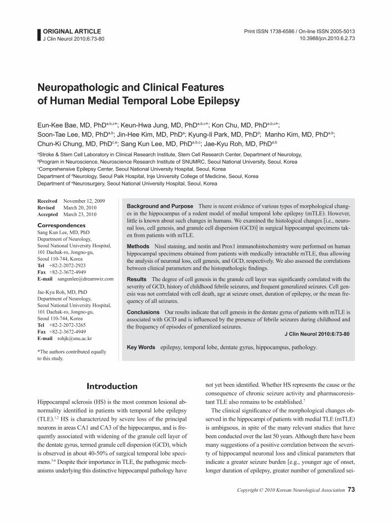
cells/HPF)”, grade 3 was “a moderate number of nestin-positive cells present (10-100 cells/HPF)”, and grade 4 was “a high num-ber of nestin-positive cells present (>100 cells/HPF).” The mean and SD values were calculated from the scored sections for each area separately (modified from Pirttilä et al.28). GCD was also measured on a scale of 1-4, where grade 1 describes epi-leptic qualities and a nondispersed appearance, grade 2 de-scribes granule cell bodies dispersed into the molecular layer forming an irregular outer border, grade 3 describes double-layer granule cell bodies organized into two layers, and grade 4 describes predominantly granule cell death (Fig. 1).3,28
Statistical analysisThe data are presented as mean±SD values. The Pearson cor-relation coefficient was determined to assess the associations between variables. Two groups were compared by chi-square analysis with arbitrary dichotomization, using SPSS for win-dows (version 15.0; SPSS Inc, Chicago, IL, USA). A value of p<0.05 was considered significant.
Results
Clinical characteristics of the patientsThe clinical characteristics of the 26 patients, who comprised
15 men and 11 women who were aged 32.07±9.29 years (ran-ge, 16-43 years) at the time of surgery, are listed in Table 1. The age at the onset of epilepsy was 11.11±6.98 years (range, 1-27 years), and the duration of epilepsy at the time of collec-tion was 17.76±10.58 years (range, 1-42 years). All patients had complex partial seizures, and the frequency of seizures per month was 4.25±5.98 (range, 0.5-30). In 16 patients (61.5%), secondary generalization frequently occurred more than once per month. A history of childhood FS was reported in 13 patients (50%), and 7 patients reported other relevant ca-usative childhood events (IPI), for example head trauma, men-ingitis, encephalitis, and Reye syndrome. No patient reported a history of SE. Most of the patients (80.7%) became seizure-free after their epilepsy surgery.
Pathological findingsThe pathological grading scores of Nissl (for neuronal loss), nestin (for cell genesis), and Prox1 (for GCD) are listed in Ta-ble 2, based on the grading system presented in Fig. 1. Most pa-tients showed a moderate-to-severe amount of neuronal loss: gra-de 3 in 12 patients (46.2%), grade 2 in 10 patients (38.5%), and grade 1 in 1 patient (3.8%). Most patients showed a mild-to-moderate amount of cell genesis: grade 1 in 8 patients (30.8%), grade 2 in 6 patients (23.1%), grade 3 in 11 patients (42.3%),
Fig. 1. Histopathological results of the semiquantitative scoring system. Nissl staining (for neuronal loss) shows (A) gra-de 1, 20% of neurons lost, (B) grade 2, 20-50% of neurons lost, and (C) grade 3 >50% of neurons lost. Nestin immunos-taining (for cell genesis) shows (D) grade 1, few positive cells present (<3 cells/HPF), (E) grade 2, a few positive ce-lls present (3-10 cells/HPF), (F) grade 3, a moderate number of positive cells pre-sent (10-100 cells/HPF), and (G) grade 4, a high number of positive cells pres-ent (>100 cells/HPF). Prox1 immunos-taining (for GCD) shows (H) grade 2, gra-nule cell bodies dispersed into the mo-lecular layer forming an irregular outer border, (I) grade 3, double-layer granule cell bodies organized in two layers, and (J) grade 4, granule cell death prevails. Sca-le bar=100 μm. GCD: granule cell disper-sion.

Neuropathologic Features of TLE
76 J Clin Neurol 2010;6:73-80
and grade 4 in 1 patient (3.8%). All patients exhibited some de-gree of GCD: grade 2 in 13 patients (50%), grade 3 in 7 patients (26.9%), and grade 4 in 6 patients (23.1%). With regard to the extrahippocampal pathological findings, only one patient ex-hibited apparent cortical dysplasia on magnetic resonance imaging (MRI) and macroscopic examination. However, 11 pa-tients (42.3%) exhibited MRI-negative microscopic dyspla-sia in the resected temporal lobe specimens on histological ex-amination.
Association between clinical and pathological va-riablesThere were significant correlations between cell genesis and GCD (r=0.43, p=0.03) and between neuronal loss and GCD (r=0.73, p<0.01). Clinical variables, including age at onset, age at surgery, duration of epilepsy, and mean frequency of preop-erative seizures, were not correlated with the pathological vari-ables, including neuronal loss, cell genesis, and GCD (Table 3). We dichotomized the results of nestin immunoreactivity into a
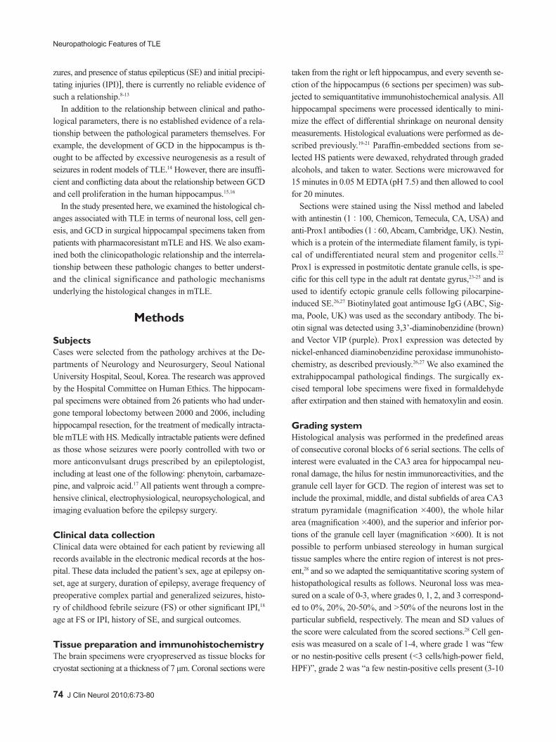
mild degree of cell genesis (grades 1 and 2) and a moderate-to-severe degree (grades 3 and 4). Moderate-to-severe cell gene-sis occurred more frequently in patients with a history of FS (p=0.03)(Fig. 2) and in those with frequent generalized seizures (p=0.026, both by chi-square test)(Fig. 2). Neuronal loss and GCD were not significantly correlated with FS or with the fre-quency of generalized seizures. The presence of temporal micro-scopic dysplasia was not significantly correlated with the oth-er pathological findings, including neuronal loss, cell genesis, or GCD, or with any clinical variable including the history of FS and frequent generalized seizures.
Discussion
We investigated the histological changes (neuronal loss, cell genesis, and GCD) in the hippocampus from mTLE patients. Cell genesis, measured as the number of nestin-positive cells in the granule cell layer, was significantly correlated with the severity of GCD, history of childhood FS, and frequent gen-
Table 1. Clinical parameters of medial temporal lobe epilepsy patientsPatient
no.Sex
Onset age (years)
Surgery age (years)
Epilepsy duration (years)
Seizure frequency (times/month)
Secondary generalization
FS IPIAge at
IPI (years)01 F 01 43 42 8 F – Head trauma 0102 M 04 23 19 3.5 R + – 0003 F 04 37 33 4 F – – 0004 F 16 28 12 8 F + – 0005 M 14 27 13 0.5 R + Head trauma 1206 F 11 19 08 5 R – Reye syndrome 0307 F 01 18 17 1 F + – 0108 M 13 21 08 0.5 F + Meningitis 0309 M 27 39 12 2.5 F – Encephalitis 0610 M 06 20 14 30 F + – 0011 M 16 51 35 1.5 F – – 0012 M 11 37 26 10 R – – 0013 F 12 16 04 1 F + – 0014 M 15 27 12 1 F + – 0015 M 20 31 11 1 F – Encephalitis 1016 M 03 24 21 2 F + – 0317 F 10 42 32 10 R – – 0018 F 08 33 25 2 R + – 0019 F 25 25 01 2 F + – 0020 F 06 20 14 0.5 R – – 0021 M 02 29 27 3 F – – 0222 M 16 22 06 ? R – – 0023 M 16 21 05 1 F + – 0024 M 10 36 26 3.5 F – – 0025 F 16 33 17 5 R + – 0026 M 06 28 22 1 R – Encephalitis 05
Not all clinical data were available for every patient. For secondary generalization, F (frequent) means more than once per month and R (rare) means less than once per month.M: male, F: female, FS: febrile seizures, IPI: initial precipitating injuries.

Bae EK et al.
www.thejcn.com 77
eralized seizures. However, cell genesis was not associated with the extent of neuronal loss, age of seizure onset, duration of epi-lepsy, or the mean frequency of seizures in the preoperative period. In addition, the presence of microscopic cortical dys-plasia in the surgical temporal lobe specimens was not corre-lated with other pathologic parameters including the degree of neuronal loss, cell genesis, and GCD. These results suggest that newly generated granule cells lead to GCD and are likely to be influenced by FS in childhood and by frequent episodes of generalized seizures.
Granule cell neurons are generated throughout life from a population of continuously dividing progenitor cells that reside in the subgranular zone of the rodent dentate gyrus.29 This also seems to occur in humans, because new neurons, as identified by bromodeoxyuridine staining, are generated from dividing progenitor cells in the dentate gyrus of the adult human brain.30 Prolonged seizure activity markedly increases neurogenesis in
the dentate gyrus of adult rats. Pulse-chase bromodeoxyuridine labeling and immunohistochemistry for immature neuronal markers show that many newly generated neurons migrate in chains from the dentate subgranular zone to ectopic locations in the hilus and molecular layer after pilocarpine-induced SE.26 Thus, one hypothesis is that GCD in humans with TLE results from enhanced neurogenesis induced by prolonged seizures. Although our results support this hypothesis, some recent studi-es have shown contrary results. For example, in a rodent model of epilepsy after kainate injection and in the hippocampi from patients with TLE, GCD does not result from increased neuro-genesis, but rather from abnormal migration of mature gran-ule cells along a radial glial scaffold, most likely caused by lo-cal reelin deficiency.15,31 Nestin, which is an indicator of cell genesis, is expressed in the neuroglial cells in addition to undif-ferentiated precursors of neurons.22 Therefore, nestin positivity cannot be attributed entirely to neurogenesis, and this may ex-plain some of the differences between our data and those of previous studies.
We demonstrated increased cell genesis in patients with fre-quent generalized seizures. Likewise, seizure-induced neuro-
Table 2. The pathological grading scores of Nissl staining, and nes-tin and Prox1 immunohistochemistry, and the presence of cortical dysplasia in human temporal lobe surgical specimens
Patientno.
Cell genesis(Nestin)
Neuronal loss (Nissl)
Granule cell dispersion
(Prox1)
Cortical dysplasia in the temporal
lobe surgical specimen
01 1 1 2 +02 2 3 3 +03 2 2 2 +04 1 1 2 –05 3 2 2 –06 1 3 3 –07 3 2 2 –08 2 3 3 +09 3 3 4 +10 3 3 2 +11 3 3 4 +12 1 3 4 +13 3 2 3 +14 3 2 3 –15 3 1 2 –16 3 3 4 –17 1 2 2 +18 2 1 2 –19 2 2 2 –20 1 2 2 –21 3 3 3 –22 1 2 2 –23 3 3 4 +24 4 3 4 –25 2 3 3 –26 1 2 2 –
Table 3. Pearson correlation coefficients between clinical vari-ables and pathological results
Cell genesis (Nestin)
Neuronal loss (Nissl)
Granule cell dispersion (Prox1)
Surgery ageCorrelation -0.06 -0.11 0.16p 0.76 0.59 0.45
Onset ageCorrelation 0.12 0.01 0.19p 0.56 0.97 0.35
Epilepsy durationCorrelation -0.1 -0.07 0.04p 0.61 0.72 0.85
Seizure frequency (Per month)
Correlation -0.11 0.13 -0.17p 0.6 0.52 0.41
Cell genesis (Nestin)
Correlation – 0.32 0.43p – 0.12 0.03*
Neuronal loss(Nissl)
Correlation 0.32 – 0.73p 0.12 – <0.01*
Granule cell dispersion (Prox 1)
Correlation 0.43 0.73 –p 0.03* <0.01* –
*Denotes significant correlation.

Neuropathologic Features of TLE
78 J Clin Neurol 2010;6:73-80
genesis in the dentate gyrus has been proven in various ani-mal models, such as, pilocarpine-induced SE,20,21,32 kainic acid injection,33 and amygdala kindling.34 Seizure-induced neu-rogenesis has also been described in adults16 and in children with mTLE.35,36 Although the role of seizure-induced neurogene-sis in the pathophysiology of TLE is uncertain, it is possible that newly generated cells contribute to the formation of GCD and ectopic granule cells in the hilus. Seizure-induced cell pro-liferation and the likelihood of developing spontaneous recu-rrent seizures following pilocarpine-induced SE are reduced by the antimitotic agent cytosine-b-D-arabinofuranoside in adult rodent models, which suggests that hippocampal cell pro-liferation plays a proepileptic rather than a compensatory role.20
Experimentally prolonged FS results in late-onset limbic (temporal lobe) epilepsy,37 but the epileptogenic potential of prolonged FS in humans remains unclear. Although retrospec-tive analysis has implicated early-life FS as a risk factor for the development of TLE in humans,38 whether early-life FS actu-ally causes TLE or is simply indicative of another, perhaps gene-
tically determined vulnerability that eventually results in TLE, cannot be determined in correlative clinical studies.37 Moreover, the epileptogenic mechanisms underlying FS remain unknown, but might involve enduring changes at the molecular and func-tional levels, such as alterations in neurotransmitter receptors or voltage-gated ion channels, and might not involve neuronal loss.39 Although the effects of FS on cell proliferation in the den-tate gyrus have been described in animal models,40,41 there is little evidence of this in humans. We found a positive correla-tion between early-life FS and ongoing cell proliferation in adult human hippocampi taken from TLE patients, but further par-allel human and animal studies are needed to demonstrate the role of altered cell proliferation after FS and other epileptogen-ic mechanisms of FS.
We found a positive correlation between the severity of HS (i.e., neuronal loss) and the presence of GCD, which is in ac-cordance with the results of recent studies5,6,13 and with the ori-ginal work by Houser.3 Considering that the presence of GCD was not correlated with clinical factors such as duration of epi-lepsy or frequency of seizures, GCD might be more closely linked to the pathological process of HS rather than being a manifestation of severe temporal lobe seizures.6 We also found no correlation between clinical variables relating to seizure bur-den and the severity of HS. This is a conflicting area, and most pathological or longitudinal MRI studies have suggested a correlation between the severity of hippocampal neuronal loss and the duration of epilepsy.13 However, few studies have thor-oughly determined of the burden of seizures,13 and one limita-tion to our study is that we did not know the exact total number of generalized and partial seizures. Despite this limitation, we found no evidence that HS occurs as a consequence of recur-rent seizures in patients with TLE.
Microscopic cortical dysplasia or microdysgenesis mani-fests as minor abnormalities on histological examination, even when the MRI or macroscopic examination reveals no abnor-malities.42 Microscopic cortical dysplasia may appear in 20-45% of surgical specimens from mTLE patients with HS.43-45 We found microscopic cortical dysplasia in 11 patients (42.3%), a rate similar to that of previous reports, suggesting that it is a relatively commonly associated finding in HS. It has been su-ggested that this represents a preexisting susceptibility factor that renders the affected brain vulnerable to the development of mTLE after IPI or FS, but the reciprocal relationship re-mains unknown.43 Recent data show that microscopic cortical dysplasia is not related to clinical parameters such as IPI or FS, or to the histological characteristics of HS;43-45 our results are consistent with these previous findings. Hence, the role of mi-croscopic cortical dysplasia is currently obscure, and future studies are needed to reveal the complexities.
The semiquantitative scoring system that we applied has
Fig. 2. The distribution of (A) patients with or without a history of FS and (B) with frequent or rare generalized seizures, relative to the degree of nestin positivity (cell genesis). A mild degree of cell genesis was classified as the pathological grading score of nestin of 1 or 2, and a moderate-to-severe degree of cell genesis was classified as grade 3 or 4. Cell genesis was significantly higher in patients with a history of FS (p=0.030) and with frequent general-ized seizures (p=0.026). FS: febrile seizures.
B
16
14
10
6
4
2
14
12
8
6
4
2
0Rare
Generalized seizures
Frequent
Cell genesis; mildCell genesis; moderate to severe
p=0.026
A
14
10
12
76
8
6
4
2
0No
FS
Yes
Cell genesis; mildCell genesis; moderate to severe
p=0.030
1
12

Bae EK et al.
www.thejcn.com 79
some limitations. We were unable to quantify real cell counts for neuronal loss and cell genesis, or to measure the real thick-ness of granule cell layer for GCD, because the tissue sections of the surgical specimens did not precisely correspond. Our re-sults may thus be inconclusive; nevertheless, we think that our results might be suggestive of the neuropathologic features of human specimens.
In conclusion, our study showed that increased cell genesis is correlated with the severity of GCD in the human hippocam-pal dentate gyrus of medically intractable mTLE patients, sup-porting the view that newly generated granule cells might lead to GCD. The degree of cell genesis was also related to the his-tory of childhood FS and frequent generalized seizures, but was not significantly associated with the degree of neuronal loss or other clinical variables, such as the age at onset, dura-tion of epilepsy, or the mean frequency of all seizures.
Conflicts of InterestThe authors have no financial conflicts of interest.
AcknowledgementsThis study was supported by the Ministry of Health and Welfare (no. A060452), Republic of Korea.
Dr. J-K. Roh was supported by a grant from Seoul National University Hospital, South Korea (no. 2120040140).
REFERENCES1. Babb TL, Brown WJ. Pathological findings in epilepsy. In: Engel J
Jr. New York: Raven Press, 1987;511-540.2. Meencke HJ, Veith G. Hippocampal sclerosis in epilepsy. In: Luders
H. Epilepsy surgery. New York: Raven Press, 1991;705-715.3. Houser CR. Granule cell dispersion in the dentate gyrus of humans
with temporal lobe epilepsy. Brain Res 1990;535:195-204.4. Lurton D, El Bahh B, Sundstrom L, Rougier A. Granule cell disper-
sion is correlated with early epileptic events in human temporal lobe epilepsy. J Neurol Sci 1998;154:133-136.
5. El Bahh B, Lespinet V, Lurton D, Coussemacq M, Le Gal La Salle G, Rougier A. Correlations between granule cell dispersion, mossy fiber sprouting, and hippocampal cell loss in temporal lobe epilepsy. Epi-lepsia 1999;40:1393-1401.
6. Thom M, Sisodiya SM, Beckett A, Martinian L, Lin WR, Harkness W, et al. Cytoarchitectural abnormalities in hippocampal sclerosis. J Neuropathol Exp Neurol 2002;61:510-519.
7. Blümcke I, Thom M, Wiestler OD. Ammon’s horn sclerosis: a malde-velopmental disorder associated with temporal lobe epilepsy. Brain Pathol 2002;12:199-211.
8. Cavanagh JB, Meyer A. Aetiological aspects of Ammon’s horn scle-rosis associated with temporal lobe epilepsy. Br Med J 1956;2:1403-1407.
9. Dam AM. Epilepsy and neuron loss in the hippocampus. Epilepsia 1980; 21:617-629.
10. Babb TL, Brown WJ, Pretorius J, Davenport C, Lieb JP, Crandall PH. Temporal lobe volumetric cell densities in temporal lobe epilepsy. Ep-ilepsia 1984;25:729-740.
11. Mathern GW, Adelson PD, Cahan LD, Leite JP. Hippocampal neuron damage in human epilepsy: Meyer’s hypothesis revisited. Prog Brain Res 2002;135:237-251.
12. Fuerst D, Shah J, Kupsky WJ, Johnson R, Shah A, Hayman-Abello B, et al. Volumetric MRI, pathological, and neuropsychological pro-
gression in hippocampal sclerosis. Neurology 2001;57:184-188.13. Thom M, Zhou J, Martinian L, Sisodiya S. Quantitative post-mortem
study of the hippocampus in chronic epilepsy: seizures do not inevita-bly cause neuronal loss. Brain 2005;128:1344-1357.
14. Jessberger S, Römer B, Babu H, Kempermann G. Seizures induce pro-liferation and dispersion of doublecortin-positive hippocampal pro-genitor cells. Exp Neurol 2005;196:342-351.
15. Fahrner A, Kann G, Flubacher A, Heinrich C, Freiman TM, Zentner J, et al. Granule cell dispersion is not accompanied by enhanced neuro-genesis in temporal lobe epilepsy patients. Exp Neurol 2007;203:320-332.
16. Thom M, Martinian L, Williams G, Stoeber K, Sisodiya SM. Cell pro-liferation and granule cell dispersion in human hippocampal sclero-sis. J Neuropathol Exp Neurol 2005;64:194-201.
17. Wiebe S, Blume WT, Girvin JP, Eliasziw M; Effectiveness and Efficien-cy of Surgery for Temporal Lobe Epilepsy Study Group. A randomized, controlled trial of surgery for temporal-lobe epilepsy. N Engl J Med 2001;345:311-318.
18. Mathern GW, Babb TL, Vickrey BG, Melendez M, Pretorius JK. The clinical-pathogenic mechanisms of hippocampal neuron loss and sur-gical outcomes in temporal lobe epilepsy. Brain 1995;118:105-118.
19. Chu K, Kim M, Jung KH, Jeon D, Lee ST, Kim J, et al. Human neu-ral stem cell transplantation reduces spontaneous recurrent seizures following pilocarpine-induced status epilepticus in adult rats. Brain Res 2004;1023:213-221.
20. Jung KH, Chu K, Kim M, Jeong SW, Song YM, Lee ST, et al. Contin-uous cytosine-b-D-arabinofuranoside infusion reduces ectopic gran-ule cells in adult rat hippocampus with attenuation of spontaneous recurrent seizures following pilocarpine-induced status epilepticus. Eur J Neurosci 2004;19:3219-3226.
21. Jung KH, Chu K, Lee ST, Kim J, Sinn DI, Kim JM, et al. Cyclooxy-genase-2 inhibitor, celecoxib, inhibits the altered hippocampal neuro-genesis with attenuation of spontaneous recurrent seizures following pi-locarpine-induced status epilepticus. Neurobiol Dis 2006;23:237-246.
22. Kruglyakova EP, Khovryakov AV, Shikhanov NP, Maccann GM 2nd, Vael’ I, Kruglyakov PP, et al. Nestin-expressing cells in the human hip-pocampus. Neurosci Behav Physiol 2005;35:891-897.
23. Pleasure SJ, Collins AE, Lowenstein DH. Unique expression patterns of cell fate molecules delineate sequential stages of dentate gyrus de-velopment. J Neurosci 2000;20:6095-6105.
24. Elliott RC, Khademi S, Pleasure SJ, Parent JM, Lowenstein DH. Dif-ferential regulation of basic helix-loop-helix mRNAs in the dentate gy-rus following status epilepticus. Neuroscience 2001;106:79-88.
25. Bagri A, Gurney T, He X, Zou YR, Littman DR, Tessier-Lavigne M, et al. The chemokine SDF1 regulates migration of dentate granule cells. Development 2002;129:4249-4260.
26. Parent JM, Elliott RC, Pleasure SJ, Barbaro NM, Lowenstein DH. Ab-errant seizure-induced neurogenesis in experimental temporal lobe epi-lepsy. Ann Neurol 2006;59:81-91.
27. McCloskey DP, Hintz TM, Pierce JP, Scharfman HE. Stereological methods reveal the robust size and stability of ectopic hilar granule cells after pilocarpine-induced status epilepticus in the adult rat. Eur J Neurosci 2006;24:2203-2210.
28. Pirttilä TJ, Manninen A, Jutila L, Nissinen J, Kälviäinen R, Vapalahti M, et al. Cystatin C expression is associated with granule cell dispersion in epilepsy. Ann Neurol 2005;58:211-223.
29. Kuhn HG, Dickinson-Anson H, Gage FH. Neurogenesis in the dentate gyrus of the adult rat: age-related decrease of neuronal progenitor proliferation. J Neurosci 1996;16:2027-2033.
30. Eriksson PS, Perfilieva E, Björk-Eriksson T, Alborn AM, Nordborg C, Peterson DA, et al. Neurogenesis in the adult human hippocampus. Nat Med 1998;4:1313-1317.
31. Heinrich C, Nitta N, Flubacher A, Müller M, Fahrner A, Kirsch M, et al. Reelin deficiency and displacement of mature neurons, but not neu-rogenesis, underlie the formation of granule cell dispersion in the epi-

Neuropathologic Features of TLE
80 J Clin Neurol 2010;6:73-80
leptic hippocampus. J Neurosci 2006;26:4701-4713.32. Scharfman HE, Goodman JH, Sollas AL. Granule-like neurons at the
hilar/CA3 border after status epilepticus and their synchrony with area CA3 pyramidal cells: functional implications of seizure-induced neu-rogenesis. J Neurosci 2000;20:6144-6158.
33. Gray WP, Sundstrom LE. Kainic acid increases the proliferation of gran-ule cell progenitors in the dentate gyrus of the adult rat. Brain Res 1998; 790:52-59.
34. Parent JM, Janumpalli S, McNamara JO, Lowenstein DH. Increased dentate granule cell neurogenesis following amygdala kindling in the adult rat. Neurosci Lett 1998;247:9-12.
35. Blümcke I, Schewe JC, Normann S, Brüstle O, Schramm J, Elger CE, et al. Increase of nestin-immunoreactive neural precursor cells in the dentate gyrus of pediatric patients with early-onset temporal lobe epi-lepsy. Hippocampus 2001;11:311-321.
36. Takei H, Wilfong A, Yoshor D, Armstrong DL, Bhattacharjee MB. Evidence of increased cell proliferation in the hippocampus in children with Ammon’s horn sclerosis. Pathol Int 2007;57:76-81.
37. Dubé C, Richichi C, Bender RA, Chung G, Litt B, Baram TZ. Tempo-ral lobe epilepsy after experimental prolonged febrile seizures: pro-spective analysis. Brain 2006;129:911-922.
38. Cendes F, Andermann F. Do febrile seizures: promote temporal lobe epilepsy? Retrospective studies. In: Baram TZ, Shinnar S. San Diego:
Academic Press, 2002;78-88.39. Dube CM, Brewster AL, Richichi C, Zha Q, Baram TZ. Fever, fe-
brile seizures and epilepsy. Trends Neurosci 2007;30:490-496.40. Lemmens EM, Lubbers T, Schijns OE, Beuls EA, Hoogland G. Gen-
der differences in febrile seizure-induced proliferation and survival in the rat dentate gyrus. Epilepsia 2005;46:1603-1612.
41. Bender RA, Dubé C, Gonzalez-Vega R, Mina EW, Baram TZ. Mossy fiber plasticity and enhanced hippocampal excitability, without hip-pocampal cell loss or altered neurogenesis, in an animal model of prolonged febrile seizures. Hippocampus 2003;13:399-412.
42. Armstrong DD. The neuropathology of temporal lobe epilepsy. J Neuropathol Exp Neurol 1993;52:433-443.
43. Kasper BS, Stefan H, Paulus W. Microdysgenesis in mesial temporal lobe epilepsy: a clinicopathological study. Ann Neurol 2003;54:501-506.
44. Diehl B, Najm I, LaPresto E, Prayson R, Ruggieri P, Mohamed A, et al. Temporal lobe volumes in patients with hippocampal sclerosis with or without cortical dysplasia. Neurology 2004;62:1729-1735.
45. Kalnins RM, McIntosh A, Saling MM, Berkovic SF, Jackson GD, Briellmann RS. Subtle microscopic abnormalities in hippocampal sclerosis do not predict clinical features of temporal lobe epilepsy. Epilepsia 2004;45:940-947.