neurosurgeryneurosurgery. outline a & p pathology diagnostics/pre-operative testing...
TRANSCRIPT

NeurosurgeryNeurosurgery
Outline• A & P• Pathology• Diagnostics/Pre-operative Testing• Medications/Anesthesia• Positioning/Prepping/Draping• Supplies/Instrumentation/Equipment• Dressings/Drains/Post-op Care• Procedures: Carpal Tunnel Release,
Craniotomy, Cervical Discectomy, Lumbar Discectomy, Ventroperitoneal Shunt

Nervous System• Functions:• Senses changes in environment• Interprets changes• Stimulates movement to respond
to these changes
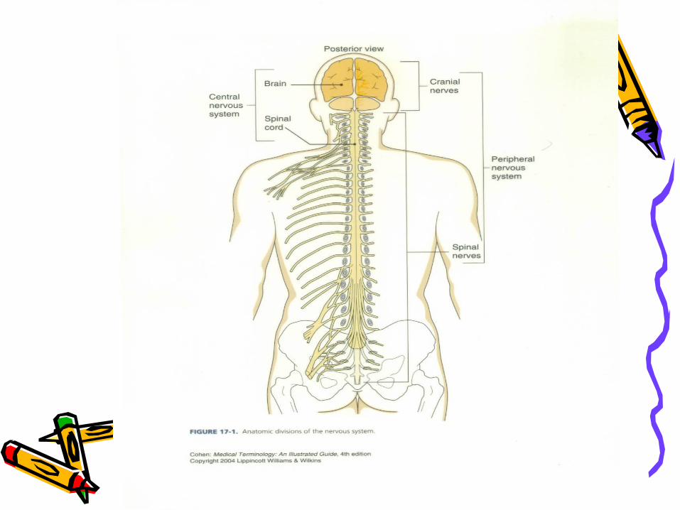
Organization of the Nervous System
• Two systems:1. CNS Central Nervous System• Two major parts: Brain and Spinal
Cord
2. PNS Peripheral Nervous System• Everything else

Peripheral Nervous System
• Two major parts:• Afferent Nervous System • Sensory neurons take info from
PNS to CNS
• Efferent Nervous System• Motor neurons take info from CNS
to PNS
Efferent Nervous System
• Motor nervous system• 2 parts:1. Somatic Nervous System• Skeletal muscle control• Conscious control2. Autonomic Nervous System• Cardiac muscle, smooth muscle, and glands• Unconscious control• Has 2 divisions:• Sympathetic Division• Parasympathetic Division
Autonomic Nervous System
• Sympathetic vs. Parasympathetic• Controlled by hypothalamus and medulla
oblongata• Both are different nerves going to the same
effector or target• Are antagonistic• Parasympathetic = decreased skeletal blood
flow, increased organ blood flow (quietly eating)
• Sympathetic = increased skeletal blood flow, decreased organ blood flow (eatus interruptus by a bear!) Also called fight or flight; prepares body for emergencies
Spinal Cord• Functions: • Info to and from the brain• Integration of reflexes
• Location:• Begins at foramen magnum and
extends to 2nd lumbar• About 16-18” in length
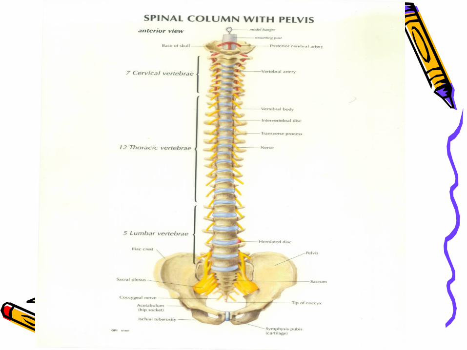
Spinal Cord Support Structures
• Vertebra • 33 total• 7 cervical• 12 thoracic• 5 lumbar• Sacrum formed by 5 fused bones• Coccyx formed by 4 fused bones
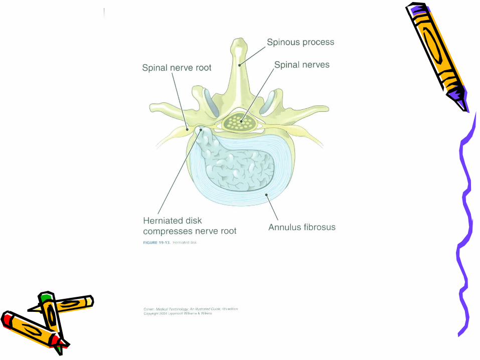
Intervertebral Disks• Separate vertebrae• Outer layer is tough and called the
annulus fibrosis• Inner core is soft and called the
nucleus pulposus • Bear stress incurred with body
weight and when lifting
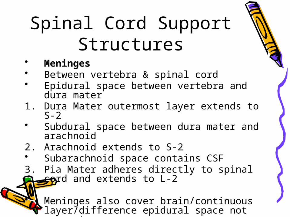
Spinal Cord Support Structures
• Meninges • Between vertebra & spinal cord• Epidural space between vertebra and dura
mater1. Dura Mater outermost layer extends to S-2• Subdural space between dura mater and
arachnoid 2. Arachnoid extends to S-2• Subarachnoid space contains CSF3. Pia Mater adheres directly to spinal cord and
extends to L-2
• Meninges also cover brain/continuous layer/difference epidural space not present

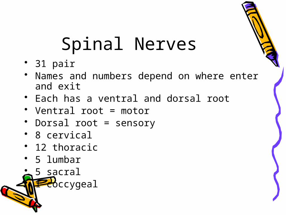
Spinal Nerves• 31 pair• Names and numbers depend on where enter
and exit• Each has a ventral and dorsal root• Ventral root = motor• Dorsal root = sensory• 8 cervical• 12 thoracic• 5 lumbar• 5 sacral• 1 coccygeal
Brain • Protected by the cranium or skull

Brain • 4 major parts:• Brain stem• Diencephalon• Cerebellum• Cerebrum
Weight about 3 lbs.


Support Structures of the Brain
1. Meninges • Continuous layer with spinal cord• NO epidural space
Support Structures of the Brain
2. Cerebrospinal fluid (CSF)• About 800ml produced each day by the
choroid plexus, a specialized set of capillaries
• Circulates inside subarachnoid space through central canal of spinal cord and the ventricles of the brain
• Reabsorbed in arachnoid villus found in the parietal lobe
• Functions as a shock absorber and circulates nutrients
Support Structures of the Brain
3. Blood Brain Barrier• Specialized set of capillaries exclusive
to the central nervous system• Less permeable than any other
capillaries in the body• Advantage = keeps out unwanted
chemicals• Disadvantage = difficult to diffuse
materials out, hence difficulty in treating conditions such as encephalitis
Brain Stem• 3 parts:• Medulla oblongata• Pons • Midbrain
Medulla oblongata• Contains:• 5 of 12 cranial nerves• Pyramids: function only with motor
neurons/a crossing of the spinal nerve impulses
• Reflex centers: hiccupping, sneezing, coughing
• Vital reflex centers:• Cardiac center – heart rate• Vasoconstrictor center-BP via blood
vessel diameter control• Respiratory center - breathing
Pons • Above medulla• Switching point for motor neurons• Respiratory center
Midbrain or Mesencephalon
• Above pons• Involuntary eye and head
movement in response to auditory stimuli
Diencephalon • 2 parts:• Thalmus• Hypothalmus
Thalmus • Relay center for sensory
information• Interprets stimuli for example pain
from changes in temperature (hot stove)
• 1st level of reasoning occurs here• Recognizes crude touch NOT
localized touch
Hypothalmus • Controls large number of subconscious
functions• Controls most of Autonomic nervous
system • Where endocrine and nervous systems
interface• Homeostasis regulation of the body• Controls: body temp, thirst, hunger,
sleep and waking habits, psychosomatic disorders, rage and aggression
Cerebellum • 2nd largest part of the brain• Primarily a motor area• Controls skeletal muscles,
subconsciously• Receives sensory input from eyes,
muscles, joints, and inner ear• Posture, balance, coordination,
equilibrium• Muscle sense tells body where other
parts are
Cerebrum • Largest part of brain• Motor/sensory/association area• 4 Lobes: frontal, parietal,
occipital, temporal• Each controls a specific function
be it motor or sensory• Limbic system: controls
emotion/functions in cerebral cortex and diencephalon
• See page 970 Figure 24-4 in Price

Cerebrum Lobes’ Function
• Frontal• Memory,
abstract thinking, ethics, judgement, emotion, expressive speech, motor
• Parietal • Sensory,
receptive speech, written word
• Temporal • Auditory,
olfactory• Occipital• Visual cortex• Visual
association
Cranial Nerves• All originate in the brain stem
EXCEPT the 1st and 2nd
• Classified as sensory or mixed (sensory and motor) nerves
• Directly off of brain• Do not go through the spine• Identified by Roman numerals and
names
Cranial NervesI. Olfactory - sense of smellII. Optic – sense of sight/visionIII. Occulomotor – eyeball, eyelid movement (medial,
inferior, superior rectus, inferior oblique), pupil constriction, lens accommodation
Muscle sense for eyeballIV. Trochlear – eyeball movement (superior oblique) Muscle sense for eyeballV. Trigeminal – masseter muscle control Sensory part has 3 branches: ophthalmic
(forehead to corner of eye), maxillary (corner of eye to upper lip/teeth), and mandibular (lower lip/teeth/tongue)
All three convey sense of touch, pain and temp changes
VI. Abducens - same as IV eyeball movement (lateral rectus) and eyeball muscle sense
FYI: EOM formula LR6(SO4)3

Cranial NervesVII. Facial- facial muscles, lacrimal and salivary glands
anterior 2/3 of tongue (taste)VIII. Vestibulocochlear -last of totally sensory nerves;
has 2 branches: vestibular conveys balance and cochlear which conveys sense of hearing
IX. Glossopharyngeal -salivary gland secretion and posterior 1/3 of tongue
X. Vagus – internal organ control motor and sensory; originates in medulla and goes down through neck into chest and abdomen
XI. Accessory – controls head and neck movement, speech, and muscle sense for the head
XII. Hypoglossal – tongue muscles: swallowing, speech, muscle sense for tongue
Neuro Pathology

Cervical Spine Pathology
• Very serious• Can have severe consequences related
to all of the spinal cords’ nerve pathways
• Spondylosis is osteophyte or bone spur formation in the spinal canal
• Cervical disk extrusion acute or chronic• Treatment errs on the side of caution
due to potential extreme consequences of surgical intervention

Thoracic Pathology• Spondylosis• Extrusion of disk
Lumbar Pathology• Spondylosis• Stenosis• Spondylolithesis • Disk extrusion
Neoplasms/Tumors • Two types:• Primary • Originate in nervous tissue or
meninges• Secondary • Are metastasized from other parts
of the body
• May be classified as benign or malignant
Tumors • Benign tumors:• “Craniopharyngiomas, epidermoids,
hemangiomas, menigiomas, acoustic neuromas, and pituitary microadenomas” Price, 2004
• Malignant tumors:• “Astrocytes or gliomas” Price, 2004
• Benign usually excisable via craniotomy• Malignant normally cannot be completely
removed but efforts are made to remove most
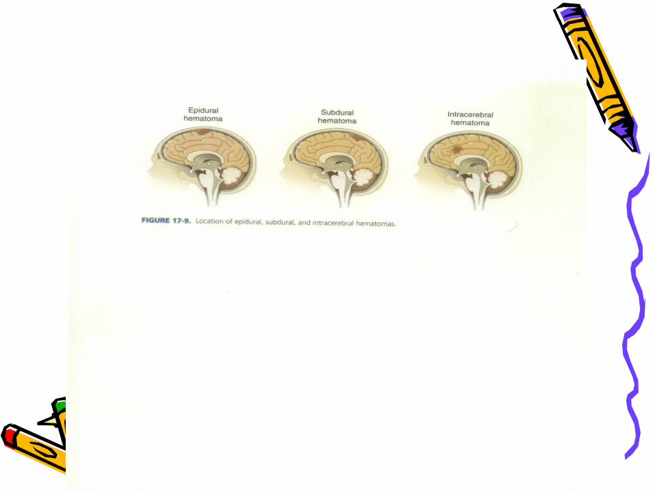
Head Trauma • Includes; • Scalp lacerations, fractures,
hematomas (epidural or subdural), and brain injuries

Spinal Cord Trauma• Vertebral Fracture• Vertebral Dislocation • Herniated disk into spinal canal• Laceration from GSW or MVA
Cerebrovascular Disease
• #3 cause of death in US• Symptoms reflect ischemia (TIAs)
or hemorrhage• Intracranial aneurysm• Arteriovenous malformations• Brain hemorrhage • Stroke or cerebrovascular accident
(CVA)

Congenital Pathology• Craniosynotosis “premature
closure of the cranial sutures” Price, 2004
• Hydrocephalus result of obstructed CSF flow
• Spina bifida

Infection • Abscess• Subdural empyema• Post-op infection

Spinal Cord Tumors• Intramedullary in the spinal cord• Intradural in dura, outside spinal
cord• Extradural outside spinal cord Price, 2004
Peripheral Nerve Pathology
• Carpal tunnel syndrome - compression of the median nerve
• Ulnar nerve compression – compression of ulnar nerve by the ligament of Osborne
Price, 2004

Diagnosis • History and physical• Symptoms usually specific to area of
pathology• Electroencephalogram (EEG)• X-ray• Myelogram • CAT Scan• MRI• Cerebral arteriograms

Medications• Lidocaine 1% with epinephrine• Topical hemostatic agents: gelfoam,
avitene, surgicel, bone wax• Antibiotic irrigants• Topical papaverine for prevention of
spasm during intracranial artery surgery
• Methyl methacrylate with cranioplasty• Heparin saline irrigation again with
intracranial artery surgery• Contrast solutions with cerebral
arteriography• Gliadel wafers (tumor bed of
glioblastoma)
Anesthesia • General • Could be local with MAC for minor
laceration suturing
Positioning • Cranial Surgery• Supine primarily,
with a specialty headrest and or fixation devices
• Can be lateral or semi-lateral
• Sitting• Prone• Varies with location
of access
• Spinal surgery• Anterior procedures
require supine• Posterior procedures
require prone

Preps • Will require shave especially on
head• Varies with surgeon preference:
betadine, alcohol, chlorohexidine • Care taken NOT to get in patient’s
eyes or facial orifices
Draping • Toweled out• Adhesive type drape• Specialty drapes: laparotomy,
thyroid, craniotomy, lumbar• Stockinette for peripheral
procedures
Supplies • Marking pen• Disposable bi-polar cord• Monopolar pencil/bovie• Cottonoids/patties• Raney clips• Hemostatic clips• Shunt catheters, tubing, connectors• Cotton balls• Hemovac drain• Nerve stimulator• Telfa• Microscope drape• C-Arm drape• Ultrasound wand drape
Instruments • Minor tray if laminectomy and
craniotomy trays do not have basic instrumentation
• Laminectomy tray • Craniotomy tray• Basic ortho tray• Plates and screws• Specialty self-retaining retractor
trays: Greenburg

Miscellaneous Instrumentation
• See pages 987-990 in PRice
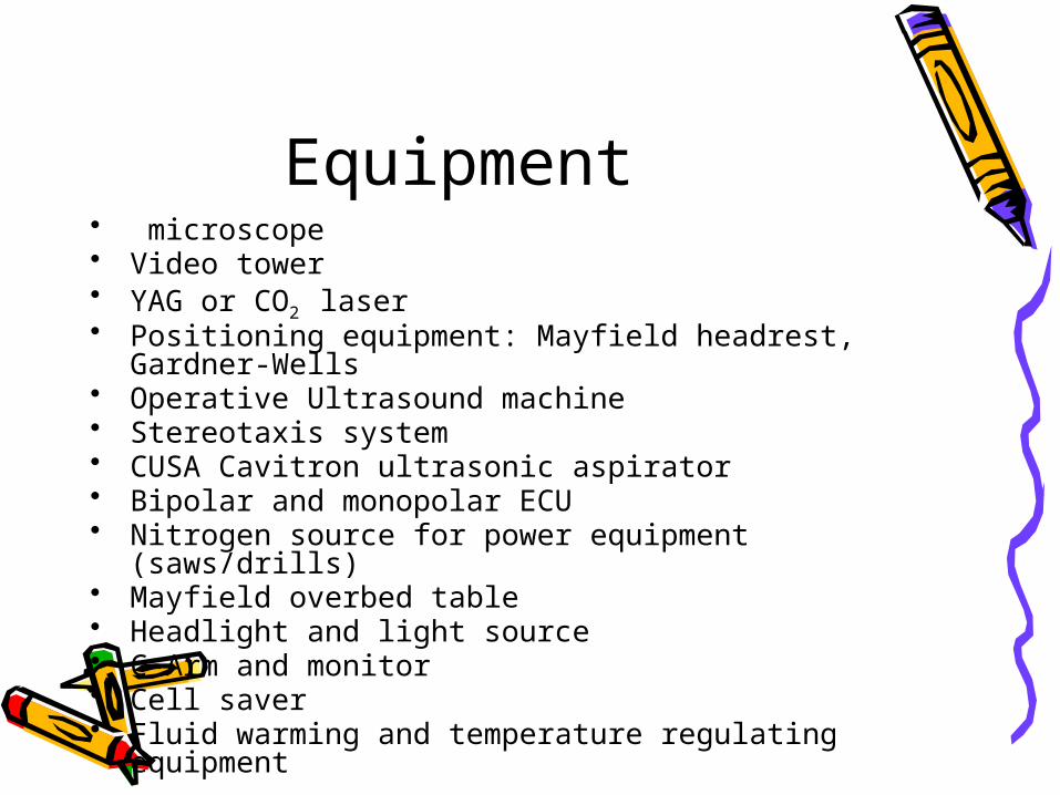
Equipment • microscope• Video tower• YAG or CO2 laser• Positioning equipment: Mayfield headrest, Gardner-
Wells • Operative Ultrasound machine• Stereotaxis system• CUSA Cavitron ultrasonic aspirator• Bipolar and monopolar ECU• Nitrogen source for power equipment (saws/drills)• Mayfield overbed table• Headlight and light source• C-Arm and monitor• Cell saver• Fluid warming and temperature regulating equipment
Dressings/Drains/Post-op Care
• Dressings surgeon preference• Drains surgeon preference• Post-op care: keep field sterile
until patient has left the OR• Careful with moving patient to
avoid patient injury and hemorrhage
Post-operative Complications
• Infection• Hemorrhage• Nerve damage• CSF leakage• Meningitis • Neurological deficits