neutron diffraction studies of human tooth enamel
TRANSCRIPT
Archs oral Bid. Vol. 15, pp. 47-63, 1969. Pergamon Press. Printed in Great Britain.
NEUTRON DIFFRACTION STUDIES OF HUMAN TOOTH ENAMEL
R. A. YOUNG and S. SPOONER
Georgia Institute of Technology, Atlanta, Georgia 30332, U.S.A.
Summary-In what is thought to be the first application of the technique in the hard tissue field, neutron diffraction study of human tooth enamel has recontirmed that the inorganic portion is basically an hydroxyapatite-like material. However, compared to hydroxyapatite, the tooth enamel shows (i) an apparent 20 per cent or greater deficiency of hydroxyl hydrogen, with probable substitution of other ions on or near the hexad axis, and (ii) an overall excess of hydrogen. Amorphous calcium phosphate, octacalcium phosphate, adsorbed water and structural hydrogen in unusual locations are shown not to be present in major degree, though the possible existence of minor components of OCP (- 2 wt “/,) and hydrogen involved with PO4 groups (e.g. 1 wt %) are not ruled out. It is therefore left to a combination of residual protein content and entrapped free water to account for the substantial quantity of hydrogen present external to the hydroxyapatite component.
The particular usefulness of neutron diffraction and scattering techniques for study of the amount, location, and, possibly, binding of the hydrogen present, whether or not structurally incorporated, is pointed out.
INTRODUCTION
THE PRESENT study is believed to be the first in which neutron diffraction techniques have been applied to the study of hard tissue. Neutrons are scattered more strongly by hydrogen than by many heavier elements. Thus, various biologically-interesting calcium-phosphate compounds, which incorporate hydrogen in different ways, can be more readily distinguished with neutron diffraction than with X-ray diffraction. As is shown here, neutron diffraction does indeed show that the crystalline fraction of this human tooth enamel specimen, while still “hydroxyapatite” in a relaxed sense of the word, is less similar to pure hydroxyapatite than has previously been indicated by X-ray diffraction studies. In addition, the strong incoherent scattering of neutrons by hydrogen has proved to be an especially useful phenomenon for the study of the hydrogen present, whether or not incorporated in a crystalline form.
The fundamental differences in the processes involved in the scattering of X-rays and of neutrons (BACON, 1962) make the two techniques complementary. Neutrons are scattered from the atomic nuclei (and from “magnetic electrons”, which are not important in this study) while X-rays are scattered almost entirely by the electrons surrounding the nuclei. One resultant difference is that the single-atom scattering amplitude for X-rays (atomic form-factor,f) falls off strongly with increasing scattering angle, whereas the single-atom coherent nuclear-scattering amplitude for neutrons (coherent nuclear-scattering length, b) is effectively independent of angle for the wavelengths ordinarily used in diffraction studies. A second resultant difference is that
47

48 R. A.YOLJNG AND KSPOONER
f increases monotonically with increasing atomic number Z (in fact, in Thomson- electron scattering units f = Z at zero scattering angle), whereas b shows no regular dependence on Z. Hydrogen (Z = I), for example, scatters neutrons coherently as strongly as does iridium (Z = 77), nearly half as strongly as does uranium and more than half as strongly as does calcium. A third resultant difference is that neutron scattering depends strongly on the particular isotope of an element while X-ray scattering has shown no such dependence. This effect is illustrated dramatically in the case of hydrogen. The scattering amplitude for neutron scattering from normal hydrogen (HI) is -0.38 x IO-l2 cm and that for deuterium (HZ) is 0.65 x IO-l2 cm. The difference in sign makes this one of the most marked of the isotopic effects, which arise from nuclear spin-dependent resonance scattering. The isotopic effect also includes a dependence on the nuclear spin state which, except under special conditions, is a random effect giving rise to a large incoherent scattering of neutrons from hydro- gen. In fact, this incoherent scattering dominates the diffuse scattering of neutrons in any hydrogenous material. These features of X-ray and neutron scattering are summarized in Table 1 for several elements which play a role in the structure of human tooth enamel and related minerals.
TABLE 1. X-RAY AND NEUTRON SCATTERING PROPERTIES OF SELECTED ELEMENTS.* LISTED ARE THE X-RAY FORM FACTOR AT ZERO SCATTERING
ANGLE v;(o)], THE NEUTRON COHERENT SCATTERING AMPLITUDE (b) AND THE INCOHERENT SCATTERING CROSS SECTION (I&).
Element Atomic f,(O) number IO-‘%rn
b (%d 10-‘2cm barns
Hydrogen I 0.28 -0.378 79.80 Deuterium I 0.28 0.65 2.20 Carbon 6 1.69 0.661 0.01 Oxygen 8 2.25 0.577 0.04 Fluorine 9 2.82 0.55 0.20 Phosphorus 15 4.28 0.53 0.10 Calcium 20 5.60 0.49 0.20
*Taken from BACON (1962), p. 31.
Differences in scattering are especially marked in the case of hydrogen and, hence, lend themselves to the problem at hand. Several critical points for neutron scattering may be summarized; (1) strong scattering by hydrogen, making it discernible in the presence of heavy atoms, (2) strong dependence on isotope, making deuteration a useful tool for the study of hydrogenous materials and (3) an incoherent scattering effect which dominates the diffuse scattering from biological calcium phosphate materials containing or incorporating hydrogen.
EXPERIMENTAL PROCEDURES
Specimen preparation
Synthetic hydroxyapatite Ca,,(PO,),(OH), was prepared by standard aqueous- solution methods (HAYEK and STADLMANN, 1955) and then fired at 1000” C to remove
NEUTRON DIFFRACTION STUDIES OF HUMAN TOOTH ENAMEL 49
water. Pooled specimens of human enamel were prepared from teeth recently extracted in the Atlanta, Georgia area. Dentine was mechanically removed and the external enamel surface was burnished to remove a thin surface layer. The enamel caps so prepared from non-carious portions of the teeth were then crushed and the material with density greater than 2.95 g/cm” was selected with a heavy liquid flotation method. Bits of the dense material showing U.V. fluorescence were removed under a low power microscope and the product was pulverized to pass a 320 mesh sieve. In the course of the work, one-half of the tooth enamel sample was given a deuteration treatment consisting of boiling it in heavy water in a reflux vessel for 240 hr. Concurrently, the other half of the sample was similarly boiled in ordinary water for the same period of time.
Neutron dtyraction
Powdered specimens for neutron diffraction studies were packed in thin-walled aluminum cylinders 2 in. or, in the deuteration studies, i in. dia. About 17 g of enamel, representing the harvest from about 150 teeth, were used in the original sample. Such a massive sample of fine powder would be unlikely to show preferred orientation effects and random orientation has been verified experimentally. The thermal neutron beam was provided by the Georgia Tech Research Reactor, a heavy-water moderated nuclear reactor of the CP-5 type, then operating at a power level of 1 mW. The 200 reflection of a large (+ x 2 x 6 in.) single crystal of copper was used to monochroma- tize the incident beam. A neutron wavelength of 1.54 A was used to facilitate the comparison of neutron diffraction patterns with the familiar X-ray patterns and also to disperse the diffraction peaks for their better resolution. To further improve the resolution in this relatively complex powder diffraction pattern, Soller-slit collimators having a 10 min-of-arc nominal resolution were used both in front of the mono- chromating crystal and between the scattering sample and the neutron detector. As a result, neutron diffraction patterns were obtained with the diffraction-line resolution approaching that in the X-ray diffraction patterns.
RESULTS
Pattern comparisons
For convenient comparison, an X-ray and a neutron diffraction pattern of a particular preparation of human tooth enamel are shown in the upper part of Fig. 1. Diffraction peaks occur at the same set of angles in each pattern, since the X-ray and neutron wavelengths were the same. However, because the scattering powers of the constituent elements for neutrons differ from those for X-rays, the relative peak intensities within each pattern are distinctly different. A neutron diffraction pattern of synthetic hydroxyapatite made under similar experimental conditions is shown in the lower portion of Fig. 1. The neutron diffraction patterns of tooth enamel and of hydroxyapatite are sufficiently similar to corroborate the X-ray identification of the crystalline portion of tooth enamel as an hydroxyapatite-like material. However, there are several important differences, both in the diffraction peak intensities and in
A.O.B. 15/1-D

50 R.A. YOUNG AND S. SPOONER
0. Neutrons
2’5 -
I OO'
I I I I I I 20” 40. 60’
28
FIG. 1. X-ray and neutron diffraction patterns of human tooth enamel and synthetic hydroxyapatite, (H.A.).
the background scattering, between the neutron diffraction patterns of tooth enamel and of hydroxyapatite.
Missing hydrogen
We will first consider the diffraction peak intensities. We note that the most obvious differences are in the relative intensities of the first and third peaks, which have the Miller-Bravais indices (Throughout this paper, the superabundant index has been omitted and it is to be understood that, for apatite, the three indices specified refer to the Bravais hexagonal axes, not the Miller rhombohedral axes.) 100 and 110, respectively. In the tooth enamel pattern these peaks are reduced by a factor of two in comparison to the counterpart peaks in the hydroxyapatite pattern. The absence of such obvious differences between hydroxyapatite and tooth enamel in the X-ray patterns suggested that these differences may be associated with the hydrogen atoms in the structure. Deuteration was therefore undertaken to point up the hydrogen-dependent features of the neutron scattering pattern.
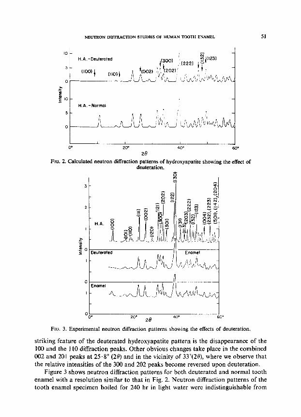
The neutron diffraction patterns expected for both deuterated and normal hydroxy- apatite were calculated on the basis of the structural parameters given by SUDAR- SANA and YOUNG (1969). The results are shown in Fig. 2. (In the deuteration studies some of the high resolution of the early diffraction patterns was given up in favour of increased intensity needed to compensate for the reduction in sample size.) The most
NEUTRON DIFFRACTION STUDIES OF HUMAN TOOTH ENAMEL 51
IO
H.A.-Dcuterotsd
5 (loo)
t CllO,j
Of--- za
.Z
E g IO
H.A. -Normal 5
0
FIG. 2. Calculated neutron diffraction patterns of hydroxyapatite showing the effect of deuteration.
9 =
3-
2-
za - f
so. c Deutcratcd
I -
0 0" 20”
28 400 60’
FIG. 3. Experimental neutron diffraction patterns showing the effects of deuteration.
striking feature of the deuterated hydroxyapatite pattern is the disappearance of the 100 and the 110 diffraction peaks. Other obvious changes take place in the combined 002 and 201 peaks at 25.8” (20) and in the vicinity of 33”(28), where we observe that the relative intensities of the 300 and 202 peaks become reversed upon deuteration.
Figure 3 shows neutron diffraction patterns for both deuterated and normal tooth enamel with a resolution similar to that in Fig. 2. Neutron diffraction patterns of the tooth enamel specimen boiled for 240 hr in light water were indistinguishable from
52 R.A. YOUNG AND S. SPOONER
those of the material before boiling treatment. Thus it was concluded that the changes observed in the patterns from the sample boiled in heavy water were due strictly to deuteration and not to any extraneous effects associated with the boiling treatment. Deuteration produced a marked reduction in the 100 and the 110 peak intensities, as expected, though perhaps it did not cause them to vanish. Similarly, the ratio of intensities of the 300 and the 202 peaks was changed markedly by deuteration, though not quite to the end points to be expected from complete deuteration of a pure hydroxyapatite.
Comparing the normal enamel and normal hydroxyapatite patterns, it is seen that the intensities in the tooth enamel patterns could be produced by partial deuteration of hydroxyapatite. This cannot be the true situation, obviously, but any element having a positive scattering amplitude replacing H (or OH) on or near the hexad axis would have a roughly similar effect on the intensities. For hkO-type reflections the effect would be the same no matter where, on the hexad axis, the substituting atom was located. For the low order reflections such as the 100 and the 110 peaks, the substi- tuting atoms would not even have to be accurately located on the hexad axis in order to produce a roughly similar effect on the intensities. It seems quite possible that such substitution may be responsible for the fact that the intensities of several of the most sensitive peaks in the deuterated tooth enamel have not quite reached their values for complete deuteration, even though boiling for 240 hr in heavy water would otherwise have been sufficient.
Strictly speaking, the neutron diffraction data do not show directly that hydrogen has necessarily been replaced; what the experimental data do show directly is that the sum of the various positive and negative scattering amplitudes from atoms on or near the hexad axis has been increased in the positive direction. Many different combinations of substitutions, additions, and removals of ions could produce the same result in the hk0 neutron diffraction intensities.
However, in view of chemical expectations as well as the deuteration experiments, we offer the following interpretation of the neutron diffraction hk0 intensity differences between tooth enamel and hydroxyapatite: In tooth enamel, some of the hydrogen appropriate to hydroxyapatite has been removed, most probably by substitution near the hexad axis but, possibly, also by substitutions elsewhere in the structure which alter the charge balance enough to permit the H- to be released. Two of the several substitution possibilities are CO3 and F. CO, is one of the more obvious because CO3 is known to be present in tooth enamel (NEUMAN and NEUMAN, 1953; CARLSTROM, 1955) and because there is other evidence that it can substitute on or near the hexad axis (ELLIOTT, 1964; BONEL and MONTEL, 1966) as well as at other locations (ELLIOTT, 1964; SMITH and LEHR, 1966). However, the present analysis has not gone far enough to permit unambiguous identification of any substituting ion.
Nonetheless, some simple calculations regarding obvious possibilities may be of interest. Working with the model of substitution on or near the hexad axis, one can estimate the degree to which a particular ion would have to substitute in order to account for the observed intensity differences. A first approximation can be obtained by (1) first calculating the degree of deuteration required to produce the observed
NEUTRON DIFFRACTIONSTUDIESOF HUMANTOOTH ENAMEL 53
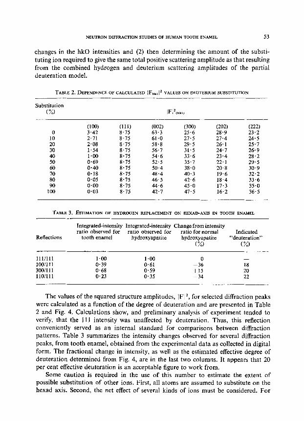
changes in the hk0 intensities and (2) then determining the amount of the substi- tuting ion required to give the same total positive scattering amplitude as that resulting from the combined hydrogen and deuterium scattering amplitudes of the partial deuteration model.
TABLE ~.DEPENDENCEOFCALCULATED IF,,k,[2~~~~~~~~ DEUTERIUM SUBSTITUTION
Substitution (%I
0 10 20 30 40
z 70 80 90
100
(1W (111) (002) (300) (202) (222) 3.42 8.75 63.3 25.6 28.9 23.2 2.71 8.75 61.0 27.5 27.4 24.5 2.08 8.75 58.8 29.5 26.1 25.1 1.54 8.75 56.7 31.5 24.7 26.9 l-00 8.75 54.6 33.6 23.4 28.2 0.69 8.75 52.5 35.7 22.1 29.5 0.40 8.75 50.4 38.0 20.8 30.9 0.18 8.75 48.4 40.3 19.6 32.2 o-05 8.75 46.5 42.6 18.4 33.6 0.00 8.75 44.6 45.0 17.3 35.0 o-03 8.75 42.7 47.5 16.2 36.5
TABLE 3. ESTIMATION OF HYDROGEN REPLACEMENT ON HEXAD-AXIS IN TOOTH ENAMEL
Reflections
Integrated-intensity Integrated-intensity Change from intensity ratio observed for ratio observed for ratio for normal Indicated
tooth enamel hydroxyapatite hydroxyapatite “deuteration” (%I (%I
lll/lll 1.00 1.00 0 - lOO/lll o-39 O-61 -36 18 300/111 0.68 0.59 +15 20 llO/lll O-23 0.35 -34 22
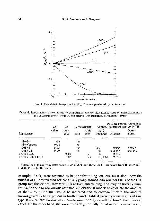
The values of the squared structure amplitudes, IF 12, for selected diffraction peaks were calculated as a function of the degree of deuteration and are presented in Table 2 and Fig. 4. Calculations show, and preliminary analysis of experiment tended to verify, that the 111 intensity was unaffected by deuteration. Thus, this reflection conveniently served as an internal standard for comparisons between diffraction patterns. Table 3 summarizes the intensity changes observed for several diffraction peaks, from tooth enamel, obtained from the experimental data as collected in digital form. The fractional change in intensity, as well as the estimated effective degree of deuteration determined from Fig. 4, are in the last two columns. It appears that 20 per cent effective deuteration is an acceptable figure to work from.
Some caution is required in the use of this number to estimate the extent of possible substitution of other ions. First, all atoms are assumed to substitute on the hexad axis. Second, the net effect of several kinds of ions must be considered. For
R. A.YOVNG AND SSPOONER
Percer.t deuterium
FIG. 4. Calculated changes in the IF,,, !* values produced by deuteration.
TABLE 4. REPLACEMENTS HAVING EQUIVALENT INFLUENCES ON hk0 REFLECTIONS OF I-IYDROXYAPATITE
IFALLATOMSSIJBSTITUTEDON THEHEXAD AXIS(NEVTRON DIFFRACTIONCASE)
Replacement
ab
(Site) Ab
(Unit cell)
Possible amount thought to ‘% replacement Approx. be present (wt “/)* in TE
Unit wt % Lrter Site cells required Average layers
H+D l-03 20 H +Vacancy O-38 55 OH +F o-35 60 2.3 0.02* >0*3* OH +CI 0.79 26 1.8 0.2-0.4 0.5-0.7
2 OH+CO, 2.00 21 1.2 2 to 3 2 OH +CO, +HzO I.82 24 1’ 3(CO3) 2 to 3
*Data for F taken from BRUDEVOLD et al. (1967), and those for Cl are taken from BESIC et al. (1969). TE = tooth enamel.
example, if CO3 were assumed to be the substituting ion, one must also know the number of H ions released for each CO3 group formed and whether the 0 of the OH group remains or not. However, it is at least entertaining, and may be usefully illus- trative, for one to use various assumed substitutional models to calculate the amount of that substitution that would be indicated and to compare it with the amount known generally to be present in tooth enamel. Table 4 presents some results of this type. It is clear that fluorine alone can account for only a small fraction of the observed effect. On the other hand, the amount of COs, normally found in tooth enamel would
NEUTRON DIFFRACTION STUDIES OF HUMAN TOOTH ENAMEL 55
be quite enough (if located on or near the hexad axis) whether or not the substitution of each CO3 group was accompanied by the formation of a water molecule. It is also possible that, at the same time, some chlorine is present on the hexad axis, as that is its normal location in chlorapatite and some chlorine is expected to be present.
Excess hydrogen
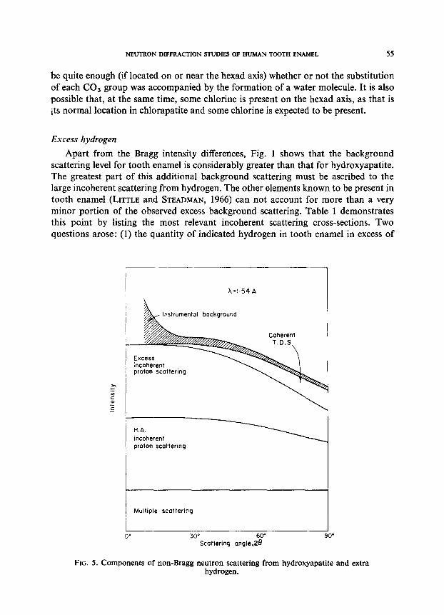
Apart from the Bragg intensity differences, Fig. 1 shows that the background scattering level for tooth enamel is considerably greater than that for hydroxyapatite. The greatest part of this additional background scattering must be ascribed to the large incoherent scattering from hydrogen. The other elements known to be present in tooth enamel (LITTLE and STEADMAN, 1966) can not account for more than a very minor portion of the observed excess background scattering. Table 1 demonstrates this point by listing the most relevant incoherent scattering cross-sections. Two questions arose: (1) the quantity of indicated hydrogen in tooth enamel in excess of
Instrumental background
proton scattering
H.A. incoherent proton scattering
Multiple scattering
30” 60”
Scattering angle,28 00
FIG. 5. Components of non-Bragg neutron scattering from hydroxyapatite and extra hydrogen.

56 R. A. YOUNG AND S. SPOONER
that in hydroxyapatite and (2) the location of it, particularly in view of the Bragg- intensities analysis suggesting a deficiency of H at its normal location in the hydroxy-
apatite portion of tooth enamel. The quantity of hydrogen present was determined from the diffuse scattering intensity after background scattering due to instrumental background, to thermal diffuse scattering and to multiple scattering were first extracted. To assess the location of the excess hydrogen, we tested several models and made use of the deuteration results.
Figure 5 shows qualitatively the various components in the background scattering.
The instrumental background was determined experimentally with the use of empty sample holders. Diffuse scattering of thermal neutrons arises from both elastic inco-
herent and inelastic incoherent scattering by the hydrogen and from coherent inelastic scattering (TDS) arising from the thermal vibrations of the lattice. A strict calculation
of the inelastic scattering is exceedingly complex and, in part, is presently impossible
because knowledge of lattice vibrations in hydroxyapatite is inadequate. However, it
can be shown (EGELSTAFF, 1965) that the inelastic scattering contributions approach zero at small angle, as is indicated in Fig. 5. Further, at zero scattering angle the elastic incoherent scattering, (which, with multiple scattering and instrumental background, remains as the principal diffuse scattering contribution), is proportional to the total number of hydrogen atoms present and is independent of hydrogen-atom dynamics.
The multiple-scattering correction is a measure of the probability that a once- scattered neutron will be scattered again before it leaves the sample. Since scattering is basically isotropic, multiple scattering will have the effect of increasing the scattering
emerging along the short dimension of the specimen at the expense of that along the
long dimension. In this case the cylindrical specimens of height h and radius R were mounted with the cylinder axis perpendicular to the plane in which the scattering
angle was measured. Thus the effect of multiple scattering was to increase the measured
background scattering. BLECH and AVERBACH (1966) have shown that for such a geometric arrangement, the multiple scattering cross-section, a,,,,, is approximated by
where us is the total scattering cross-section, a, is the sum of the scattering and absorption cross-sections and S is obtained from tables of S vs. R/h and pR where p is the experimental linear attenuation coefficient. Since the absorption cross-section is
very small compared to a, (because the incoherent scattering cross-section is so large) the approximation may be used,
US S UP?lS m---
457 1--
Table 5 gives the parameters needed for completion of the individual multiple scattering calculations for each of the samples. The pR values were determined experimentally by transmission measurements.
For quantitative study, we selected the background in the neighbourhood of the 111 peak because (1) it was at sufficiently low angles that, in a first approximation,

r
NEUTRONDIFFRACTlONSlUDIFSOFHUMANTOO~ENAMEL 57
TABLE 5. PARAMETERS FORTHEDETERMINATION OF MULTIPLE SCATTERING
Specimen R/h PR
Hydroxyapatite 0.30 0.042 Octacalcium phosphate* 0.30 0.160 Tooth enamel 0.30 0.185 Tooth enamel A 0.25 O-092 Tooth enamel B (deuterated A) 0.25 0.070
*We thank Dr. W. E. BROWN for the loan of this specimen.
6
0.040 o-134 0.152 0.085 0.067
we could neglect TDS and inelastic incoherent scattering and (2) the 111 was un- affected by deuteration and therefore provided an internal calibration for comparison of deuterated with undeuterated specimens. Figure 6 shows the data from several specimens. The magnitudes of the statistical errors in counting are indicated by the figure. The applicability of the multiple-scattering corrections was verified by experi- ments with specially devised cylindrical samples in which cadmium discs divided the
1
L
Scattering angle
FIG. 6. The 111 Bragg peak and immediate background used for hydrogen-content analysis. A-hydroxyapatite; B-tooth enamel, fine collimation; C-tooth enamel, coarse
collimation; D-deuterated tooth enamel, coarse collimation.
cylindrical powder sample into several short cylinders. The error in the experimental verification of the multiple-scattering correction was estimated at 15 per cent and the random error from counting statistics was estimated at 4 per cent in the case of tooth enamel and 6 per cent in the case of hydroxyapatite. These values lead to +5 per cent and *6 per cent for the precision of the determination of hydrogen incoherent scatter- ing from tooth enamel and hydroxyapatite, respectively.
58 R. A. YOUNG AND S. SPOONER
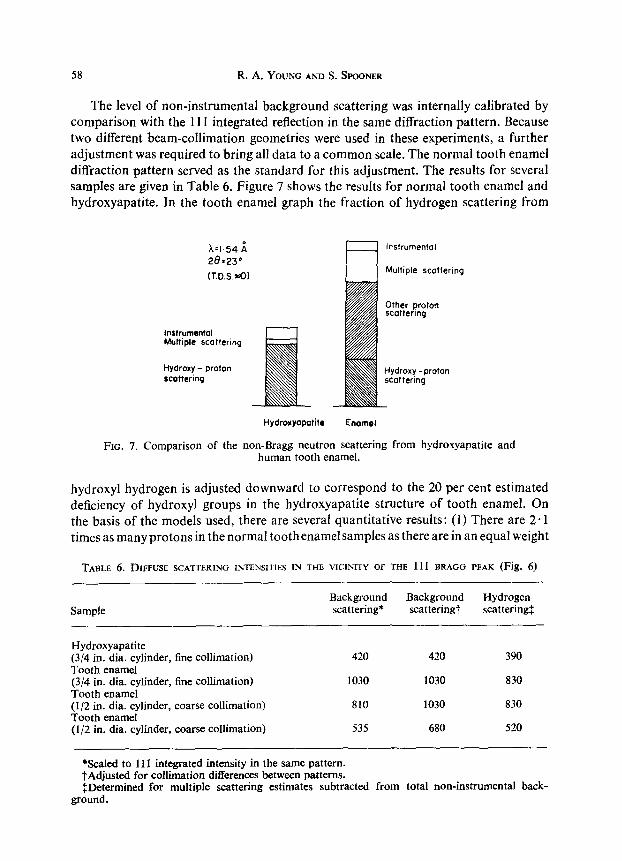
The level of non-instrumental background scattering was internally calibrated by comparison with the 111 integrated reflection in the same diffraction pattern. Because two different beam-collimation geometries were used in these experiments, a further adjustment was required to bring all data to a common scale. The normal tooth enamel diffraction pattern served as the standard for this adjustment. The results for several samples are given in Table 6. Figure 7 shows the results for normal tooth enamel and hydroxyapatite. In the tooth enamel graph the fraction of hydrogen scattering from
X’I.54 il
28=23*
(T.D.S.Hl)
Instrumental Multiple Scattering
Hydroxy - proton scattering
Hydroxyapatite Enamel
FIG. 7. Comparison of the non-Bragg neutron scattering from hydroxyapatite and human tooth enamel.
hydroxyl hydrogen is adjusted downward to correspond to the 20 per cent estimated deficiency of hydroxyl groups in the hydroxyapatite structure of tooth enamel. On the basis of the models used, there are several quantitative results: (1) There are 2.1 times as many protons in the normal toothenamelsamples as there are in an equal weight
TABLE 6. DIFFUSE SCATTERING INTENSITIES IN THE VICINITY OF THE 111 BRAGG PEAK (Fig. 6) _~~ _
Sample Background scattering*
Background scattering?
Hydrogen scattering:
Hydroxyapatite (3/4 in. dia. cylinder, fine collimation) 420 420 390 Tooth enamel (3/4 in. dia. cylinder, fine collimation) 1030 1030 830 Tooth enamel (l/2 in. dia. cylinder, coarse collimation) 810 1030 830 Tooth enamel (l/2 in. dia. cylinder, coarse collimation) 535 680 520
*Scaled to 111 integrated intensity in the same pattern. fAdjusted for collimation differences between patterns. fDetermined for multiple scattering estimates subtracted from total non-instrumental back-
ground.
NEUTRON DIFFRACTION STUDIES OF HUMAN TOOTH ENAMEL 59
of hydroxyapatite. (2) There are about 2.3 times as many protons present in tooth enamel as the Bragg-intensity analysis indicated are present in the crystalline hydroxy- apatite portion of it. (3) On deuteration, only about 35 per cent of the hydrogen atoms exchanged. This closely approximates the amount thought to be in the hydroxyapatite lattice in the normal tooth enamel sample.
The remaining problem is the location of the excess hydrogen. If 20 per cent is taken as the measure of the hydrogen deficiency in the hydroxyapatite portion of tooth enamel, there are nearly twice as many protons to be accounted for off the normal hydroxyapatite position as are on them, for a total amount of 1.3 times the stoichio- metric amount that is either not in the “hydroxyapatite” structure or is in unusual locations within it. Several models for hard tissues and Ca-deficient hydroxyapatite have been suggested which would involve excess hydrogen and which can be tested here for their ability to account, alone, for all of the observations.
(1) Amorphous fraction. POSNER and co-workers (e.g. TERMINE, 1966; TERMINE and POSNER, 1966; TERMINE and POSNER, 1967; WEBER, EANES and GERDES, 1967; BIENEN- STOCK and POSNER, 1968) have demonstrated the existence of an amorphous Ca phosphate material in bone and in early precipitation from solutions which can be used for preparation of hydroxyapatite, though not in mature tooth enamel.
With the wavelength used here, such an amorphous material should give a scattering curve with a broad maximum in the neighbourhood of 29” (20). No such maximum occurs in the non-Bragg scattering from the tooth enamel specimens.
(2) Adsorbed water. B.E.T. measurements of surface area give 1.5 m2/g for the hydroxyapatite specimen and 5 03 m’/g for the tooth enamel specimen. These surface areas are too small to permit adsorbed water to provide the excess hydrogen in observed quantitites.
(3) H-bonds and structural H. Several authors (POSNER and PERLOFF, 1957; POSNER, STUTMAN and LIPPINCOTT, 1960) have suggested the formation of hydrogen bonds, or structural incorporation of hydrogen, particularly in association with low Ca/P ratios. BERRY (1967) has discussed several of the possibilities. Structural hydro- gen in the quantity necessary to account for all of the “excess” hydrogen, because it would be localized at specific crystallographic positions, would be expected to have a profound effect on the Bragg intensities in neutron diffraction patterns. However, all major intensity differences between the hydroxyapatite and the tooth enamel patterns seem to be explicable on the basis of substitutions for hydrogen on the hexad axis, as shown by calculation and by the deuteration effect of making the tooth enamel and hydroxyapatite patterns much more similar. It is difficult to see why other structurally incorporated hydrogen should not exchange with deuterium to some significant degree when that on the hexad axis does. But if such hydrogen were present and did not so exchange, then the neutron diffraction of deuterated tooth enamel and hydroxy- apatite should be less, rather than more, similar than are the patterns of the undeuterated materials. Thus we conclude that the maximum possible amount of structural incorporation of additional hydrogen could not account for more than a minor fraction of the observed excess hydrogen. (It should be specifically noted, however, that the evidence evaluated here does not rule out the relatively small amount
60 R.A.YouNG ANDS.SPOONER
of hydrogen required for hydrogen bonding in, for example, WINAND’S (1961) formula for calcium-deficient apatite, Ca,,_,H,(PO,),(OH),_,.)
(4) Octacalcium phosphate, Ca,H,(PO,),. 5H20. BROWN (1966) has suggested that octacalcium phosphate (OCP) may be an important intermediary step in the formation of biological hydroxyapatite (HA) and, further, that OCP and HA may be inter- layered on (100) planes to some degree. The amount of OCP required to provide the observed excess hydrogen would be 24 wt % (and would result in a density, p, of 3.02 g/cm3, using pocp = 2.58 g/cm3 and pHa = 3.15 g/cm3). The 100 reflection of OCP is especially strong. Within an experimental error <3 per cent, no OCP-100 peak occurs in the neutron diffraction pattern of tooth enamel, nor are any other peaks of the OCP pattern identified in the tooth enamel pattern. However, the other peaks in the OCP pattern are relatively weak. Further, Brown notes that only a 50 per cent hydrolysis of OCP into HA causes the 100 peak of OCP to disappear from an X-ray powder pattern. For this reason and because of the special interest in OCP-HA relationships we look more carefully for evidence of even small amounts of OCP.
The interlayering of OCP with HA is expected (BROWN, 1966) to occur on (100) planes of both. An effect of random interlayering of two unequally thick layers is to cause the associated lowest-order diffraction peaks, in this case the 100 peaks, to become one peak at a position intermediate between the two normal positions, the position being dependent on the relative amounts of the two kinds of layers (HEN- DRICKS and TELLER, 1942). For small amounts of one kind of layer, the shift is nearly linearly related to the amount present. Thus the absence of the 100 reflection of OCP from its proper position does not, in itself, indicate the absence of OCP interlayered with HA. However, neither is there any peak in the region between the normal position of 100 [$A and 100ocp, and any shifting of the lOOnA is, at least, not obvious. Thus one may conclude that not nearly enough OCP is present to account for the observed excess hydrogen.
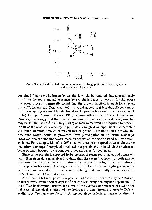
To digress for a moment, because the OCP interlayering idea seems intrinsically important, detailed examination of the 100 ,{A for any sign of shifting, even if arising from only part of the sample, seemed in order. Consequently both peak position and peak breadth (a tendency for interlayering in one part of the specimen but not others would broaden the peak) were examined in detail. Figure 8 shows the peak-breadth data. There is no evidence of any special broadening of the 100 other than that occurring for all 100 reflections by reason of the tooth enamel crystallites being thin in that direction. There is, possibly, a shift toward lower angles of about 0.1” (20). If real and due to OCP interlayering, on a linear approximation the shift would correspond to about 2 per cent of the layers being OCP. Although the linear approxi- mation is an oversimplification, nonetheless it helps to demonstrate that our data show that only a very little OCP could be present.
(5) Protein. Even though the enamel specimens were chosen in such a way as to minimize their content of organic protein material, it is to be expected that some of the organic matrix is present, e.g., in interprismatic spaces. It is also possible that much of the hydrogen in the protein component would be bonded to carbon and therefore would be unlikely to exchange with deuterium (HINE, 1956). If the protein component
NEUTRON DIFFRACTION STUDIES OF HUMAN TOOTH ENAMEL 61
06 -
% L
g 0.4-
c;
0.2 -
01 I I I
0 0.1 02 0.3 0.4
Tan he,
FIG. 8. The full width at half maximum of selected Bragg peaks in the hydroxyapatite and tooth enamel patterns.
contained 7 per cent hydrogen by weight, it would be required that approximately 4 wt % of the tooth enamel specimen be protein in order to account for the excess hydrogen. Since it is generally found that the protein fraction is much lower (e.g., O-4 wt %, LITTLE and CASCIANI, 1966), it would appear that less than 20 per cent of the excess hydrogen should be attributed to the protein fraction of the tooth enamel.
(6) Entrapped water. MYERS (1965), among others (e.g. LITTLE, CUETTO and ROWLEY, 1962) suggested that enamel contains free water entrapped in regions that may be as small as 25 A dia. Only 2 wt ‘A of such water would be required to account for all of the observed excess hydrogen. Little’s weight-loss experiments indicate that this much, or more, free water may in fact be present. It is not at all clear why and how such water should be prevented from participation in deuterium exchange. However, one can imagine several possibilities which can not be ruled out by present evidence. For example, MYER’S (1965) small volumes of entrapped water might escape deuterium exchange if completely enclosed in a protein sheath in which the hydrogen, being strongly bonded to carbon, could not exchange for deuterium.
Since some protein is expected to be present, it seems reasonable, and consistent with all neutron data as analyzed to date, that the excess hydrogen in tooth enamel may arise from two unequal contributions, a small one from tightly bound hydrogen in the protein fraction and a larger one from the loosely bound hydrogen in water entrapped and occluded from deuterium exchange but essentially free in respect to thermal motions of the molecules.
A distinction between protons in protein and those in free-water may be obtained, in future work, from another aspect of neutron scattering, the angular dependence of the diffuse background. Briefly, the slope of the elastic component is related to the tightness of chemical binding of the hydrogen atoms through a pseudo-Debye- Waller-type “temperature factor”. A steepel slope reflects a weaker binding. A
62 R. A. YOUNG AND S. SPOONER
temporary lack of equipment capability, for separate determination of elastic and
inelastic contributions, and experimental error in the diffuse background determin-
ations (5 per cent from counting statistics and, probably another 5 per cent for possible error in the multiple-scattering correction) prevented quantitative evaluation of the relevant slopes at the present time.
Acknowledgemenfs-This work was supported in part by the U.S. Public Health
Service through NIH-NIDR Grant DE-01912.
R&urn&-On a pen& que la premiere utilisation de la diffraction neutronique pour I’ttude de 1’Cmail dentaire humain reconfirmerait que la partie inorganique ttait constit- uCe par un matCriel roisin de l’hydroxylapatite. Cependant, par rapport B cette derni&, I’Cmail dentaire ptisente (i) une dCficience de 20 pourcent ou plus d’hydro&ne hydroxyi8, avec substitution probable d’autres ions sur ou pr& de I’axe “hexad” et (ii) un excks global d’hydrogkne. Le phosphate de calcium amorphe, le phosphate octocalcique, de I’eau absorb6 et de l’hydrogbne de structure, en position anormale, ne sont pas pt&ents en grande quantit6, bien que I’existence possible de composCs mineurs tels que OCP (_ 2 % poids) et de l’hydrogkne en rapport avec des groupes PO, (environ 1% poids) ne puisse &tre rejetCe. C’est done une combinaison de prot6ine r&siduelle et de l’eau libre emprisonn&, qui pourrait rendre compte de la quantitt d’hydrogbne, situ6 & I’exttrieur du compo& hydroxyleapatite.
L’utilitO de la diffraction neutronique et des techniques voisines pour 1’6tude de la quantitC, situation, et liens possibles de I’hydrog+ne prksent, incorport ou non dans la structure cristalline est ainsi dkmontrte.
Zusammenfassung-Erste Untersuchungen mit der Neutronendiffraktionstechnik im Bereich der Zahnhartsubstanzen haben bestltigt, dal3 der anorganische Anteil des menschlichen Zahnschmelzes im wesentlichen aus einem hydroxylapatittihnlichem Material besteht. Im Vergleich zum Hydroxylapatit zeigt der Schmelz jedoch (i) in der GrGBenordnung von 20 Prozent oder mehr ein Fehlen von Wasserstoffionen an der Hydroxylgruppe, die vermutlich durch andere Ionen ersetzt sind, und (ii) einen Gesamt- iiberschul3 von Wasserstoffionen. Amorphes Kalziumphosphat, Oktakalziumphosphat, adsorbiertes Wasser und an ungewiihnlicher Stelle gebundener Wasserstoff konnten nicht in nennenswerten Mengen nachgewiesen werden, obwohl nicht ausgeschlossen werden konnte, daB mZjglicherweise kleinere Mengen Oktakalziumphosphat (- 2 Gewichtspro- zente) und von PO,-Gruppen umlagerter Wasserstoff (_ 1 Gewichtsprozent) vorhanden sind. Es bleiben daher sowohl ein Restproteingehalt als such eingeschlossenes freies Wasser fiir den auRerhalb der Hydroxylapatitstrukturen gelegenen Wasserstoff verant- wortlich.
Es wird die besondere Brauchbarkeit der Neutronendiffraktion und der Neutro- nenstreuungfi.ir Untersuchungen iiber Menge, Lokalisation und miiglicherweise Bindung des vorhandenen Wasserstoffs im oder aul3erhalb des Kristallgefiiges dargestellt.
REFERENCES
BACON, G. E. 1962. Neutron Diflkzction (2nd ed.) Oxford University Press, London. BERRY, E. E. 1967. The structure and composition of some calcium deficient apatites. J. inorg. nucf.
Chem. 29, 317-327. BESIC, F. C., KNOWLES, C. R., WIEMANN, M. R. JK. and KELLER, 0. 1969. Electron probe micro-
analysis of noncarious enamel and dentin and calcified tissues in mottled teeth. J. dent. Res. 48, 131-139.
BIENENSTOCK, A. and POSNER, A. S. 1968. Calculations of the x-ray intensities from arrays of small crystallites of hydroxyapatites. Archs biochem. biophys. 124, 604415.
BLECH. I. A. and AVERBACH, B. L. 1965. Multiple scattering of neutrons in vanadium and copper. P/I~s. Rec. 137, 1113-l 116.

NEUTRONDIFFRACTIONSTUDIESOFIiUMANTOOTHENAhfEL 63
BONEL, G. and MONTEL, G. 1966. Introduction of COO ions into the calcium apatite lattice. C. r. h&d. S&~nc. Acad. Sci., Paris, 1010-1013.
BROWN, W. E. 1966. Crystal growth of bone mineral. Clin. Orthop. No. 44,205-216. BRUDEVOLD, F. and SGREMARK, R. 1967. Structural and Chemical Organization of Teeth (edited by
MILES, A. W.) Vol. II. Chap. II, p. 254. Academic Press, New York. CARLSTROM, D. 1955. X-ray crystallographic studies on apatites and calcified structures. Acta radiol.
Suppl. 121. EGELSTAFF, P. A. 1965. Thermal Neutron Scattering. Academic Press, London. ELLIOTT, J. C. 1964. Thesis, The Crystallographic Structure of Dental Enamel and Related Apatites.
Ph.D thesis Univ. of London. HAYEK, E. and STADLMANN, W. 1955. Preparation of pure hydroxyapatite for adsorption uses.
Angew. Chem. 67,327. HENDRICKS, S. B. and TELLER, E. 1942. X-ray interference in partially ordered layer lattices. J. them.
Phys. 10, 147-167. HINE, .I. Physical Organic Chemistry. McGraw-Hill, New York. LITTLE, M. F., CUETTO, E. S. and ROWLEY, J. 1962. Chemical and physical properties of altered and
sound enamel, I. Ash, Ca, P, C02, N, water, microradiolucency, and density. Archs oral Btol. 7, 173-184.
LISLE, M. F. and STEADMAN, L. T. 1966. Chemical and physical properties of altered and sound enamel, IV. Trace element composition. Archs oral Biol. 11, 273-278.
LITTLE, M. F. and CASCIANI, F. S. 1966. The nature of water in sound human enamel. Archs oral Biol. 11, 565-571.
MYERS, H. M. 1965. Trapped water of dental enamel. Nature, Land. 206, 713-714. NEWMAN, W. F. and NEUMAN, M. W. 1953. The nature of the mineral phase of bone. Chem. Rev. 53,
l-45. POSNER, A. S. and PERLOFF, A. 1957. Apatites deficient in vibalent cations. J. Res. natn. Bur. Stand.
S&279-286. POSNER, A. S., STUTMAN, J. M. and LIPPINCOTT, E. R. 1960. Hydrogen bonding in calcium-deficient
apatite. Nature, Land. 188,486-487. SMITH, J. P. and LEHR, J. R. 1966. An x-ray investigation of carbonate apatites. J. agric. Fd Chem. 14,
342-349. SUDARSANAN, K. and YOUNG, R. A. 1969. Significant precision in crystal structural details: Holly
Springs hydroxyapatite. Acta Cryst. in press. TERMINE, J. D. 1966. Thesis, Amorphous Calcium Phosphate. The Second Mineral of Bone. Cornell
University Medical College, New York. TERMINE, J. D. and POSNER, A. S. 1966. Infra-red analysis of rat bone: age dependency of amorphous
and crystalline mineral fractions. Science, N. Y. 153, 1523-1525. TERMINE, J. D. and POSNER, A. S. 1967. Amorphous-crystalline interrelations in bone mineral.
Calc. Tissue Res. 1, 8-23. WEBER, J. C., EANES, E. D. and GERDES, R. J. 1967. Electron microscope study of non-crystalline
calcium phosphate. Archs biochem biophys. 120,723-724. WINAND, L. 1961. I%ude physico-chimique du phosphate tricalcique hydrate et de l’hydroxylapatite.
Ann. Chim. 6,941-967.