neutron instrumentation - neutron... · summary • neutron instrument concepts – time-of-flight...
TRANSCRIPT
Neutron Instrumentation
Ken Andersen
Oxford School on Neutron Scattering5th September 2019
Summary
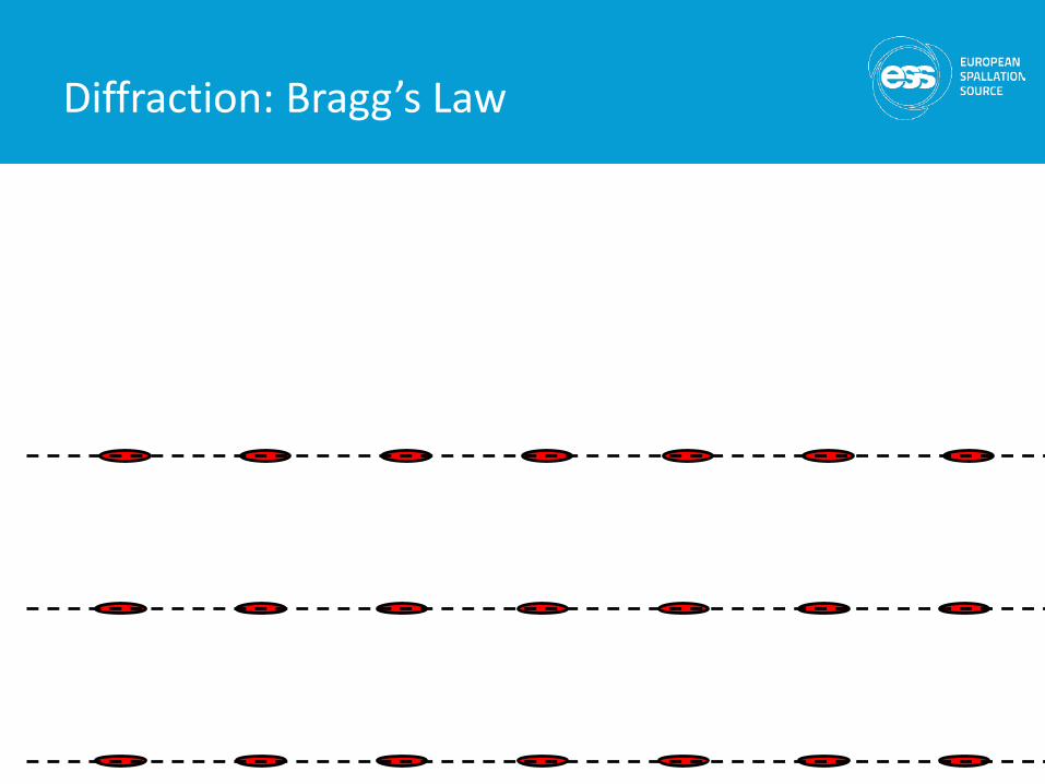
• Neutron instrument concepts– time-of-flight– Bragg’s law
• Neutron Instrumentation– guides– monochromators– shielding– detectors– choppers– sample environment– collimation
• Neutron diffractometers• Neutron spectrometers
2
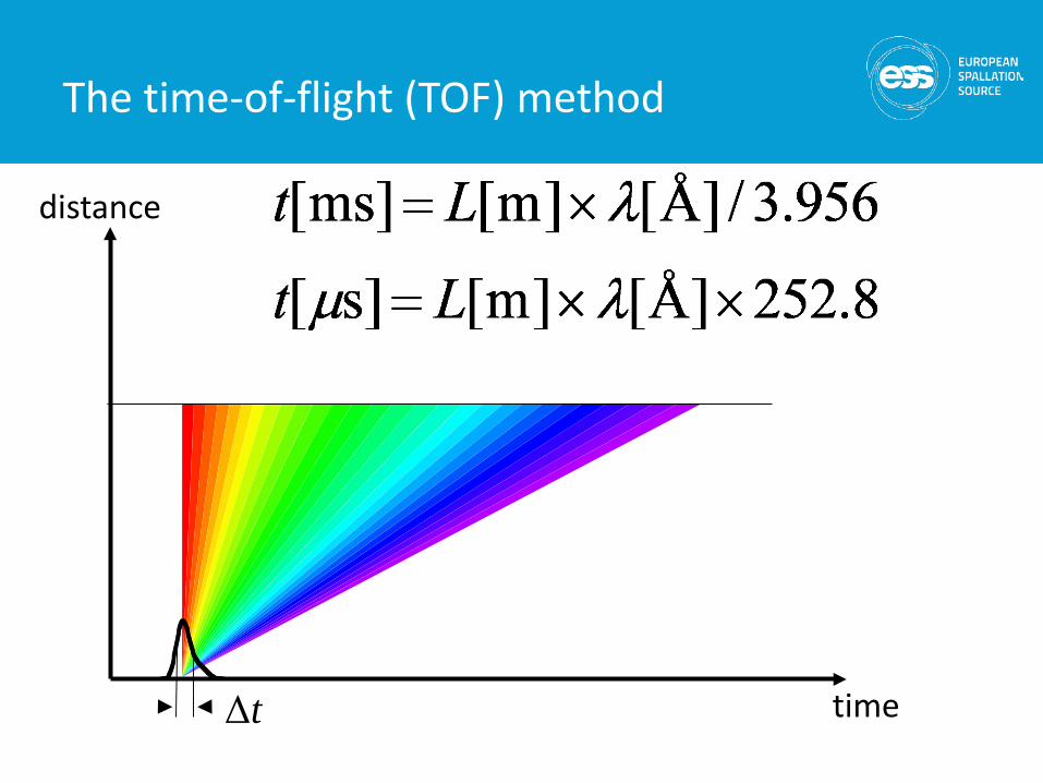
The time-of-flight (TOF) method
distance
time
Δt
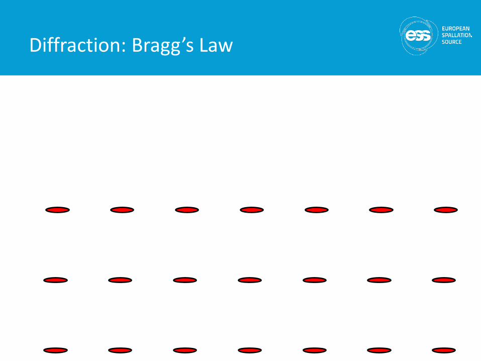
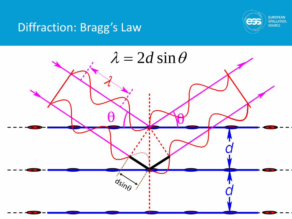
Diffraction: Bragg’s Law
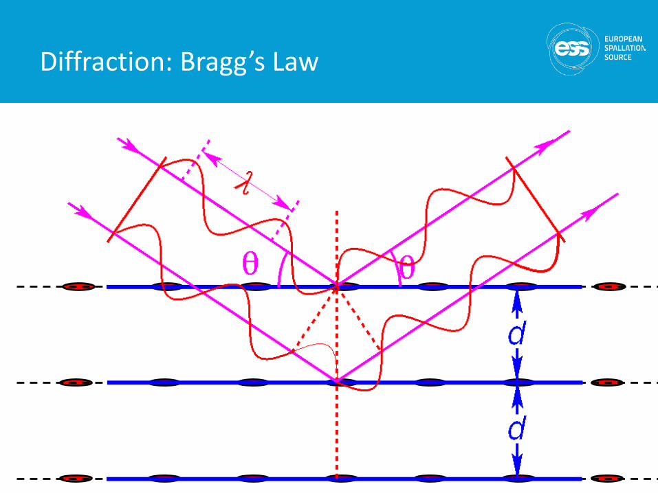
Diffraction: Bragg’s Law
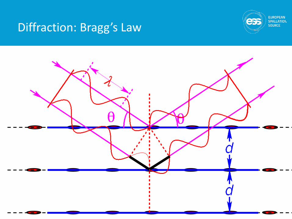
Diffraction: Bragg’s Law
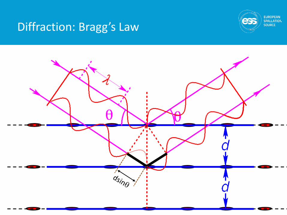
Diffraction: Bragg’s Law
Diffraction: Bragg’s Law
Diffraction: Bragg’s Law
θλ sin2d=
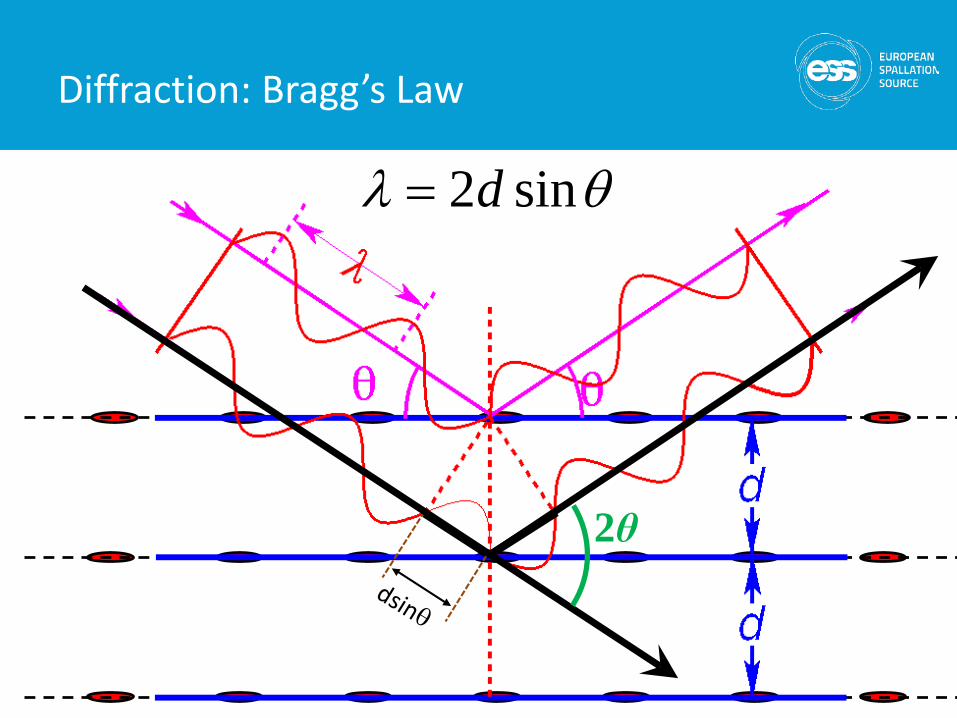
Diffraction: Bragg’s Law
2θ
θλ sin2d=
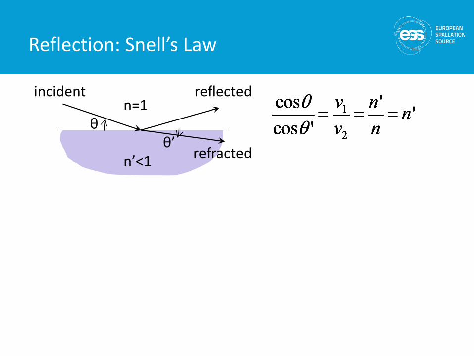
n=1
n’<1
incident
refracted
reflected
θ
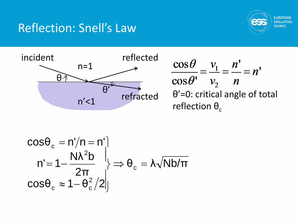
Reflection: Snell’s Law
θ’
n=1
n’<1
incident
refracted
reflected
θ
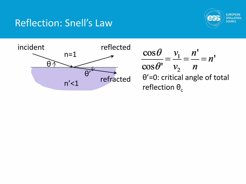
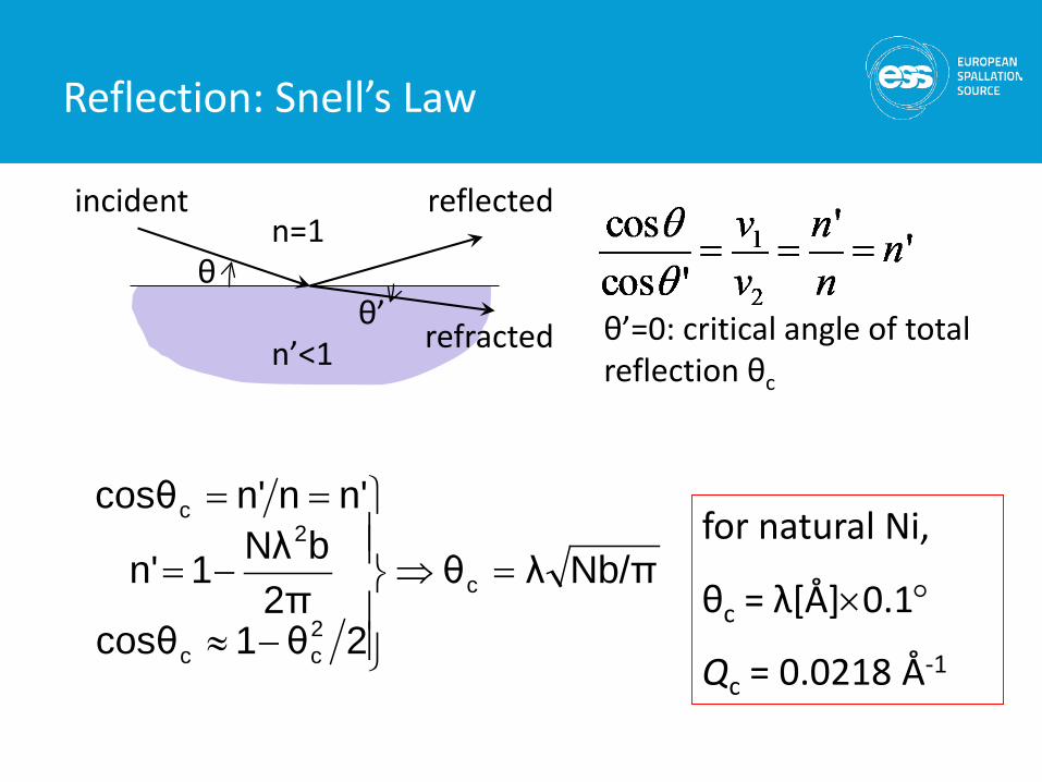
Reflection: Snell’s Law
θ’ θ’=0: critical angle of total reflection θc
n=1
n’<1
incident
refracted
reflected
θ
Reflection: Snell’s Law
θ’ θ’=0: critical angle of total reflection θc
Nb/πλθ
2θ1cosθ2π
bNλ1n'
n'nn'cosθ
c
2cc
2c
=⇒
−≈
−=
==
n=1
n’<1
incident
refracted
reflected
θ
Reflection: Snell’s Law
θ’ θ’=0: critical angle of total reflection θc
Nb/πλθ
2θ1cosθ2π
bNλ1n'
n'nn'cosθ
c
2cc
2c
=⇒
−≈
−=
==for natural Ni,
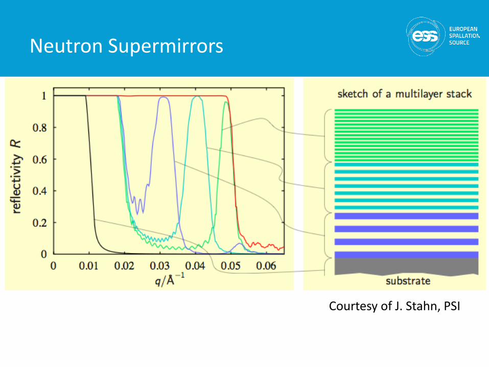
θc = λ[Å]×0.1°
Qc = 0.0218 Å-1
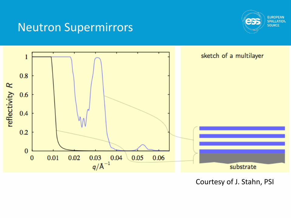
Courtesy of J. Stahn, PSI
Neutron Supermirrors
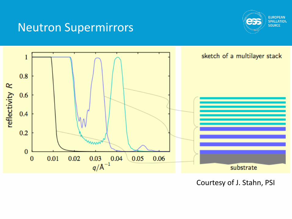
Courtesy of J. Stahn, PSI
Neutron Supermirrors
Courtesy of J. Stahn, PSI
Neutron Supermirrors
Neutron Supermirrors
18
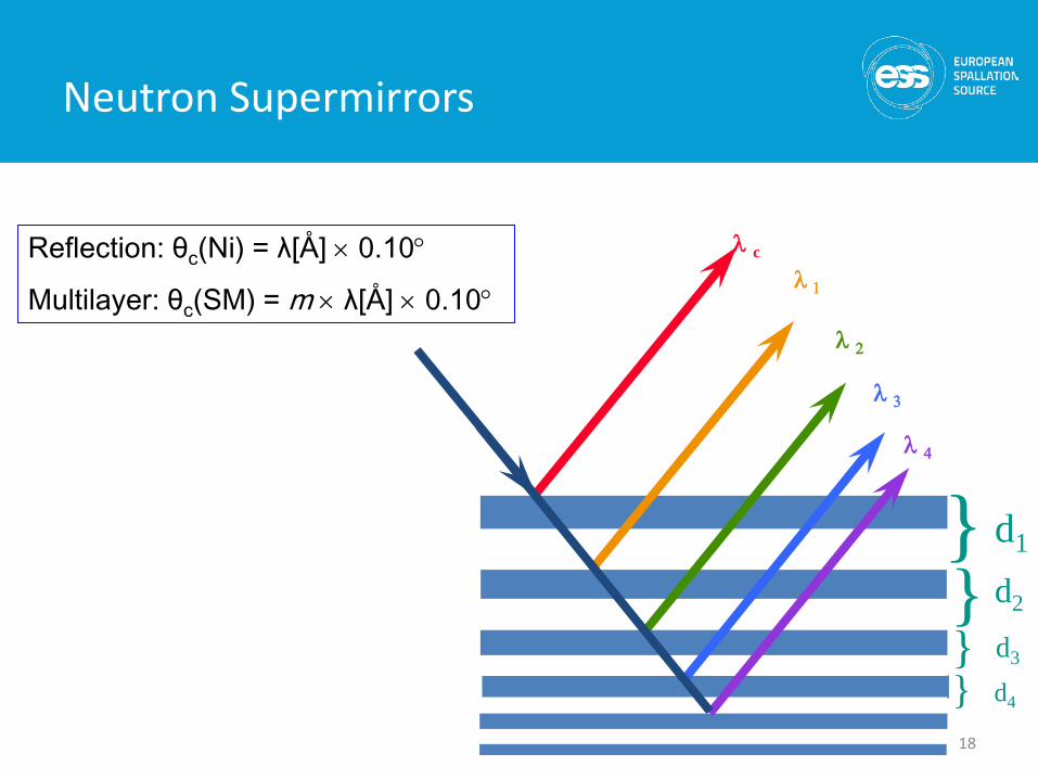
d1}
λ 1
λ 2
λ 3
λ 4
λ c
d2} d3
} d4
}
Reflection: θc(Ni) = λ[Å] × 0.10°
Multilayer: θc(SM) = m × λ[Å] × 0.10°
Neutron Supermirrors
19
}
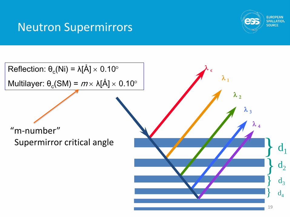
λ 1
λ 2
λ 3
λ 4
λ c
} } }
Reflection: θc(Ni) = λ[Å] × 0.10°
Multilayer: θc(SM) = m × λ[Å] × 0.10°
“m-number” Supermirror critical angle d1
d2
d3
d4
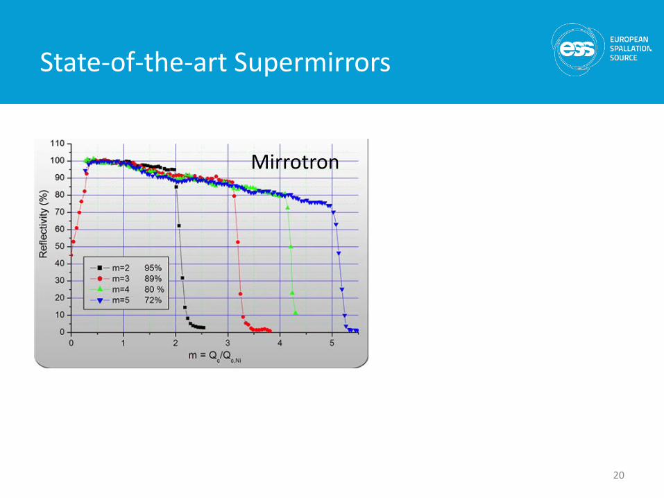
State-of-the-art Supermirrors
20
Mirrotron
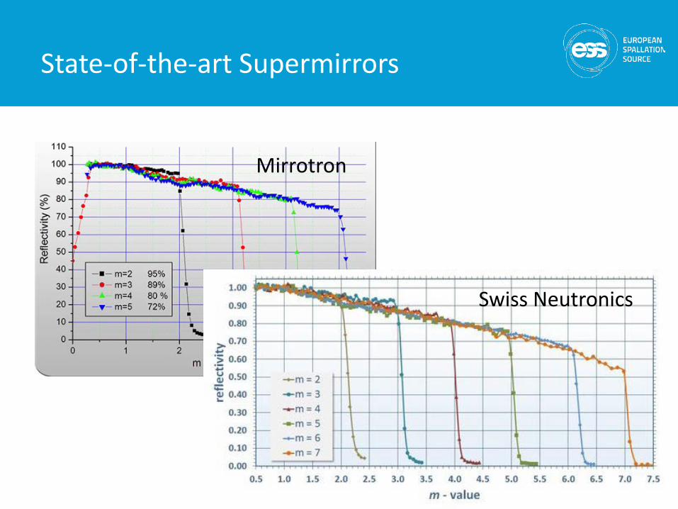
State-of-the-art Supermirrors
21
Mirrotron
Swiss Neutronics
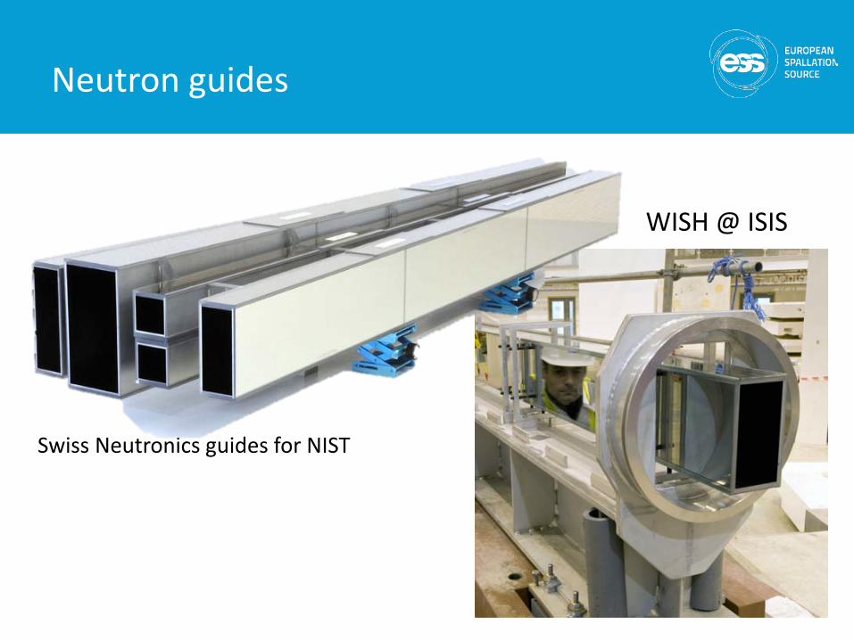
WISH @ ISIS
Neutron guides
Swiss Neutronics guides for NIST
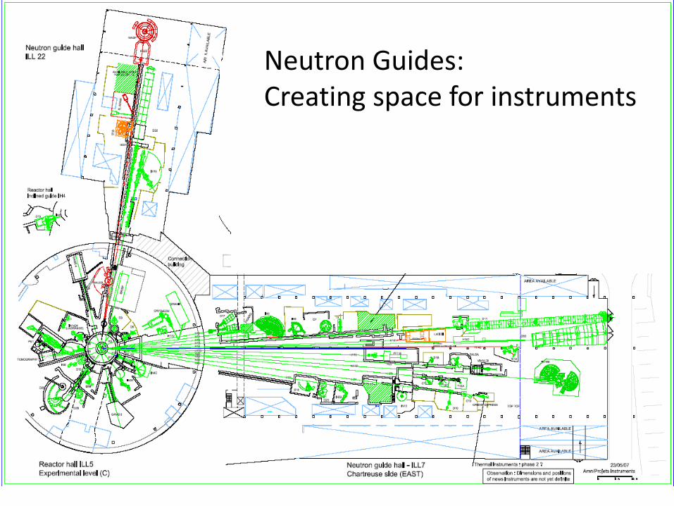
Distribution by GuidesNeutron Guides: Creating space for instruments
Background Reduction
• Distance:– move away from fast-
neutron source ~ 1/R2
Guides can be used to reduce background
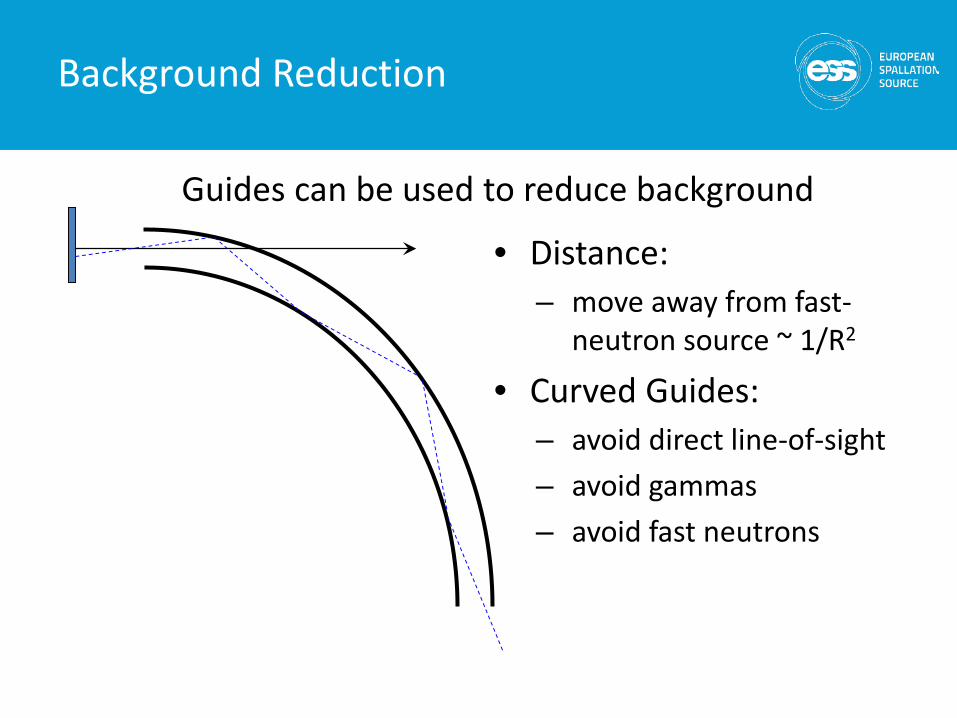
Background Reduction
• Distance:– move away from fast-
neutron source ~ 1/R2
• Curved Guides: – avoid direct line-of-sight– avoid gammas– avoid fast neutrons
Guides can be used to reduce background
Focusing
26
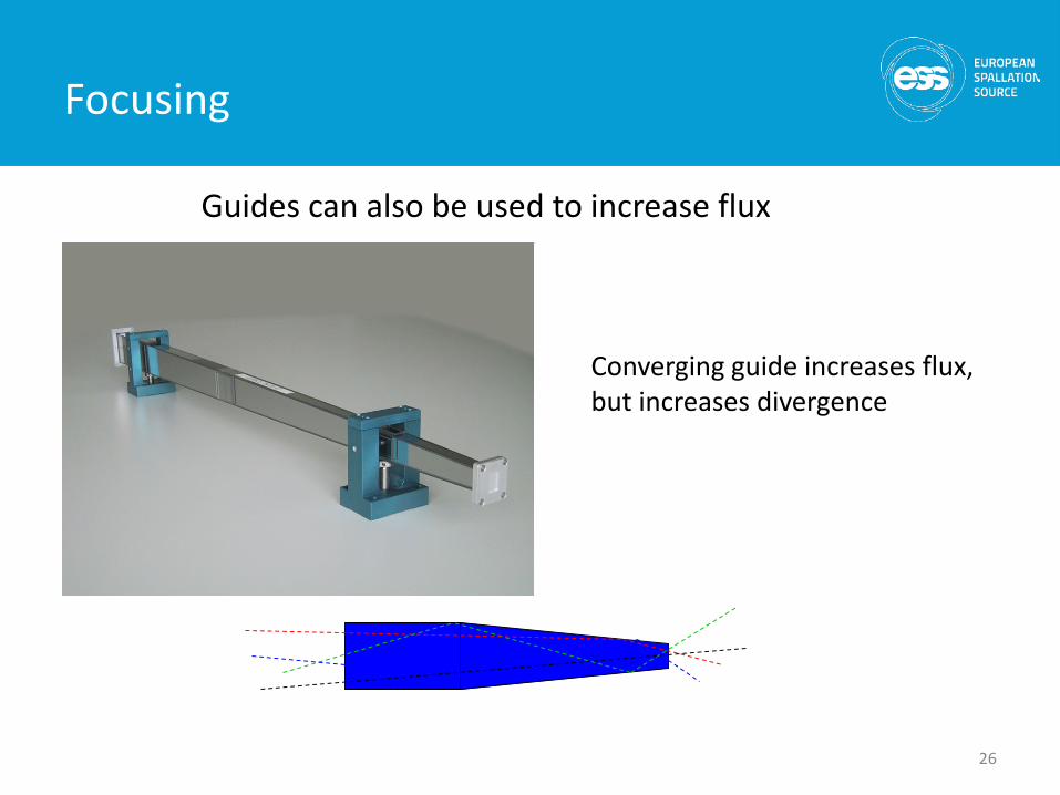
Converging guide increases flux, but increases divergence
Guides can also be used to increase flux
Shielding
• Shielding functions: – allow safe operation– keep background down– reduce activation
• Radiation components:– Slow neutrons– Fast neutrons– Gammas
27
Shielding: radiation units and numbers
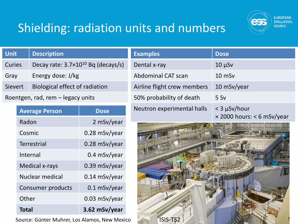
Unit Description
Curies Decay rate: 3.7×10¹⁰ Bq (decays/s)
Gray Energy dose: J/kg
Sievert Biological effect of radiation
Roentgen, rad, rem – legacy units
28
Average Person Dose
Radon 2 mSv/year
Cosmic 0.28 mSv/year
Terrestrial 0.28 mSv/year
Internal 0.4 mSv/year
Medical x-rays 0.39 mSv/year
Nuclear medical 0.14 mSv/year
Consumer products 0.1 mSv/year
Other 0.03 mSv/year
Total 3.62 mSv/yearSource: Günter Muhrer, Los Alamos, New Mexico
Examples Dose
Dental x-ray 10 μSv
Abdominal CAT scan 10 mSv
Airline flight crew members 10 mSv/year
50% probability of death 5 Sv
Neutron experimental halls < 3 μSv/hour× 2000 hours: < 6 mSv/year
ISIS-TS2
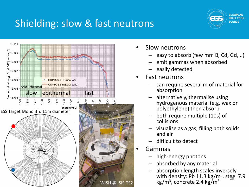
Shielding: slow & fast neutrons
29
slow epithermal fast
• Slow neutrons– easy to absorb (few mm B, Cd, Gd, ..)– emit gammas when absorbed– easily detected
• Fast neutrons– can require several m of material for
absorption– alternatively, thermalise using
hydrogenous material (e.g. wax or polyethylene) then absorb
– both require multiple (10s) of collisions
– visualise as a gas, filling both solids and air
– difficult to detect• Gammas
– high-energy photons– absorbed by any material– absorption length scales inversely
with density: Pb 11.3 kg/m3, steel 7.9 kg/m3, concrete 2.4 kg/m3
cold thermal
ESS Target Monolith: 11m diameter
WISH @ ISIS-TS2
10-1410-1110-910-710-210-1
Tim
e [s
]
10-12
10-9
10-15
10-6
10-3
1
103
10-6
Particle Physics
Spin-echo
Imaging
Reflectometry
10-10 10-12 10-1510-810-5 10-6
Backscattering
TOF & Crystal Spectroscopy
SANSDiffraction
Ener
gy [m
eV]
Length [m]
10210-1 110-3 10110-2
103Wavevector Q [Å-1]
10-3
10-1410-1110-910-710-210-1
Tim
e [s
]
10-12
10-9
10-15
10-6
10-3
1
103
10-6
Particle Physics
Spin-echo
Imaging
Reflectometry
10-10 10-12 10-1510-810-5
Visib
le
Ligh
t
InfraredLight
FTIRRaman
Brillouin
Dynamic Light Scattering
Laser PCS
10-6
Inelastic X-ray Scattering
NM
RµS
R
Diel
ectr
ic S
pec.Backscattering
UV & X-ray FEL: Pump & Probe studies
NMR Imaging Triangulation NMR
X-ray Imaging SAXS, X-ray Refl. X-ray Diffraction EXAFS & XANES
MicroscopyOptical Microscopy
Scanning TechniquesSNOM AFM STM
TOF & Crystal Spectroscopy
Infr
a-re
d
SANSDiffraction
Ener
gy [m
eV]
Length [m]
10210-1 110-3 10110-2
103
X-ray Spec.
X-ray PCS
Wavevector Q [Å-1]
10-3
X-rays
NMR
Optical Nanoscopy Electron Microscopy
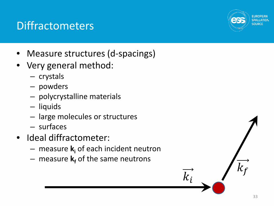
Diffractometers
• Measure structures (d-spacings)• Very general method:
– crystals– powders– polycrystalline materials– liquids– large molecules or structures– surfaces
• Ideal diffractometer: – measure ki of each incident neutron– measure kf of the same neutrons
33
𝑘𝑘𝑖𝑖𝑘𝑘𝑓𝑓
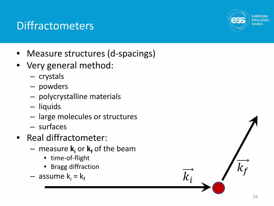
Diffractometers
• Measure structures (d-spacings)• Very general method:
– crystals– powders– polycrystalline materials– liquids– large molecules or structures– surfaces
• Real diffractometer: – measure ki or kf of the beam
• time-of-flight• Bragg diffraction
– assume ki = kf
34
𝑘𝑘𝑖𝑖𝑘𝑘𝑓𝑓
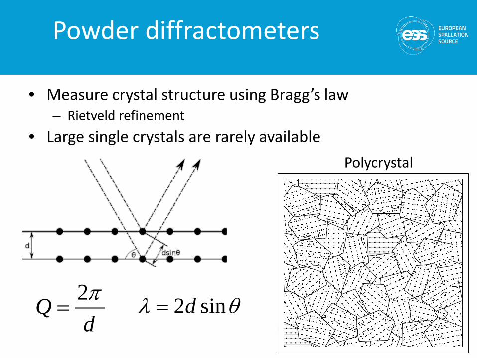
Powder diffractometers
Polycrystal
θλ sin2d=d
Q π2=
• Measure crystal structure using Bragg’s law– Rietveld refinement
• Large single crystals are rarely available
distance
time
sample
detector
Δt
θλ sin2d=
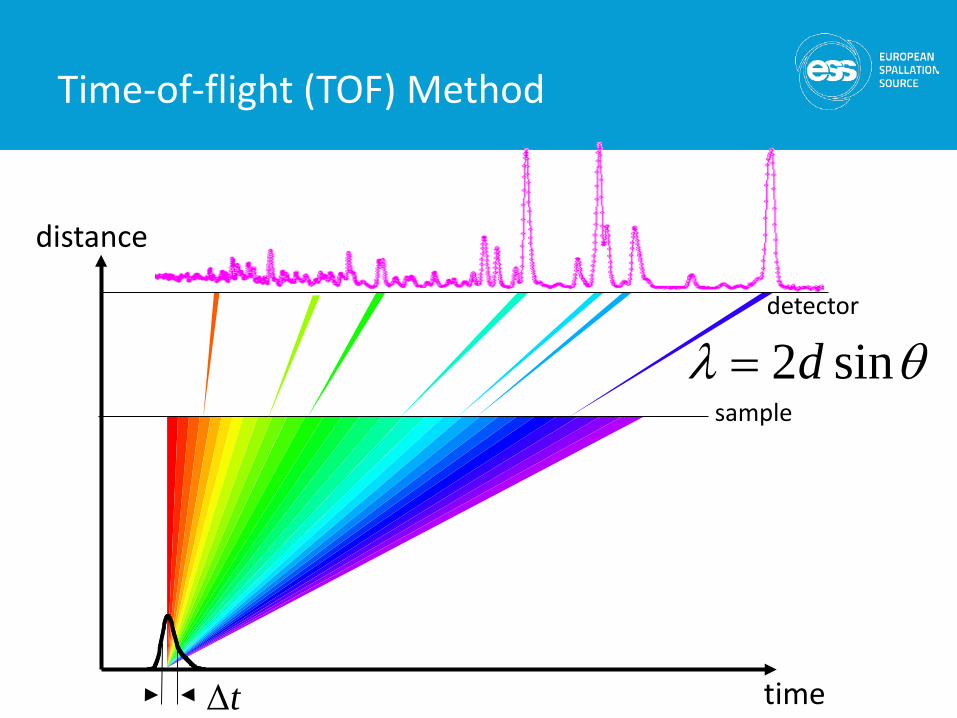
Time-of-flight (TOF) Method

distance
time
sample
detector
Δt
θλ sin2d=
Time-of-flight (TOF) Method

distance
time
sample
detector
Δt
θλ sin2d=
Time-of-flight (TOF) Method
time
Δt
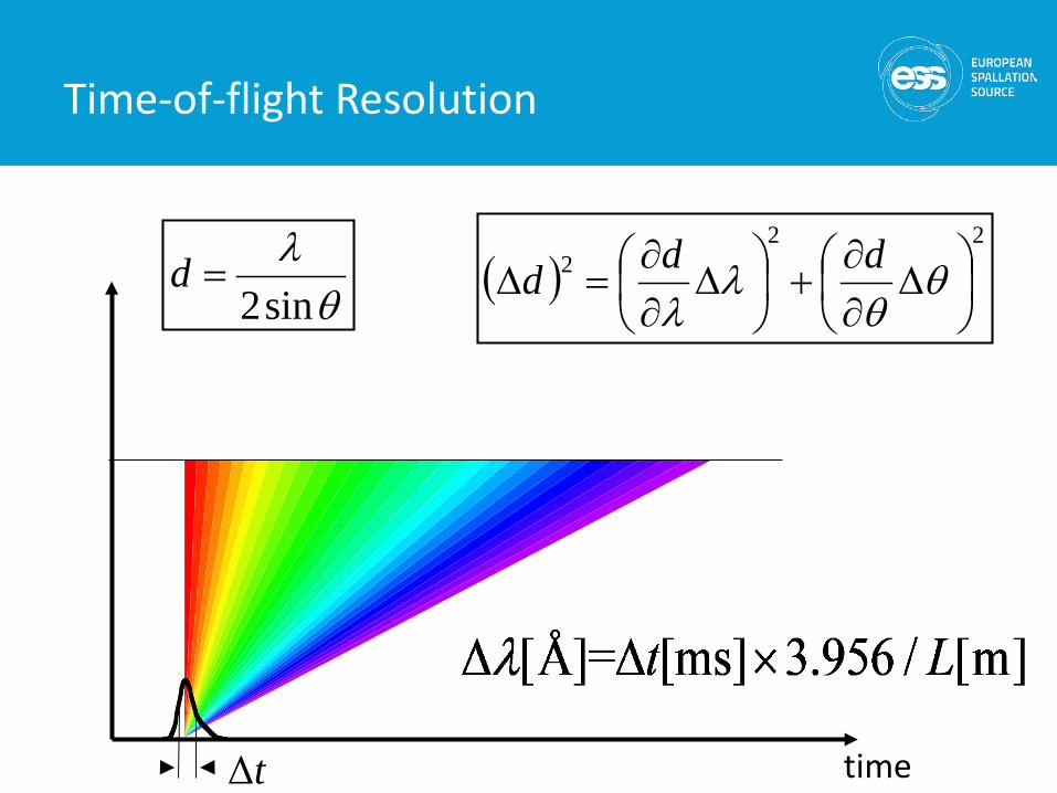
Time-of-flight Resolution
( )22
2
∆
∂∂
+
∆
∂∂
=∆ θθ
λλ
dddθλ
sin2=d
time
Δt
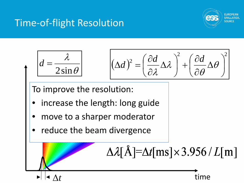
Time-of-flight Resolution
( )22
2
∆
∂∂
+
∆
∂∂
=∆ θθ
λλ
dddθλ
sin2=d
To improve the resolution:• increase the length: long guide• move to a sharper moderator• reduce the beam divergence
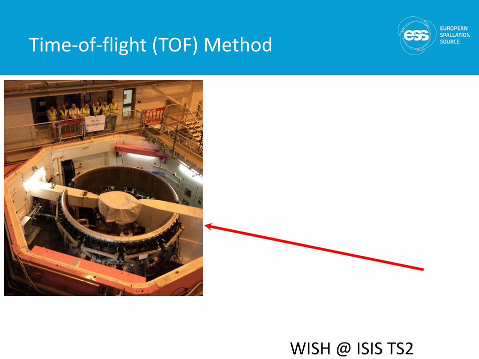
WISH @ ISIS TS2
Time-of-flight (TOF) Method
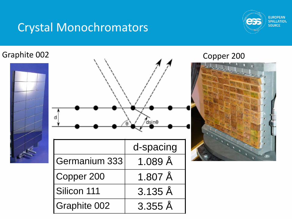
Copper 200Graphite 002
d-spacingGermanium 333 1.089 ÅCopper 200 1.807 ÅSilicon 111 3.135 ÅGraphite 002 3.355 Å
Crystal Monochromators
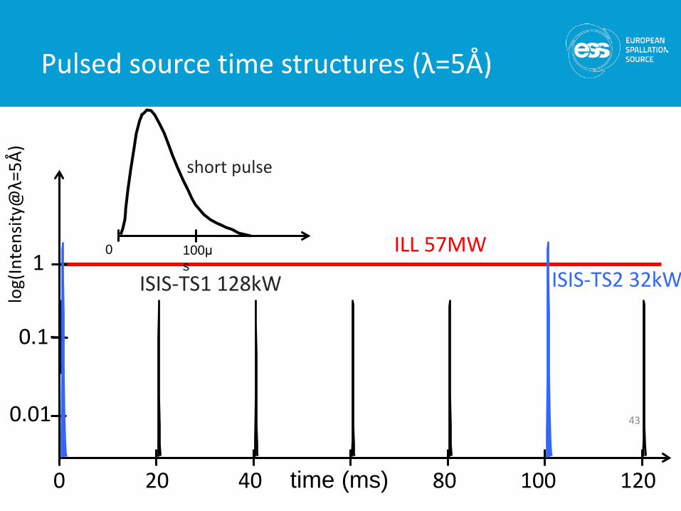
Pulsed source time structures (λ=5Å)
43
log(
Inte
nsity
@λ=
5Å)
0 20 40 80 100 120 time (ms)
1
0.1
0.01
ISIS-TS1 128kW ISIS-TS2 32kW100μs
0
short pulse
ILL 57MW
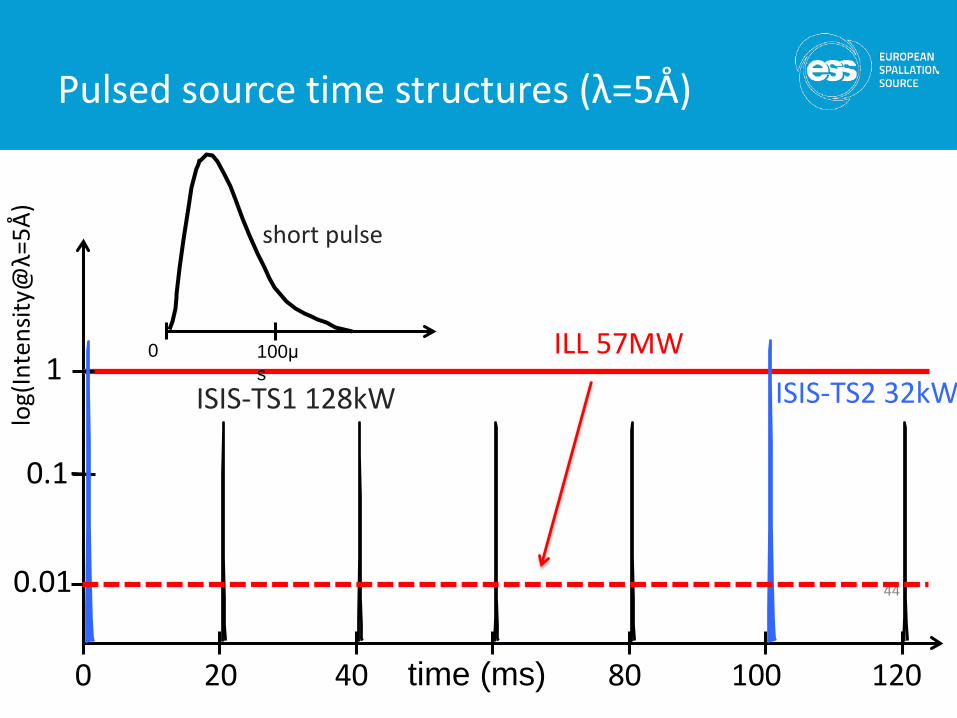
Pulsed source time structures (λ=5Å)
44
log(
Inte
nsity
@λ=
5Å)
0 20 40 80 100 120 time (ms)
1
0.1
0.01
ISIS-TS1 128kW ISIS-TS2 32kW100μs
0
short pulse
ILL 57MW
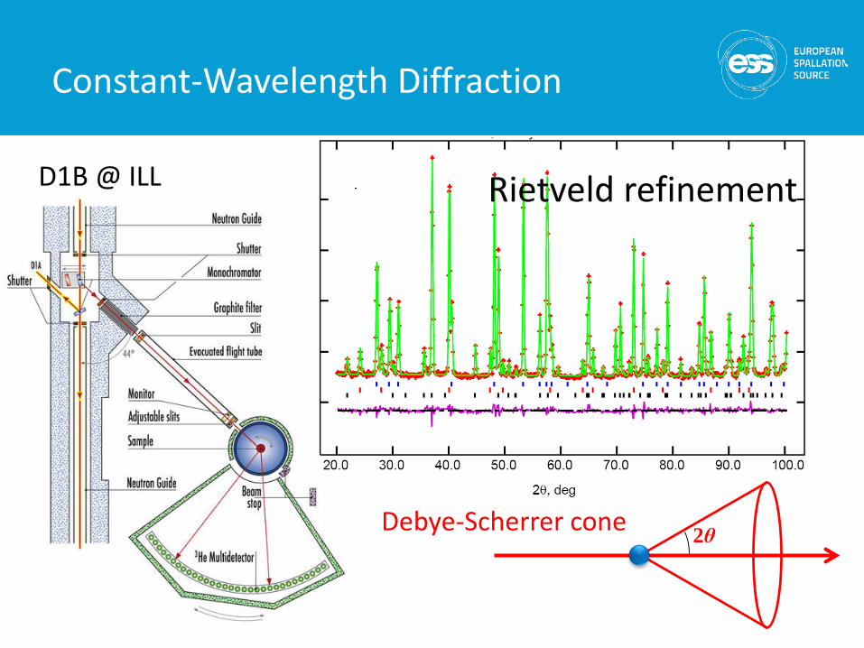
D1B @ ILL
Constant-Wavelength Diffraction
Rietveld refinement
2θDebye-Scherrer cone
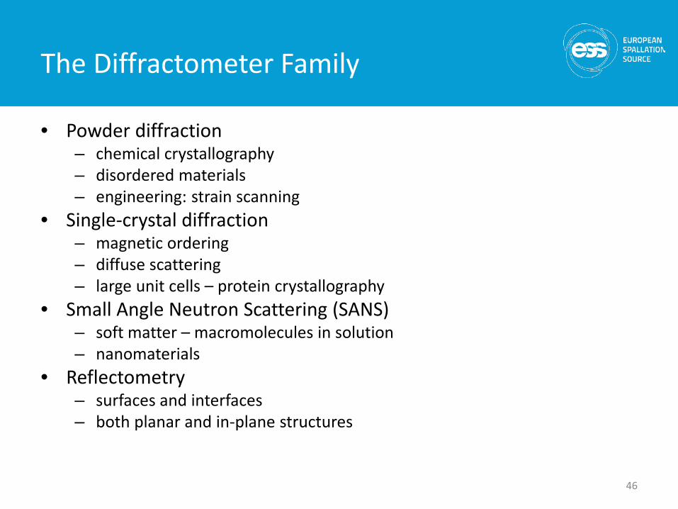
The Diffractometer Family
• Powder diffraction– chemical crystallography– disordered materials– engineering: strain scanning
• Single-crystal diffraction– magnetic ordering– diffuse scattering– large unit cells – protein crystallography
• Small Angle Neutron Scattering (SANS)– soft matter – macromolecules in solution– nanomaterials
• Reflectometry– surfaces and interfaces– both planar and in-plane structures
46
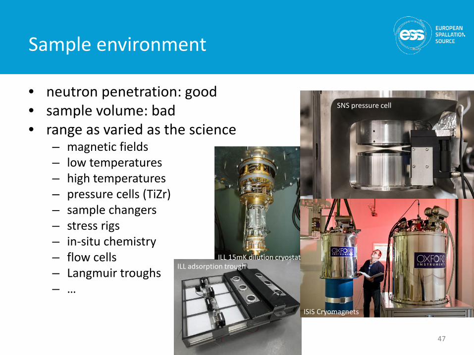
Sample environment
• neutron penetration: good• sample volume: bad• range as varied as the science
– magnetic fields– low temperatures– high temperatures– pressure cells (TiZr)– sample changers– stress rigs– in-situ chemistry– flow cells– Langmuir troughs– …
47
ILL adsorption trough
ISIS Cryomagnets
SNS pressure cell
ILL 15mK dilution cryostat
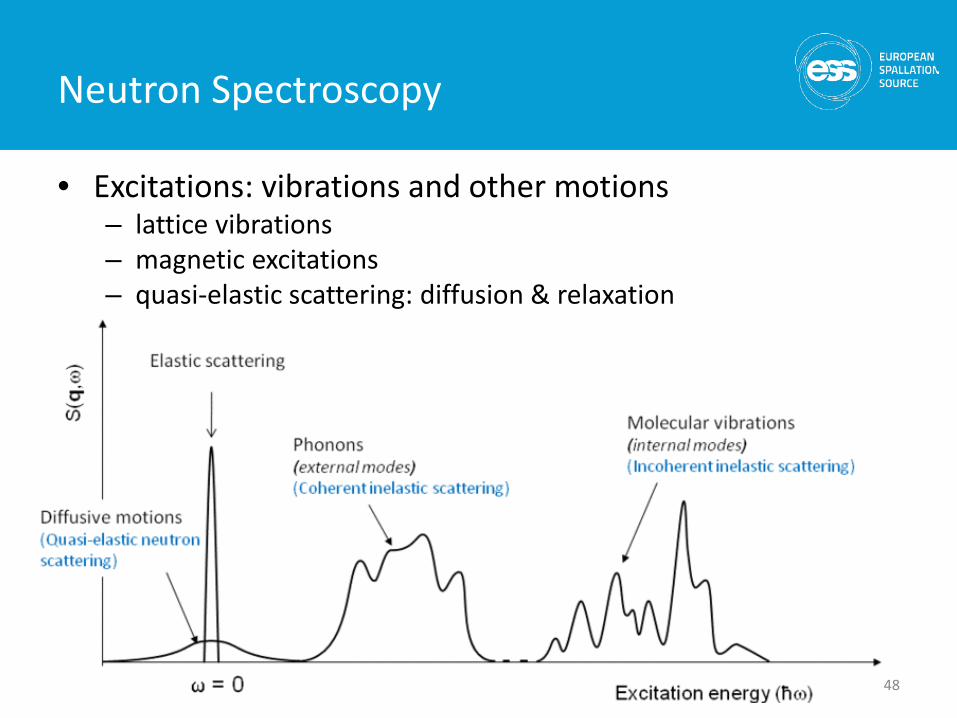
Neutron Spectroscopy
• Excitations: vibrations and other motions– lattice vibrations– magnetic excitations– quasi-elastic scattering: diffusion & relaxation
48
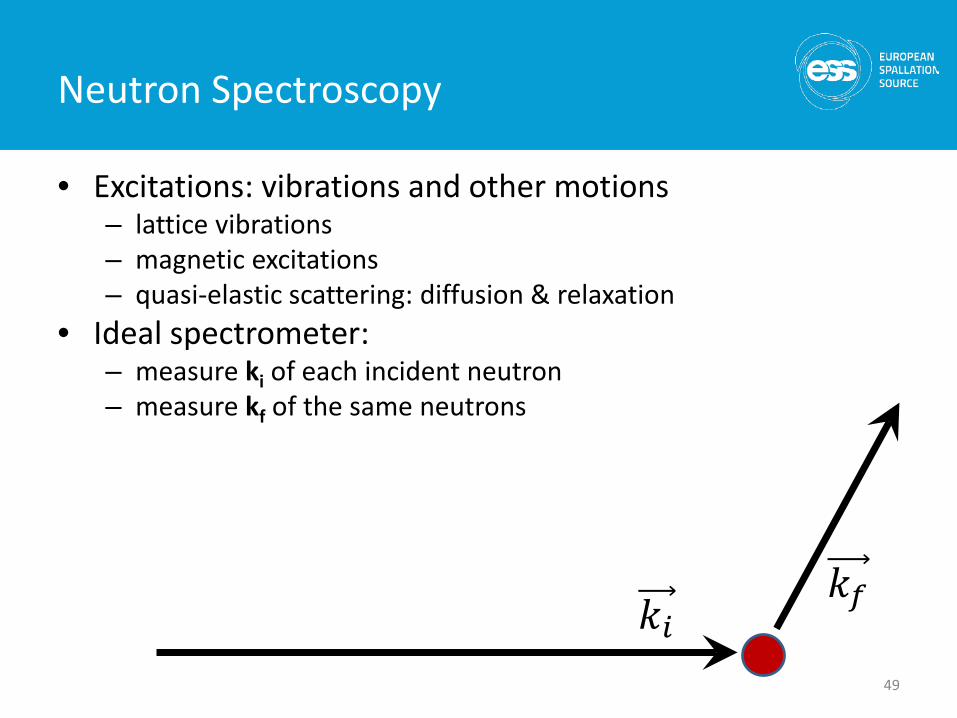
Neutron Spectroscopy
• Excitations: vibrations and other motions– lattice vibrations– magnetic excitations– quasi-elastic scattering: diffusion & relaxation
• Ideal spectrometer: – measure ki of each incident neutron– measure kf of the same neutrons
49
𝑘𝑘𝑖𝑖𝑘𝑘𝑓𝑓
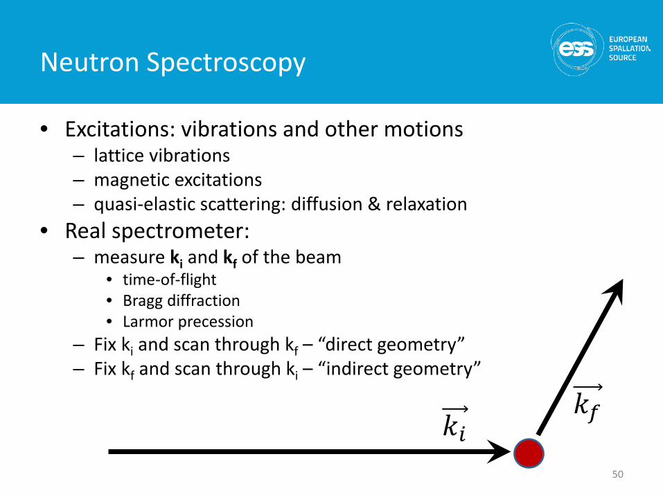
Neutron Spectroscopy
• Excitations: vibrations and other motions– lattice vibrations– magnetic excitations– quasi-elastic scattering: diffusion & relaxation
• Real spectrometer: – measure ki and kf of the beam
• time-of-flight• Bragg diffraction• Larmor precession
– Fix ki and scan through kf – “direct geometry”– Fix kf and scan through ki – “indirect geometry”
50
𝑘𝑘𝑖𝑖𝑘𝑘𝑓𝑓
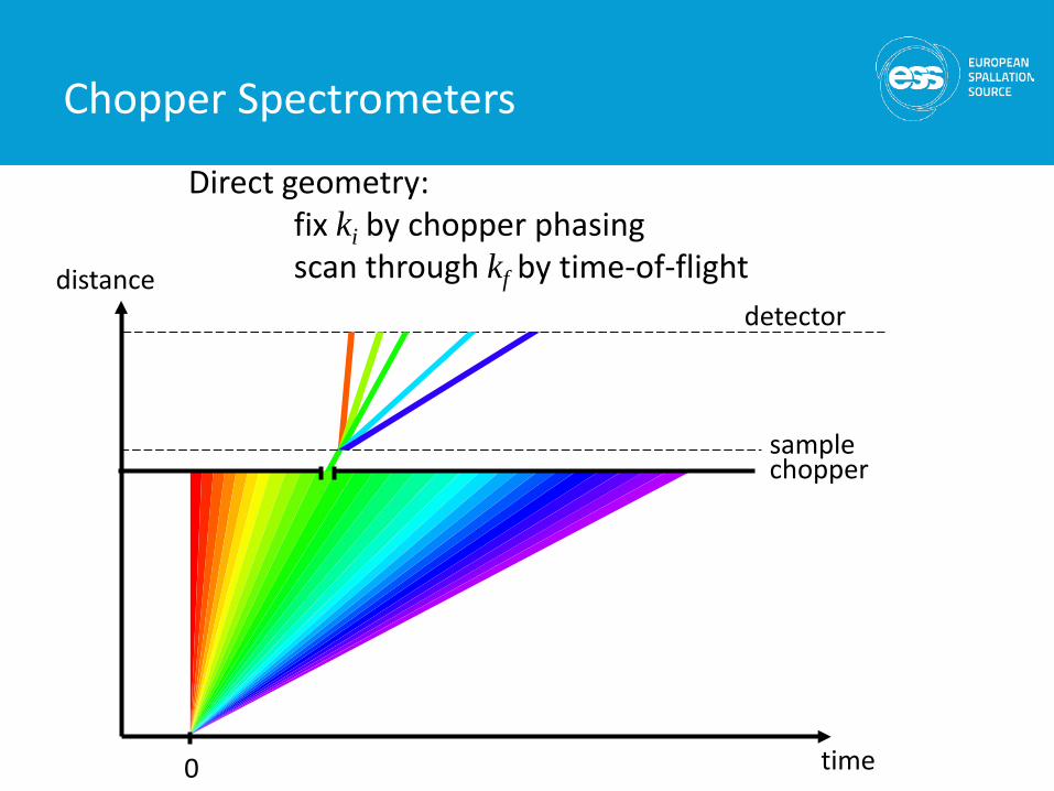
distance
time
chopper
detector
sample
0
Direct geometry: fix ki by chopper phasingscan through kf by time-of-flight
Chopper Spectrometers
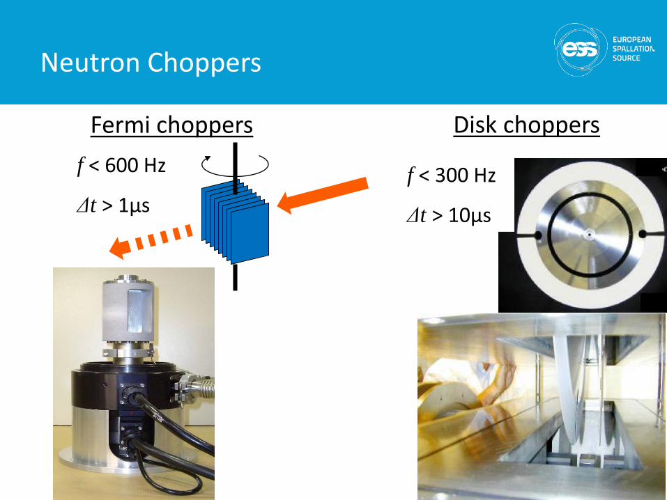
Disk choppersFermi choppers
f < 300 Hz
Δt > 10μs
f < 600 Hz
Δt > 1μs
Neutron Choppers
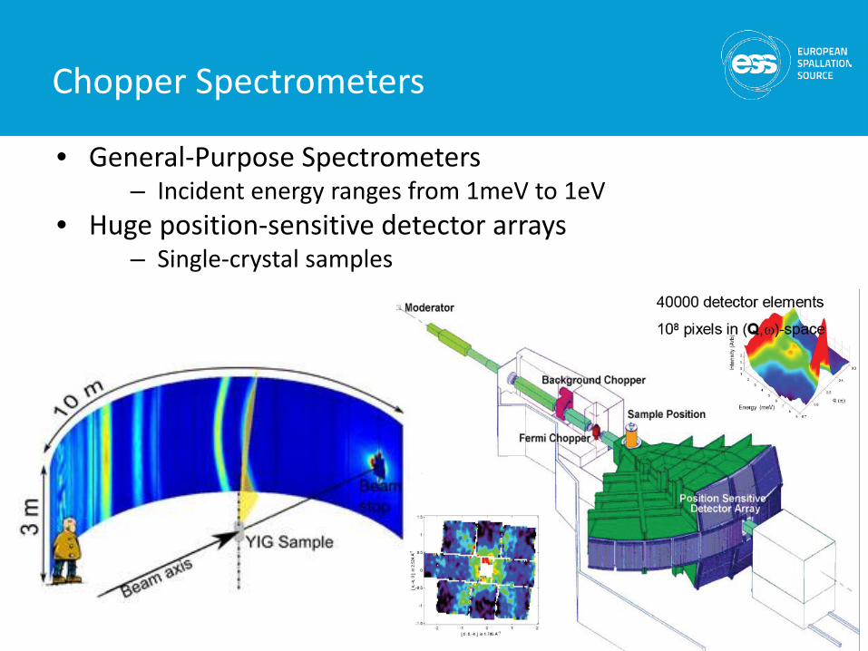
• General-Purpose Spectrometers– Incident energy ranges from 1meV to 1eV
• Huge position-sensitive detector arrays– Single-crystal samples
Chopper Spectrometers
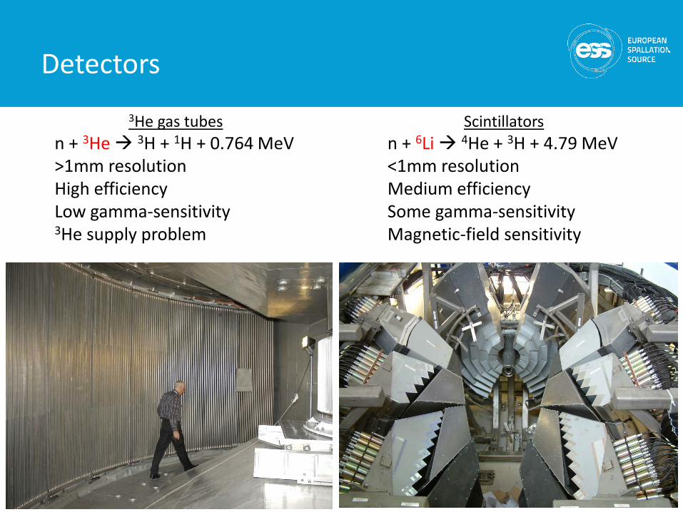
3He gas tubesn + 3He 3H + 1H + 0.764 MeV>1mm resolutionHigh efficiencyLow gamma-sensitivity3He supply problem
Scintillatorsn + 6Li 4He + 3H + 4.79 MeV<1mm resolutionMedium efficiencySome gamma-sensitivityMagnetic-field sensitivity
Detectors
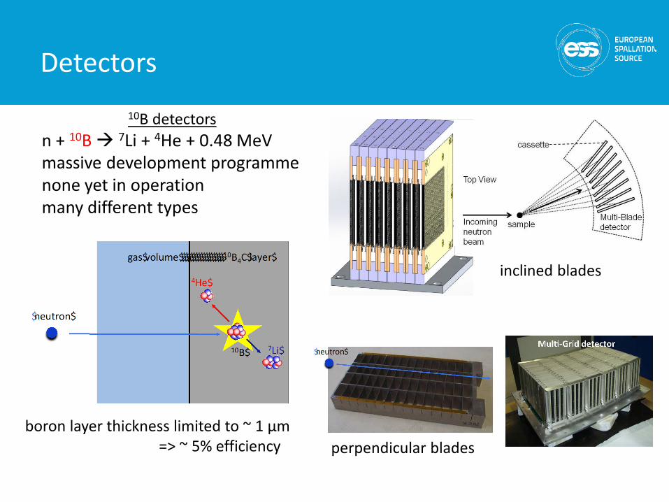
10B detectorsn + 10B 7Li + 4He + 0.48 MeVmassive development programmenone yet in operationmany different types
Detectors
inclined blades
perpendicular bladesboron layer thickness limited to ~ 1 μm
=> ~ 5% efficiency
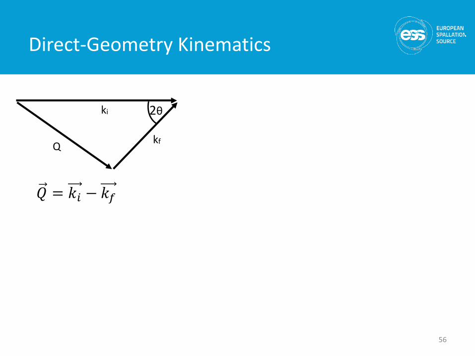
Direct-Geometry Kinematics
56
ki
kfQ
2θ
𝑄𝑄 = 𝑘𝑘𝑖𝑖 − 𝑘𝑘𝑓𝑓
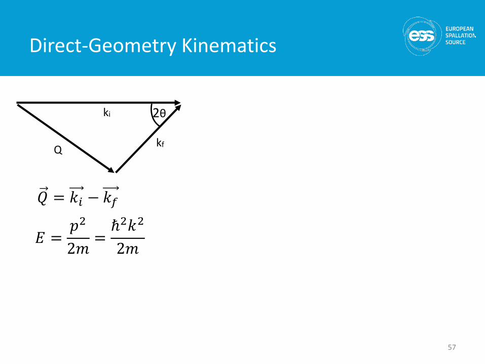
Direct-Geometry Kinematics
57
ki
kfQ
2θ
𝐸𝐸 =𝑝𝑝2
2𝑚𝑚=ℏ2𝑘𝑘2
2𝑚𝑚
𝑄𝑄 = 𝑘𝑘𝑖𝑖 − 𝑘𝑘𝑓𝑓
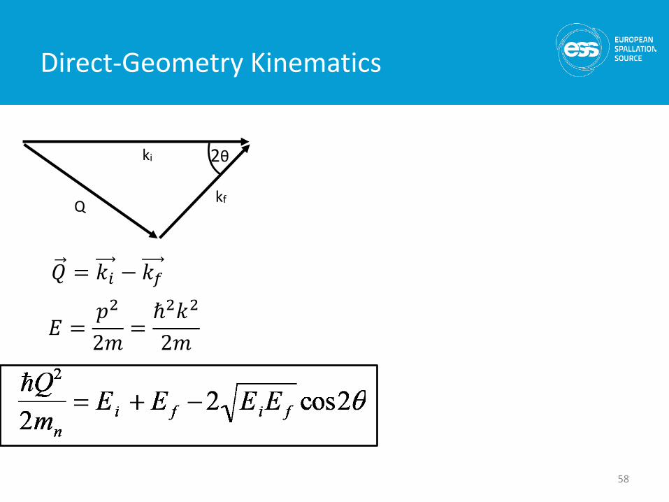
Direct-Geometry Kinematics
58
ki
kfQ
2θ
𝐸𝐸 =𝑝𝑝2
2𝑚𝑚=ℏ2𝑘𝑘2
2𝑚𝑚
𝑄𝑄 = 𝑘𝑘𝑖𝑖 − 𝑘𝑘𝑓𝑓
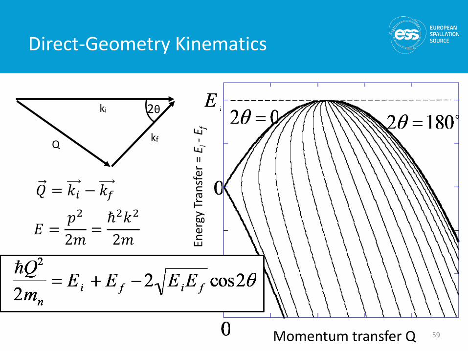
Direct-Geometry Kinematics
59Momentum transfer Q
ki
kfQ
2θ
Ener
gy T
rans
fer =
Ei-
E f
𝐸𝐸 =𝑝𝑝2
2𝑚𝑚=ℏ2𝑘𝑘2
2𝑚𝑚
𝑄𝑄 = 𝑘𝑘𝑖𝑖 − 𝑘𝑘𝑓𝑓
distance
time
detector
sample
0
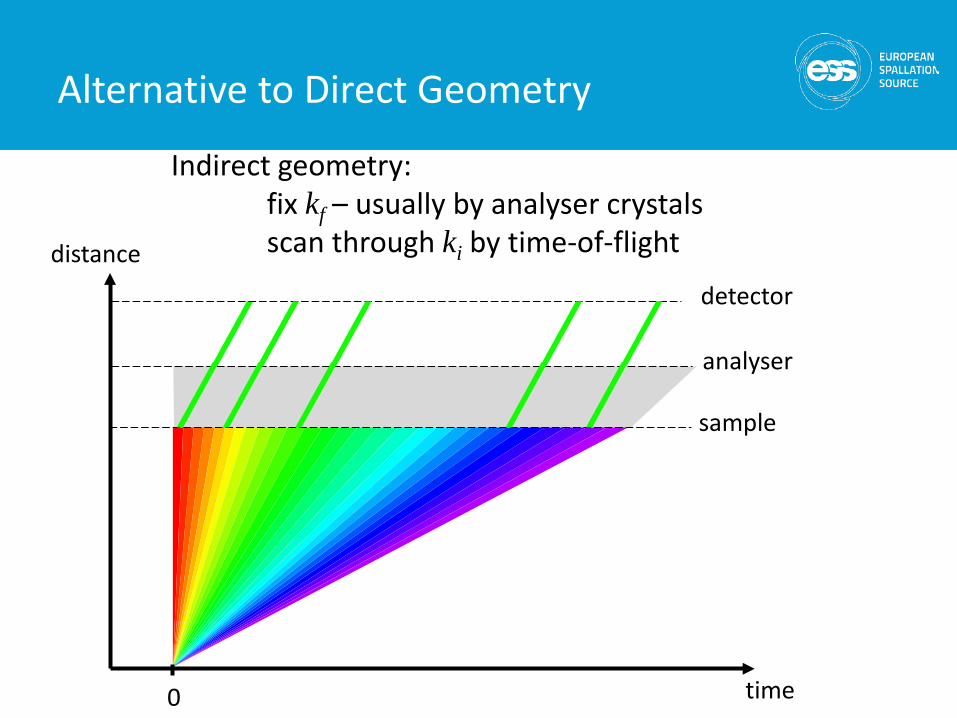
Indirect geometry: fix kf – usually by analyser crystalsscan through ki by time-of-flight
analyser
Alternative to Direct Geometry
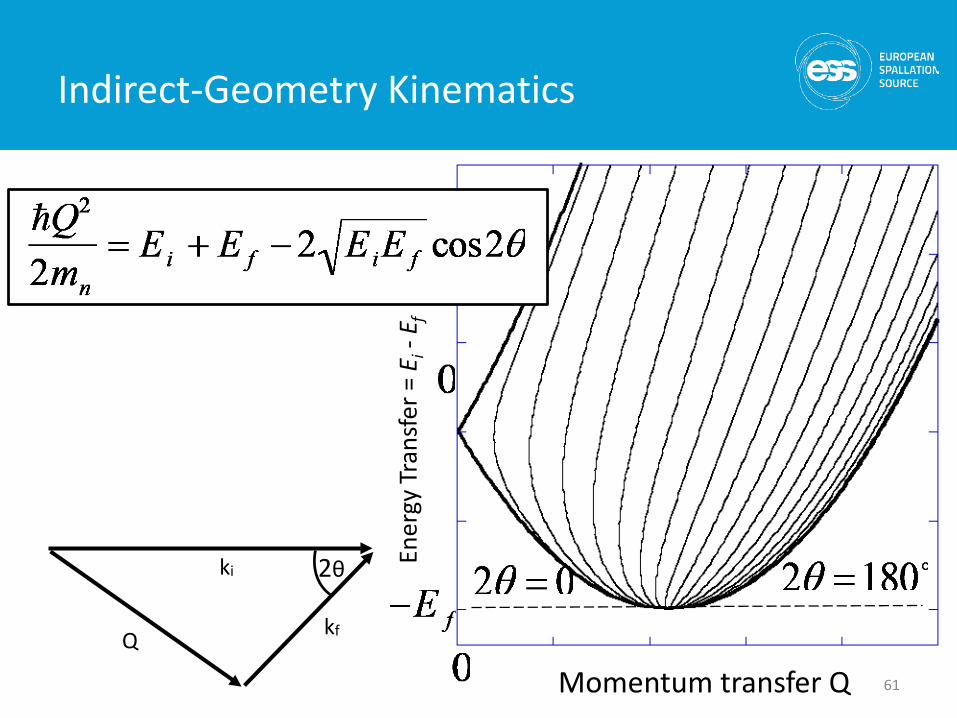
Indirect-Geometry Kinematics
61Momentum transfer Q
ki
kfQ
2θ Ener
gy T
rans
fer =
Ei-
E f
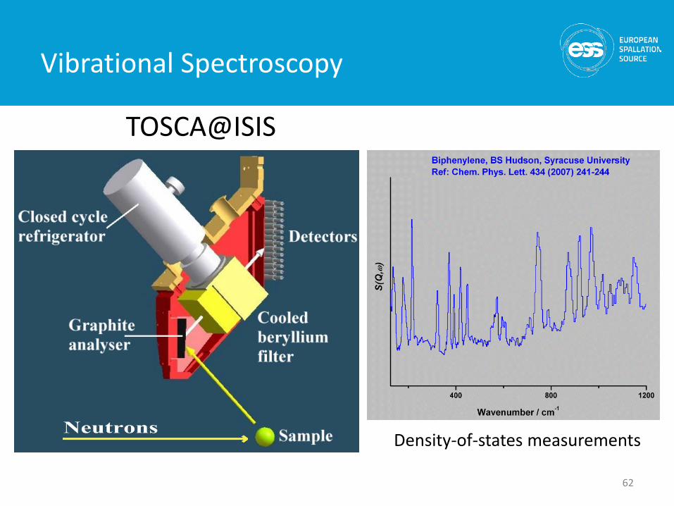
Vibrational Spectroscopy
62
TOSCA@ISIS
Density-of-states measurements
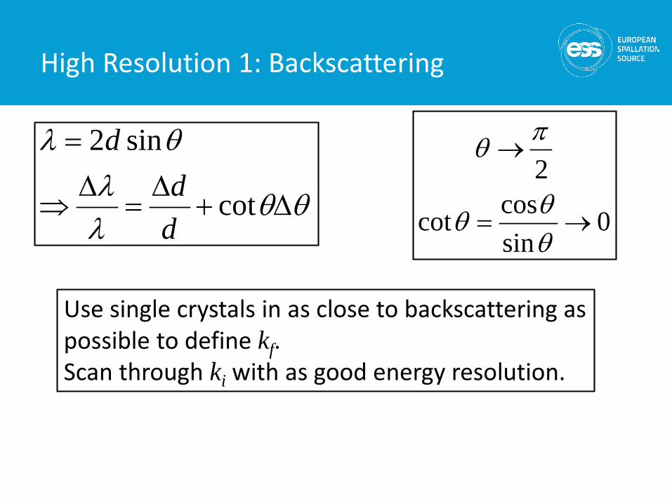
θθλλ
θλ
∆+∆
=∆
⇒
=
cot
sin2
dd
d
0sincoscot
2
→=
→
θθθ
πθ
Use single crystals in as close to backscattering as possible to define kf. Scan through ki with as good energy resolution.
High Resolution 1: Backscattering
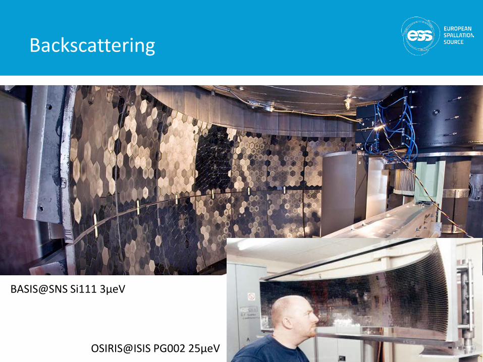
Backscattering
BASIS@SNS Si111 3μeV
OSIRIS@ISIS PG002 25μeV
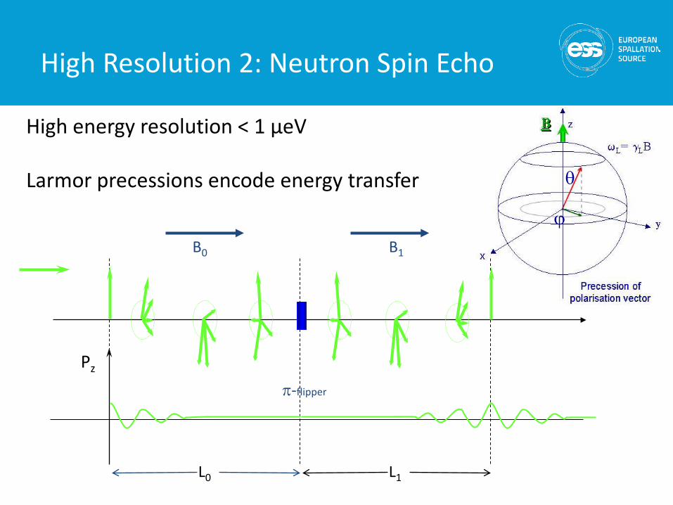
ϕ
θ
B0 B1
L0 L1
Pz
π-flipper
High energy resolution < 1 μeV
Larmor precessions encode energy transfer
High Resolution 2: Neutron Spin Echo
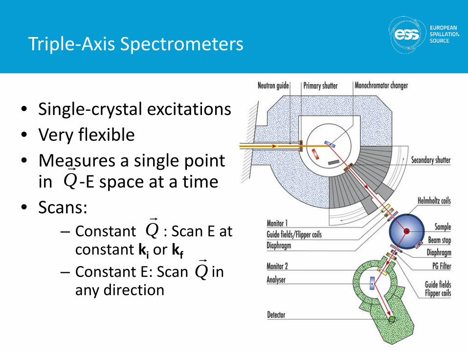
• Single-crystal excitations• Very flexible• Measures a single point
in -E space at a time• Scans:
– Constant : Scan E at constant ki or kf
– Constant E: Scan in any direction
Q
Q
Q
Triple-Axis Spectrometers
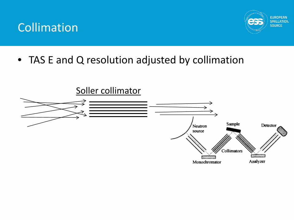
Collimation
• TAS E and Q resolution adjusted by collimation
Soller collimator
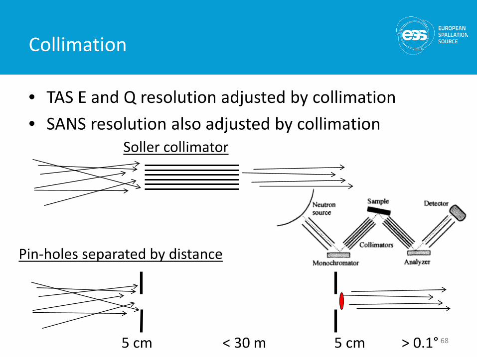
Collimation
• TAS E and Q resolution adjusted by collimation• SANS resolution also adjusted by collimation
68
Soller collimator
Pin-holes separated by distance
> 0.1°5 cm 5 cm< 30 m
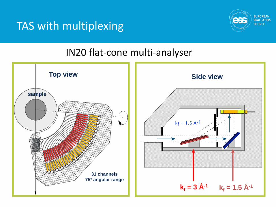
TAS with multiplexing
Top view
sample
31 channels75º angular range
kf = 3 Å-1 kf = 1.5 Å-1
Side view
IN20 flat-cone multi-analyser
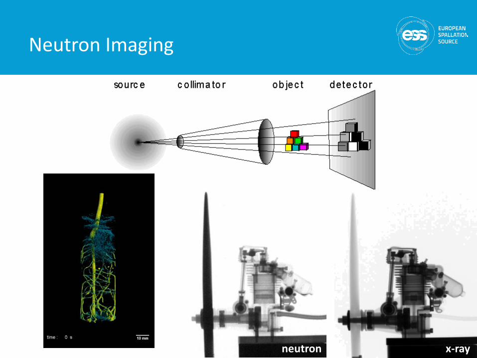
Neutron Imaging
70neutron x-ray
Summary
• Neutron instrument concepts– time-of-flight– Bragg’s law
• Neutron Instrumentation– guides– monochromators– shielding– detectors– choppers– sample environment– collimation
• Neutron diffractometers– powder diffraction
• Neutron spectrometers– direct and indirect geometry time-of-flight– backscattering– triple-axis– spin-echo
• Imaging
71

Thank you!
725th June 2015