neutrophilic dermatosis confined to the lymphedematous …€¦ · lymphedema is a common sequelae...
TRANSCRIPT
Letter to the Editor
Vol. 26 No. 3, 2014 411
Received June 4, 2013, Accepted for publication June 14, 2014
Corresponding author: You Chan Kim, Department of Dermatology, Ajou University School of Medicine, 206 WorldCup-ro, Yeongtong-gu, Suwon 443-749, Korea. Tel: 82-31-219-5190, Fax: 82-31-219-5189,E-mail: [email protected]
This is an Open Access article distributed under the terms of the Creative Commons Attribution Non-Commercial License (http:// creativecommons.org/licenses/by-nc/3.0) which permits unrestrictednon-commercial use, distribution, and reproduction in any medium, provided the original work is properly cited.
http://dx.doi.org/10.5021/ad.2014.26.3.411
Neutrophilic Dermatosis Confined to the Lymphedematous Area
Ji-Youn Park, Hee Young Kang, You Chan Kim
Department of Dermatology, Ajou University School of Medicine, Suwon, Korea
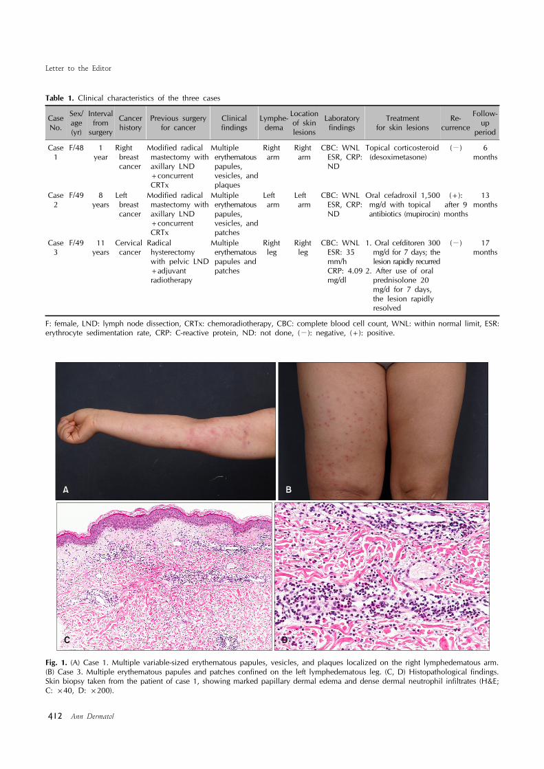
Dear Editor:Lymphedema is a common sequelae after cancer surgery with lymph node dissection1. A lymphedematous limb, which is prone to the development of infections or tumors, suggests an alteration in regional immune com-petence2. Neutrophilic dermatosis on postmastectomy lymphedema (NDPL) is a newly suggested disease by Demitsu and Tadaki3 It is also referred to as localized Sweet’s syndrome (SS) because of the histological similarities between the two conditions4. Herein, we report additional cases of neutrophilic dermatosis con-fined within a lymphedematous site and a review of the disease entity.All three cases involved female patients, two of whom were breast cancer patients who underwent modified radical mastectomy with axillary lymph node dissections (Table 1). The third patient had undergone radical hysterectomy with pelvic lymph node dissection for cervical cancer. Lymphedema was confined to the lymph- node-dissected limb developed in all three patients after the surgery. All of them rapidly developed erythematous rashes on their lymphedematous limb (Fig. 1A, B). Skin biopsy revealed marked papillary dermal edema and dense dermal neutrophil infiltrates, consistent with the histopathologic features of SS (Fig. 1C, D). The laboratory findings, including white blood cell count, percentage of neutrophils, erythrocyte sedimentation rate (ESR), and
C-reactive protein (CRP), were within the normal ranges, except in case 3 (ESR: 35 mm/h; CRP: 4.09 mg/dl). Each case was treated with topical or oral corticosteroid, or oral antibiotics. In case 3, the lesion recurred rapidly after initially being treated with oral antibiotics. After the administration of oral corticosteroid, the lesion rapidly resolved within 1 week. On the basis of the characteristic distribution of the lesion and the histopathologic features, a diagnosis of ‘neutrophilic dermatosis on the lymphede-matous area’ was made.Neutrophilic dermatosis or SS localized on the area of lymphedema is rare, and only 11 cases have been reported1,3,4. The clinical presentations of previously reported cases were erythematous papules, plaques, and vesicles on the lymphedematous arm after a mastectomy, which are consistent with our cases1,3,4. The patho-mechanism cannot be fully demonstrated; however, the vulnerability of the lymphedematous area seems to be the main factor1,2,5. The stasis of protein-rich lymphatic fluid contains numerous cytokines that might attract neutrophils and also result in the impairment of immune sur-veillance2,5. Because this dermatosis shows typical clini-cohistopathological findings of SS, they are considered a localized variant of SS4. However, other systemic pre-sentations, such as leukocytosis, neutrophilia, or fever, were less frequent than in typical SS. Therefore, several other reports suggested the use of the new term NDPL1,3. Other clinical differential diagnosis included cellulitis or erysipelas. Contrary to cellulitis or erysipelas, the lesion was confined only to the lymphedematous area and was well treated with oral or topical corticosteroid1,4. Although the 11 reports to date were cases of a lesion on a lymphedematous arm after a mastectomy, our report in-cludes one case of a lesion that had developed on the lymphedematous leg after a hysterectomy for cervical cancer. Consequently, our cases suggest a novel point that a lesion could develop on any lymphedematous limb after
Letter to the Editor
412 Ann Dermatol
Table 1. Clinical characteristics of the three cases
Case No.
Sex/age(yr)
Interval from
surgery
Cancer history
Previous surgery for cancer
Clinical findings
Lymphe-dema
Location of skin lesions
Laboratory findings
Treatment for skin lesions
Re-currence
Follow-up
period
Case 1
F/48 1 year
Right breast cancer
Modified radical mastectomy with axillary LND +concurrent CRTx
Multiple erythematous papules, vesicles, and plaques
Right arm
Right arm
CBC: WNL ESR, CRP: ND
Topical corticosteroid (desoximetasone)
(−) 6 months
Case 2
F/49 8years
Left breast cancer
Modified radical mastectomy with axillary LND +concurrent CRTx
Multiple erythematous papules, vesicles, and patches
Left arm
Left arm
CBC: WNL ESR, CRP: ND
Oral cefadroxil 1,500 mg/d with topical antibiotics (mupirocin)
(+): after 9 months
13 months
Case 3
F/49 11 years
Cervical cancer
Radical hysterectomy with pelvic LND +adjuvant radiotherapy
Multiple erythematous papules and patches
Right leg
Right leg
CBC: WNL ESR: 35 mm/h CRP: 4.09 mg/dl
1. Oral cefditoren 300 mg/d for 7 days; the lesion rapidly recurred2. After use of oral prednisolone 20 mg/d for 7 days, the lesion rapidly resolved
(−)
17 months
F: female, LND: lymph node dissection, CRTx: chemoradiotherapy, CBC: complete blood cell count, WNL: within normal limit, ESR:erythrocyte sedimentation rate, CRP: C-reactive protein, ND: not done, (−): negative, (+): positive.
Fig. 1. (A) Case 1. Multiple variable-sized erythematous papules, vesicles, and plaques localized on the right lymphedematous arm. (B) Case 3. Multiple erythematous papules and patches confined on the left lymphedematous leg. (C, D) Histopathological findings. Skin biopsy taken from the patient of case 1, showing marked papillary dermal edema and dense dermal neutrophil infiltrates (H&E; C: ×40, D: ×200).
Letter to the Editor
Vol. 26 No. 3, 2014 413
Received March 13, 2013, Revised March 27, 2013, Accepted for publication June 17, 2013
Corresponding author: Seong-Jin Kim, Department of Dermatology, Chonnam National University Hospital, Chonnam National University Medical School, 42 Jebong-ro, Dong-gu, Gwangju 501-757, Korea. Tel: 82-62-220-6683, Fax: 82-62-222-4058, E-mail: seongkim @jnu.ac.kr
This is an Open Access article distributed under the terms of the Creative Commons Attribution Non-Commercial License (http:// creativecommons.org/licenses/by-nc/3.0) which permits unrestricted non-commercial use, distribution, and reproduction in any medium, provided the original work is properly cited.
lymph node dissection. Therefore, we suggest the term ‘neutrophilic dermatosis on the lymphedematous area’ rather than NDPL.
REFERENCES
1. Lee CH, Lee HC, Lu CF, Hsiao CH, Jee SH, Tjiu JW. Neutrophilic dermatosis on postmastectomy lymphoedema:
a localized and less severe variant of Sweet syndrome. Eur J
Dermatol 2009;19:641-642.2. Mallon E, Powell S, Mortimer P, Ryan TJ. Evidence for
altered cell-mediated immunity in postmastectomy lymphoe-
dema. Br J Dermatol 1997;137:928-933.3. Demitsu T, Tadaki T. Atypical neutrophilic dermatosis on the
upper extremity affected by postmastectomy lymphedema:
report of 2 cases. Dermatologica 1991;183:230- 233.4. García-Río I, Pérez-Gala S, Aragüés M, Fernández-Herrera J,
Fraga J, García-Díez A. Sweet's syndrome on the area of
postmastectomy lymphoedema. J Eur Acad Dermatol Vene-reol 2006;20:401-405.
5. Ruocco E, Puca RV, Brunetti G, Schwartz RA, Ruocco V.
Lymphedematous areas: privileged sites for tumors, infec-tions, and immune disorders. Int J Dermatol 2007;46:662.
http://dx.doi.org/10.5021/ad.2014.26.3.413
Nipple Eczema: A Diagnostic Challenge of Allergic Contact Dermatitis
Sun Kyung Kim, Young Ho Won, Seong-Jin Kim
Department of Dermatology, Chonnam National University Medical School, Gwangju, Korea
Dear Editor:Nipple eczema, considered mostly as a minor manifesta-tion of atopic dermatitis, may have unknown causes. However, its clinical course and pattern often make it difficult to differentiate its underlying causes such as irri-tation or sensitization. Nevertheless, allergic contact der-matitis must be considered an important cause of nipple eczema.In the present study, we analyzed the patch test results from pateints of nipple eczema by using the Korean stan-dard series comprising 25 antigens (Chemotechnique Diagnostics, Malmo, Sweden). Antigens were carefully
added into an IQ Ultra chamberⓇ (Chemotechnique Dia-gnostics) which is made of additive-free polyethylene plastic foam with a filter paper incorporated, and stuck to the backs of the patients. Results were recorded 30 minutes after patch removal (as usual), and the patients were re-evaluated 48 hours later. On the basis of the recommendations of the International Contact Dermatitis Research Group, a reading of +1 (patients with erythema-tous papules and edema but without any vesicles) or higher was deemed a positive response.Among a total of 12 patients (all women) who were patch tested, 5 were clearly diagnosed with atopic dermatitis on