neutrophil–lymphocyte ratio for the prediction of
TRANSCRIPT

RESEARCH Open Access
Neutrophil–lymphocyte ratio for theprediction of histological chorioamnionitisin cases of preterm premature rupture ofmembranes: a case-control studyGreta Balciuniene1,2, Greta Kvederaite-Budre1, Violeta Gulbiniene1,2, Irena Dumalakiene3, Rita Viliene3,Ingrida Pilypiene2, Grazina S. Drasutiene1,2 and Diana Ramasauskaite1,2*
Abstract
Background: The neutrophil-lymphocyte ratio (NLR) is easily calculated blood test parameter, which can be used asmarker to predict many inflammatory disorders. The aim of this study was to assess and compare the NLR inmaternal blood with the white blood cell (WBC) count and C-reactive protein (CRP) concentration for theprediction of histological chorioamnionitis.
Methods: This was a case-control study of 137 woman with preterm premature rupture of membranes (PPROM) ata gestational age between 22+ 0 and 34+ 6 weeks. Blood samples, collected less than 48 h before delivery and atleast 48 h after the administration of corticosteroids, were selected for the analysis. The NLR was calculated bydividing the number of neutrophils by the number of lymphocytes. Chorioamnionitis was diagnosed by thehistopathological evaluation of placental membranes and chorionic plate.
Results: Patients with diagnosed histological chorioamnionitis (HCA) had significantly higher levels of WBC, CRPand NLR (p-value < 0.001). Levels of WBC, CRP and NLR predicted HCA with an area under the curve (AUC) of 0.81,0.81 and 0.89, respectively. NLR had statistically significantly higher AUC than WBC, but no significant difference wasfound between AUCs of NLR and CRP. The cut-off level of NLR was found to be 5,97, which had a sensitivity of77 % and a specificity of 95 %.
Conclusion: NLR has a good predictive value for HCA and could be used as an additional diagnostic marker forpredicting histological chorioamnionitis in cases with preterm premature rupture of membranes before 34 weeks ofgestation.
Keywords: chorioamnionitis, preterm premature rupture of membranes, neutrophil-lymphocyte ratio, white bloodcell, C-reactive protein
© The Author(s). 2021 Open Access This article is licensed under a Creative Commons Attribution 4.0 International License,which permits use, sharing, adaptation, distribution and reproduction in any medium or format, as long as you giveappropriate credit to the original author(s) and the source, provide a link to the Creative Commons licence, and indicate ifchanges were made. The images or other third party material in this article are included in the article's Creative Commonslicence, unless indicated otherwise in a credit line to the material. If material is not included in the article's Creative Commonslicence and your intended use is not permitted by statutory regulation or exceeds the permitted use, you will need to obtainpermission directly from the copyright holder. To view a copy of this licence, visit http://creativecommons.org/licenses/by/4.0/.The Creative Commons Public Domain Dedication waiver (http://creativecommons.org/publicdomain/zero/1.0/) applies to thedata made available in this article, unless otherwise stated in a credit line to the data.
* Correspondence: [email protected] of Obstetrics, and Gynecology, Institute of Clinical Medicine, Faculty ofMedicine of Vilnius University, M.K. Ciurlionio st. 21, 03101 Vilnius, Lithuania2Center of Obstetrics and Gynecology, Vilnius University Hospital SantarosKlinikos, Santariskiu st. 2, 08661 Vilnius, LithuaniaFull list of author information is available at the end of the article
Balciuniene et al. BMC Pregnancy and Childbirth (2021) 21:656 https://doi.org/10.1186/s12884-021-04101-z

BackgroundPreterm premature rupture of the membranes (PPROM)is the spontaneous rupture of foetal membranes duringpregnancy prior to 37 weeks of gestation. One of themost serious complications of PPROM is chorioamnio-nitis, which complicates almost half of all PPROM cases[1] and is associated with increased risks of neonatalmorbidity and mortality [2, 3]. Induction of labour is themost effective intervention for protecting the foetusagainst intrauterine infection, but preterm delivery, par-ticularly before 32 weeks of gestation, is associated withhigh risks of neonatal morbidity and mortality becauseof foetal immaturity [3, 4]. Thus, when choosing themethod of PPROM management, the benefits of preg-nancy prolongation and the risk of chorioamnionitismust be balanced.The white blood cells (WBC) and C-reactive protein
(CRP) are inflammatory markers used worldwide for theearly diagnosis of chorioamnionitis. A systemic reviewand meta-analysis performed by Sabogal with colleaguesdemonstrated the low sensitivity (51 %) and specificity(65 %) of leucocytosis for the diagnosis of histologicchorioamnionitis (HCA) [5]. Another systemic review byTrochez-Martinez and colleagues showed controversialresults for the prediction value of CRP for chorioamnio-nitis [6]. Other serum inflammatory markers, such assoluble intercellular adhesion molecule-1, interleukin-6,matrix metalloproteinase-9, tissue inhibitor ofmetalloproteinases-1, angiopoietin-2, and insulin likegrowth factor binding protein-2 have also been found tobe associated with chorioamnionitis, but these laboratorytests are not performed routinely [7–9].The neutrophil–lymphocyte ratio (NLR) is a marker of
systemic inflammation that is calculated by dividing theabsolute neutrophilic count by the absolute lymphocytecount [10]. The NLR may be used to diagnose various dis-eases, such as congenital infections and COVID-19 infec-tion and as the diagnostic and prognostic tool for sepsis[10–12]. It is an inexpensive, universal biomarker derivedfrom a routinely performed complete blood count.The aim of this study was to assess and compare the
NLR in maternal blood with the WBC and CRP concen-tration to determine the best maternal blood inflamma-tory marker for predicting HCA.
Materials and methodsThis case–control study included 185 patients who werediagnosed with PPROM prior to 34 weeks of gestationand were admitted to Vilnius University HospitalSantaros Klinikos between July 2017 and July 2019. Thestudy was approved by the Vilnius Regional BiomedicalResearch Ethics Committee (2017-07-04 No. 158200-17-931-434), and all participants signed an informed con-sent form before enrolment.
The inclusion criteria were as follows: (1) maternalage ≥ 18 years; (2) singleton gestation; (3) gestational age22+ 0–33+ 6 weeks; (4) diagnosed with PPROM; and (5)the absence of a maternal hypertensive disorder, gesta-tion diabetes and intrahepatic cholestasis. Exclusion cri-teria included (1) multiple gestations; and (2) foetalmalformations.Clinical characteristics such as maternal age, gravidity,
parity, gestational age, newborn birthweight, Apgarscore, and umbilical cord arterial pH were retrievedfrom the patients’ medical records.According to the institution’s standard protocol, all
patients with diagnosed PPROM prior to 34 weeks ofgestation were managed with antibiotics, a single courseof antenatal corticosteroids, and tocolytics. Intravenousampicillin 2 g and oral erythromycin 250 mg every 6 hwere used for 48 h. The patients were then placed onoral amoxicillin 500 mg every 8 h and erythromycin 250mg every 6 h to complete a 7-day course of antibiotictherapy. Two doses of 12 mg of dexamethasone givenintramuscularly 12 h apart were used for acceleratingfetal lung maturation. We analysed blood samples col-lected 24–48 h before delivery. Additionally, due to thepresence of corticosteroid-induced leucocytosis, we onlyanalysed blood samples that were collected at least 48 hafter the administration of corticosteroids. Thus, 137participants were included in the final analysis (Fig.1).According to the institution’s standard protocol, all
postpartum placentas were examined histologically afterpreterm delivery. Classification of placental lesions wasbased on Amsterdam Placental Workshop Group criteria[13], grading and staging of the inflammation was per-formed according to the diagnostic criteria proposed bythe Perinatal Section of the Society for Pediatric Path-ology [14].The occurrence of histological chorioamnioni-tis was determined based on the presence of maternalneutrophil infiltration in the amnion, chorion, and par-ietal decidua. Based on the histological analysis, patientswere grouped into the histological chorioamnionitisgroup (Group I), when acute placental infectious inflam-matory lesions were found, or the control group withoutthese histological changes (Group II).Data analysis was performed using R package (version
4.0.3) (R Core Team, 2020). The distribution of the datawas determined by the Shapiro–Wilk test. All continu-ous variables were not normally distributed and are pre-sented as the medians and interquartile ranges (IQRs).The categorical data are expressed as frequencies andpercentages. Differences were compared between thetwo groups using the Mann–Whitney U test for con-tinuous variables and the Pearson Chi-square test forcategorical variables. Receiver operating characteristic(ROC) curves were constructed to estimate the ability ofvariables to differentiate between the groups. The area
Balciuniene et al. BMC Pregnancy and Childbirth (2021) 21:656 Page 2 of 7

under the curve (AUC) was calculated to indicate theaverage sensitivity of a marker over the entire ROCcurve. The DeLong test was used to compare the AUCsof the different models. The best cut-off values to pre-dict the outcome were determined by the Youden index.A multivariate logistic regression analysis was performedto evaluate independent prognostic factors associatedwith HCA. p-Values of < 0.05 were considered statisti-cally significant for all tests.
ResultsThe study included 137 patients with preterm prematurerupture of the membranes prior to 34 weeks of gesta-tion: 52 patients in Group I and 85 patients in Group II.The clinical characteristics of the patients and their dis-tribution within the groups are listed in Table 1. Mater-nal age, gravidity, parity, gestational age, neonatalbirthweight, and umbilical cord arterial pH were similarbetween groups and did not differ statistically. Newbornsin Group I, where histological chorioamnionitis was di-agnosed, had lower Apgar scores (p-value = 0.01). Also,the time from steroid administration to delivery was lon-ger in Group I than in Group II (p-value = 0.008).The white blood cell (WBC), neutrophil and lympho-
cyte counts, C-reactive protein (CRP) concentration, andthe NLR are shown in Table 2. The WBC and neutrophilcounts, and CRP concentration were statistically signifi-cantly higher in Group I than in Group II. The NLR wasalso higher in Group I, whereas the level of lymphocyteswas lower in Group I than in Group II.To assess the relative importance of blood inflamma-
tory markers in the prediction of HCA, we performed amultiple logistic regression analysis. CRP and NLR werefound to be the independent variables and were signifi-cantly associated with the occurrence of HCA (Table 3).
ROC curves were constructed to determine the abilityof CRP, WBC and NLR to differentiate between groups.Figure 1 shows a comparison of the ROC curves for theprediction of histological chorioamnionitis. CRP andNLR predicted the occurrence of histological chorioam-nionitis with AUCs of 0.81 and 0.89, respectively. TheDeLong test demonstrated no statistically significant dif-ference between the ROC curves of CRP and NLR (p-value = 0.08). Additionally, we constructed a ROC curveof the WBC and compared it with the ROC curve of theNLR. The results were approaching an acceptable sig-nificance level (p-value = 0.053). Moreover, we assessedthe effectiveness of CRP as an additional diagnosticmarker to NLR for predicting the occurrence of HCA.This model predicted the occurrence of HCA with anAUC of 0.9 but did not differ significantly from theAUC of NLR alone (p-value = 0.38).The optimum cut-off values for CRP, WBC and NLR
were identified using the Youden index. Regarding theprediction of histological chorioamnionitis, the cut-offvalue was found to be 8.5 mg/L for CRP, 12.62 × 10⁹/Lfor WBC and 5.97 for NLR. The sensitivity, specificity,positive predictive value (PPV), and negative predictivevalue (NPV) of the markers are demonstrated inTable 4.
DiscussionThe NLR has been proposed as an additional infectionmarker and a potential parameter for predicting bacterialinfection. The NLR increases following the progressionof inflammatory disease: chemokines release neutrophilsfrom the bone marrow and increase their life span in theblood [15, 16], while increased levels of adrenocortico-tropic hormone, cortisol, catecholamines, and corticoste-roids reduce the lymphocyte count [17].
Table 1 Characteristics of Patients According to the Groups. PPROM, preterm premature rupture of the membranes; Group I,patients with diagnosed histological chorioamnionitis; Group II, patients without diagnosed histological chorioamnionitis
Characteristics Group I (n = 52) Group II (n = 85) p-value
Age of mother (years) 31 (28–34) 31 (27–35) 0.89
Primigravida, n (%) 18 (34.6) 36 (42.4) 0.34
Multigravida, n (%) 34 (65.4) 49 (57.6) 0.34
Primiparous, n (%) 23 (44.2) 46 (54.1) 0.26
Multiparous, n (%) 29 (55.8) 39 (45.9) 0.26
Gestational age at birth (weeks) 32 (27–34) 33 (31–34) 0.06
Birthweight (g) 1770 (1170–2203) 1925 (1510–2310) 0.12
Apgar score < 7 at 5 min, n (%) 6 (11.5) 1 (1.2) 0.01
Umbilical cord arterial pH 7.35 (7.29–7.4) 7.32 (7.26–7.37) 0.08
Clinical chorioamnionitis, n (%) 4 (7.7) 1 (1.2) 0.05
Latency between PPROM and delivery (hours) 148.3 (91.1-246.5) 93.5 (65.1-166.05) 0.007
Time from steroid administration to delivery (hours) 134.5 (78.05–233) 89 (52.01–152) 0.008
Balciuniene et al. BMC Pregnancy and Childbirth (2021) 21:656 Page 3 of 7

Our study demonstrates that an increased NLR is anappropriate indicator for the prenatal diagnosis of HCA.According to the ROC curve analysis, the prognosticvalue of NLR does not differ significantly from that ofthe CRP, but it has greater prognostic value than theWBC and may be used as additional marker to predictHCA. The optimal NLR cut-off value to predict HCAwas found to be 5.97 with a sensitivity of 77 % and a spe-cificity of 95 %.To our knowledge, other published studies on the use
of the NLR in the prediction of chorioamnionitis havebeen sporadic [18, 19]. Kim and colleagues evaluated thepredictive value of the NLR in the placental inflamma-tory response and found that the NLR had a better diag-nostic value than the maternal serum CRP or WBCcounts. The optimal NLR cut-off value to predict theplacental inflammatory response was found to be 6.48,similar to our result, but that study included all patientswho underwent a preterm delivery between 24 and 37weeks of gestation. Additionally, those authors showedthat patients with a high NLR were at risk of impendingpreterm delivery in the context of a normal CRP level,and NLR together with CRP can help to predict poorpregnancy outcomes in patients with a placental inflam-matory response [18].Recent studies focused on the ability of the NLR to
predict preterm delivery. Kim and colleagues found thata combined model consisting of cervical length and NLRhad a higher predictive value for spontaneous pretermdelivery than using cervical length alone or other inflam-matory markers such as CRP and WBC [20]. A study byAkgun et al. demonstrated that an elevated NLR is asso-ciated with preterm delivery and lower birthweight. Theauthors hypothesized that an elevated NLR would be
affected by the maternal hyperinflammatory state thatled to foetal growth restriction and early initiation of de-livery [21].Patients with an imminent preterm delivery prior to
34 weeks of gestation receive corticosteroid therapy toaccelerate foetal lung development. Corticosteroids areknown to increase the WBC and predominantly the neu-trophil count. The biological effects that contribute tothe increase in neutrophils are the release of immatureneutrophils from the bone marrow into the circulation,the demargination of neutrophils from the endovascularlining, the delayed migration of neutrophils into tissue,and the lower rate of apoptosis [22–24]. Leucocytosis isinduced for up to 24–48 h after corticosteroid adminis-tration [25]. Thus, the NLR is a misleading predictivefactor of infection during this period.The NLR changes during some pregnancy-related dis-
orders. The NLR may increase in preeclampsia, gesta-tional diabetes mellitus, and intrahepatic cholestasis[26–28]. Furthermore, Orgul and colleagues evaluatedhow the NLR changes in pregnant women who are ad-ministrated magnesium sulfate for foetal neuroprotec-tion and found that the NLR increased 6 h after startingmagnesium sulfate [29]. Thus, the NLR should be evalu-ated carefully in the presence of pregnancy-related disor-ders, such as preeclampsia, gestational diabetes mellitus,and intrahepatic cholestasis and in cases where magne-sium sulfate treatment occurs.
Practical recommendationOur clinical expectation is that the NLR together withother maternal blood inflammatory markers will im-prove the prediction of histological chorioamnionitis.NLR analysis is simple, inexpensive, and easily obtained.
Table 2 The levels of blood inflammatory markers. Data are presented as medians (interquartile ranges). Group I, patients withdiagnosed histological chorioamnionitis; Group II, patients without diagnosed histological chorioamnionitis
Blood inflammatory marker Group I (n = 52) Group II (n = 85) p-value
White blood cells (cells x 109/L) 14.58 (12.56–16.6) 10.68 (8.78–12.57) < 0.001
Neutrophils (cells x 109/L) 12 (9.75–13.9) 7.5 (6.1–9.44) < 0.001
Lymphocytes (cells x 109/L) 1.5 (1.13–1.9) 2 (1.6–2.4) < 0.001
C-reactive protein (mg/L) 15.5 (8.71–38.38) 3.66 (2.23–6.73) < 0.001
Neutrophil–lymphocyte ratio 8.01 (6.18–9.72) 4.09 (3.3–4.93) < 0.001
Table 3 Adjusted risk factors for the occurrence of histological chorioamnionitis in patients with preterm rupture of the membranesprior to 34 weeks of gestation. CI, confidence interval
Blood inflammatory markers Adjusted odds ratio (95% CI) p-value
White blood cells 1.84 (0.35–9.7) 0.47
Neutrophils 1.69 (0.06–2.77) 0.35
Lymphocytes 0.21 (0.41–61.49) 0.21
C-reactive protein 1.07 (1.03–1.1) < 0.001
Neutrophil-lymphocyte ratio 2.59 (1.85–3.62) < 0.001
Balciuniene et al. BMC Pregnancy and Childbirth (2021) 21:656 Page 4 of 7
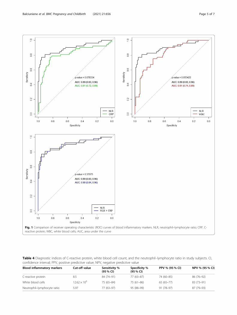
Fig. 1 Comparison of receiver operating characteristic (ROC) curves of blood inflammatory markers. NLR, neutrophil–lymphocyte ratio; CRP, C-reactive protein; WBC, white blood cells; AUC, area under the curve
Table 4 Diagnostic indices of C-reactive protein, white blood cell count, and the neutrophil–lymphocyte ratio in study subjects. CI,confidence interval; PPV, positive predictive value; NPV, negative predictive value
Blood inflammatory markers Cut-off value Sensitivity %(95% CI)
Specificity %(95% CI)
PPV % (95% CI) NPV % (95% CI)
C-reactive protein 8.5 84 (74–91) 77 (63–87) 74 (60–85) 86 (76–92)
White blood cells 12.62 × 10⁹ 75 (65–84) 75 (61–86) 65 (65–77) 83 (73–91)
Neutrophil–lymphocyte ratio 5.97 77 (63–97) 95 (88–99) 91 (78–97) 87 (79–93)
Balciuniene et al. BMC Pregnancy and Childbirth (2021) 21:656 Page 5 of 7

Strengths and limitationsTo the best of our knowledge, this is the first study toevaluate the NLR for the prediction of HCA in patientswith PPROM prior to 34 weeks of gestation. Addition-ally, this study excluded blood samples affected by theadministration of corticosteroids and cases with pre-eclampsia, gestational diabetes mellitus, and intrahe-patic cholestasis. The limitation of our study is itsretrospective design. Further prospective cohort stud-ies are required to evaluate our result and to producestronger evidence. Moreover, chorioamnionitis wasdefined by histological examination of the placenta.Although histological chorioamnionitis is the goldstandard for diagnosing intrauterine infections [30],there is controversy as to whether histological chor-ioamnionitis is correlated with higher rates of neo-natal morbidity and mortality [30–32].
ConclusionsThe NLR has a good predictive value for the occurrenceof HCA and could be used as an additional diagnosticmarker for predicting histological chorioamnionitis incases with preterm premature rupture of the membranesprior to 34 weeks of gestation.
AbbreviationsNLR: neutrophil-lymphocyte ratio; WBC: white blood cell; CRP: C-reactiveprotein; HCA: histological chorioamnionitis; PPROM: preterm prematurerupture of the membranes; IQR: interquartile range; ROC: receiver operatingcurve; AUC: area under the curve
AcknowledgementsWe thank the study participants for their support of the study.
Authors' contributionsD.R. designed the study, together with G.S.D and I.D. G.B, V.G, and R.V.applied for ethical approval and collected data. G.B. analysed the data withsupport from G.K.B and V.G. G.B and G.K.B wrote the main manuscript text,prepared tables and figures. The article was revised and edited by I.P., G.S.D.,I.D., and D.R. All authors approved the version to be published.
FundingThis study was funded by the Research Council of Lithuania under grant No.S-MIP-19-57.
Availability of data and materialsThe data that support the findings of this study are available on requestfrom corresponding author. The data are not publicly available due toprivacy or ethical restrictions.
Declarations
Ethics approval and consent to participateThis study was approved by the Vilnius Regional Bio-medical Research EthicsCommittee (2017-07-04 No. 158200-17-931-434), and all participants signedan informed consent form before enrolment. All methods were performed inaccordance with the relevant guidelines and regulations (Declaration ofHelsinki).
Consent for publicationNot applicable.
Competing interestThe authors declare that they have no competing interests.
Author details1Clinic of Obstetrics, and Gynecology, Institute of Clinical Medicine, Faculty ofMedicine of Vilnius University, M.K. Ciurlionio st. 21, 03101 Vilnius, Lithuania.2Center of Obstetrics and Gynecology, Vilnius University Hospital SantarosKlinikos, Santariskiu st. 2, 08661 Vilnius, Lithuania. 3Department ofImmunology, State Research Institute Centre for Innovative Medicine,Santariskiu st. 5, 08410 Vilnius, Lithuania.
Received: 9 May 2021 Accepted: 29 August 2021
References1. DiGiulio DB, Romero R, Kusanovic JP, Gomez R, Kim CJ, Seok KS, et al.
Prevalence and diversity of microbes in the amniotic fluid, the fetalinflammatory response, and pregnancy outcome in women with pretermpre-labor rupture of membranes. Am J Reprod Immunol. 2010;64:38–57.
2. Mittendorf R, Montag AG, MacMillan W, Janeczek S, Pryde PG, Besinger RE,et al. Components of the systemic fetal inflammatory response syndrome aspredictors of impaired neurologic outcomes in children. Am J ObstetGynecol. 2003;188:1438–46.
3. Zanardo V, Vedovato S, Cosmi E, Litta P, Cavallin F, Trevisanuto D, et al.Preterm premature rupture of membranes, chorioamnion inflammatoryscores and neonatal respiratory outcome: PPROM, chorioamnionitis andneonatal respiratory outcome. BJOG Int J Obstet Gynaecol. 2010;117:94–8.
4. Rogers EE, Hintz SR. Early neurodevelopmental outcomes of extremelypreterm infants. Semin Perinatol. 2016;40:497–509
5. Catano-Sabogal CP, Fonseca J, Garcia-Perdomo HA. Validation of DiagnosticTests for Histologic Chorioamnionitis: Systematic Review and Meta-Analysis.Eur J Obstet Gynecol Reprod Biol. 2018;228:13–26.
6. Trochez-Martinez RD, Smith P, Lamont RF. Use of C-reactive protein as apredictor of chorioamnionitis in preterm prelabour rupture of membranes: asystematic review. BJOG. 2007;114:796–801.
7. Zou L, Zhang H, Zhu J, Zhu J. The value of the soluable intercellularadhesion molecule-1 levels in matermal serum for determination of occultchorioamnionitis in premature rupture of membranes. J Huazhong Univ SciTechnolog Med Sci. 2004;24:154–7.
8. Martinez-Portilla RJ, Hawkins-Villarreal A, Alvarez-Ponce P, Chinolla-ArellanoZL, Moreno-Espinosa AL, Sandoval-Mejia AL, et al. Maternal SerumInterleukin-6: A Non-Invasive Predictor of Histological Chorioamnionitis inWomen with Preterm-Prelabor Rupture of Membranes. Fetal Diagn Ther.2019;45:168–75.
9. Park JW, Park KH, Kook SY, Jung YM, Kim YM. Immune biomarkers inmaternal plasma to identify histologic chorioamnionitis in women withpreterm labor. Arch Gynecol Obstet. 2019;299:725–32.
10. Martins EC, Silveira L da F, Viegas K, Beck AD, Fioravantti-Junior G,Cremonese RV, et al. Neutrophil-lymphocyte ratio in the early diagnosis ofsepsis in an intensive care unit: a case-control study. Rev Bras Ter Intensiva.2019;31:63–70.
11. Ozel A, Davutoglu EA, Yurtkal A, Madazli R. How do platelet-to-lymphocyteratio and neutrophil-to-lymphocyte ratio change in women with pretermpremature rupture of membranes, and threaten preterm labour? J ObstetGynaecol. 2020;40:195–9.
12. Imran MM, Ahmed U, Usman U, Ali M, Shaukat A, Gul N. Neutrophil/Lymphocyte Ratio – A Marker of COVID-19 Pneumonia Severity. Int J ClinPract. 2021;00:e13698.
13. Khong T, Mooney Y, Ariel EE, Balmus I, Boyd NCM, Brundler TK, et al.Sampling and Definitions of Placental Lesions Amsterdam PlacentalWorkshop Group Consensus Statement. Arch Pathol Lab Med. 2016;140:698–713.
14. Redline RW, Faye-Petersen O, Heller D, Qureshi F, Savell V, Vogler C,Amniotic Fluid Infection Nosology Committee Society for PediatricPathology Perinatal Section. Amniotic infection syndrome: nosology andreproducibility of placental reaction patterns. Pediatr Dev Pathol. 2003;6:435–48.
15. Davis JM, Albert JD, Tracy KJ, Calvano SE, Lowry SF, Shires GT, et al.Increased neutrophil mobilization and decreased chemotaxis during cortisoland epinephrine infusions. J Trauma. 1991;31:725–31.
16. Summers C, Rankin SM, Condliffe AM, Singh N, Peters AM, Chilvers ER.Neutrophil kinetics in health and disease. Trends Immunol. 2010;31:318–24.
17. Matalka KZ, Sidk A. Academic Stress — Influence on Leukocyte Distribution,Cortisol, and Prolactin. Lab Med. 1998;29:697–701.
Balciuniene et al. BMC Pregnancy and Childbirth (2021) 21:656 Page 6 of 7

18. Kim MA, Lee YS, Seo K. Assessment of Predictive Markers for PlacentalInflammatory Response in Preterm Births. PLoS One. 2014;9:e107880.
19. Singareddy A, Lee ASE, Sweeney PL, Finkle AE, Williams HL, Buchanan PM,et al. Elevated neutrophil-lymphocyte ratios in extremely preterm neonateswith histologic chorioamnionitis. J Perinatol. 2021; https://doi.org/10.1038/s41372-021-00964-4.
20. Kim MA, Lee BS, Park YW, Seo K. Serum markers for prediction ofspontaneous preterm delivery in preterm labour. European journal ofclinical investigation. 2011;41:773–80.
21. Akgun N, Namli Kalem M, Yuce E, Kalem Z, Aktas H. Correlations of maternalneutrophil to lymphocyte ratio (NLR) and platelet to lymphocyte ratio (PLR)with birth weight. The Journal of Maternal-Fetal & Neonatal Medicine. 2017;30:2086–91.
22. Nakagawa M, Terashima T, D’yachkova Y, Bondy GP, Hogg JC, van Eeden SF.Glucocorticoid-induced granulocytosis: contribution of marrow release anddemargination of intravascular granulocytes. Circulation. 1998;98:2307–13.
23. Weber PSD, Toelboell T, Chang L-C, Tirrell JD, Saama PM, Smith GW, et al.Mechanisms of glucocorticoid-induced down-regulation of neutrophil L-selectin in cattle: evidence for effects at the gene-expression level andprimarily on blood neutrophils. J Leukoc Biol. 2004;75:815–27.
24. Liles WC, Dale DC, Klebanoff SJ. Glucocorticoids inhibit apoptosis of humanneutrophils. Blood. 1995;86:3181–8.
25. Bauer ME, Price LK, MacEachern MP, Housey M, Langen ES, Bauer ST.Maternal leukocytosis after antenatal corticosteroid administration: asystematic review and meta-analysis. J Obstet Gynaecol. 2018;38:210–6.
26. Kurtoglu E, Kokcu A, Celik H, Tosun M, Malatyalioglu E. May ratio ofneutrophil to lymphocyte be useful in predicting the risk of developingpreeclampsia? A pilot study. J Matern Fetal Neonatal Med. 2015;28:97–9.
27. Sahbaz A, Cicekler H, Aynioglu O, Isik H, Ozmen U. Comparison of thepredictive value of plateletcrit with various other blood parameters ingestational diabetes development. J Obstet Gynaecol. 2016;36:589–93.
28. Kirbas A, Biberoglu E, Daglar K, Iskender C, Erkaya S, Dede H, et al.Neutrophil-to-lymphocyte ratio as a diagnostic marker of intrahepaticcholestasis of pregnancy. Eur J Obstet Gynecol Reprod Biol. 2014;180:12–5.
29. Orgul G, Agbal T, Celen S, Caglar AT. Neuroprotective magnesium sulfateadministration increases maternal Neutrophil-to-Lymphocyte Ratio, Platelet-to-Lymphocyte Ratio and Systemic Immune-Inflammation Index. ArchGynecol Obstet. 2020; https://doi.org/10.1007/s00404-020-05866-y.
30. Pugni L, Pietrasanta C, Acaia B, Merlo D, Ronchi A, Ossola, MW, et al.Chorioamnionitis and neonatal outcome in preterm infants: A clinicaloverview. J. Matern Fetal Neonatal Med. 2016;29,1525–9.
31. Romero R, Chaemsaithong P, Docheva N, Korzeniewski SJ, Kusanovic JP,Yoon BH, et al. Clinical chorioamnionitis at term VI: Acute chorioamnionitisand funisitis according to the presence or absence of microorganisms andinflammation in the amniotic cavity. J. Perinat Med. 2016;44,33–51.
32. Ocheke N, Ocheke IE, Agaba PA, Imadde GE, Silas OA, Ajetunmobi OI, et al.Maternal and Neonatal Outcomes of Histological Chorioamnionitis. J WestAfr Coll Surg. 2016;6,1–14.
Publisher’s NoteSpringer Nature remains neutral with regard to jurisdictional claims inpublished maps and institutional affiliations.
Balciuniene et al. BMC Pregnancy and Childbirth (2021) 21:656 Page 7 of 7