new kinorhyncha from florida coastal waters - springer · new kinorhyncha from florida coastal...
TRANSCRIPT

ORIGINAL ARTICLE
New Kinorhyncha from Florida coastal waters
Marıa Herranz • Nuria Sanchez • Fernando Pardos •
Robert P. Higgins
Received: 4 May 2013 / Revised: 8 August 2013 / Accepted: 11 September 2013 / Published online: 2 October 2013
� Springer-Verlag Berlin Heidelberg and AWI 2013
Abstract Four new species of Kinorhynchs are described
from the West Atlantic coast off Fort Pierce, Florida, USA.
They are the following: Antygomonas gwenae n. sp.,
Echinoderes riceae n. sp., Echinoderes adrianovi n. sp. and
Pycnophyes norenburgi n. sp. All species were collected at
the same locality called ‘‘20 miles station.’’ Samples were
processed for standard granulometric data, yielding an
estimated average particle diameter of 250 lm. The diag-
nostic characters and the general morphology of the new
species are discussed in depth as well as the diversity and
distribution of Kinorhyncha in the area.
Keywords Kinorhyncha � Meiofauna � Florida �Echinoderes � Antygomonas � Pycnophyes
Introduction
The kinorhynch fauna reported from the East coast of the
USA includes currently 14 species. The first three kin-
orhynch species recorded and described in the area were
Echinoderes remanei Blake 1930; Pycnophyes frequens
Blake 1930; and Kinorhynchus mainensis Blake 1930.
These three species were found to form a common
assemblage from Maine to Massachusetts (Wieser 1960;
Higgins 1964a, 1965). Two additional species, Campylo-
deres sp. reported as Campyloderes macquariae Johnston
1938 by Higgins (1980) and posteriorly assigned to Cam-
pyloderes cf. vanhoeffeni by Neuhaus and Sørensen (2013);
and Centroderes spinosus Reinhard 1881 recognized as a
new species of Centroderes by Neuhaus, Pardos, Sørensen
and Higgins (in preparation), were found later by Higgins
(1982) in the same area. The latter species was not iden-
tified correctly, and his recording represents a yet unde-
scribed species. Blake’s three species are replaced by three
similar species in the area stretching from Beaufort, North
Carolina to Florida. They include Pycnophyes beaufort-
ensis Higgins 1964; Kinorhynchus langi Higgins 1964; and
Echinoderes bookhouti Higgins 1964 (see Higgins 1964b).
The kinorhynch fauna of Bermuda reported by Higgins
(1982) includes Echinoderes bispinosus Higgins 1982;
Echinoderes bermudensis Higgins 1982; Kinorhynchus
fimbriatus Higgins 1982; and a new species of Centroderes.
All of the above species were collected subtidally, but in
1977, two new cyclorhagid kinorhynchs were found in
intertidal habitats. Echinoderes sublicarum Higgins 1977
was found in bryozoan colonies on pilings in a tidal stream
in the North River Inlet, South Carolina, and Echinoderes
coulli Higgins 1977 and was found in the mud of this same
intertidal creek area. This latter species was the first kin-
orhynch published with a complete series of juvenile
developmental stages (see Higgins 1977).
The knowledge of the marine fauna in Atlantic Florida
waters has been improved along many years by the
research activities developed through the Smithsonian
Marine Station at Fort Pierce, FL. It became well known
among researchers that these waters constitute a kind of hot
spot for meiobenthic communities. Accordingly, study sites
were established in 5-mile increments seaward from Fort
Pierce, Florida, between 1975 and 1995 by R. P Higgins, a
Communicated by H.-D. Franke.
M. Herranz (&) � N. Sanchez � F. Pardos
Dpto. Zoologıa y Antropologıa Fısica, Facultad de Biologıa,
Universidad Complutense de Madrid, C/Jose Antonio Novais 2,
28040 Madrid, Spain
e-mail: [email protected]
R. P. Higgins
122 Strawbridge Court, Asheville, NC 28803, USA
123
Helgol Mar Res (2014) 68:59–87
DOI 10.1007/s10152-013-0369-9

research effort that focused mainly on the phylum Kin-
orhyncha. Several new species appeared but only Ze-
linkaderes floridensis Higgins 1990 was described
formally. Z. floridensis is a cyclorhagid kinorhynch, which
represented a new family, the Zelinkaderidae (see Higgins
1990). This species occurs primarily at depths around
140 m in the muddy sand found 20 miles offshore. More
recently, M. V. Sørensen collected at different localities off
Fort Pierce and described four new species including a new
genus: Echinoderes spinifurca Sørensen et al. 2005, Ze-
linkaderes brightae Sørensen et al. 2007, Antygomonas
paulae Sørensen 2007 and Tubulideres seminoli Sørensen
et al. 2007 (see Sørensen et al. 2007).
The purpose of this paper is to describe the remaining
species from Bob Higgins’ old samplings from the deepest
and richest of the stations hereafter referred to as ‘‘20 miles
station’’ combining his results with new records from
recent samplings.
Materials and methods
Sediment samples were taken in August 1993 from one
single station: the ‘‘20 miles site,’’ 27�300N, 79�560W at
140-m depth (Fig. 1) from sandy mud using a Higgins
anchor dredge (Higgins and Thiel 1988) from the R/V
Sunburst (Smithsonian Marine Station at Fort Pierce). The
anchor dredge was designed to sample only the upper few
centimeters of sediment, thereby eliminating the need for
processing large volumes of otherwise uninhabited sedi-
ment. Additional material was collected in August 2011, at
the same station from the same R/V and using the same
dredge.
Sediment samples for granulometric studies were wet-
sieved using fresh water to remove salt through a series of
geological sieves with 0.5-phi (u) intervals. Phi (u) = -
log2 of mesh size in mm (1,000, 500, 250, 125 and 63 lm
mesh). A mechanical shaker was used for a period of
15 min. The material retained on each sieve was placed in
disposable aluminum weighing pans and dried in an oven
at 80 �C for 24 h. After drying, each fraction of the
material was weighted. The sediment weight fractions
(calculated in percent of the total sample) (Fig. 2a) were
transformed into a cumulative frequency series and then
plotted as a cumulative frequency curve (Fig. 2b). In the
latter, the medium particle diameter, i.e., the u value cor-
responding to the 50 % point of the cumulative scale (Md
u or u 50), is estimated. The ‘‘quartile deviation’’ (QD)
expresses the number of phi (u) units lying between the
upper and lower quartile diameters Q1 and Q3 (Fig. 2b).
Thus, sediment with a small spread between the quartiles,
i.e., a small QD u, is regarded as being ‘‘well sorted’’
(Higgins and Thiel 1988).
In the samples from 1993, kinorhynchs were sorted
under a stereomicroscope, fixed in 10 % formalin and, after
24 h, transferred to 20 % Carosafe � (Carolina Biological
Supply, Co.). Selected specimens were later transferred to
2 % glycerin in 70 % ethanol, which was allowed to
evaporate over a period of about 5 days, thereby leaving
the specimens in glycerin only. From the glycerin, each
specimen was transferred to a drop of Hoyer’s 150
mounting medium (Higgins and Thiel 1988) on the base
coverslip of an H–S slide mount (Higgins and Thiel 1988;
Shirayama et al. 1993). A 12-mm-diameter, circle cover
glass was placed on the mounting medium and manipulated
to orient the specimen.
mi
km
NORTH ATLANTIC OCEANN
0 m100200300
20 m
0 m100200300
40 m30 m 50 m 100 m 150 m 200 m 250 m
10
10
20
10 m
Fort PierceFlorida
USA
C
A
B
20
27°26’20’’N 80°20’8’’W
Fig. 1 Map showing a Florida (inset), western North Atlantic Ocean. b Locality of the study off Fort Pierce Station corresponding to miles off
shore indicated by numbers in oval. c Transect showing depths from 0 to 300 m with vertical line corresponding to the 20 miles station
60 Helgol Mar Res (2014) 68:59–87
123

Specimens collected in 2011 were fixed in 10 %
formalin. For light microscopy (LM) selected specimens
were later dehydrated through a graded series of ethanol
and transferred to glycerin prior to mounting in Fluoro-
mount G�. All specimens were examined and photo-
graphed using an Olympus BX51 microscope with
differential interference contrast optics equipped with an
Olympus Colorview DP70 camera. Measurements were
made using Olympus Cell A software (Olympus,
Europe).
Not all species described yielded specimens suitable for
scanning electron microscopy (SEM) studies. The available
specimens for SEM were dehydrated through a graded
series of ethanol and critical point dried. The dried speci-
mens were mounted on aluminum stubs, sputter coated
with gold and examined with a JEOL JSM-6335F field
emission scanning electron microscope.
Terminology for head, neck and trunk morphology
follows Neuhaus and Higgins (2002), Sørensen and
Pardos (2008) for cyclorhagids and Sanchez et al. (2011)
for homalorhagids. The number and distribution of
introvert appendages has been studied both by rings and
sectors using polar diagrams following Sørensen and
Pardos (2008). The terminology used to describe the
different kinds of scalids follows both Brown (1989) and
Higgins (1990), recently revised and unified by Neuhaus
(2012).
Results
Granulometry
Results of the sediment analyses (Fig. 2) show that a high
percentage of the dry weight of the sediment is represented
by grain size fractions ranging from 500 to 62 lm, with
62 lm occurring as the most frequent grain size consti-
tuting 23 % of the sediment, followed by 125 lm with
22 % and 250 lm with 17 %. Grain fractions from 250 to
62 lm constitute around 64 % of the total sediment.
Cumulative dry weight graphic shows a higher slope in
between 250 and 62 lm as well, showing a mean particle
size around 250 lm. The grain size fractions less repre-
sented are the smallest \62 lm and the biggest 2,000 lm
with a 2.8 and 3.4 %, respectively. However, the amount of
the fraction smaller than 62 lm is not totally reliable due to
the use of 63-lm mesh to take the samples. That means that
part of the finest fraction could have been washed out
during the collection in the periphery of the dredge.
Taxonomic account
Antygomonas gwenae n. sp. (Figs. 3, 4; Tables 1, 2)
Order Cyclorhagida (Zelinka 1896) Higgins 1964
Family Antygomonidae Adrianov and Malakhov 1994
Genus Antygomonas Nebelsick 1990
Diagnosis
Antygomonas with a very sclerotized cuticle. Anterior edge
of the first trunk segment with a conspicuous notch in
middorsal and midventral positions extending half of the
segment length. Trunk segments 2–10 with distinct ter-
gosternal junctions. Middorsal spines from segments 1–4
and 10 are flexible and thin, while those from segments 5–9
and 11 are conical, robust and stout. Segment 2 with a pair
of lateroventral cuspidate spines located close to a pair of
much smaller, flexible and hairy acicular spines. Segments
3 through 9 with lateroventral acicular spines; segments 5,
8 and 9 additionally with pair of lateroventral, lateral
accessory or ventrolateral cuspidate spines. Acicular spines
become very robust and stout from segment 5 in advance
especially in segments 8 and 9. Cuspidate spines in lat-
eroventral position on segments 5 and 9; cuspidate spines
on segment 8 in lateral accessory position. Segment 10
with laterodorsal acicular spines flexible in males and stout
and straight in females. Lateral terminal spines much
shorter than lateral terminal accessory spines. Females with
minute midventral conical projection of segment 9. Males
differing from females by the presence of crenulated pos-
terior half of middorsal and subdorsal spines of segment 10
0
5
10
15
20
25
2000 1000 500 250 125 62 <62
Dry
Wt
(%)
Grain size fraction (µm)
20 miles site sediment
0
20
40
60
80
100
120
2000 1000 500 250 125 62 <62
Cu
mu
lati
ve D
ry W
t (%
)
Grain size fraction (µm)
20 miles site sediment
Q3Md=~250
Q1
A
B
Fig. 2 Grain size fractions plotted: a against dry weight (%)
b against cumulative dry weight (%). Md medium particle diameter.
Q1,3 quartile diameters
Helgol Mar Res (2014) 68:59–87 61
123

and the lack of a pair of strongly sclerotized gonopores on
segment 10.
Etymology
This species is named in honor Gwen L. Higgins, Bob
Higgins’ wife of through 50 years.
Type material
Holotype: adult male collected in August 1993 at the 20
miles station, Fort Pierce, off the Floridian West coast
(Fig. 1) from sandy mud, mounted in Hoyer’s medium.
Allotype: adult female, same collecting data as holotype,
mounted in Hoyer’s medium. Paratype: adult female, same
A B
C D
E
ltsltas
md9
md1
ijlslss
ldss
ss3
rth
ac6
ac2
sp
ff
spf
go
mts
md11
pa
lds10
lds10
cu2tsj
pdss
Fig. 3 Antygomonas gwenae n. sp. line art illustration. a Male
ventral view. b Male dorsal view. c Female ventral detail of segments
10–11. d Female dorsal detail of segments 10–11. e Midterminal
spine full length. ac acicular spine, cu cuspidate spine, ff free flap, go
gonopore, ijl intersegmentary joint line, lds laterodorsal spine, ldss
laterodorsal sensory spot, ltas lateral terminal accessory spine, lts
lateral terminal spine, md middorsal spine, mts midterminal spine, pa
papillae, pdss paradorsal sensory spot, rth roof tile hairs, ss3 sensory
spot type 3, slss sublateral sensory spot, sp sieve plate, spf secondary
pectinate fringe, tsj tergosternal junctions. Digits refer to the segment
number. Scale bar 100 lm
62 Helgol Mar Res (2014) 68:59–87
123

Fig. 4 Light micrographs showing details in neck and trunk
morphology of Antygomonas gwenae n. sp. holotypic male USNM
1196402 (a–e) and allotypic female USNM 1196402 (f). a Neck and
segments 1–3, dorsal view. b Neck and segments 1–3 ventral view.
c Segments 6–8, dorsal view. d Segments 6–8 ventral view. eSegments 9–11, dorsal view. f Segments 9–11 ventral view. ac
acicular spine, cu cuspidate spine, ff free flap, go gonopore, ijl
intersegmentary joint line, lds laterodorsal spine, ltas lateral terminal
accessory spine, lts lateral terminal spine, md middorsal spine, mdn
middorsal notch, mdp middorsal placid, mts midterminal spine, mvn
midventral notch, mvp midventral placid, sa smooth area, sp sieve
plate, spf secondary pectinate fringe, tsj tergosternal junctions. Digits
after abbreviations refer to segment number. White circles indicate
sensory spots (solid line) and papillae in females (dotted line)
Helgol Mar Res (2014) 68:59–87 63
123

collecting data as holotype and allotype, mounted in Ho-
yer’s medium. Holotype and allotype were deposited at the
National Museum of Natural History (Smithsonian Insti-
tution) under accession numbers USNM 1196402 and
USNM 1196403, respectively. The single paratype is
stored in the Meiofauna Laboratory collection at the Fac-
ultad de Biologıa, Universidad Complutense de Madrid
under accession number K15/31.
Table 1 Measurements of adult Antygomonas gwenae n. sp. (in lm)
Holotype male Paratype male Allotype female Holotype male Paratype male Allotype female
TL 356 372 264 MD5 24 23 25
SW 40 62 62 MD6 24 26 27
SW/TL (%) 11.2 16.7 23.5 MD7 30 22 28
MSW-8 76.8 78.8 78.0 MD8 28 23 28
MSW/TL (%) 21.6 21.1 29.5 MD9 28 25 30
MTS 352 200 288 MD10 32 25 35
MTS/TL (%) 98.8 53.2 109.0 LDS10 33 35 28
S1 42 38 40 MD11 40 31 49
S2 31 28 26 LVS2 (cu) 24 22 18
S3 32 31 29 LVS2 (ac) 13 11 13
S4 34 34 32 LVS3 22 18 20
S5 37 35 35 LVS4 24 21 22
S6 38 38 35 VLS5 (cu) 28 22 24
S7 38 42 36 LVS5 (ac) 23 24 26
S8 41 42 40 LVS6 28 26 28
S9 42 44 42 LVS7 31 30 30
S10 44 43 40 LVS8 (ac) 32 30 30
S11 30 32 42 LAS8 (cu) 25 28 28
MD1 23 22 24 VLS9 (cu) 28 32 31
MD2 21 23 23 LVS9 (ac) 30 23 24
MD3 23 23 24 LTS11 40 41 41
MD4 24 25 25 LTAS11 89 80 80
Numbers, where inserted, indicates segment number
ac Acicular spine, cu cuspidate spine, las lateral accessory spine, lds laterodorsal spine, ltas lateral terminal accessory spine, lts lateral terminal
spine, lvs lateroventral spine, md middorsal spine, msw maximum sternal width, mts midterminal spine, sw standard width, s1–s11 segment
lengths of trunk segments 1–11, tl trunk length, vls ventrolateral spine. Holotypic male in boldface
Table 2 Summary of nature and location of sensory spots, spines and papillae arranged by series in Antygomonas gwenae n. sp
Segments MD PD SD LD ML SL LA LV VL VM
1 ac ss ss ss
2 ac ss ss cu, ac
3 ac ss ss ss ac ss
4 ac ss ss ac ss
5 ac ss ss ac cu
6 ac ss ss ac ss
7 ac ss ss ss ac ss
8 ac ss cu ac ss
9 ac ss ss ac cu pa$
10 ac ss ss ac, ss ss
11 ac, mts ss, ss ltas lts ss
ac Acicular spine, cu cuspidate spine, la lateral accessory, ld laterodorsal, ltas lateral terminal accessory spine, lts lateral terminal spine, lv
lateroventral, md middorsal, ml midlateral, mts midterminal spine, pa papilla, pd paradorsal, sd subdorsal, sl sublateral, ss sensory spot, vl
ventrolateral, vm ventromedial, $ female condition of sexually dimorphic character
64 Helgol Mar Res (2014) 68:59–87
123

Description
Adult specimens consisting of a head, a neck and eleven
trunk segments (Figs. 3, 4) Measurements and dimensions
are given in Table 1. A summary of cuticular characters
(spines, tubules, gland outlets and sensory spots) locations
is given in Table 2.
Mouth cone and introvert armature could not be exam-
ined in detail in any of the prepared specimens.
Neck with 16 placids of unequal shape, width and
length. Midventral and middorsal placids are triangular and
elongated measuring 6 lm wide at their bases and 22 lm
long, narrower than all others (Figs. 3a, b, 4a, b). Laterally
adjacent placids in the dorsal side still triangular being
14 lm wide, longer mesially than laterally but overall
slightly longer than middorsal and midventral placids.
Ventral placids narrower measuring 8 lm in width and
20 lm in length. Remaining placids more rectangular,
nearly equal in size, 10 lm wide and 12 to 16 lm long.
The cuticle between adjacent placids is more flexible,
folding inwards and giving the false appearance of inter-
stitial placids. No trichoscalid plates could be observed.
Segment 1, 42 lm long at lateral margins, 26 lm long
middorsally and midventrally, forming an anterior broad
notch (Figs. 3a, b, 4a, b). The cuticle in this and the fol-
lowing segments seems to be thicker and stronger than in
other Antygomonas species. Middorsal spine short, hairy
and flexible originating near the posterior edge of the
segment in a notched margin and flanked by paradorsal
sensory spots in this and the following segments. Paired
sensory spots are furthermore present in laterodorsal and
ventromedial positions (Figs 3a, b, 4a, b). Sensory spots in
this and the following segments are rounded with short
papillae and two pores, appearing very distinct in light
microscopy; paradorsal sensory spots with a semicircular
outline (Fig. 3b). It was not possible to identify the sensory
spot type. Hence, more detailed examination through
scanning electron microscopy is required to provide an
exact map of these structures. Cuticular hairs are scattered
over the segment. The hairs seem to have a roof tile or
elongated leaf-like appearance with a perforation site, and
are scattered around the segment. Wide striated free flap
delimited anteriorly by a conspicuous ij-line in this and the
following segments (Figs. 3a, b,4c). Pectinate fringe not
apparent through LM.
Segment 2, 31 lm long at lateral margin of tergal plate,
narrowing to 25 lm long at tergosternal junction. Sternal
plates with arch-shaped anterior margins, each being ca.
16 lm wide anteriorly and 23 lm wide posteriorly
(Figs. 3a, 4b). A pair of lateroventral cuspidate spines
present, each with an associated hairy, thin and flexible
acicular spine (Figs. 3a, 4b). Middorsal spine similar to
that of previous segment. Paired sensory spots in
paradorsal and midlateral positions. The anterior part of the
tergal plate has at least four secondary pectinate fringes
arranged into parallel rows on this and the following seg-
ments (Figs. 3b, 4a). All of them consist of a wavy line of
minute, cuticular, spine-like scales; the last row is enlarged
middorsally forming a conspicuous fringe behind the
middorsal spine of each segment. Anterior to the secondary
fringes a narrow smooth area without any scales occurs;
this area is usually covered by the free posterior edge of the
preceding segment (Fig. 4a–c). Secondary pectinate fringes
on the sternal plates made by similar scales arranged into a
wavy lines making out a conspicuous smooth area
(Figs. 3a, 4d). Cuticular hairs with the same structure as on
preceding segment but present on tergal plate only. Free
flap striated and without a marked pectinate fringe as on
previous segment. Middorsal and lateroventral spines on
this and the following segments are associated with a
conspicuous notch of the free flap (Figs. 3b, 4a, c, d).
Segment 3. Sternal plates slightly trapezoid, narrowest
at anterior margins. (Figs. 3a, 4b). Flexible and hairy lat-
eroventral spines present. Middorsal spine similar to that of
previous segment. Paired sensory spots in paradorsal, lat-
erodorsal, sublateral and ventrolateral positions. Cuticular
hairs, fringes and free flap as on preceding segment.
Segment 4. Sternal plates nearly squarish. Middorsal
spine slightly longer and more robust than spine on pre-
vious segments. Lateroventral acicular spines with the
same appearance as those from segment 3. Paired sensory
spots in paradorsal, sublateral and ventrolateral positions.
Cuticular hairs, fringes and free flap as on previous
segments.
Segment 5 with a middorsal spine slightly longer and
stronger than the one of the previous segment (Fig. 4c). A
pair of ventrolateral cuspidate spines longer than those of
segment 2 closely associated with a pair of lateroventral
and stout acicular spines both present with the same length
(Fig. 3a). Both the middorsal and the lateral accessory
acicular spines with a thick-walled cuticle around a central
cavity. Paired sensory spots in paradorsal and laterodorsal
positions. Cuticular hairs, fringes and free flap as on pre-
vious segment.
Segment 6 with a stout middorsal spine with the same
length as the one in previous segment. A pair of wide and
stout lateroventral acicular spines is present (Figs. 3a, 4d).
Both the middorsal and lateroventral spines with a thick-
walled cuticle around a central cavity, in this and the fol-
lowing segments. Paired sensory spots situated on para-
dorsal, sublateral and ventrolateral positions. Cuticular
hairs, fringes and free flap as on previous segments.
Segment 7 with a stout middorsal spine (Figs. 3b, 4c). A
pair of robust lateroventral spines is present (Figs. 3a, 4d).
Sternal plates similar to those of segment 6, slightly nar-
rowing at posterior edges. Sensory spots located in
Helgol Mar Res (2014) 68:59–87 65
123

paradorsal, laterodorsal, sublateral and ventrolateral posi-
tions. Cuticular hairs, fringes and free flap as in previous
segments.
Segment 8 with a very robust middorsal spine (Fig. 3b).
Lateroventral acicular spines also very stout (Fig. 3a). Both
middorsal and lateroventral acicular spines with the same
appearance as described in previous segments. A pair of
lateral accessory cuspidate spines is present. Paired sensory
spots situated in paradorsal and ventrolateral positions.
Cuticular hairs, fringes and free flap as in previous
segments.
Segment 9 with a stout middorsal spine (Figs. 3b, 4e).
Paired ventrolateral cuspidate spines and conspicuously
robust lateroventral acicular spines present (Figs. 3a, 4f).
Paired sensory spots in paradorsal and sublateral positions.
Female with a pair of ventromedial conical protuberances
or papillae (modified gland cell outlets) (Figs. 3c, 4f).
Small oval sieve plate near lateroventral tergal margin,
long axis about 6 lm (Figs. 3a, 4f). Cuticular hairs, fringes
and free flap as on previous segments.
Segment 10 showing either a thin and crenulated mid-
dorsal spine in males or a stout middorsal spine in females
(Figs. 3b, d, 4e, f). Additionally, a pair of laterodorsal
spines crenulated in males and robust in females with the
same length of the middorsal spine is present. Paired sen-
sory spots in paradorsal, subdorsal laterodorsal and lat-
eroventral positions. The posterior margin of the tergal
plate free flap has deep notches around the middorsal and
laterodorsal spines. Female’s free flap of the sternal plates
with two ventrolateral rounded notches around the go-
nopores of segment 11; this free flap is straight in males
(Figs. 3a, c, 4f). Cuticular hairs and pectinate fringes as on
previous segments.
Segment 11 triangular, tapering posteriorly. Tergal
plate slightly longer than sternal plates without tergal
extensions. Segment appendages consist of a middorsal
spine, a midterminal spine nearly the same length as the
trunk and paired lateral terminal and lateral terminal
accessory spines (Figs. 3a, b, e, 4e, f). The lateral
accessory spines are twice as long as the lateral terminal
spines. Paired modified sensory spots (type 3) are present
at the base of the middorsal, midterminal and lateral
terminal spines (Figs. 3a, b, 4e, f). These sensory spots
consist of an area with densely set papillae, with a long
and slender tubule protruding from the center. Female
gonopores are easily recognized in LM as two strongly
cuticularized areas at the intersegmental junction between
segments 10 and 11 (Figs. 3c, 4f). Cuticular leaf-like
hairs are lacking in this segment, instead the surface is
covered by small scale-like hairs without perforation sites.
A wide and striated free flap is present only in the sternal
plates, lacking in tergal ones. Secondary fringes could not
be examined in detail.
Remarks
The new species has sixteen placids, a first trunk segment
being ring-shaped showing middorsal and midventral
anterior notches, both presence of acicular and cuspidate
spines, cuticular structures such as hairs and secondary
fringes, and characteristic notches in the posterior margins
of the trunk segments. All those characters are only shared
by species of Antygomonas. However, there are some other
important features of the new species that do not fit that
well within this genus such as the high sclerotization of the
trunk cuticle, the depth of the notches on segment 1
(around 50 % of the segment length), very short and robust
middorsal and lateroventral spines, middorsal and latero-
dorsal spines of segment 10, being crenulated only in
males, and the presence of distinct tergosternal junctions in
segments 2–11. Based on the last difference solely, one
would have to reject the assignment of the new species to
the genus Antygomonas as originally described by Nebel-
sick (1990). However, and regarding this feature, Sørensen
(2007) reported the presence of hardly recognizable ter-
gosternal divisions in A. paulae and re-examined speci-
mens of Antygomonas oreas Bauer-Nebelsick 1996,
indicating that all segments from 2 to 11 consist of one
tergal and two sternal plates. Subsequently, Sørensen et al.
(2009) confirmed this trait also for Antygomonas incomi-
tata. Thus, with the emended diagnosis based on these
examinations, we can consider the genus Antygomonas to
have segments 2–11 weakly divided into one tergal and
two sternal plates; therefore, the new species can be still
considered as belonging to Antygomonas.
Within the suborder Conchorhagae, the genera Semno-
deres and Sphenoderes share several notable characters
with the new species such as the composition of the plac-
ids, combining different lengths and widths; segment 1
with more or less developed incisions or notches that could
function similar to the clam-shell-like closing apparatus
described for Sphenoderes and Semnoderes (see Higgins
1969; Sørensen et al. 2009; Sørensen et al. 2010; Zelinka
1928); the presence of short and stout spines (only shared
with Sphenoderes species) and the presence of the conical
papillae located in the center of each sternal plate of seg-
ment 9. However, there are some noteworthy variations in
the closing apparatus: Semnoderes has a segment one
composed of two lateral halves separated by deep but not
complete incisions and a neck with narrow middorsal and
midventral placids to fit into these incisions (Sørensen et al.
2009). Sphenoderes in turn has a first segment divided into
single tergal and sternal plates and a pair of lateral plates
(Higgins 1969), showing broader incisions with also
broader middorsal and midventral placids. In case of
Sphenoderes poseidon Sørensen and Thormar 2010, the
first segment is clearly not divided, but the broader
66 Helgol Mar Res (2014) 68:59–87
123

incisions and placids are still present. The new species has
a first segment not divided but showing conspicuous
although not very deep anterior notches. Furthermore, in
the neck, the midventral and middorsal placids are con-
spicuously narrower than the remaining ones. These dif-
ferences exclude the new species to be assigned as either
Semnoderes or Sphenoderes.
The doubtful position of the new species in between
Antygomonas, Semnoderes and Sphenoderes is not unex-
pected knowing all the similarities shared by those genera
previously reported by Sørensen et al. (2009), (2010).
Surprisingly, the new species shares other characters
with species of the genus Centroderes such as the sexual
dimorphism exhibited by the laterodorsal and middorsal
spines of segment 10, being crenulated in males; this fea-
ture is not shared by any species of Antygomonas nor
Semnoderes and Sphenoderes. However, other genera such
as Wollunquaderes, Tubulideres (Sørensen et al. 2007);
Triodontoderes (Sørensen and Rho 2009); and Zelinka-
deres (Higgins 1990; Bauer-Nebelsick 1995; Sørensen
et al. 2007) show the same dimorphism. The stout and
robust middorsal and laterodorsal acicular spines of A.
gwenae n. sp. resemble those showed in Centroderes,
although species of Semnoderes and especially Spheno-
deres (S. poseidon Sørensen et al. 2010) also share this
character. Future phylogenetic analysis would hopefully
clarify whether all these traits are relevant for taxonomy
and phylogenetic relationships.
The combination of the traits displayed by the new
species makes its generic assignment problematic and
somehow resembles the situation described for Wollun-
quaderes majkenae (see Sørensen and Thormar 2010),
which has characters pointing toward Semnoderidae and
Centroderidae. We tentatively assign the new species to
the genus Antygomonas despite all the differences listed
above until phylogenetic analysis confirm or reject it.
Another alternative could be to erect a new cyclorhagid
intermediate genus to accommodate this single species,
based on all the differences showed with the genus An-
tygomonas and the similarities shared with Sphenoderes
and Semnoderes. The authors find it inadequate without a
phylogenetic background. Thus, as stated above, the new
species fits better within Antygomonas than in other
alternative genera and therefore is here assigned provi-
sionally to this genus.
Currently, the genus Antygomonas comprises three
species: Antygomonas incomitata Nebelsick 1990; A. ore-
as; and A. paulae. The main differences between these
species are basically their spine formula, or more precisely
the position of the cuspidate spines on segments 6, 8 and 9
(see Nebelsick 1990; Bauer-Nebelsick 1996; Sørensen
2007). Antygomonas gwenae n. sp. is distinguished from A.
incomitata by the lack of ventrolateral cuspidate spines on
segment 9 and lateroventral cuspidate spines on segment 8.
They also differ in length of the LTS and LTAS being
equal in A. incomitata while different in A. gwenae n. sp.
which shows much shorter LTS. A. oreas and A. gwenae n.
sp. can be discriminated by the position of the cuspidate
spines on segment 9, which is lateroventral in A. gwenae n.
sp. and lateral accessory in A. oreas. Also the TL of A.
oreas is around 50 lm shorter than A. gwenae n. sp. A.
paulae and A gwenae n. sp. have exactly the same spine
formula, but they can be easily discriminated by the body
dimensions with A. paulae being 100 lm longer than A.
gwenae n. sp. and by the overall appearance of the mid-
dorsal and lateroventral spines stout and short in A. gwenae
n. sp. and long and flexible in A. paulae. A. gwenae n. sp.
can furthermore be differentiated from A. paulae by the
sensory spot pattern showing additional sensory spots in
laterodorsal position on segments 3 and 5 and in sublateral
positions on segments 3, 6, 7 and 9. The lack of ventral
notches of segments 1, 3, 4 and 6–8 occurring in A. paulae
and lacking in A. gwenae and the different length of the
LTAS and LTS equal in A. paulae are as well important
features to distinguish both species (see Sørensen 2007).
Echinoderes adrianovi n. sp. (Figs. 5, 6; Tables 3, 4)
Order Cyclorhagida (Zelinka 1896) Higgins 1964
Family Echinoderidae Zelinka 1894
Genus Echinoderes Claparede 1863
Diagnosis
Echinoderes with long middorsal spines on segments 4–8
increasing in length from segments 4–7, middorsal spine of
segment 8 shorter than the one on segment 7; long sub-
dorsal and ventrolateral tubules on segment 2; lateroventral
tubules on segment 5; lateroventral spines on segments
6–9; lateral accessory tubule on segment 8; lateral acces-
sory spines on segment 11 in females; lateral terminal
spines on segment 11.
Etymology
This species is named in honor of Dr. Andrey V. Adrianov,
Vice Director, Institute of Marine Biology, Far East
Branch, Russian Academy of Science, Vladivostok, Russia,
a student, mutual friend and colleague of R.P. Higgins.
Type material
Holotype: adult female collected on August 1993 at the 20
miles site, Fort Pierce, off the Floridian West coast (Fig. 1)
27�300N, 790560W, 140 m depth from sandy mud, mounted
in Hoyer’s medium, deposited at National Museum of
Helgol Mar Res (2014) 68:59–87 67
123

Natural History (Smithsonian Institution) under accession
number USNM 1196401.
Description
Holotypic female with head, neck and 11 trunk segments
(Figs. 5a, b, 6a). See Table 3 for measurements and dimen-
sions. A summary of location of cuticular characters (spines,
tubules, gland outlets and sensory spots) is given in Table 4.
No specimens were available for SEM examination; thus, it
was not possible to identify unambiguously all minor cuticular
structures such as sensory spots and gland cell outlets. These
structures are reported when apparent through LM; however,
the lack of mention in the description of some segments should
not be interpreted as a confirmation of their absence.
The head consists of a retractable mouth cone and an
introvert. Inner and outer armature could not be examined
in detail.
A B
gco1
ijl
vlt2
lvt5
lat8
lvs9
te
ltas
lts
md4
tp
sdt2
ldss
Fig. 5 Echinoderes adrianovi
n. sp. line art illustration.
a Ventral view. b Dorsal view.
ijl intersegmentary joint line, lat
lateral accessory tubule, ldss
laterodorsal sensory spot, ltas
lateral terminal accessory spine,
lts lateral terminal spine, lvs
lateroventral spine, lvt
lateroventral tubule, md
middorsal spine, gco1 glandular
cell outlet type 1, sdt subdorsal
tubule, te tergal extension, tp
trichoscalid plate, vlt
ventrolateral tubule. Digits refer
to the segment number. Scale
bar 100 lm
68 Helgol Mar Res (2014) 68:59–87
123

The neck consists of 16 placids all measuring 12 lm in
length; midventral placid 10 lm wide at the base; and
8 lm distally where it expands laterally at its margins
(Figs. 5a, 6c); remaining placids measure 6 lm at their
bases and 3 lm distally. All placids articulate with the first
trunk segment. Six trichoscalid plates present, 2 ventral on
placids 1 and 16 and 4 dorsal on placids 6, 8, 10 and 12
(Figs. 5a, b, 6c, d). Ventral trichoscalid plates are
triangular with rounded corners and wider than dorsal ones
which have a triangular outline (Figs. 6c, d).
Trunk
The trunk has 11 segments, with segments 1 and 2 con-
sisting of a closed cuticular ring, and segments 3–11 with
one tergal and two sternal plates (Fig. 5a).
Fig. 6 Light micrographs showing details in neck and trunk
morphology of Echinoderes adrianovi n. sp. holotypic female,
USNM 1196401. a Dorsal view. b Segments 5–10 left side, ventral
view. c Neck, dorsal view. d Neck lateroventral view. e Segments
1–3, dorsal view. f Segments 1–3, ventral view. ijl intersegmentary
joint line, lat lateral accessory tubule, ltas lateral terminal accessory
spine, lts lateral terminal spine, lvs lateroventral spine, lvt
lateroventral tubule, md middorsal spine, mdp middorsal placid,
mvp midventral placid, pl placid, sdt subdorsal tubule, sp sieve plate,
te tergal extension, tp trichoscalid plate, tr trichoscalid, vlt ventro-
lateral tubule. White circles indicate glandular cell outlets type 1
(dotted line) and sensory spots (solid line). Digits after abbreviations
refer to segment number
Helgol Mar Res (2014) 68:59–87 69
123
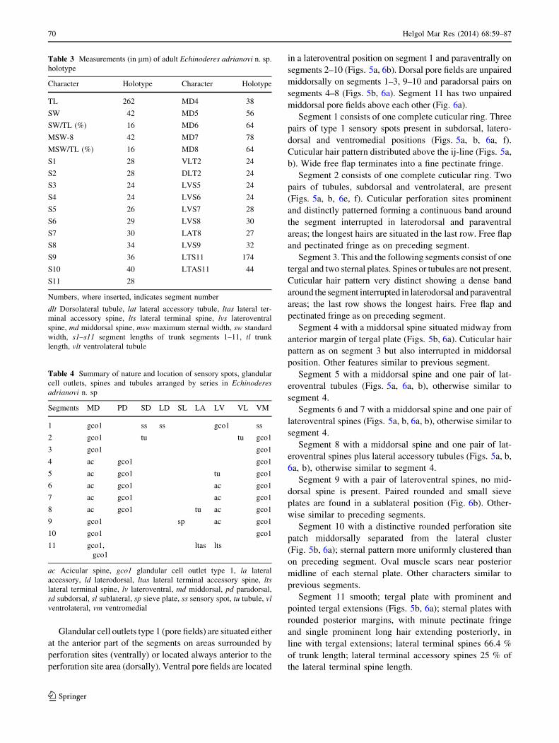
Glandular cell outlets type 1 (pore fields) are situated either
at the anterior part of the segments on areas surrounded by
perforation sites (ventrally) or located always anterior to the
perforation site area (dorsally). Ventral pore fields are located
in a lateroventral position on segment 1 and paraventrally on
segments 2–10 (Figs. 5a, 6b). Dorsal pore fields are unpaired
middorsally on segments 1–3, 9–10 and paradorsal pairs on
segments 4–8 (Figs. 5b, 6a). Segment 11 has two unpaired
middorsal pore fields above each other (Fig. 6a).
Segment 1 consists of one complete cuticular ring. Three
pairs of type 1 sensory spots present in subdorsal, latero-
dorsal and ventromedial positions (Figs. 5a, b, 6a, f).
Cuticular hair pattern distributed above the ij-line (Figs. 5a,
b). Wide free flap terminates into a fine pectinate fringe.
Segment 2 consists of one complete cuticular ring. Two
pairs of tubules, subdorsal and ventrolateral, are present
(Figs. 5a, b, 6e, f). Cuticular perforation sites prominent
and distinctly patterned forming a continuous band around
the segment interrupted in laterodorsal and paraventral
areas; the longest hairs are situated in the last row. Free flap
and pectinated fringe as on preceding segment.
Segment 3. This and the following segments consist of one
tergal and two sternal plates. Spines or tubules are not present.
Cuticular hair pattern very distinct showing a dense band
around the segment interrupted in laterodorsal and paraventral
areas; the last row shows the longest hairs. Free flap and
pectinated fringe as on preceding segment.
Segment 4 with a middorsal spine situated midway from
anterior margin of tergal plate (Figs. 5b, 6a). Cuticular hair
pattern as on segment 3 but also interrupted in middorsal
position. Other features similar to previous segment.
Segment 5 with a middorsal spine and one pair of lat-
eroventral tubules (Figs. 5a, 6a, b), otherwise similar to
segment 4.
Segments 6 and 7 with a middorsal spine and one pair of
lateroventral spines (Figs. 5a, b, 6a, b), otherwise similar to
segment 4.
Segment 8 with a middorsal spine and one pair of lat-
eroventral spines plus lateral accessory tubules (Figs. 5a, b,
6a, b), otherwise similar to segment 4.
Segment 9 with a pair of lateroventral spines, no mid-
dorsal spine is present. Paired rounded and small sieve
plates are found in a sublateral position (Fig. 6b). Other-
wise similar to preceding segments.
Segment 10 with a distinctive rounded perforation site
patch middorsally separated from the lateral cluster
(Fig. 5b, 6a); sternal pattern more uniformly clustered than
on preceding segment. Oval muscle scars near posterior
midline of each sternal plate. Other characters similar to
previous segments.
Segment 11 smooth; tergal plate with prominent and
pointed tergal extensions (Figs. 5b, 6a); sternal plates with
rounded posterior margins, with minute pectinate fringe
and single prominent long hair extending posteriorly, in
line with tergal extensions; lateral terminal spines 66.4 %
of trunk length; lateral terminal accessory spines 25 % of
the lateral terminal spine length.
Table 3 Measurements (in lm) of adult Echinoderes adrianovi n. sp.
holotype
Character Holotype Character Holotype
TL 262 MD4 38
SW 42 MD5 56
SW/TL (%) 16 MD6 64
MSW-8 42 MD7 78
MSW/TL (%) 16 MD8 64
S1 28 VLT2 24
S2 28 DLT2 24
S3 24 LVS5 24
S4 24 LVS6 24
S5 26 LVS7 28
S6 29 LVS8 30
S7 30 LAT8 27
S8 34 LVS9 32
S9 36 LTS11 174
S10 40 LTAS11 44
S11 28
Numbers, where inserted, indicates segment number
dlt Dorsolateral tubule, lat lateral accessory tubule, ltas lateral ter-
minal accessory spine, lts lateral terminal spine, lvs lateroventral
spine, md middorsal spine, msw maximum sternal width, sw standard
width, s1–s11 segment lengths of trunk segments 1–11, tl trunk
length, vlt ventrolateral tubule
Table 4 Summary of nature and location of sensory spots, glandular
cell outlets, spines and tubules arranged by series in Echinoderes
adrianovi n. sp
Segments MD PD SD LD SL LA LV VL VM
1 gco1 ss ss gco1 ss
2 gco1 tu tu gco1
3 gco1 gco1
4 ac gco1 gco1
5 ac gco1 tu gco1
6 ac gco1 ac gco1
7 ac gco1 ac gco1
8 ac gco1 tu ac gco1
9 gco1 sp ac gco1
10 gco1 gco1
11 gco1,
gco1
ltas lts
ac Acicular spine, gco1 glandular cell outlet type 1, la lateral
accessory, ld laterodorsal, ltas lateral terminal accessory spine, lts
lateral terminal spine, lv lateroventral, md middorsal, pd paradorsal,
sd subdorsal, sl sublateral, sp sieve plate, ss sensory spot, tu tubule, vl
ventrolateral, vm ventromedial
70 Helgol Mar Res (2014) 68:59–87
123

Remarks
Currently forty-one species of Echinoderes share the
same middorsal spine pattern (segments 4–8). Of these,
twenty-one species show ventrolateral tubules on seg-
ment 2, lateroventral tubules on segment 5 and latero-
ventral spines on segments 6–9. Only one species out of
these twenty-one, Echinoderes kanni Thormar and
Sørensen 2010, described from the Solomon Islands,
share the pattern of spines/tubules with E. adrianovi n.
sp. Despite the coincidence of spine/tubule formula and
body proportions, both species can be differentiated by
the length of these spines and tubules: E. kanni shows all
middorsal spines increasing progressively in length,
while E. adrianovi n. sp. shows the middorsal spine of
segment 8 shorter than the one on previous segment
being equal in length with the one on segment 6. The
middorsal spines of segments 4–7 in E. adrianovi n. sp.
are around 10 lm longer than in E. kanni, except for the
middorsal spine on segment 8 which is 20 lm shorter.
The length of the ventrolateral and dorsolateral tubules
of segment 2 show big differences measuring twice as
much in E. adrianovi n. sp.; tubules of segments 5 and 8
are also longer in E. adrianovi n. sp. but not as long as
those on segment 2. Lateroventral spines show differ-
ences as well being slightly longer in E. adrianovi n. sp.
except for the lateroventral spine on segment 9 much
longer in E. kanni. Lateral terminal and lateral terminal
accessory spines around 15 lm longer in E. adrianovi
n. sp.
Another species that strongly resembles E. adrianovi n.
sp. is Echinoderes parrai GaOrdonez et al. 2008, described
from the North coast of Spain (GaOrdonez et al. 2008).
This is the only other species of the genus Echinoderes
combining 5 middorsal spines, dorsal tubules on segment 2
and lateral accessory tubules on segment 8. However, it
lacks ventrolateral tubules on segment 2, it has much
shorter LTS and LTAS and shows a bulbous appearance of
the anteriormost segments; therefore, it is very easily dis-
criminated from E. adrianovi n. sp.
Two other species of the same genus, Echinoderes
spinifurca and Echinoderes horni Higgins 1983, are
reported from the Atlantic coast of Fort Pierce, Florida,
USA (Sørensen et al. 2005). Both have been found closer
to shore, from the coarser sediments of the 5 miles station.
E. horni is otherwise known only from its type locality,
Carrie Bow Cay, Belize. E. adrianovi n. sp. is easily dif-
ferentiated from E. spinifurca because of the very long
tergal extensions and the lack of tubules laterodorsally on
segment 2 and lateral accessory on segment 8 of the latter
species, and from E. horni because this latter lacks mid-
dorsal spines.
Echinoderes riceae n. sp. (Figs. 7, 8, 9, 10; Tables 5, 6)
Diagnosis
Echinoderes with middorsal spines on segments 4, 6 and 8
gradually increasing in length posteriorly; ventrolateral
tubules on segment 2; lateroventral tubules on segment 5;
lateroventral spines on segments 7, 8 9; lateral accessory
tubules on segment 8.
Etymology
This species is named in honor of Dr. Mary E. Rice,
Emeritus Research Zoologist at the Smithsonian Marine
Station at Fort Pierce, Florida, for a whole life dedicated to
the marine research.
Type material
Holotype: adult female collected on August 1993 at the
20 miles site, Fort Pierce, off the Floridian West coast
(Fig. 1) 27�300N, 790560W, 140 m depth from sandy mud,
mounted in Hoyer’s medium, deposited at the National
Museum of Natural History (Smithsonian Institution) under
accession number USNM 1196386. Allotype: adult male,
same collecting data as holotype, mounted in Hoyer’s
medium, deposited at National Museum of Natural History
(Smithsonian Institution) under accession number USNM
1196396. Paratypes, 9 males and 9 females same collecting
data as holotype, mounted in Hoyer’s medium. Thirteen
paratypes were deposited at the National Museum of Nat-
ural History under accession numbers USNM 1196387-95
and USNM 1196397-99. Remaining five paratypes are
preserved in the Meiofauna Laboratory collection at the
Facultad de Biologıa, Universidad Complutense de Madrid
under accession numbers K15/31-36.
Additional material
Additional material was collected in August 2011 at the 20
miles site, Fort Pierce, off the Floridian West coast
27�30,840N 79� 54,860W; 152 m depth from fine mud. Ten
specimens were studied in LM mounted in Fluoromount
G�, and 5 were prepared and examined with SEM. This
material is deposited in the Meiofauna Laboratory collec-
tion at the Facultad de Biologıa, Universidad Complutense
de Madrid under accession numbers K15/1-10.
Description
Adult specimens consist of a head, a neck and eleven trunk
segments (Figs. 7a, b, 9a, b). Measurements and dimen-
sions are given in Table 5. A summary for location of
sensory spots, spines, tubules and glandular cell outlets is
provided in Table 6.
Helgol Mar Res (2014) 68:59–87 71
123

The head consists of a retractable mouth cone and an
introvert (Fig. 8). The mouth cone has nine, long and
pointed outer oral styles divided into two segments. The
bases have a conspicuous long fringe composed of at least
five fringe tips flanked by a pair of spikes (Fig. 10a); the
inner armature of the mouth cone could not be examined
in detail. The introvert has seven rings of spinoscalids and
one additional ring of trichoscalids (Fig. 8). Ring 01 has
ten primary spinoscalids consisting of a short basal sheath,
equipped with two rows of long fringes and a long end
piece. Ring 02 is formed by 10 laterally compressed
spinoscalids, all formed by a basal part covered with a
long (more than half of the length of the scalid) and
smooth sheath that terminates into a short fringe with
wide fringe tips (Fig. 10b). Ring 03 with 20 spinoscalids
all with conspicuous sheaths around their bases and a
A B
C D
ps
tp
gco1
ijl
vlt2
md4
te
ltas
lts
lvt5
lat8
lvs8
lvs9
p1p2
p3
ff
Fig. 7 Echinoderes riceae n.
sp. line art illustration. a Ventral
view. b Dorsal view. c Male
detail segment 10–11 ventral
view. d Male detail segments
10–11, ventral view. ff free flap,
ijl intersegmentary joint line, lat
lateral accessory tubule, ldss
laterodorsal sensory spot, ltas
lateral terminal accessory spine,
lts lateral terminal spine, lvs
lateroventral spine, lvt
lateroventral tubule, md
middorsal spine, p1-3 penile
spines, gco1 glandular cell
outlet type 1, ps perforation
sites, te tergal extension, tp
trichoscalid plate, vlt
ventrolateral tubule. Digits refer
to the segment number. Scale
bar 100 lm
72 Helgol Mar Res (2014) 68:59–87
123

distal fringe. These sheaths have rounded edges with a
characteristic flexible spine in the middle (Fig. 10b).
Rings 04 and 05 consist of 10 and 20 spinoscalids,
respectively (Fig. 8); all resemble those of ring 03 but
without the conspicuous sheaths. Ring 06 is formed by 6
spinoscalids, shorter than those in preceding rings and also
showing shorter sheaths. Ring 07 has 20 leaf-like scalids
with a wide and hairy base from where several flexible
elongations arise (Fig. 10b). Six trichoscalids are present
on their respective trichoscalid plates (Figs. 8, 9a, b, 10b).
The introvert can also be described as divided into ten
sectors defined by radii drawn through primary spinosca-
lids, so each sector is delimited by two consecutive pri-
mary spinoscalids (Fig. 8). The midventral sector is
numbered as 1, followed clockwise by sectors 2–10. All
sectors, except the middorsal sector 6, contain one oral
style in the mouth cone. Even sectors contain 8 spino-
scalids (other than primary ones) except for sector 6 with
10 spinoscalids. Uneven sectors contain 10 spinoscalids
except for sectors 5 and 7, which also contain a tricho-
scalid that prevents the presence of one of the leaf-like
scalids. The arrangement of the spinoscalids shows double
diamonds in uneven sectors and sector 6 and quincunxes
in the remaining even sectors formed by spinoscalids of
rows 03–05 (Fig. 8). Trichoscalid plates with trichoscalids
are situated on sectors 2, 4, 5, 7, 8 and 10. See Fig. 8 for a
complete summary of oral styles, scalids and placid
locations.
S1
S10
S9
S8
S7
S6
S5
S4
S3
S2
? ??
? ???
?
?
? ?
??
??
? ???
?
Scalid and style arrangement
Ring/Sector 1 2 3 4 5 6 7 8 9 10 Total00 oos 1 1 1 1 1 0 1 1 1 1 901 psp 1 1 1 1 1 1 1 1 1 1 1002 sps 1 1 1 1 1 1 1 1 1 1 1003 sps 2 2 2 2 2 2 2 2 2 2 2004 sps 1 1 1 1 1 1 1 1 1 1 1005 sps 2 2 2 2 2 2 2 2 2 2 2006 sps 1 0 1 0 1 1 1 0 1 0 607 ls 3 2 3 2 2 3 2 2 3 2 2408 tr 0 1 0 1 1 0 1 1 0 1 6
Total scalids 10 9 10 9 10 10 10 9 10 9 115
Fig. 8 Diagram of mouth cone,
introvert and placids showing
distribution of oral styles and
scalids in Echinoderes riceae n.
sp. ‘‘Double diamonds’’ are
marked in the table with double
lines, and quincunxes are
marked with dotted lines. ls
leaf-like scalid, oos outer oral
style, psp primary spinoscalid,
S1-10 sectors, sps spinoscalid, tr
trichoscalid
Helgol Mar Res (2014) 68:59–87 73
123

Neck with 16 placids, 13 lm long, conventionally
numbered clockwise from the midventral one at sector 1.
Midventral placid 10 lm wide at base, distal margin of
placid nearly the same width. Lateral placids, 6 lm wide at
base, 3 lm wide at distal margin. All placids articulate
with the first trunk segment. Trichoscalid plates appear on
dorsal placids 6, 8, 10, 12 and on ventral placids 2 and 16.
Dorsal trichoscalid plates are rounded and small (6 lm),
while ventral plates are larger and triangular, with enlarged
bases (9 lm).
Fig. 9 Light micrographs showing details in neck and trunk
morphology of Echinoderes riceae n. sp. Non-types. a Male dorsal
view. b Male ventral view. c Detail of segments 5–7, ventral view,
left side. d Male, detail of segments 8–11, ventral view, right side.
e Detail segments 1–3, ventral view. f Female details segments 10–11,
ventral view, focus is on the ltas. g Male detail of segments 10–11
ventral view, left side. ijl intersegmentary joint line, lat lateral
accessory tubule, ldt laterodorsal tubule, ltas lateral terminal
accessory spine, lts lateral terminal spine, lvs lateroventral spine, lvt
lateroventral tubule, md middorsal spine, mdp middorsal placid, ms
muscular scar, mvp midventral placid, po protonephridial opening,
p1-3 penile spines, te tergal extension, tp trichoscalid plate, vlt
ventrolateral tubule. White circles indicate glandular cell outlets type
1 and (dotted line) and sensory spots (solid line). Digits after
abbreviations refer to segment number
74 Helgol Mar Res (2014) 68:59–87
123

Fig. 10 Scanning electron micrographs showing trunk morphology
and cuticular details in Echinoderes riceae n. sp. a Detail of mouth cone,
asterisk marks middorsal position. b Detail introvert, sectors 4–5.
c Introvert, neck and segments 1–8, dorsal view. d Detail of neck and
trunk segments 1–4, ventral view. e. Detail of trunk segments 5–8,
lateral view. f Detail of protonephridial opening on segment 9, lateral
view. g. Female detail of trunk segments 9–11, dorsal view. h. Male
detail of trunk segments 8–11, laterodorsal view. i. Male detail of penile
spines on segment 11, lateral view. I introvert, lat lateral accessory
tubule, ldt laterodorsal tubule, ls leaf-like scalid, ltas lateral terminal
accessory spine, lts lateral terminal spine, lvs lateroventral spine, mc
mouth cone, md middorsal spine, mvp midventral placid, po protone-
phridial opening, oos outer oral style, p1-3 penile spines, psp primary
spinoscalid, sps spinoscalid, spf secondary pectinate fringe, te tergal
extension, tr trichoscalid, vlt ventrolateral tubule. White circles indicate
glandular cell outlets type 1 (dotted line) and sensory spots (solid line).
Digits after abbreviations refer to segment number or introvert ring
number. Lambda symbols (K) mark attachment points of scalids
Helgol Mar Res (2014) 68:59–87 75
123

Trunk
Type 1 glandular cell outlets (pore fields) are ventrally
situated at the anterior part of the segments, or dorsally
located always anterior to the perforation sites (Figs. 7a, b,
9a, b). Ventral pore fields are located in a ventrolateral
position on segment 1, paraventral position on segments
2–10. Dorsal pore fields are unpaired middorsally on seg-
ments 1, 2, 3, 5, 7, 10 and paired in a paradorsal position on
segments 4, 6, 8, 9.
Secondary pectinate fringe present in the anterior mar-
gin of segments 2–11 as a continuous belt of short fringe
tips (Figs. 10d, e).
Segment 1 consists of one complete cuticular ring. Pairs
of subdorsal and laterodorsal sensory spots flanked by two
or three long cuticular hairs emerging from rounded per-
foration sites present. Those are the only cuticular hairs
noted both dorsally or ventrally in the segment (Figs. 7a, b,
9a, e, 10c, d) giving the cuticle a smooth appearance.
Sensory spots on this segment are big, rounded with
numerous papillae and two pores. Posterior margin with a
short and well-developed pectinate fringe.
Segment 2 consists in one complete cuticular ring. A
pair of ventrolateral tubules present. Each tubule consists
of a short and smooth basal part, and a longer distal part
with two small longitudinal, wing-like lateral projections
(Figs. 7a, 9e, 10d). Paired sensory spots present in ven-
tromedial and laterodorsal positions. Sensory spots in this
and the following segments are smaller and more oval
showing less papillae. These papillae increase in length at
Table 5 Measurements (in lm) for adults of Echinoderes riceae, n. sp
Character Type n Range Average SD
$ # $ # $ # $ # $ #
TL 240 222 9 10 206–240 196–248 226 224 10.9 16.0
SW (%) 28 32 8 8 26–38 29–37 30 33 4.3 3.4
SW/TL (%) 11.7 14.4 8 8 12–17 12–18 13 15 1.9 1.8
MSW-10 44 42 7 8 40–48 38–45 44 41 2.4 2.3
MSW/TL 18.3 18.9 7 8 18–22 17–21 19 18 1.2 1.4
S1 23 24 8 8 23–26 23–28 24 25 1.3 1.6
S2 23 22 8 8 21–24 20–26 23 23 1.3 1.8
S3 22 24 8 8 21–27 22–26 24 24 2.1 1.4
S4 24 24 8 8 23–28 22–28 25 24 2.3 1.3
S5 25 26 8 8 25–30 24–32 27 28 2.2 1.4
S6 28 26 8 8 26–33 26–34 28 30 2.7 2.3
S7 32 32 8 8 27–34 28–34 31 32 2.6 2.3
S8 31 34 8 8 30–34 30–34 33 30 1.5 1.6
S9 32 34 8 8 31–34 30–33 32 21 1.2 1.4
S10 28 32 8 8 27–30 29–22 28 32 1.5 1.4
S11 18 22 8 8 18–20 19–38 19 33 0.7 0.9
MD4 24 24 9 9 17–26 20–34 23 26 3.4 5.1
MD6 50 48 9 9 32–56 38 46 49 7.6 5.2
MD8 64 60 7 10 60–70 57–44 63 62 3.6 2.9
VLT2 11 l6 9 10 10–16 10–15 12 15 2.4 2.1
LVS5 16 16 9 10 12–21 10 16 15 2.9 1.9
LVS7 20 20 9 10 18–28 20–26 21 21 3.0 1.4
LVS8 24 24 9 10 21–30 22–26 24 24 3.0 1.4
LAT8 16 19 7 10 16–21 14–32 18 20 2.0 2.5
LVS9 28 27 8 10 24–30 30–64 27 27 2.0 2.0
LTS11 108 130 7 10 100–114 92–105 109 121 4.0 9.6
LTAS11 26 N/A 9 N/A 25–30 49 27 N/A 1.7 N/A
Numbers, where inserted, indicates segment number
lat Lateral accessory tubule, ltas lateral terminal accessory spine, lts lateral terminal spine, lvs lateroventral spine, md middorsal spine, msw
maximum sternal width, n number of measured specimens, s1–s11 segment lengths of trunk segments 1–11, sd standard derivation, sw standard
width, tl trunk length, vlt ventrolateral tubule
76 Helgol Mar Res (2014) 68:59–87
123

the posterior side of the sensory spot, with usually one very
long seta or spine pointing backwards and reaching far
beyond the sensory spot limit (Fig. 10f). Cuticular hairs on
this and the following segments quite long, emerging from
bracteated perforation sites. Cuticular hair pattern extend-
ing from the midventral area toward the tergal–sternal
junctions; a small area of cuticular hairs without perfora-
tion sites is present paraventrally (Fig. 10d). Tergal plates
with a median belt of perforation sites (Fig. 7b). Pectinate
fringe slightly longer than on previous segment.
Segment 3 consists of one tergal and two sternal plates
as do the remaining segments to the 11th. (Figs. 7a, 9b).
Spines or tubules are not present. A pair of sensory spots is
present in a subdorsal position. Cuticular hair pattern and
pectinate fringe as on previous segment.
Segment 4 with a middorsal spine (Figs. 7b, 9a, 10c),
no sensory spots present, cuticular hair pattern similar to
that of previous segments. Pectinate fringe well
developed.
Segment 5 with one pair of lateroventral tubules
(Figs. 7a, 9b, c, 10e). Paired sensory spots in ventromedial,
subdorsal and midlateral positions. Cuticular hair patterns
and pectinate fringe are similar to the preceding segments.
Segment 6 with a middorsal spine (Figs. 7b, 9a, 10c).
No lateroventral spines or tubules present (Figs. 7a, 9c,
10e). Paired ventromedial, paradorsal and midlateral sen-
sory spots. Cuticular hair pattern and pectinate fringe as in
segment 5.
Segment 7 with one pair of lateroventral spines
(Figs. 7a, 9c, 10e). Paired subdorsal and midlateral sensory
spots. Cuticular hair patterns and pectinate fringe as in
preceding segments.
Segment 8 with a middorsal spine (Figs. 7b, 9a), one
pair of lateroventral spines, and lateral accessory tubules
slightly separated from the lateroventral spines (Figs. 7a,
9d, 10e). A pair of paradorsal sensory spots present.
Cuticular hair pattern and pectinate fringe similar to that of
previous segments.
Segment 9 with one pair of lateroventral spines
(Figs. 7a, 9b, d). Paired sensory spots present in ventro-
lateral, laterodorsal, subdorsal and paradorsal positions.
Paired protonephridial openings present in sublateral
positions; those openings are not sieve-plate-like, but are
formed by a small opening surrounded by few pores and
several papillae (Figs. 9d, 10f). Cuticular hair pattern and
pectinate fringe similar to that of previous segments.
Segment 10 without spines. Males and females with a
pair of laterodorsal tubules (Figs. 7b, d, 9a). Paired ven-
trolateral and subdorsal sensory spots present, the latter
with a long conspicuous hair emerging (Fig. 10h). The
posterior margin of the segment has a free flap with a
midventral extension covering partially segment 11. The
primary pectinate fringe is very short ventrolaterally, but
increases in length toward the midventral position. Cutic-
ular hair pattern more widely separated dorsally, and
restricted to slightly smaller area ventrally.
Segment 11 with one pair of long and flexible lateral
terminal spines (Figs 7a, b, 10g). Paradorsal sensory spots
are present adjacent to the middorsal fringed area of the
tergal plates. No cuticular hairs with perforation sites
noted; short pectinate fringe present. Tergal extensions are
pointed and with a small notch on the medial side (Figs. 7a,
b, 9f, 10g, h). Their margins have cuticular hair-like
extensions and fringes that are very long midlaterally and
anterior to the insertion of the lateral terminal spines
(Fig. 10g, h). Sternal plates short and triangular with frin-
ged margins. Females with paired lateral terminal acces-
sory spines (Figs. 7a, b, 9f, 10g); males with three pairs of
Table 6 Summary of nature and location of sensory spots, glandular cell outlets, spines and tubules arranged by series in Echinoderes riceae n.
sp
Segments MD PD SD LD ML SL LA LV VL VM
1 gco1 ss ss gco1
2 gco1 ss tu ss, gco1
3 gco1 ss gco1
4 ac gco1 gco1
5 gco1 ss ss tu ss, gco1
6 ac gco1, ss ss ss, gco1
7 gco1 ss ss ac gco1
8 ac gco1, ss tu ac gco1
9 gco1, ss ss ss sp ac ss gco1
10 gco1 ss tu ss gco1
11 ss ltas lts
ac Acicular spine, gco1 glandular cell outlet type 1, la lateral accessory, ld laterodorsal, ltas lateral terminal accessory spine, lts lateral terminal
spine, lv lateroventral, md middorsal, ml midlateral, pd paradorsal, sd subdorsal, sl sublateral, sp sieve plate, ss sensory spot, tu tubule, vl
ventrolateral, vm ventromedial
Helgol Mar Res (2014) 68:59–87 77
123

penile spines (P1–P3) instead. P1 and P3 longer than P2
which is shorter and many times thicker (Figs. 7c, d, 9d, g,
10h, i).
Remarks
Echinoderes riceae n. sp. is one of 12 species in the genus
having middorsal spines on segments 4, 6 and 8. From
these, 6 species (Echinoderes abbreviatus Higgins 1983;
Echinoderes higginsi Huys and Coomans 1989; Echino-
deres hispanicus Pardos et al. 1998; Echinoderes kristen-
seni Higgins 1985; Echinoderes riedli Higgins 1978; and
Echinoderes wallaceae Higgins 1983) share the presence
of lateroventral tubules on segment 2 and lateroventral
spines on segments 6–9 with a lateral accessory tubule on
segment 8 (see Higgins 1978, 1983, 1985; Huys and
Coomans 1989; Pardos et al. 1998). However, E. riceae n.
sp. shows a unique spine formula lacking the lateroventral
spine on segment 6; thus, it is easily discriminated from all
other similar species. Another relevant feature is the nearly
complete absence of cuticular hairs on segment 1. Except
for the cuticular hairs associated with the dorsal sensory
spots, the surface of this segment is ventrally smooth giv-
ing it a special appearance only shared by few echinoderid
species such as Echinoderes coulli Higgins 1977. This
species show great differences in spine/tubule formula with
E. riceae lacking middorsal or lateroventral spines and only
showing lateroventral tubules on segments 5 and 8 (see
Higgins 1977); therefore, there is no way to confuse them.
However, the presence of cuticular hairs is a feature that
has to be used with caution because not all species
described (mostly old descriptions) contain this informa-
tion in detail.
Three other kinorhynchs of this genus are known from
the area, E. adrianovi n. sp., E. horni and E. spinifurca. As
noted earlier, E. adrianovi n. sp. have middorsal spines on
segments 4–8, lateroventral spines on segments 6–9, sub-
dorsal and ventrolateral tubules on segment 2 and lateral
accessory tubules on segment 8. E. horni has no middorsal
spines, and E. spinifurca, although sharing its spine for-
mula with 11 other species, has remarkably long terminal
tergal extensions.
Introvert
Echinoderes is the most diverse genus within the phylum
Kinorhyncha containing to date 77 species (Neuhaus
2012); unfortunately, only 6 of them have a detailed
description of the introvert: Echinoderes applicitus Ost-
mann et al. 2012; Echinoderes capitatus Zelinka 1928;
Echinoderes cernunnos Sørensen et al. 2012; Echinoderes
microaperturus Sørensen et al. 2012, E. spinifurca and E.
tchefouensis Lou 1934 (Nebelsick 1993; Ostmann et al.
2012; Sørensen and Pardos 2008; Sørensen et al. 2012b).
Regarding the introvert structure, the echinoderid species
with the greatest resemblance with E. riceae n. sp is E.
applicitus showing identical uneven sectors and even sec-
tors 4 and 8. The only differences are the number and
distribution of leaf-like scalids in sectors 2 and 10 and the
lack of a spinoscalid in ring 06 of sector 6. Despite the
similarities showed by the introvert characters in E. riceae
n. sp and E. applicitus, trunk features are completely dif-
ferent. Another species, but from a different genus, show-
ing a similar scalid arrangement to E. riceae n. sp is
Meristoderes macracanthus Herranz et al. 2012 (see Her-
ranz et al. 2012); differences occur with respect to the
number of scalids in the posteriormost rings and in M.
macracanthus, its lack of leaf-like lateral scalids on sectors
where trichoscalids are present (2, 4, 5, 7, 10). This feature
makes the arrangement of scalids within the sector asym-
metrical; a trait which is also shared by E. riceae n. sp, E.
applicitus, E. microaperturus, E. cernunnos, E. tchefou-
ensis in sectors 5 and 7 as well as Meristoderes herranzae
Sørensen et al. 2012 (see Sørensen et al. 2012a) in sectors
2, 4, 8, 10, although the last four species have less spino-
scalids in even sectors.
The scalid pattern of E. riceae n. sp. is unique. However,
the arrangement of scalids by sectors with ‘‘double dia-
monds’’ in uneven sectors and ‘‘quincunxes’’ in the even
ones (Fig. 8) seems to be rather common in the echinoderid
genera Echinoderes, Cephalorhyncha and Meristoderes,
and also in other genera such as Antygomonas, Dracoderes,
Centroderes or Pycnophyes. The number of scalids of E.
riceae n. sp (115) seems to be the highest ever reported,
showing more scalids in the uneven numbered sectors. The
presence of more scalids in the uneven sectors was previ-
ously reported by Sørensen et al. (2012b) as a common
feature in the majority of species among Kinorhyncha.
Nevertheless, the taxonomic and phylogenetic significance
of the introvert characters, mainly type, number and
arrangement of scalids, need to be tested and evaluated
thoroughly, and thus, the resemblances reported should be
used with caution.
Pycnophyes norenburgi n. sp. (Figs. 11, 12, 13; Tables 7,
8)
Order Homalorhagida (Zelinka 1896) Higgins 1964
Family Pycnophyidae Zelinka 1896
Genus Pycnophyes Zelinka 1907
Diagnosis
Pycnophyes with middorsal elevations on segments 1–9
flanked by paradorsal sensory spots. These sensory spots
are associated with butterfly-like intracuticular atria on
78 Helgol Mar Res (2014) 68:59–87
123

segments 1–6 and with middorsal intracuticular atria on
segments 7–9. Middorsal elevations less conspicuous on
segments 7–9. Unpaired paradorsal seta on segments 4 and
6. Laterodorsal setae on segments 2–9, paralateral ones on
segment 1, lateroventral ones on segments 2, 4, 6, 8 and 10
(the latter only in males), ventrolateral ones on segment 5
and ventromedial ones on segments 2–9 (absent on seg-
ment 2 in males). Conspicuous elongated paraventral
muscular scars on segments 2–10, only visible with LM.
Type 1 sensory spots in paradorsal positions on segments
1–9; any other sensory spot belongs to type 2. Trunk sur-
face appears finely perforated in LM.
Etymology
This species is named in honor of Dr. Jon Norenburg, head
of Zoology Department at the National Museum of Natural
History, Smithsonian Institution, Washington DC, for his
continuous contribution and support of meiofauna research
worldwide.
Type material
Thirteen specimens (4 males and 9 females) mounted in
Fluoromount G� were studied with LM, and 4 specimens
ff
mspms
esp
mvp
t
pas
vms
lvs
lvssvls
spfvmss
ms
vp
tsjpo
vlssa
dpl
sdsspls
me
pdss
lds
ldsspds
spf
ms
dpnp
ica
cr
lts
A B
C D
ps
Fig. 11 Line art illustrations of
Pycnophyes norenburgi n. sp.
a Male, ventral view. b Male,
dorsal view. c Female, segments
1–2, ventral view. d Female,
segments 10–11, ventral view.
a apodeme, anteromesial
thickenings of ventral
pachycyclus, cr cuticular ridge,
dp dorsal pachycycli, dpl dorsal
placid, esp episternal plate, ff
free flap, ica intracuticular atria
of sensory spot, lds laterodorsal
seta, ldss laterodorsal sensory
spot, lts lateroterminal spine, lvs
lateroventral seta, lvss
lateroventral sensory spot, me
middorsal elevation, ms
muscular scar, msp midsternal
plate, mvp midventral process,
np naked patch, pas peg and
socket joint, pds paradorsal seta,
pdss paradorsal sensory spot, pls
paralateral seta, po
protonephridial opening, ps
penile spine, sdss subdorsal
sensory spot, spf secondary
pectinate fringe, t tube, tsj
tergosternal junction, vms
ventromedial seta, vmss
ventromedial sensory spot, vls
ventrolateral seta, vlss
ventrolateral sensory spot, vp
ventral pachycyclus. Scale bar
100 lm
Helgol Mar Res (2014) 68:59–87 79
123

(3 males and 1 female) were studied with SEM. Holotype:
adult male collected on August 2011 at the 20 miles sta-
tion, Fort Pierce, off the Floridian West coast (Fig. 1)
27�30,840N 79�54,860 W; 152 m depth from fine mud,
deposited at the National Museum of Natural History
(Smithsonian Institution) under accession number USNM
1207950. Allotype: adult female, same collecting data as
holotype, deposited at the National Museum of Natural
History under accession number USNM 1207884. Para-
types: 1 male and 4 females, same collecting data as
holotype, deposited at the National Museum of Natural
History under accession numbers USNM
1207885–1207889. The remaining material is deposited in
the Meiofauna Laboratory collection at the Facultad de
Biologıa, Universidad Complutense de Madrid, accession
numbers K15/37-43.
Additional material
Because of the close resemblance of the new species with
Pycnophyes frequens, topotype specimens of this latter
species were loaned from the Smithsonian Institution and
checked with LM. This material includes two males and
two females collected by R. P. Higgins on June 13, 1962, in
Salisbury Cove, Mount Desert Island, Maine, position
44�260N, 68�170W; 15 m depth, gray mud, deposited in the
Fig. 12 Light micrographs showing details in neck and trunk
morphology of Pycnophyes norenburgi n. sp. a Allotypic female,
USNM 1207884, ventral view. b Allotypic female, USNM 1207884,
dorsal view. c Male, dorsal placids. d Male, sternal plates of segments
3–5. e Female, spermatophore with a tangled ball of sperm inside.
f Holotypic male, USNM X, dorsal view of segments 4–8. g Female,
spermatophore overview attached to the last segment of the trunk
between the two lateroterminal spines. Sensory spots are marked with
dotted circlets. Setae are marked with circlets. dpl dorsal placids, ica
intracuticular atria of sensory spot, ms muscular scar, nls net-like
structure, pds paradorsal seta, ts trichoscalid, vms ventromedial seta,
vp ventral pachycyclus. Digits after abbreviations refer to segment
numbers
80 Helgol Mar Res (2014) 68:59–87
123

Fig. 13 Scanning electron micrographs showing trunk morphology
and cuticular details in Pycnophyes norenburgi n. sp. a Male, lateral
view. b Male, dorsal view. c Male, lateroventral view. d Male, dorsal
placids. e Male, lateral view of segment 4. f Male, dorsal view of
segment 7. g Male, ventral view of segment 7. h Male, lateral view of
segment 9, protonephridial opening. i Male, dorsal view of segment 6.
j Male, lateroventral view of segments 1–2. k Male, ventral view of
segment 9. ci cilium, dpl dorsal placid, hme hairy middorsal elevation,
lvs lateroventral seta, mvp midventral process, pdss paradorsal
sensory spot, po protonephridial opening, sdss subdorsal sensory
spot, spf secondary pectinate fringe, t tube, tsj tergosternal junction,
vmms ventromedial muscular scar, vms ventromedial seta, vmss
ventromedial sensory spot, vlss ventrolateral sensory spot. Sensory
spots are marked with dotted circlets. Setae are marked with circles
Helgol Mar Res (2014) 68:59–87 81
123

marine invertebrate collection of US National Museum,
under accession numbers W30973–W30974 and
1185888–1185889. Loaned specimens were in poor con-
dition and were subsequently remounted in glycerin–par-
affin (two specimens) and Fluoromount G� (two
specimens). The specimen under accession number
1185888, male, was assigned as neotype. Moreover, seven
specimens of P. frequens collected from Woods Hole
(Massachusetts) loaned for examination from the Natural
History Museum of Denmark (accession numbers ZMUC
KIN-00667 to KIN 00673).
Description
Measurements and dimensions of the examined specimens
are summarized in Table 7. The distribution of cuticular
setae and sensory spots is summarized in Table 8.
Mouth cone and introvert armature could not be exam-
ined in detail in any of the prepared specimens.
Neck with 4 dorsal and 2 ventral placids. All placids
seem robust with a concave surface. Medial two dorsal
placids wide and rectangular, whereas the lateral two dorsal
placids are narrower, with a square profile (Figs. 11b, 12c,
13d). Ventral placids smaller than dorsal ones. Ventral
placids widely rectangular, extending from the midventral
line to the middle region of the episternal plates. All
placids without trichoscalid plates, joined and articulated
with the anterior edge of the first trunk segment. Placids
continue anteriorly and merge with the thin and flexible
cuticle of the introvert.
Trunk with 11 segments (Figs. 11a, b, 12a, b, 13a, c).
First segment with one tergal and three ventral plates.
Remaining segments with one tergal and two sternal plates.
The segment width is fairly constant, with the maximum
width at segment 8, turning narrower posteriorly. Cuticle
appears thick, pachycycli and peg and socket joints appear
well developed on all segments, the latter less conspicuous
on segment 10. Most trunk surface, except pairs of naked
oval patches located laterodorsally and ventromedially,
covered by short, scale-like cuticular hairs without perfo-
ration sites (Figs. 11a, b). These cuticular hairs are longer
on the middorsal elevation and on the first half of the
segment than on the posterior one. In LM, the cuticle
appears finely punctuated (Fig. 12f), probably perforated
by numerous intracuticular pores. However, these do not
reach the surface that appears smooth with SEM, except for
the scale-like hairs referred to above (Figs. 13f, g). Tergal
plates of segments 1–6 with paradorsal sensory spots
Table 7 Measurements (in lm) for adults of Pycnophyes norenburgi n. sp
Character n Range Average SD
$ # $ # $ # $ #
TL 5 4 601–669 646–738 635 710 28.1 42.9
SW10 3 1 129–141 129 135 – 8.9 –
SW/TL (%) 3 1 19–23 0.18 0.21 – 0.03 –
MSW-8 3 1 151–152 155 152 – 0.03 –
MSW/TL (%) 3 1 0.23–0.25 0.21 0.24 – 0.02 –
LTS/TL 3 2 0.16–0.19 0.17 0.17 0.17 0.02 0.01
S1 3 1 103–109 104 107 – 4.1 –
S2 3 1 62–67 68 64 – 3.1 –
S3 3 1 64—69 63 66 – 3.8 –
S4 3 1 58–70 70 64 – 8.7 –
S5 3 1 59–68 69 63 – 5.9 –
S6 3 1 63–71 74 67 – 5.9 –
S7 3 1 69–70 78 69 – 1.3 –
S8 3 1 74–77 81 76 – 2.5 –
S9 3 1 67–73 80 70 – 4.1 –
S10 3 1 70–78 86 74 – 5.8 –
S11 N/A N/A N/A N/A N/A N/A N/A N/A
LTS11 4 2 108–122 120–133 115 125 7.2 0.04
SPL 5 0 97–125 – 108 – 13.3 –
SPW 5 0 103–136 – 121 – 13.3 –
SWT 5 0 8–13 – 11 – 1.7 –
lts Lateral terminal spine, msw maximum sternal width, n number of measured specimens, sd standard deviation, spl spermatophore length, spw
spermatophore width, sw standard width, s1–s11 segment lengths of trunk segments 1–11, swt spermatophore wall thickness, tl trunk length
82 Helgol Mar Res (2014) 68:59–87
123

associated with conspicuous butterfly-like intracuticular
atria; middorsal elevations never surpasses the posterior
edge of the segment (Figs. 12f, 13f, i). Tergal plates of
segments 7–9 with inconspicuous middorsal elevations and
middorsal unpaired intracuticular atria, not butterfly-like
(Figs. 11b, 12f, 13f). Two pairs of muscular scars, one
conspicuous and elongated in paraventral position and one
more rounded, oblique and less clear, in subdorsal position
(Fig. 12d). Free flaps striated longitudinally, only visible
with LM. Minute pectinate fringe, only visible with SEM.
Secondary pectinate fringes located in the anteriormost
region of the segments.
Segment 1 with lateral margins of tergal plate projecting
anteriorly into horn-like extensions. Tergal anterior edge of
segment smooth, not denticulated, followed by a net-like
area, not always well developed (Fig. 12c). Tergal plate
with middorsal elevation flanked by one pair of paradorsal
sensory spots associated with intracuticular atria. One pair
of paralateral setae and three additional pairs of sensory
spots, one subdorsal and two laterodorsal. From these lat-
ter, one is located near the anterior margin of the segment,
and the other is very close to the already mentioned para-
lateral setae. All sensory spots on this and the following
segments (except the paradorsal ones), belong to type 2.
They are oval and elongated, with two pores, median and
anterior, surrounded by numerous cuticular papillae. A
cilium emerges from the median pore, while the anterior
pore opens on a short elevated tube. The paradorsal sensory
spots share an elongated area of cuticular papillae. Their
cuticular papillae are smaller, surrounding a single pore
through which a cilium emerges (Fig. 13i). Ventral side
with one trapezoidal midsternal plate and two episternal
plates, all of them with an inconspicuous cuticular orna-
mentation in its anterior region, weakly concave. Posterior
margin of midsternal plate with a pointed midventral pro-
cess (Fig. 13j). Each episternal plate with a curved,
crescentic muscular scar in the middle region of the plate
and two ventromedial sensory spots, one located at the
anterior third of the segment, and one at the posterior third.
Pectinate fringe on the posterior edge of the segment
reduced, only detectable with SEM.
Segment 2 showing a tergal plate with a middorsal
elevation flanked by one pair of paradorsal sensory spots
with conspicuous intracuticular atria. One pair of latero-
dorsal and lateroventral setae. Four additional pairs of
sensory spots located in the middle region of the tergal
plate: one subdorsal, two laterodorsal and one lateroven-
tral. One of the laterodorsal sensory spots appears mesial
to the laterodorsal seta and the other one laterally, close
to the paralateral line (Fig. 11b). Lateroventral sensory
spots are smaller and narrower than others and appear
located adjacent to the tergosternal junctions (Fig. 13e, h).
One pair of subdorsal oval muscular scars, inconspicuous
and only visible with LM, located anterior to the sub-
dorsal sensory spots, between this sensory spot and one
laterodorsal-naked oval patch. Two secondary pectinate
fringes run parallel and close to the anterior edge of the
segment (Fig. 13f). The anteriormost one shows two
subdorsal indentations pointing backwards, while the
posterior one has three similar indentations, two subdorsal
and one middorsal, this latter deeper and pointed
(Figs. 11b, 13f). Sternal plates with a pair of ventromedial
sensory spots and a pair of paraventral elongated mus-
cular scars. A pair of ventromedial naked oval patches
appears adjacent to the muscular scars. Females further-
more with a pair of ventromedial setae (Fig. 11c). Males
with one pair of ventromedial tubes, big and short
(Figs. 11a, 13j). Longitudinal band of densely packed
cuticular hairs along the tergosternal junctions (Figs. 13e,
h). Two secondary fringes near and parallel to the anterior
margin of the segment. The anteriormost extends along
the whole ventral margin of the segment and shows
ventrolateral indentations. The posteriormost without
indentations and running from the tergosternal junctions
to the ventromedial naked patches. Pectinate fringe as on
previous segment.
Table 8 Summary of nature
and location of cuticular
features arranged by series for
adults of Pycnophyes
norenburgi n. sp
f female condition of sexually
dimorphic character, ld
laterodorsal, lts lateroterminal
spine, lv lateroventral, m male
condition of sexually dimorphic
character, p penile spine, pd
paradorsal, pl paralateral, sd
subdorsal, se setae, ss sensory
spot, t tube, vl ventrolateral, vm
ventromedial, 1 unpaired
cuticular structure
Segments PD SD LD PL LV VL VM
1 ss ss ss, ss se ss, ss
2 ss ss ss, se, ss se, ss ss, se(f), t(m)
3 ss ss ss, se ss ss, se
4 se(1), ss ss se, ss se, ss ss, se
5 ss ss ss, se ss se ss, se
6 se(1), ss ss se, ss se, ss ss, se
7 ss ss, ss ss, se, ss ss ss, se
8 ss ss, ss se, ss se, ss ss, se
9 ss ss, ss se(m), ss, ss ss ss ss, se
10 ss, ss ss se(m) ss
11 lts
Helgol Mar Res (2014) 68:59–87 83
123

Segment 3 showing a tergal plate with middorsal ele-
vation, one pair of paradorsal sensory spots associated with
intracuticular atria similar to those on previous segments.
One pair of laterodorsal setae located more laterally than
the same on previous segment. Three additional pairs of
sensory spots, one subdorsal, one laterodorsal and one
lateroventral. The laterodorsal pair of sensory spots appears
in the same position as the laterodorsal setae of segment 2
(Figs. 11b, 12b, 13a). Lateroventral sensory spots at the
same position as those on the previous segment. One pair
of subdorsal muscular scars located anterior to the sub-
dorsal sensory spots and one pair of laterodorsal-naked
oval patches. Sternal plates with one pair of ventromedial
setae located slightly anterior to the sensory spots
(Fig. 11a).
Segment 4 showing a tergal plate with a middorsal
elevation, one pair of paradorsal sensory spots associated
with intracuticular atria similar to those on previous seg-
ments. A single, unpaired paradorsal seta on the right side
of the middorsal elevation (Figs. 11b, 12f). One pair of
laterodorsal and lateroventral setae, the former appears in
the same position as the laterodorsal sensory spots of
segment 3 (Figs. 11b, 12b, 13a). Three pairs of sensory
spots, one subdorsal, one laterodorsal and one lateroven-
tral, the latter same as on previous segments (Fig. 13e).
Laterodorsal sensory spots appear in the same position as
the laterodorsal setae of segment 3 (Fig. 13a). One pair of
subdorsal muscular scars and one pair of laterodorsal-
naked oval patches as those on segment 3. Sternal plates as
on segment 3 (Figs. 11a, 12d).
Segment 5 with tergal and sternal plates as those on
segment 3 but with a pair of ventrolateral setae.
Segment 6 with a tergal plate similar to that on segment
4 but with the single, unpaired paradorsal seta on the left
side of the middorsal elevation (Figs. 11b, 12f, 13i). Ster-
nal plates as those on segment 4.
Segment 7 showing a tergal plate with middorsal ele-
vation less conspicuous than those on previous segments.
Middorsal intracuticular atria pointed, not butterfly-like
(Fig. 12f). One pair of laterodorsal setae. Six pairs of tergal
sensory spots, one paradorsal, two subdorsal (Fig. 11b,
13f), two laterodorsal, on both sides of the laterodorsal
seta; and one lateroventral same as on previous segments.
Sternal plates similar to those on segment 3, with the
ventromedial sensory spots mesially displaced (Figs. 11a,
12a, 13b, g).
Segment 8 showing a tergal plate with middorsal ele-
vation, intracuticular atria and paradorsal sensory spots
similar to those on segment 7. Without paradorsal setae
(Fig. 12f). Pairs of laterodorsal and lateroventral setae, the
former located in the same position as the mesial latero-
dorsal sensory spots of segment 7 (Figs. 11b, 12b, 13a).
Four additional pairs of sensory spots: two subdorsal, one
laterodorsal in the same position as the laterodorsal setae of
segment 7, and one lateroventral same as on previous
segments (Figs. 12b, 13a). Sternal plates as on segment 7.
Segment 9 with tergal plate same as on segment 8 but
with an additional pair of laterodorsal sensory spots. Lat-
erodorsal setae present in males only. Protonephridial
opening located paralaterally, not sieve-like but formed by
several minute tubes with bevelled endings and surrounded
by few short cuticular hairs (Fig. 13h). Sternal plates
similar to those on segment 7 but with one pair of ven-
trolateral sensory spots (Figs. 11a, 13c, k).
Segment 10 showing a tergal plate without middorsal
elevation or intracuticular atria. One pair of lateroventral
setae present in males only (Figs 11a, d). With one pair of
subdorsal muscular scars and three pairs of sensory spots:
two subdorsal and one laterodorsal, the latter near the
posterior margin of the segment (Fig. 12b). Three pairs of
longitudinal cuticular ridges near anterior margin of the
segment, two subdorsal and one laterodorsal. Sternal plates
with one pair of ventromedial sensory spot near the pos-
terior margin of the segment (Fig. 13c) and one pair of
elongated ventromedial muscular scars similar to those on
previous segments but reversed and more oblique. One pair
of anteromedial apodemes near anterior margin. Females
with the apodemes displaced. One pair of ventromedial
cuticular ridges. Posterior margin of the segment rounded
and moderately projecting ventromedially (Fig. 13c).
Segment 11 showing tergal and sternal plates without
any remarkable cuticular features, only a pair of latero-
terminal cuticular processes on the tergal plate and one pair
of robust lateroterminal spines. Males with two pairs of
flexible penile spines of equal size. Some of the studied
female specimens bear spermatophores, they are spherical
capsules covered by debris, tightly held at the posterior end
of the trunk, between the two lateroterminal spines that
appear to embrace it (Figs. 12e, g). These spermatophores
show a thick wall slightly wider in the middle region than
in the lateral areas (Fig. 12e). Measurement data on these
spermatophores are included in Table 7. In all cases, the
capsules contained a tangled ball of sperm inside without
any recognizable arrangement.
Remarks
The new species shares the presence of paradorsal setae
exclusively on segments 4 and 6 with only two species: Py-
cnophyes cryopygus Higgins and Kristensen 1988 and Pycn-
ophyes emarginatus Higgins 1983. However, P. cryopygus
presents laterodorsal setae on segments 2, 4, 6–9, whereas the
new species has this kind of setae on segments 2–9. Moreover,
P. cryopygus differs from the new species by the presence of
middorsal processes and lateroventral setae on segments 2–4,
6, 8 and 10. Although P. cryopygus and the new species have
84 Helgol Mar Res (2014) 68:59–87
123

ventromedial setae on segments 3–9, P. cryopygus has these
setae on segment 3–6 lined-up and the setae on segment 7–9
are mesially displaced, whereas the new species shows always
all of them lined-up, exactly in the same point on each seg-
ment. Finally, the distribution of sensory spots and muscular
scars in both dorsal and ventral sides is different in both spe-
cies (Higgins and Kristensen 1988).
Pycnophyes emarginatus and Pycnophyes norenburgi n.
sp. share the presence of middorsal setae on segments 4 and
6, laterodorsally on segments 2–8 and ventromedially on
segments 3–9 (Higgins 1983). However, P. emarginatus
lacks laterodorsal setae on segment 9 and ventrolateral
ones on segment 5, and it also differs from the new species
regarding the position of the ventromedial setae with rel-
ative to the sensory spots (these setae appear mesial to
sensory spots) and the distribution of lateroventral setae
and sensory spots (Higgins 1983).
Nevertheless, P. norenburgi n. sp. shows most resem-
blance with P. frequens, described from the East coast of
USA and redescribed by Higgins (1965). From the infor-
mation provided in the original description (Blake 1930)
and the redescription (Higgins 1965), the distribution pat-
tern of sensory spots and setae cannot be stated reliably.
Consequently, the authors requested both topotype material
and recently collected specimens from Woods Hole
(Massachusetts). Examination of this material allowed a
proper identification of the relevant cuticular structures and
their position. The protuberances identified by Higgins
(1965) as ‘‘sensory hair’’ on segment 5 in ventrolateral
position as well as the middorsal ones on segments 4, 6 and
8 correspond to setae (the latter are actually located in
paradorsal position, not middorsally). Contrarily, the pro-
tuberances observed ventrolaterally on the episternal plates
and along the tergal plate of segment 10 are actually sen-
sory spots and not setae. P. frequens and the new species
described herein share many similarities: the finely perfo-
rated trunk surface; the ventral cuticular ornamentation on
segment 1; shape and position of paraventral muscular
scars (segments 2–10); the naked oval patches next to these
paraventral muscular scars, the appearance of pachycycli,
peg and socket joints and anteromesial apodemes; the
middorsal elevations on segments 1–9 associated with in-
tracuticular atria, the unpaired paradorsal setae, laterodor-
sal setae on segments 2–9, lateroventral ones on segments
2, 4, 6, 8 and 10 (the latter only in males), ventrolateral
ones on segment 5 and ventromedial ones on segments 3–9
(males also with a pair of setae on segment 2). Neverthe-
less, P. frequens bears paradorsal setae on segments 4, 6
and 8, lateroventral ones on segment 9 and laterodorsal
ones on segment 9 in both sexes, whereas P. norenburgi n.
sp. lacks paradorsal setae on segment 8 and lateroventral
ones on segment 9, as well as laterodorsal ones on segment
9 in females. Moreover, despite both species show latero-
dorsal sensory spots not aligned (sensory spots of segments
3, 5 and 7 appear slightly mesially displaced), all the
specimens of P. norenburgi n. sp. have alternation as to the
position between laterodorsal setae and sensory spots of
these three segments, so neither laterodorsal setae nor
sensory spots appear aligned, while P. frequens has the
laterodorsal setae lined-up. Furthermore, the tergal anterior
ornamentation of the first segment with a net-like pattern in
P. norenburgi n. sp. lacking it in P. frequens, the number
and shape of secondary pectinate fringes, and the distri-
bution of longitudinal cuticular ridges are different in both
species. All these characters were consistently found in all
the specimens studied from Florida and support the erec-
tion of the new species P. norenburgi n. sp.
The presence of specimens bearing a spherical capsule
with sperm inside attached to the segment 11 is not new;
such structure, identified as spermatophore, was observed
by several authors in different kinorhynch species, most of
them homalorhagids: Centroderes spinosus, Kinorhynchus
ilyocryptus (Higgins 1961), K. langi, Kinorhynchus phyl-
lotropis Brown and Higgins 1983, Pycnophyes dentatus
(Reinhard 1881), Pycnophyes flaveolatus Zelinka 1928, P.
frequens, Pycnophyes greenlandicus Higgins and Kristen-
sen 1988 and Pycnophyes kielensis Zelinka 1928 (see
Nyholm 1947; Higgins 1965, 1974; Brown 1983; Kris-
tensen and Higgins 1991; Adrianov and Malakhov 1999;
Neuhaus 2012). Data on number of individuals bearing
such capsules is available only for K. phyllotropis (see
Brown 1983), P. dentatus and P. kielensis (see Neuhaus
2012). The incidence detected was similar for these three
species, 1 of 88 females, while in the present work, about
50 out of 100 collected specimens had this structure.
However, only few specimens kept it attached after the
fixing process, perhaps explaining the low incidence
reported in the literature. In all cases, the spermatophores
were observed attached to females, with sperm inside as a
tangled ball suggesting that the sperm could have started
the migration into the female body through the gonopores
(Brown 1983).
However, the knowledge about these spermatophores
and their function in reproduction is still limited, as well as
how males produce the spermatophores, how these are
transferred from males to females and how and where the
fertilization occurs (Neuhaus 1999). Also, the role of the
so-called penile spines of males and their special tubes of
segment 2 is to be determined, as well as the female ovi-
position (Nyholm 1947; Lang 1963; Brown 1983; Kris-
tensen and Higgins 1991; Neuhaus and Higgins 2002).
Future studies, probably through culturing techniques,
would shed light into the reproductive processes of
kinorhynchs.
Helgol Mar Res (2014) 68:59–87 85
123

Conclusion
To date, studies in the Atlantic Floridian waters have
reported a total of 7 species of kinorhynchs belonging to 4
different genera: A. paulae, E. bookhouti, E. horni, E.
spinifurca, T. seminoli, Z. brightae and Z. floridensis. From
these only E. bookhouti was not found in Fort Pierce. The
present contribution adds four new species to the area: A.
gwenae n. sp, E. adrianovi n. sp., E. riceae n. sp. and P.
norenburgi n. sp. Very recent samplings in the region have
revealed additional, yet undescribed kinorhynch species
(Herranz pers. obs.), showing that the area still holds a high
diversity of Kinorhyncha to unveil.
The 20 miles station seems to be a special locality; its
particular type of sediment called ‘‘green/gray mud’’ shows
a highly diverse kinorhynch fauna with 5 different genera
and 6 different species: Z. floridensis, Centroderes sp. 1, E.
adrianovi n. sp. E. riceae n. sp. A. gwenae n. sp. and P.
norenburgi n. sp. All these species were found on this
locality exclusively and are not present in nearby localities
with different sediments. It should be noted that Z. florid-
ensis, E. riceae and P. norenburgi n. sp. were also very
abundant.
Acknowledgments The authors are grateful for the assistance of Dr.
Mary Rice and Dr. Jon Norenburg, Smithsonian Institution, who acted
as scientific advisors to M. Herranz during her research visit to the
Smithsonian Marine Station at Fort Pierce (SMSFP) and have been
colleagues and friends of R. P. Higgins for many years. Dr. Valerie
Paul (Head Scientist) and the staff of the SMSFP provided us with
excellent working facilities and technical support. The loan of topo-
type material of Pycnophyes frequens by the Smithsonian Institution
is greatly acknowledged. Dr. B. Neuhaus, Museum fur Naturkunde,
Berlin, kindly remounted two loaned specimens of P. frequens. The
generous contribution of Dr. M. V. Sørensen, Natural History
Museum of Denmark, for his loan of specimens of P. frequens from
Woods Hole, Massachusetts, is deeply appreciated. The staff of the
Centro Nacional de Microscopıa Electronica is acknowledged for the
use of the SEM. This work was financed by the research project CGL
2009-08928 (Ministerio de Ciencia y Tecnologıa, Government of
Spain) to FP and a Link Foundation grant to MH. This publication is
Smithsonian Marine Station no. 926.
References
Adrianov AV, Malakhov VV (1999) Kinorhyncha. In: Jamieson BGM
(ed) Reproductive biology of invertebrates. Vol. IX, Part A.
Wiley, Chichester, pp 193–211
Bauer-Nebelsick M (1995) Zelinkaderes klepali sp.n. from shallow
water sands of the Red Sea. Ann Nat Mus Wien 97B:57–74
Bauer-Nebelsick M (1996) Antygomonas oreas sp. n., a new deep sea
kinorhynch from the Pacific Ocean (Kinorhyncha: Cyclorhag-
ida). Ann Nat Mus Wien 98B:5–22
Blake CH (1930) Three new species of worms belonging to the order
Echinodera. Biol Surv Mt Desert Reg 4:3–10
Brown R (1983) Spermatophore transfer and subsequent sperm
development in a homalorhagid Kinorhynch. Zool Scr
12:257–266
Brown R (1989) Morphology and ultrastructure of the sensory
appendages of a kinorhynch introvert. Zool Scr 18(4):471–482
Brown R, Higgins RP (1983) A new species of Kinorhynchus
(Homalorhagida, Pycnophyidae) from Australia with a rede-
scription and range extension of other Kinorhyncha from the
South Pacific. Zool Scr 12:161–169
Ga Ordonez D, Pardos F, Benito J (2008) Three new Echinoderes
(Kinorhyncha, Cyclorhagida) from North Spain, with new
evolutionary aspects in the genus. Zool Anz 247:95–111
Herranz M, Thormar J, Benito J, Sanchez N, Pardos F (2012)
Meristoderes gen. nov., a new kinorhynch genus, with the
description of two new species and their implications for
echinoderid phylogeny (Kinorhyncha: Cyclorhagida, Echinoder-
idae). Zool Anz 251:161–179
Higgins RP (1961) Three new homalorhagid kinorhynchs from the
San Juan Archipelago Washington. J Elisha Mitchell Sci Soc
77:81–88
Higgins RP (1964a) Redescription of the kinorhynch Echinoderes
remanei (Blake, 1930) Karling 1954. Trans Am Microsc Soc
83:243–247
Higgins RP (1964b) Three new Kinorhyncha from the North Carolina
coast. Bull Mar Sci Gulf Caribb 14(3):479–493
Higgins RP (1965) The homalorhagid Kinorhyncha of northeastern
U.S. coastal waters. Trans Am Microsc Soc 84:65–72
Higgins RP (1969) Indian Ocean Kinorhyncha: 1. Condyloderes and
Sphenoderes, new cyclorhagid genera. Smithson Contrib Zool
14:1–13
Higgins RP (1974) Kinorhyncha. In: Giese AG, Pearse JS (eds)
Reproduction of marine invertebrates, Vol. 1: acoelomate and
pseudocoelomate metazoans. Academic Press, New York,
pp 507–518
Higgins RP (1977) Two new species of Echinoderes (Kinorhyncha)
from South Carolina. Trans Am Microsc Soc 96:340–354
Higgins RP (1978) Echinoderes gerardi n. sp. and E. riedli
(Kinorhyncha) from the Gulf of Tunis. Trans Am Microsc Soc
97(2):171–180
Higgins RP (1982) Three new species of Kinorhyncha from Bermuda.
Trans Am Microsc Soc 104:305–316
Higgins RP (1983) The Atlantic barrier reef ecosystem at Carrie Bow
Cay, Belize, Il. Kinorhyncha. Smithson Contrib Mar Sci 18:1–131
Higgins RP (1985) The genus Echinoderes (Kinorhyncha: Cyclo-
rhagida) from the English Channel. J Mar Biol Assoc UK
65:785–800
Higgins RP (1990) Zelinkaderidae, a new family of cyclorhagid
Kinorhyncha. Smithson Contrib Zool 500:1–26
Higgins RP, Kristensen RM (1988) Kinorhyncha from Disko Island,
West Greenland. Smithson Contr Zool 458:1–56
Higgins RP, Thiel L (1988) Introduction to the study of Meiofauna.
Smithsonian Institution Press, Washington DC
Huys R, Coomans A (1989) Echinoderes higginsi sp. n. (Kinorhyn-
cha, Cyclorhagida) from the southern North Sea with a key to the
genus Echinoderes Claparede. Zool Scr 18:211–221
Kristensen RM, Higgins RP (1991) Kinorhyncha. In: Harrison FW,
Ruppert EE (eds) Microscopic anatomy of invertebrates, Vol. 4:
Aschelminthes. Wiley-Liss, New York, pp 377–404
Lang K (1963) The relations between the Kinorhyncha and Priapulida
and their connection with the Aschelminthes. In: Dougherty EC
(ed) The Lower Metazoa comparative biology and phylogeny.
University of California Press, Berkeley, pp 256–262
Nebelsick M (1990) Antygomonas incomitata gen. et sp.n. (Cyclo-
rhagida, Kinorhyncha) and its phylogenetic relationships. Zool
Scr 19:143–152
Nebelsick M (1993) Introvert, mouth cone, and nervous system of
Echinoderes capitatus (Kinorhyncha, Cyclorhagida) and impli-cations for the phylogenetic relationships of Kinorhyncha.
Zoomorphology 113:211–232
86 Helgol Mar Res (2014) 68:59–87
123

Neuhaus B (1999) Kinorhyncha. In: Knobil E, Neill JD (eds)
Encyclopedia of reproduction, vol 2. Academic Press, San
Diego, pp 933–937
Neuhaus B (2012) Kinorhyncha (=Echinodera). In: Schmidt-Rhaesa
A (ed) Handbook of Zoology, Gastrotricha, Cycloneuralia and
Gnathifera, volume 1: Nematomorpha, Priapulida, Kinorhyncha,
Loricifera. Walter de Gruyter, Berlin, pp 181–348
Neuhaus B, Higgins RP (2002) Ultrastructure, biology and phyloge-
netic relationships of Kinorhyncha. Integ Comp Biol 42:619–632
Neuhaus B, Sørensen MV (2013) Populations of Campyloderes sp.
(Kinorhyncha, Cyclorhagida): One global species with signifi-
cant morphological variation? Zool Anz 252:48–75
Nyholm KG (1947) Studies in Echinoderida. Arkiv Zool
39A(14):1–36
Ostmann A, Nordhaus I, Sørensen MV (2012) First recording of
kinorhynchs from Java, with the description of a new brackish
water species from a mangrove-fringed lagoon. Mar Biodiversity
42:79–91
Pardos F, Higgins RP, Benito J (1998) Two new Echinoderes
(Kinorhyncha, Cyclorhagida) from Spain, including a reevalu-
ation of kinorhynch taxonomic characters. Zool Anz
237:195–208
Reinhard W (1881) Uber Echinoderes und Desmoscolex der Umgeg-
end von Odessa. Zool Anz 4:588–592
Sanchez N, Pardos F, Herranz M, Benito J (2011) Pycnophyes
dolichurus sp. nov. and P. aulacodes sp. nov. (Kinorhyncha,
Homalorhagida, Pycnophyidae), two new kinorhynchs from
Spain with a reevaluation of homalorhagid taxonomic characters.
Helgol Mar Res 65:319–334
Shirayama Y, Kaku T, Higgins RP (1993) Double-sided microscopic
observation of meiofauna using an H-S-slide. Benthos Res
44:41–44
Sørensen MV (2007) A new species of Antygomonas (Kinorhyncha:
Cyclorhagida) from the Atlantic coast of Florida, USA. Cah Biol
Mar 48:155–168
Sørensen MV, Pardos F (2008) Kinorhynch systematics and biol-
ogy—an introduction to the study of kinorhynchs, inclusive
identification keys to the genera. Meiofauna Mar 16:21–73
Sørensen MV, Rho HS (2009) Triodontoderes anulap gen. et sp.
nov.—a new cyclorhagid kinorhynch genus and species from
Micronesia, UK. J Mar Biol Assoc 89(6):1269–1279
Sørensen MV, Thormar J (2010) Wollunquaderes majkenae gen. et sp
nov.—a new cyclorhagid kinorhynch genus and species from the
Coral Sea, Australia. Mar Biodiv 40:261–275
Sørensen MV, Heiner I, Ziemer O (2005) A new species of
Echinoderes from Florida (Kinorhyncha: Cyclorhagida). Proc
Biol Soc Wash 118:499–508
Sørensen MV, Heiner I, Ziemer O, Neuhaus B (2007) Tubulideres
seminoli gen. et n. sp. and Zelinkaderes brightae n. sp.
(Kinorhyncha, Cyclorhagida) from Florida. Helgol Mar Res
61:247–265
Sørensen MV, Heiner I, Hansen JG (2009) A comparative morpholog-
ical study of the kinorhynch genera Antygomonas and Semnoderes
(Kinorhyncha: Cyclorhagida). J Morphol 63:129–147
Sørensen MV, Rho HS, Kim D (2010) A new species of the rare
genus Sphenoderes (Cyclorhagida, Kinorhyncha), with differen-
tial notes on S. indicus Higgins, 1969. Mar Biol Res 6:472–484
Sørensen MV, Rho HS, Min WG, Kim D, Chang CY (2012a)
Occurrence of the newly described kinorhynch genus Meristo-
deres (Cyclorhagida: Echinoderidae) in Korea, with the descrip-
tion of four new species. Helgol Mar Res 67:291–319
Sørensen MV, Rho HS, Min WG, Kim D, Chang CY (2012b) An
exploration of Echinoderes (Kinorhyncha: Cyclorhagida) in
Korean and neighboring waters, with the description of four new
species and a redescription of E. tchefouensis Lou, 1934.
Zootaxa 3368:161–196
Thormar J, Sørensen MV (2010) Two new species of Echinoderes
(Kinorhyncha: Cyclorhagida) from the Solomon Islands. Meio-fauna Mar 18:67–96
Wieser W (1960) Benthic studies in Buzzards Bay, TI. The
meiofauna. Limnol Oceanogr 5:121–137
Zelinka C (1896) Demonstration von Tafeln der Echinoderes-
Monograhie. Verhandlungen der deutschen Zoologischen Ges-
selschaft 6:197–199
Zelinka C (1928) Monographie der Echinodera. Verlag Wilhelm
Engelmann, Leipzig
Helgol Mar Res (2014) 68:59–87 87
123