new mass spectrometry techniques for studying physical
TRANSCRIPT

UC IrvineUC Irvine Previously Published Works
TitleNew mass spectrometry techniques for studying physical chemistry of atmospheric heterogeneous processes
Permalinkhttps://escholarship.org/uc/item/41h069c4
JournalInternational Reviews in Physical Chemistry, 32(1)
ISSN0144-235X 1366-591X
AuthorsLaskin, JuliaLaskin, AlexanderNizkorodov, Sergey A
Publication Date2013-03-01
DOI10.1080/0144235X.2012.752904 Peer reviewed
eScholarship.org Powered by the California Digital LibraryUniversity of California

New mass spectrometry techniques for studying physical chemistry ofatmospheric heterogeneous processes
Julia Laskina*, Alexander Laskinb and Sergey A. Nizkorodovc
aChemical and Materials Sciences Division, Pacific Northwest National Laboratory, Richland, WA99352, USA; bEnvironmental Molecular Sciences Laboratory, Pacific Northwest National Laboratory,Richland, WA 99352, USA; cDepartment of Chemistry, University of California, Irvine, CA 92697, USA
(Received 14 September 2012; final version received 21 November 2012)
Ambient particles and droplets have a significant effect on climate, visibility andhuman health. Once formed, they undergo continuous transformations through con-densation and evaporation of water, uptake of low-volatility organic molecules andphotochemical reactions involving various gaseous and condensed-phase species inthe atmosphere. These transformations determine the physical and chemical proper-ties of airborne particles, such as their ability to absorb and scatter solar radiationand nucleate cloud droplets. The complexity, heterogeneity and size of ambientparticles make it challenging to understand the kinetics and mechanisms of theirformation and chemical transformations. Mass spectrometry (MS) is a powerfulanalytical technique that enables detailed chemical characterisation of both small andlarge molecules in complex matrices. This capability makes MS a promising tool forstudying chemical transformations of particles and droplets in the atmosphere. Thisreview is focused on new and emerging experimental MS-based approaches forunderstanding the kinetics and mechanisms of such transformations. Some of thetechniques discussed herein are best suited for ambient samples, while the otherswork best in laboratory applications. However, in combination, they provide acomprehensive arsenal of methods for characterisation of particles and droplets. Inaddition, we emphasise the role of fundamental physical chemistry studies in thedevelopment of new methods for chemical analysis of ambient particles anddroplets.
Keywords: mass spectrometry; ambient particles; ambient droplets; reaction kineticsand mechanisms; depth profiling
Contents PAGE
1. Introduction 129
2. Chemical analysis of substrate-deposited particles and surfaces 1312.1. Ambient surface ionisation techniques 1312.2. Secondary ion mass spectrometry 1412.3. Secondary neutral mass spectrometry 1442.4. Laser desorption ionisation mass spectrometry 1462.5. Laser ablation mass spectrometry 147
3. Depth profiling 148
*Corresponding author. Email: [email protected]
International Reviews in Physical Chemistry, 2013Vol. 32, No. 1, 128–170, http://dx.doi.org/10.1080/0144235X.2012.752904
� 2013 Manuscript Authored by Battelle Memorial Institute Under Contract Number DE-ACOS-76RL01830 with the US Departmentof Energy. The US Government retains and the publisher, by accepting this article for publication, acknowledges that the US Govern-ment retains a non-exclusive, paid-up, irrevocable, world-wide license to publish or reproduce the published form of this manuscript, orallow others to do so for US Government purposes.
Dow
nloa
ded
by [
The
UC
Irv
ine
Lib
rari
es]
at 1
4:36
25
Febr
uary
201
3
4. Chemical analysis of airborne droplets 1514.1. Field-induced droplet ionisation mass spectrometry 1514.2. Mass spectrometric studies of the reactivity at the air–water interface in
microdroplets 1544.3. Laser abblation-mass spectrometry of levitated and supported droplets 1584.4. Ion-mobility studies of nanodroplets 159
5. Summary and outlook 162
Acknowledgements 163
References 164
1. Introduction
Ambient particles are complex mixtures that often contain both organic and inorganic compo-nents. The components may be uniformly distributed within particles, forming an internallymixed aerosol, or separated from each other, forming an externally mixed aerosol. Further-more, various components in a given particle often separate into domains with different phasestates, for example, a solid inorganic core fully or partially engulfed by a liquid organic phase[1,2]. Chemical reactivity of such complex systems is a strong function of their size,morphology, chemical composition and surface properties. Reactions taking place on surfacesof ambient particles are increasingly believed to play an important role in atmosphericchemistry [3]. For example, reactive uptake of hydroxyl radicals (OH) and ozone (O3) maylead to oxidation of the surface organics, thereby altering a particle’s hygroscopic properties[4]. Photochemical reactions involving surface-active photosensitisers may lead to unconven-tional oxidation of volatile organic compounds (VOCs), such as isoprene, causing secondaryorganic aerosol (SOA) formation [5]. Because the sizes of atmospheric particles vary fromseveral nanometers for freshly nucleated particles to several hundred nanometers for accumu-lation-mode particles to several microns for mechanically generated particles, studying theirphysicochemical properties with a single analytical method is especially challenging.Therefore, a variety of complementary analytical approaches have been developed for bothbulk and single-particle characterisation of ambient particles [6,7].
Aqueous droplets found in clouds and fogs are generally larger than particles, typicallyranging from a fraction of a micrometer to several millimeters in diameter. The distinctionbetween a ‘particle’ and a ‘droplet’ is a matter of semantics. In this review, the term ‘particles’will refer to atmospheric particulates with a relatively small fraction of water, and ‘droplets’will denote particulates that are almost entirely aqueous. Although they are dominated by water,droplets also contain a number of dissolved organic and inorganic components, as well as sus-pended insoluble particulates, bioorganism debris and even living organisms [9]. Fogs tend tocontain smaller droplets with significantly larger concentrations of the solutes, especially inurban areas [10]. The air–water interface separating the surrounding gas phase and the droplet’sinterior represents a unique environment, which not only serves as a gateway to the gas-dropletexchange but also promotes chemical processes that do not occur in the bulk environment[11,12]. For example, there is significant experimental evidence of SOA formation and ageing
International Reviews in Physical Chemistry 129
Dow
nloa
ded
by [
The
UC
Irv
ine
Lib
rari
es]
at 1
4:36
25
Febr
uary
201
3

occurring in the aqueous atmospheric droplets [13]. In addition, the organic solutes, especiallythe amphiphilic and non-polar organic compounds, are significantly enhanced at the dropletsurface [14]. The size of the air–water interface, equivalent to just several molecular layers, andits highly dynamic nature places significant demands on the selectivity and sensitivity of themethods required to probe this delicate environment.
Mass spectrometry (MS) is uniquely suited for comprehensive molecular characterisationof complex systems. The complexity of atmospheric particles and their wide size distributionpresents a significant challenge for MS analysis on a particle-by-particle basis. Nevertheless,MS has been widely used for both online characterisation of ambient particles and dropletsand offline analysis of particles collected on substrates and bulk samples of cloud and fogwaters. The analytical performance and numerous applications of these techniques have beenextensively reviewed [15–27] and will not be discussed in this review article. Instead, we willfocus on the role of MS in studying physical chemistry of ambient particles and droplets witha special emphasis placed on characterisation of their surface properties.
The advent of ambient surface ionisation techniques [28–33] that enable direct analysis ofaerosol samples collected on substrates has opened up new opportunities for studying themechanisms and reactivity of organic aerosols (OA). Although these techniques have beendeveloped for applications in biology and forensics, they have been recently used forcharacterisation of the composition and reactivity of atmospheric aerosols. Examples includeapplications of high-resolution mass spectrometry (HR-MS) coupled to desorption electro-spray ionisation (DESI) [34], nanospray desorption electrospray ionisation (nano-DESI) [35],atmospheric solid analysis probe (ASAP)-MS [36], extractive electrospray ionisation (EESI)[37] and liquid extraction surface analysis [38].
A number of other exciting new developments in the MS of droplets have made it possi-ble to selectively probe the processes occurring at the air–water interface. These methods takeadvantage of the fact that ions produced from surface-active species in charged or highlypolarised droplets naturally originate from the droplet surface. This review will highlight threeespecially promising droplet MS methods: (1) field-induced droplet ionisation MS (FIDI-MS)[39,40], (2) MS of microjets [41] and (3) charge-assisted laser desorption/ionisation (CALDI)[42], in which surface chemistry is probed using either electrospray or laser-based ionisationtechniques. An alternative approach to probing surface properties in droplets is to reduce thedroplet size to several nanometers. The fraction of surface-bound molecules in droplets ofsuch small sizes becomes comparable to that of bulk molecules. The caveat to this approachis that the chemical composition of the surface also changes with the particle size as a resultof the Kelvin effect and increased competition for the surfaces sites. Furthermore, molecular-level effects become important as the droplet size is pushed into the domain of large molecu-lar clusters. We will highlight several novel applications of ion-mobility spectrometry (IMS)coupled in tandem with MS to study the properties of nanodroplets.
Despite significant advancements in soft ionisation MS techniques, their sensitivity is stillinsufficient for studying chemical transformations of complex particles on a single-particle basis.In contrast, MS approaches that generate ions through laser ablation (LA) or bombardment ofsurfaces using energetic primary ion beams demonstrate great potential for single-particleanalysis [43]. However, the energetic interrogation of surfaces results in facile fragmentation ofmolecules, which presents a significant challenge for understanding and interpreting theexperimental data. For example, LA coupled with MS is commonly used for the elemental anal-ysis of surfaces [44]. However, because LA is a non-linear process, a fundamental understanding
130 J. Laskin et al.
Dow
nloa
ded
by [
The
UC
Irv
ine
Lib
rari
es]
at 1
4:36
25
Febr
uary
201
3

of physicochemical phenomena accompanying laser–surface interactions [45] is essential for thedevelopment of improved quantification using this technique. In addition, depth profiling ofparticles and surfaces using LA relies on reproducible formation and the shape of the craterformed during laser irradiation [46]. Similar challenges are associated with understanding andcontrolling ion formation through sputtering [47].
In this review, we will describe new and emerging MS techniques for studying thekinetics and mechanisms of chemical transformations of ambient particles and droplets in theatmosphere. In the future, these techniques may provide understanding of the effect of particleheterogeneity on the kinetics and mechanisms of gas-to-particle reactions in the atmosphere.The review is organised as follows. First, we introduce ambient surface ionisation techniquessuitable for bulk characterisation of particles collected on substrates. Next, we describe recentdevelopments in MS-based surface analysis techniques for chemical analysis of individualparticles on surfaces. In the last section, we discuss new MS approaches for studying chemis-try on surfaces of liquid droplets and nanodroplets. This review is by no means exhaustive.Rather, it complements several excellent recent reviews [15–26] describing figures of meritand analytical applications of more traditional MS-based techniques for on- and off-lineaerosols analysis.
2. Chemical analysis of substrate-deposited particles and surfaces
2.1. Ambient surface ionisation techniques
Over the last few years, a large group of ambient surface ionisation MS techniques have beendeveloped for chemical characterisation of organic and biological analytes on surfaces [28].These techniques enable rapid and sensitive detection of substrate-deposited molecules with-out sample pre-treatment and, therefore, offer a unique opportunity for studying physicalchemistry of substrate-bound particles and environmental surfaces. Physical principles ofclose to 30 variations of ambient surface ionisation techniques and their applications havebeen extensively reviewed [28–30,33,48–51]. Their distinguished features are defined byspecific designs of the corresponding ambient surface sampling/ionisation sources that usecommon ionisation principles, such as electrospray, photoionisation, plasma and chemicalionisation. Understanding environmental effects of aerosols, their sources, formation andatmospheric chemistry relies on a fundamental knowledge of the relationship between theirmolecular composition and their chemical and physical properties. Ambient surface ionisationtechniques produce molecular ions without significant fragmentation and are ideally suited forstudying chemical transformations of organic molecules resulting from physicochemicalprocesses pertinent to atmospheric ageing of aerosols. While many of these techniques can beused for characterisation of particles collected on substrates, we will focus only on methodsthat have been used for studying physical and chemical transformations of ambient particlesrelevant to understanding their atmospheric chemistry.
Figure 1(a) illustrates DESI MS – the first and, arguably, most established ambient surfaceionisation technique – introduced by Cooks and co-workers in 2004 [52]. In this technique,simultaneous sampling and ionisation of analyte molecules take place when chargedelectrospray droplets impact a sample positioned in close proximity to a mass spectrometerinlet. Analyte desorption in DESI is typically attributed to a ‘droplet pickup’ process [28,53],in which the initial wetting of the sample and dissolution of analyte is followed by a subse-quent release of secondary charged droplets containing solvated analyte molecules [54–56]. In
International Reviews in Physical Chemistry 131
Dow
nloa
ded
by [
The
UC
Irv
ine
Lib
rari
es]
at 1
4:36
25
Febr
uary
201
3

a typical DESI experiment, a nebulising gas (e.g. nitrogen) accelerates the incoming dropletsand provides the necessary momentum to the secondary droplets for their transfer to a massspectrometer inlet. In addition to the ‘droplet pickup’ mechanism, an indirect ionisationprocess has been recently described by Wood et al. [57]. Specifically, in that study, the‘droplet pickup’ mechanism was suppressed by placing a physical barrier between the spraytip and mass spectrometer inlet. Despite the presence of the barrier, they observed analytesignal in a mass spectrometer. Microscopy imaging indicated that the ion signal was producedfrom Taylor cones formed at the ends of charged rivulets streaming away from the impactarea. Depending on the source geometry and the sample’s position with respect to the massspectrometer inlet, ionisation through ‘charged rivulets’ formed at a distance from the impactarea can become the only mechanism responsible for the DESI signal [57]. As such, thepresence of ‘charged rivulets’ is detrimental to the technique’s performance because it resultsin transport of the analyte on the substrate. Furthermore, the formation of the secondary
Figure 1. (Colour online) Schematic drawings of two ambient surface ionisation techniques used for theanalysis of OA samples: (a) DESI and (b) nano-DESI. Reproduced with permission from Ref. [18].
132 J. Laskin et al.
Dow
nloa
ded
by [
The
UC
Irv
ine
Lib
rari
es]
at 1
4:36
25
Febr
uary
201
3

charged droplets from the rivulets is a poorly controlled process that reduces the efficiency ofthe analyte transfer to the mass spectrometer inlet, hence reducing the sensitivity and thestability of the MS signal.
More efficient transfer of the dissolved analyte can be accomplished by placing a self-aspirating capillary between the sample and the instrument. This approach is used in a nano-DESI source [35] shown schematically in Figure 1(b). In nano-DESI, analyte molecules aredesorbed into a solvent bridge formed between two glass capillaries brought into close proxim-ity to a solid or liquid sample. The primary capillary delivers a solvent to the surface, while thesecondary nanospray capillary removes the solvent and transfers it to a mass spectrometer inlet.When a field gradient is created at the mass spectrometer inlet, a Taylor cone is formed at thetip of the secondary capillary, resulting in nanospray ionisation of the dissolved analyte. The pri-mary capillary supplies solvent at a flow rate adjusted to maintain the liquid bridge and providestable source operation. This approach separates desorption and ionisation processes and offersimproved sampling efficiency and signal stability [35]. In addition, the size of the probed areaunder the liquid bridge can be precisely controlled for chemical imaging applications [58].Similar to ESI [59], positive ion mode DESI and nano-DESI result in the formation ofprotonated molecules [M+H]+ and molecules cationised on metals, such as sodium [M+Na]+
adducts, while deprotonated [M�H]� molecules are observed in the negative ion mode.The unique traits of DESI and other ambient surface ionisation MS techniques that enable
analysis of substrate-deposited analytes without sample preparation have resulted in wide-spread applications, including analysis of pharmaceutical compounds, biological samples,forensics, explosives, crude oil, polymers, fragrances and other complex analytes[30–33,50,60]. Recently, DESI and nano-DESI have been used for understanding physicalchemistry of atmospheric aerosols and their molecular-level reaction chemistry [34,61–70].Traditional methods of collecting airborne particles in field and laboratory studies by filteringand impaction are ideally suited for subsequent analyses using ambient surface ionisationapproaches. However, thus far, relatively few ambient surface ionisation techniques have beenused for chemical analysis of collected aerosol particles. These studies (discussed herein) dem-onstrate the power of ambient surface ionisation MS techniques for understanding the physicalchemistry of ambient aerosols. When coupled with HR-MS, these techniques enable accuratemass measurements for both abundant and minor peaks in the spectrum and structuralcharacterisation of the major peaks using MSn experiments. For example, Roach et al. obtainedhigh-quality nano-DESI/HR-MS spectra of both laboratory-generated and field-collectedaerosol samples while consuming only a small amount (<10 ng) of material [69]. Furthermore,it has been demonstrated that because of the short residence time of analyte molecules in thesolvent (seconds), DESI and nano-DESI preserve [34,69] chemically labile aerosol constituentsthat cannot be detected using more traditional ESI/HR-MS analysis that expose the analytemolecules to the solvent for significantly longer periods of time (minutes).
The unique utility of DESI and nano-DESI/HR-MS approaches for understanding molecu-lar transformations of OA resulting from atmospheric ageing processes has been demonstratedin a number of studies employing targeted laboratory experiments [34,64–66,69] and analysisof samples collected in field campaigns [67,68]. For example, recent laboratory studies havedemonstrated that biogenic SOA produced by oxidation of d-limonene (limonene SOA) canbe transformed from predominantly scattering ‘white’ aerosol into light-absorbing ‘brown’aerosol as a result of gas–particle reactions between its carbonyl constituents and ammonia[34,69,71,72]. Figure 2 provides an illustration of this atmospheric ‘browning’ chemistry,
International Reviews in Physical Chemistry 133
Dow
nloa
ded
by [
The
UC
Irv
ine
Lib
rari
es]
at 1
4:36
25
Febr
uary
201
3

wherein limonene SOA collected on Teflon substrates turns from white to brown after severalhours of exposure to parts per billion (ppb) levels of gaseous ammonia in the presence ofwater vapour. DESI and nano-DESI analyses indicated the chromophores responsible for theobserved browning of limonene SOA were likely highly conjugated N-containing organiccompounds (NOC) [34,69]. These chemically labile NOC could not be detected usingtraditional ESI analysis of limonene SOA solvent extracts [71].
In a follow-up study, Nguyen et al. used nano-DESI/HR-MS to probe effects of dissolution,evaporation and redissolution of limonene SOA in the presence and absence of ammoniumsulphate on the chemical composition of SOA [65]. It has been demonstrated that waterevaporation from droplets containing common atmospheric mixtures of SOA/ammonium sul-phate can greatly accelerate reaction processes, leading to the formation of NOC, of whichsome exhibit strong light-absorbing properties. Quantitative estimates performed in that studyindicated the molar fraction of the NOC chromophores was less than 2%, while their overalleffect on the effective mass absorption coefficient in the visible wavelength range was domi-nant, resulting in the effective absorption mass coefficient for the aged limonene SOA materialin excess of 103 cm2 g�1. These results suggest that trace amounts of chromophoric compoundsmay define the SOA material’s overall optical properties [65]. These laboratory observationswere later corroborated by the results obtained for field-collected samples of SOA, where higherfractions of NOC were identified in the night samples [67,68]. Consistent with the laboratoryexperiments, higher relative humidity (RH) and lower nighttime temperatures facilitate aque-ous-phase reactions of amines/ammonia with aldehyde/carbonyl species in deliquesced particles[34]. An additional study using the nano-DESI/HR-MS approach compared the chemicalcomposition of OA formed in laboratory-controlled chamber experiments and field samplescollected at rural and urban sites during the CalNex 2010 field campaign [68]. The results ofthis comparative study indicated the composition of ambient SOA from an urban environmentexhibited substantial overlap with the chamber experiments, where model aerosol was preparedfrom photooxidation of diesel fuel fumes, suggesting a promising approach for future studies toimprove the fundamental understanding of SOA chemistry and sources.
Another example of fundamental reaction chemistry revealed by the nano-DESI/HR-MSapproach was demonstrated in a laboratory study investigating the effects of RH on the com-position and concentrations of SOA generated by photooxidation of isoprene [66]. Figure 3shows mass spectra of SOA formed under dry (2% RH) and humid conditions (90% RH).Clearly, SOA formed at high RH contains a significantly smaller number of high-MWoligomers compared to the dry conditions. This result was attributed to the shift in chemicalequilibria of the particle-phase condensation/oligomerisation reactions, such as esterification,toward the reactants. An important implication of these chemical differences could be lowerwater solubility and higher viscosity of SOA produced under dry conditions. These propertiesmay have an effect on the physical state of OA [66], leading to potential formation of semi-solid or glassy states of particles [73–75] with subsequent consequences on the kinetics andchemistry of their atmospheric oxidation, hygroscopicity, ability to nucleate cloud dropletsand ice crystals and overall lifetime of SOA particles in the atmosphere.
If corresponding chemical standards are available and their ionisation efficiency is not toosensitive to matrix effects, both DESI and nano-DESI can be employed for quantitative analy-sis of selected organic molecules present in aerosol mixtures using calibration measurements.For instance, successful quantitative detection of selected polycyclic aromatic hydrocarbons(PAHs) [62] and dicarboxylic acids [63] has been reported for field-collected samples of
134 J. Laskin et al.
Dow
nloa
ded
by [
The
UC
Irv
ine
Lib
rari
es]
at 1
4:36
25
Febr
uary
201
3

biomass burning aerosols and verified by independent gas chromatography–MS measure-ments. The use of different solvents and their mixtures may be employed for targeted analysisof different classes of organic compounds based on their solvent-specific solubility [76]. Inaddition, doping solvent with a derivatising reagent at different concentrations enablesreactive DESI and nano-DESI/HR-MS experiments, where selected analyte molecules may be
Figure 2. (Colour online) (a) Photographs of a limonene SOA sample generated by ozonolysis of d-limonene. Limonene SOA is initially white (left-hand side image) but changes color to brown (right-hand side image) after several hours of exposure to ppb levels of NH3. (b) UV–visible spectra oflimonene SOA collected during ‘browning’ reactions with NH3(g). (c) A generalised reactionmechanism of carbonyl-to-amine conversion inferred from comparative analysis of ‘white’ and ‘brown’samples using DESI/HR-MS. Reproduced with permission from Ref. [34].
International Reviews in Physical Chemistry 135
Dow
nloa
ded
by [
The
UC
Irv
ine
Lib
rari
es]
at 1
4:36
25
Febr
uary
201
3

separated from the complex mixture based on their specific chemical reactivity with thereagent [70,77,78]. For example, the Girard’s T (GT) reagent efficiently reacts withcholesterol, enabling sensitive detection of this low ionisation efficiency compound [78].Furthermore, these experiments may be used to estimate concentrations of a large subset ofindividual analyte molecules that cannot be quantified using any existing analyticalapproaches [70].
Figure 4(a) illustrates the reactive nano-DESI/HR-MS experiment performed for quantita-tive analysis of selected individual limonene SOA constituents using the GT reagent [70]. TheGT reagent [79] is an quaternary ammonium salt with the structural formula [(CH3)3N
+CH2C(=O)NHNH2]Cl
�. Once dissolved, the hydrazine group in the corresponding cation (GT+)reacts selectively with molecules (M) containing carbonyl groups, such as aldehydes andketones [79], forming either direct adducts [M+GT]+ or their dehydrated co-products[M–H2O+GT]+ [80]. Characteristic shifts in exact masses of peaks observed before and afterdoping the solvent with GT identify the reactant–product pairs produced during reactive nano-DESI experiments as indicated by black (reactants) and red (products) colors in the figure. Animportant feature of these reactions is that positively charged GT+ tag converts neutralcarbonyl compounds into charged adducts, thereby enabling uniform ionisation and
Figure 3. Stick spectra of all assigned compounds in SOA samples generated by photooxidation ofisoprene under dry (2% RH) and humid (90% RH) conditions. To facilitate spectra comparison andeliminate differences occurring from different ionisation mechanisms, the horizontal axis corresponds tomolecular weights of the neutral SOA compounds. High-MW oligomeric species in the dry sample aresignificantly more abundant. Reproduced with permission from Ref [66].
136 J. Laskin et al.
Dow
nloa
ded
by [
The
UC
Irv
ine
Lib
rari
es]
at 1
4:36
25
Febr
uary
201
3
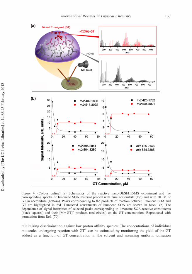
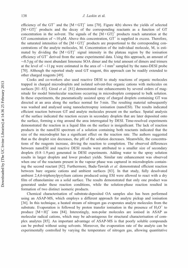
minimising discrimination against low proton affinity species. The concentrations of individualmolecules undergoing reaction with GT+ can be estimated by monitoring the yield of the GTadduct as a function of GT concentration in the solvent and assuming uniform ionisation
Figure 4. (Colour online) (a) Schematics of the reactive nano-DESI/HR-MS experiment and thecorresponding spectra of limonene SOA material probed with pure acetonitrile (top) and with 50 μM ofGT in acetonitrile (bottom). Peaks corresponding to the products of reaction between limonene SOA andGT are highlighted in red. Unreacted constituents of limonene SOA are shown in black. (b) Thedependence of signal intensities of selected peaks corresponding to limonene SOA-reactive constituents(black squares) and their [M+GT]+ products (red circles) on the GT concentration. Reproduced withpermission from Ref. [70].
International Reviews in Physical Chemistry 137
Dow
nloa
ded
by [
The
UC
Irv
ine
Lib
rari
es]
at 1
4:36
25
Febr
uary
201
3
efficiency of the GT+ and the [M+GT]+ ions [70]. Figure 4(b) shows the yields of selected[M+GT]+ products and the decay of the corresponding reactants as a function of GTconcentration in the solvent. The signals of the [M+GT]+ products reach saturation at theGT concentration of �10 μM. Above this concentration, GT+ is supplied in excess. Therefore,the saturated intensities of the [M+GT]+ products are proportional to the corresponding con-centrations of the analyte molecules, M. Concentration of the individual molecule, M, is esti-mated by dividing the [M+GT]+ signal intensity in the plateau region by the ionisationefficiency of GT+ derived from the same experimental data. Using this approach, an amount of�0.5 pg of the most abundant limonene SOA dimer and the total amount of dimers and trimersat the level of �11 pg were estimated in the area of �1mm2 sampled by the nano-DESI probe[70]. Although the reported study used GT reagent, this approach can be readily extended toother charged reagents [49].
Cooks and co-workers also used reactive DESI to study reactions of organic moleculestrapped in charged microdroplets and isolated solvent-free organic cations with molecules onsurfaces [81–83]. Girod et al. [81] demonstrated rate enhancements by several orders of mag-nitude for model bimolecular reactions occurring in microdroplets compared to bulk solution.In these experiments, the pneumatically assisted spray of charged droplets containing GT wasdirected at an area along the surface normal for 5min. The resulting material subsequentlywas washed and analysed using nanoelectrospray ionisation (nanoESI). The results indicatedefficient reaction between GT and analyte molecules present on the surface. Spatial analysisof the surface indicated the reaction occurs in secondary droplets that are later deposited ontothe surface, forming a ring around the area interrogated by DESI. Time-resolved experimentsdemonstrated the reaction in a liquid film on the surface is insignificant. The lack of reactionproducts in the nanoESI spectrum of a solution containing both reactants indicated that thesize of the microdroplet has a significant effect on the reaction rate. The authors suggestedthat as the droplet size decreases, the pH of the solution decreases. Meanwhile, the concentra-tions of the reagents increase, driving the reaction to completion. The observed differencesbetween nanoESI and reactive DESI results were attributed to a smaller size of secondarydroplets (0.8–1.9 μm) generated in DESI experiments. Adding water to the spray solutionresults in larger droplets and lower product yields. Similar rate enhancement was observedwhen one of the reactants present in the vapour phase was captured in microdroplets contain-ing the second reactant [82]. Furthermore, Badu-Tawiah et al. demonstrated efficient reactionbetween bare organic cations and ambient surfaces [83]. In that study, fully desolvatedambient 2,4,6-triphenylpyrylium cations produced using ESI were allowed to react with a dryfilm of ethanolamine on a solid surface. The results demonstrated that only one product wasgenerated under these reaction conditions, while the solution-phase reaction resulted information of two distinct isomeric products.
Chemical characterisation of substrate-deposited OA samples also has been performedusing an ASAP-MS, which employs a different approach for analyte pickup and ionisation[36]. In this technique, a heated stream of nitrogen gas evaporates analyte molecules from thesubstrate. Evaporation is followed by proton transfer ionisation in the presence of H3O
+ toproduce [M+H]+ ions [84]. Interestingly, non-polar molecules are ionised in ASAP asmolecular radical cations, which may be advantageous for structural characterisation of com-plex analytes [85]. An important advantage of ASAP-MS is that poorly soluble compoundscan be probed without using solvents. Moreover, the evaporation rate of the analyte can beexperimentally controlled by varying the temperature of nitrogen gas, allowing quantitative
138 J. Laskin et al.
Dow
nloa
ded
by [
The
UC
Irv
ine
Lib
rari
es]
at 1
4:36
25
Febr
uary
201
3

measurements of the vapour pressure of low-volatility organic compounds. These data arecritically important for predicting particle formation and ageing in atmospheric models.Figure 5 shows the results obtained in a temperature-programmed ASAP-MS experimentreported recently by Bruns et al. [86]. In that study, the technique’s performance wasdemonstrated using pyrene, a chemical standard with well-known vapour pressure, as a modelsystem. Figure 5(a) shows the experimentally measured evaporation rate (Et) as a function oftime and temperature. The value of Et, in mol s�1, is calculated using Equation (1):
Et ¼ N0 � ItItotal
ð1Þ
where N0 is the number of molecules in the probed sample calculated from the known massloading. It is a signal intensity of the pyrene peak at time t, while Itotal is its integrated valueover the entire duration of the experiment. The vapour pressure was derived from the averaged
Figure 5. (Colour online) (a) Evaporation rate as a function of time and temperature for pyrene measured ina temperature-programmed experiment using ASAP-MS detection. (b) Natural log of pyrene vapour pressureas a function of inverse temperature determined from the ASAP-MS measurements (black dots) incomparison with previously published data (coloured symbols). Reproduced with permission from Ref. [36].
International Reviews in Physical Chemistry 139
Dow
nloa
ded
by [
The
UC
Irv
ine
Lib
rari
es]
at 1
4:36
25
Febr
uary
201
3

(ET) values corresponding to isothermal time periods at different temperatures (T) using theLangmuir’s model of evaporation (Equation (2)):
PT ¼ ET
ffiffiffiffiffiffiffiffiffiffiffiffiffiffiffiffi2pRTM
p
NASTð2Þ
In this equation, R is the gas constant, M is the molar mass, NA is the Avogadro numberand ST is the surface area of the sample that was estimated based on visual observations andassuming a flattened sphere geometry. The heat of sublimation (ΔHsub) was determined fromthe slope of the Clausius–Clapeyron plot (Figure 5(b)), as shown in Equation (3):
lnPT ¼ �DHsub
RTþ Const ð3Þ
Similar experiments were performed for a number of dicarboxylic acids. Although theheats of sublimation obtained from ASAP-MS experiments were in excellent agreement withthe literature values, vapour pressures were lower than the values obtained using othertechniques by a factor of 2 and 3. The authors attributed this discrepancy to the relativelyslow diffusion of molecules during sample evaporation at atmospheric pressure (AP) com-pared to effusion in vacuum-based techniques. Nevertheless, results of the initial ASAP-MSmeasurements were quite promising. For example, ASAP-MS measurements correctly repro-duced the even/odd alterations in the vapour pressure of dicarboxylic acids as a function ofthe number of carbon atoms reported in a separate study of Cappa et al. [87]. Other sol-vent-free ambient ionisation techniques, including laserspray ionisation and its analogues[88,89] can be used for the analysis of non-polar and poorly soluble analytes in complexorganic samples.
EESI-MS [90] is another promising ambient ionisation method for online analysis of aero-sols. In EESI-MS of aerosols, a mist of charged droplets produced by electrospray is crossedwith a flow of air containing particles in front of an MS inlet. The first application of EESI-MS to α-pinene +O3 SOA by Doezema et al. [37] showed that both the particle and gas-phase components of aerosols can be detected with protonation serving as the primary ionisa-tion mechanism. They reported a significant enhancement of oligomeric compounds in SOAprepared at 50% RH in the reaction chamber compared to the SOA generated in dry air. Thiscontrasts previous observations [66,91] in isoprene high-NOx, where the oligomeric contentwas reduced in humid air. Further examination of the effects of RH on SOA molecular com-position is clearly warranted.
The field of ambient surface ionisation techniques is undergoing rapid growth both inmethod developments and applications. Thus far, few of the techniques described earlier havebeen used for chemical characterisation of aerosol samples. The following discussionhighlights several recent developments in ambient surface ionisation techniques that have thepotential for analyzing aerosol samples. For example, Nudnova et al. [92] described anambient ionisation approach, wherein neutral compounds evaporating from an organic film,either spontaneously or as a result of laser desorption, are ionised by plasma sustained insidea MS capillary inlet. The method is suitable for analysis of particle filter samples and poten-tially can be used in a surface imaging mode with spatial resolution characteristic of laserdesorption methods. Ho et al. [93] presented a paper-based surface acoustic wave (SAW)sample delivery and ionisation source, where a solvent phase organic analyte absorbed by the
140 J. Laskin et al.
Dow
nloa
ded
by [
The
UC
Irv
ine
Lib
rari
es]
at 1
4:36
25
Febr
uary
201
3
paper is atomised by SAW activated using the underlying piezoelectric element. Surfaceelectric field induced by SAW results in polarisation of the liquid film at the substrate andsubsequent charging of the atomised droplets from which gas phase ions are produced uponevaporation. Organic standards or heavy metals at concentrations as low as 1 nM weresuccessfully detected using both positive and negative ion modes, suggesting the greatpotential of this method for aerosol analysis and environmental monitoring.
Guenther et al. [94] used a method related to EESI to post-ionise electrosurgical aerosolproduced by rapid heating of biological tissues. The electrosurgical aerosol contains a detect-able amount of ions, which are dominated by fragments of phospholipids. In EESI, the ion cur-rent in the positive ion mode increased by a factor of 20–50 and produced largely intactphospholipid ions extracted from the neutral particles by the electrospray. No ion currentenhancement was observed in the negative ion mode. In a variation of the EESI-MS method,the ionising spray was charged by a corona discharge [95]. The advantage of the coronadischarge is in its ability to ionise analytes that do not typically appear in ESI mass spectra. Forexample, Hu et al. was able to detect radical cations of non-polar compounds, such as benzeneand cyclohexane, while polar compounds, such as acetone, acetonitrile and oxygenated terp-enes, appeared as protonated species in the spectrum. The method tolerates complex matrices,making it suitable for analysis of environmental samples, including extracts from aerosols.
2.2. Secondary ion mass spectrometry
In contrast with the ambient ionisation techniques, surface analysis using secondary ion massspectrometry (SIMS) is performed in vacuum. SIMS involves sputtering molecules fromsurfaces using energetic bombardment with a primary ion beam followed by the analysis ofsecondary ions using MS [96,97]. Although a variety of mass analysers can be used for thedetection of secondary ions, time-of-flight (TOF) instruments are particularly attractivebecause they enable fast acquisition of mass spectra over a broad range of mass-to-chargeratios (m/z) [98,99]. This capability is critical for detailed characterisation of the spatialdistribution of both organic and inorganic species in complex samples with good sensitivityand in a reasonable amount of time.
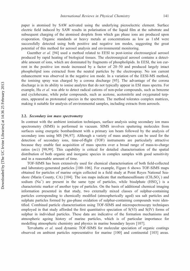
TOF-SIMS has been extensively used for chemical characterisation of both field-collectedand laboratory-generated particles [100–106]. For example, Figure 6 shows TOF-SIMS mapsobtained for particles of marine origin collected in a field study at Point Reyes National Sea-shore (Marin County, CA) [104]. The ion maps indicate that methanesulfonate (CH3SO
�3 ) and
sodium (Na+) are present in the same type of particles, while bisulphate (HSO�4 ) is a
characteristic marker of another type of particles. On the basis of additional chemical imaginginformation presented in that study, two externally mixed classes of sulphur-containingparticles corresponding to chemically modified (atmospherically aged) sea salt particles andsulphate particles formed by gas-phase oxidation of sulphur-containing compounds were iden-tified. Combined particle characterisation using TOF-SIMS and microspectroscopy techniquesemployed in that study afforded the first quantitative speciation of S(VI) and S(IV) forms ofsulphur in individual particles. These data are indicative of the formation mechanisms andatmospheric ageing history of marine particles, which is of particular importance formodelling atmospheric chemistry and physics in marine boundary layers [107].
Tervahattu et al. used dynamic TOF-SIMS for molecular speciation of organic coatingsobserved on ambient particles representative for marine [100] and continental [103] areas.
International Reviews in Physical Chemistry 141
Dow
nloa
ded
by [
The
UC
Irv
ine
Lib
rari
es]
at 1
4:36
25
Febr
uary
201
3

Their results showed the presence of palmitic and other fatty acids in the particles’ outermostlayers, corroborating predictions from earlier modelling and laboratory studies that suggestedcommon existence of the fatty acid surfactants on atmospheric particles [108,109]. Thesestudies indicated the need for a better understanding of the chemical reactivity, physical andoptical properties of surfactant-coated particles and their impact on the atmosphericenvironment.
The examples discussed earlier demonstrate the potential of TOF-SIMS for characterisingthe internal composition and surface chemistry of aerosol particles. However, the lateralresolution of the commercial instruments is insufficient for detailed mapping of the chemicalcomposition of individual particles. To address this problem, Sakamoto et al. developed a newhigh spatial resolution TOF-SIMS instrument [110]. In this apparatus, SIMS is coupled withscanning electron microscopy that enables easy localisation of particles for TOF-SIMS imag-ing. In addition, focused ion beam may be used for sectioning particles prior to analysis. Thisapproach is important for studying the internal structure of inhomogeneous particles. Theinstrument’s performance was demonstrated for two 1.7 and 5.4 μm diameter field-collectedaerosol particles.
Two other studies focused on the development of new approaches for improved sensitivityand mass resolution in SIMS. Specifically, Fletcher et al. developed a new dynamic TOF-SIMS instrument that uses a continuous primary ion beam (C60
+, 40 keV) [111]. In thisinstrument, secondary ions undergo collisional cooling in a radio frequency (RF)-only quadru-pole. The ions are subsequently transferred to a linear buncher that generates short pulses ofions for analysis in the TOF section. This process efficiently decouples secondary ionformation from mass analysis, enabling independent optimisation of mass resolution andsignificantly increasing the duty cycle, hence the instrument’s sensitivity. Furthermore, it sup-ports tandem mass spectrometry, or MS/MS, experiments critical for structural characterisationof complex molecules. In a related study, Carado et al. reported on the implementation ofSIMS on a commercial hybrid-quadrupole orthogonal TOF-MS [112]. This instrument also
Figure 6. (Colour online) TOF-SIMS maps of typical samples collected at the ground site located inPoint Reyes National Seashore: (a) Overlaid maps of the Na+ and CH3SO
�3 ions. Their close overlap
indicates formation of methanesulfonate in sea salt particles. (b) Overlaid maps of CH3SO�3 and HSO�
4ions indicate external mixing of sea salt and ammonium sulphate particles. Maps are 100 μm� 100 μm.Reproduced with permission from Ref. [104].
142 J. Laskin et al.
Dow
nloa
ded
by [
The
UC
Irv
ine
Lib
rari
es]
at 1
4:36
25
Febr
uary
201
3

uses a continuous ion beam, which significantly increases the duty cycle, and the sensitivity.The ions undergo collisional cooling and focusing in a quadrupole ion guide and are analysedusing a high-resolution orthogonal TOF-MS. Mass resolving power of �15,000 with an m/zrange up to 40,000 demonstrated with this instrument enables simultaneous chemical analysisof small and large molecules. These capabilities will be very useful for unraveling the com-plexity of individual atmospheric particles collected on substrates.
Because TOF-SIMS of atmospheric particles generates fairly complex mass spectra, thedevelopment of new data analysis tools is critical to extracting maximum information fromthe experimental data. Principal component analysis is commonly used to classify complexTOF-SIMS spectra. Recently, Kalegowda and Harmer examined the performance of severalstatistical analysis tools for classification of TOF-SIMS spectra of complex copper-iron sul-phide mineral samples [113]. Two methods, soft independent modelling of class analogy andk-nearest neighbour classification, were identified as the most accurate techniques for particleclassification. In addition, these methods enabled the classification of particles containing thesame elements but different crystal structures. These results are particularly remarkablebecause of the complexity and similarity between the experimental spectra obtained fordifferent particle types.
Cameca’s recent development of NanoSIMS instruments has opened up new opportunitiesfor accurate measurements of the elemental composition and isotope ratios. NanoSIMS is amicroprobe instrument that combines the sensitivity of dynamic SIMS with relativelyhigh mass resolution necessary for separation and unambiguous identification of isotopicpeaks (e.g. 13C14N� vs. 12C15N� at m/z 27.016 and 27.009, respectively). The combinationof high spatial resolution (down to 50 nm), high sensitivity (parts per million, ppm, in ele-mental imaging) and high mass resolution has made this instrument one of the most sensitivetools for spatially resolved elemental and isotopic analysis of complex surfaces. Althoughmost applications of NanoSIMS are related to imaging of biological materials [114–119] andsoil aggregates, several groups explored the potential of this technique for chemical character-isation of ambient particles. For example, McIntire et al. demonstrated the utility ofNanoSIMS in studying the three-dimensional (3D) structure of complex organic particles[120]. The observed increase in the oxygen-to-carbon ratio toward the interior of organicparticles produced during ozonolysis of surface bond alkenes indicated the polar groups areburied inside the particle’s hydrophobic shell.
Winteholler et al. used NanoSIMS’ high sensitivity for measuring sulphur isotope ratios ofindividual particles down to 500 nm in diameter [121]. In that study, sulphur isotope ratios weremeasured for several atmospherically relevant minerals. Particle-to-particle reproducibility of2–5% was reported for submicron particles with the lowest precision observed for particleswith complex topographies. The 34S/32S ratio varied by �15% between different mineralsamples, indicating measurable matrix effects on the observed isotope ratios. A fundamentalunderstanding of the effect of particle morphology and composition on isotope ratio measure-ment accuracy is necessary for improved performance of NanoSIMS for individual particlecharacterisation.
Recently, Harris et al. examined that sulphur isotope undergoes fractionation duringoxidation of SO2 in the gas phase, liquid phase and on mineral dust particle surfaces usingNanoSIMS of collected products [122,123]. Oxidation of sulphur dioxide to sulphate is animportant reaction in atmospheric chemistry [124]. The isotopic fractionation [125] (variationin the equilibrium distribution of the stable isotopes) typically results from slight differences
International Reviews in Physical Chemistry 143
Dow
nloa
ded
by [
The
UC
Irv
ine
Lib
rari
es]
at 1
4:36
25
Febr
uary
201
3

in bond strengths, velocities and diffusivities of molecules containing lighter vs. heavier iso-topes. For example, the slightly higher evaporation rate of H2
16O compared to H218O from
groundwater contributes to 16O enrichment of atmospheric water vapour and cloud droplets.The fractionation constant is defined as the ratio of the heavy to light isotopes in the productsdivided by the ratio of the heavy to light isotopes in the reactants. For the sulphur isotopefractionation, the fractionation constant is given by Equation (4):
a ¼ ð34S=32SÞproductsð34S=32SÞreactants
ð4Þ
When the heavy isotope reacts faster than the light isotope, the fractionation constant isgreater than one (α> 1). The fractionation factor of 1.008 at 19 °C was obtained for the gas-phase oxidation of SO2 [123]. For aqueous oxidation, the fractionation factors of 1.0151,1.0174 and 1.0118 were obtained when H2O2, O3 and the H2O2/O3 mixtures were used asoxidants, respectively [123]. The radical chain reaction catalysed by iron was the onlyreaction with a faster rate for the lighter isotope (α = 0.9894 at 19 °C). It follows that isotoperatio measurements may be useful for distinguishing metal-catalysed oxidation of SO2 fromother competing pathways. Another study by Harris et al. demonstrated that sulphur isotopefractionation can be used for understanding the kinetics and mechanisms of SO2 oxidation bymineral dust [122]. Sulphur isotope fractionation during heterogeneous oxidation of SO2 onSahara dust particles was characterised by a substantially larger fractionation factor(α= 1.0096) than aqueous oxidation in Sahara dust leachate (α = 0.9917). The authorsproposed that a radical chain reaction pathway initiated by transition metal ions leached fromthe dust is the main mechanism of aqueous SO2 oxidation in Sahara dust leachate [122]. Incontrast, heterogeneous oxidation on dust particles primarily is controlled by particlecomposition. Very different fractionation factors were reported for ilmenite/rutile (α = 1.012),feldspar (α= 0.948) and clay (α= 1.085). These results indicate that the isotope ratio measure-ments provide important constraints on the possible oxidation pathways of sulphur dioxide inthe environment and contribute to a fundamental understanding of the homogeneous andheterogeneous oxidation of SO2.
2.3. Secondary neutral mass spectrometry
Secondary neutral mass spectrometry (SNMS) relies on postionisation of the neutralmolecules sputtered during surface bombardment by the primary ion beam. Postionisationresults in significant signal enhancement and enables detection of a broader range of sputteredmolecules. When combined with soft ionisation, SNMS is particularly well suited for imagingorganic molecules in aerosol samples. Soft ionisation of sputtered organic molecules is per-formed using single-photon absorption of vacuum ultraviolet (VUV) light from a laser [126]or a tunable synchrotron radiation light source [23]. Tyler et al. [126,127] used 157 nm(7.9 eV) and 193 nm (6.4 eV) lasers for postionisation and imaging PAHs in individual field-collected aerosol particles. The samples were cryocooled to reduce the loss of semi-volatilePAHs in vacuum. Significant signal enhancement was observed for all PAHs examined in thatstudy. Postionisation using a 193 nm laser resulted in 2–50-fold increases in the ion yield,while 157 nm postionisation resulted in a 1000-fold increase in the signal. Pyrene and
144 J. Laskin et al.
Dow
nloa
ded
by [
The
UC
Irv
ine
Lib
rari
es]
at 1
4:36
25
Febr
uary
201
3

anthracene were efficiently ionised using both lasers. However, because of multiphotonionisation processes, higher fragmentation yield was obtained using the 193 nm laser. Incontrast, postionisation of naphthalene resulted in the formation of abundant fragment ions. Inaddition, an abundant [M�H]+ peak was observed when sputtered naphthalene was ionisedusing the 157 nm laser. The authors hypothesised that because the ionisation energy (IE) ofnaphthalene (8.14 eV) is greater than the energy of both lasers, postionisation of a neutral[M�H]• radical formed in the sputtering process is responsible for the formation of the[M�H]+ ion. Figure 7 shows SNMS maps of naphthalene, anthracene and pyrene in ambientaerosol particles. The images were analysed using maximum autocorrelation factors (MAFs).This statistical analysis technique identified three particle types as shown in the regions ofinterest (ROIs) panel. The combination of laser-SNMS imaging with statistical analysis toolsis ideally suited for studying surface chemistry of PAHs on ambient particle surfaces.
Imaging of smaller particles may become possible using recent instrument developmentefforts reported by Ebata et al. [128]. Their study described the development of a new laserionisation mass nanoscope, in which the primary 20 keVGa+ ion beam can be focused to adiameter of 40 nm. Sputtered neutral molecules were ionised using a femtosecond laser. Theions subsequently were analysed using a high-resolution multi-pass TOF with a massresolving power up to 40,000. In comparison with SIMS, postionisation of neutrals resultedin a 1000-fold increase in the signal of silver ions.
Takahashi et al. demonstrated the utility of wavelength-tunable postionisation of sputteredneutrals for chemically selective SNMS imaging [129]. Quasi-continuous tunable radiationfrom a synchrotron light source enabled acquisition of SNMS spectra at 10 kHz repetitionrate. Furthermore, the wavelength of the radiation may be tuned to affect selective ionisation
Figure 7. (Colour online) 75� 75 μm2, laser-SNMS (157 nm) images of ambient aerosol particles fromimpactor stage 6 (2–3.5 μm in aerodynamic diameter). The top row shows the total ion image, a red–green–blue overlay of MAF factors 1�3 and ROI corresponding to the three particle types identified inthe analysis. The lower row shows the molecular ion images for naphthalene, anthracene and pyrene.Reproduced with permission from Ref. [126].
International Reviews in Physical Chemistry 145
Dow
nloa
ded
by [
The
UC
Irv
ine
Lib
rari
es]
at 1
4:36
25
Febr
uary
201
3

of low-ionisation efficiency (IE) compounds, such as PAHs, or for efficient ionisation of allorganic compounds, including high ionisation energy aliphatic organic molecules. Finally, ithas been demonstrated that photoionisation efficiency (PIE) curves can be acquired by scan-ning the wavelength of the light. PIE curves provide important structural information for bothorganic and inorganic molecules. VUV-SNMS of monolignols [130] produced spectra domi-nated by the molecular ions. The PIE curves of fragment ions were shifted towards lowerenergies, indicating significant vibrational excitation of the sputtered neutral molecules. Thephotofragment yield can be reduced by increasing the extraction delay time. This observationwas rationalised assuming that molecules with lower internal energies also have lower kinet-ics energies.
2.4. Laser desorption ionisation mass spectrometry
A similar approach has been used for the online characterisation of airborne particles pro-duced in smog chamber experiments. These experiments typically use single-photon VUVionisation of neutral molecules formed during thermal desorption of particles [23,131,132].Tunable VUV photoionisation reduces fragmentation of SOA constituents prior to analysis,which greatly simplifies the observed mass spectra. Using this technique, Fang et al. con-ducted detailed characterisation of the composition of SOA produced through photooxidationof several VOCs in a smog chamber [133,134]. It has been demonstrated that accuratemeasurement of the ionisation energies of individual SOA constituents provides importantstructural information that is difficult to obtain using other techniques. Specifically, PIE mea-surements enable differentiation between different isomeric species [23]. Gloaguen et al.examined the kinetics of ozone reaction with sodium chloride particles coated with anthracene[135]. Ozonolysis produced oxygenated anthracene molecules with up to five oxygen atoms.The ionisation energies of all the products has been determined from the corresponding PIEcurves. However, the lack of data on the IE of oxygenated anthracene molecules made it dif-ficult to assign structures to the observed products.
Bertram and co-workers developed a broadly tunable, laboratory-based VUV lightsource and demonstrated its applications for characterisation of single particles down to300 nm in diameter when coupled to an ion trap mass spectrometer [136]. In a follow-upstudy from the same group [137], this instrument was used to characterise single-compo-nent particles of oleic acid and dihydroxybenzoic acid (DHB). In that study, particle vapori-sation was performed using a CO2 laser. The degree of fragmentation observed in massspectra was a strong function of vaporisation energy and the analyte molecule’s properties.For example, at low vaporisation energies, DHB was observed as intact ions, while signifi-cant fragmentation was observed for oleic acid. The observed fragmentation was attributedto the unimolecular dissociation of vibrationally excited molecules following the ionisationstep. Simpson et al. examined the effect of particle composition on the observed fragmenta-tion in VUV mass spectra of two-component oleic acid-oleyl alcohol particles [138]. Theyfound the degree of fragmentation of individual components in mixed particles depends onthe particle composition. Specifically, the addition of oleyl alcohol resulted in enhancedfragmentation of oleic acid. This observation was rationalised assuming that the presenceof oleyl alcohol results in more efficient heating of oleic acid during the laser vaporisationprocess. Furthermore, the degree of fragmentation did not show a linear dependence on the
146 J. Laskin et al.
Dow
nloa
ded
by [
The
UC
Irv
ine
Lib
rari
es]
at 1
4:36
25
Febr
uary
201
3

molar ratio of oleic acid in the particle, indicating that quantification using this approach ischallenging.
Geddes et al. [139] described a near-infrared (IR) laser desorption ionisation method withsufficient sensitivity to produce mass spectra of OA at realistic atmospheric mass concentra-tions (�1 μg/m3) and with good time resolution (2min). The aerosols first are collected on analuminum probe then desorbed/ionised with a 1064-nm laser pulse. Little or no fragmentationwas observed in mass spectra of model aerosols of oleic acid and SOA produced throughozonolysis of α-pinene.
2.5. Laser ablation mass spectrometry
LA-MS has been extensively used for analysing surfaces and ambient particles [45,140,141].The technique uses an intense laser beam for volatilisation and ionisation of material from asurface. LA is a highly non-linear process, in which the amounts of material removed from thesurface, the plume’s composition, and the heating of the material strongly depend on the irradi-ance (energy per unit time and area) [45,140]. In addition, the spatial profile of the laser beam,pulse duration and wavelength significantly influence the LA process [140]. Particles, mole-cules, atoms and ions are ejected from surfaces following laser irradiation [142]. Significantheating of the ablated material results in facile fragmentation of molecules and ions, making itdifficult to determine molecular and structural information based on LA mass spectra [143].However, LA is well suited for spatially resolved elemental analysis of surfaces. By far,coupling LA with inductively coupled plasma MS (LA-ICPMS) is the most popular approachfor sensitive mapping of the elemental composition of surfaces without special sample pre-treatment [45,144]. In LA-ICPMS, the laser-generated aerosol is transferred to the ICP source,where it is vaporised and ionised by the plasma. The high sensitivity and dynamic range of thistechnique enable simultaneous detection of both major and minor constituents of the sample.Although this technique has been used primarily in materials science applications, recent studiesexamined the utility of LA-ICPMS for analysis of soil and ambient particles [141,145–147].
The duration of the laser pulse plays an important role in LA-ICPMS experiments [44,148].The use of nanosecond lasers is associated with several undesirable effects, including significantmaterial redistribution, preferential evaporation of certain materials and absorption of the laserlight by the plume, that further alters the plasma composition. In contrast, femtosecond ablationcauses significantly less surface damage and dramatically suppresses the fractionation andmatrix effects in LA-ICPMS [148]. It has been demonstrated that the crater shape, plume com-position and size of ejected particles are critical factors that determine the utility of femtosecondLA for quantitative elemental mapping of materials. Further improvement in quantificationusing femtosecond LA-ICPMS requires better understanding of the dynamics of the plasmaformation following laser irradiation. Koch et al. used shadowgraphy and light scattering tovisualise shockwave propagation and particle ejection during femtosecond LA [149]. This studydemonstrated the initial gas compression following laser irradiation is followed by a secondshockwave caused by material ejection and air breakdown away from the surface. Multimodalparticle size distributions observed in these experiments were attributed to different mechanismsof particle formation in the LA plume. Interestingly, submicron particles were produced duringLA of Y:ZrO2, while LA of Li2B4O7 generated larger particles.
Several groups examined the performance of LA coupled with atomic force microscopy(AFM) or near-field optical microscopy for high-resolution chemical imaging of surfaces
International Reviews in Physical Chemistry 147
Dow
nloa
ded
by [
The
UC
Irv
ine
Lib
rari
es]
at 1
4:36
25
Febr
uary
201
3

[150–153]. It has been demonstrated that near-field LA can be used for reducing the size ofthe ablation area down to 100 nm in diameter. However, the sampling efficiency of ionsformed in the process typically is fairly low. Zhu et al. combined AP near-field LA with sam-pling and postionisation of neutrals inside the vacuum system [150,153]. Such couplingenables chemical imaging of surfaces with spatial resolution of 5 μm [153]. That studyreported the combined sampling efficiency of 10�4 [150]. Sampling efficiency is determinedby the transfer efficiency of neutrals into the vacuum system (�0.1) and the IE (10�3) obtainedusing electron impact ionisation. Although this technique has not been used to analyse smallparticles, the spatial resolution attainable using near-field LA is sufficient for such analysis.
3. Depth profiling
Several depth profiling MS techniques have been developed for applications in materials sci-ence and biology. A vast majority of these techniques rely on controlled removal of materialfrom a solid or liquid surface using laser irradiation or ion beam bombardment [154]. Despitesignificant advancements in depth profiling instrumentation, detailed characterisation ofsubmicron-sized particles still is a challenge. To the best of our knowledge, most studies haveused SIMS for depth profiling of a collection of ambient aerosol particles [105,106,155,156].In this section, we will discuss recent developments in depth profiling using MS that providethe basis to develop sensitive and robust approaches for detailed characterisation of the 3Dstructures of small particles.
Although SIMS is a fairly mature analytical technique, the formation of secondary ionsduring the energetic ion bombardment is a complex process that still is not well understood. Itis well known that surface bombardment with monoatomic and small polyatomic primary ionsresults in significant surface damage [157–159]. Other important limitations of SIMS includesubstantial fragmentation of organic molecules [159,160] and relatively low ionisation yields[161]. Significant efforts have focused on addressing these limitations using beams of clusterions [162,163]. It has been demonstrated that surface bombardment using keV C60
+ ionssignificantly reduces surface damage and increases secondary ion yields [47]. Moleculardynamics simulations indicate that, in contrast with the monoatomic primary ions, the kineticenergy of the primary C60
+ ions is deposited closer to the surface. As a result, C60+ bombard-
ment results in a more efficient removal of material from a surface while generating arelatively shallow crater (�10 nm deep) [164]. This property of cluster beam bombardment isa key to accurate depth profiling using ion beams [154,162,163] and will be discussed later.Rabbani et al. compared the performance of C60
+ and large, singly charged argon clusters fordepth profiling using SIMS [165]. The results demonstrate that, in comparison with C60, sur-face bombardment with large argon clusters results in gentler sputtering and reduces surfacedamage. However, secondary ion yields decrease with an increase in the argon cluster’s size.
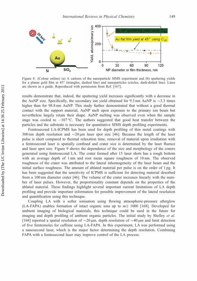
Chemical analysis of environmental particles on substrates using SIMS requires a funda-mental understanding of the dependence of the sputtering yield on particle size. Moleculardynamics simulations indicate the sputtering yield for small gold nanoparticles (AuNP) andthin gold films are enhanced by up to a factor of three compared to the bulk gold [166]. Thehighest sputtering yields were predicted for 8 nm-gold particles and 3 nm-thick gold films.Yang et al. conducted an experimental investigation of size-dependent secondary ion yieldsduring sputtering of AuNP in the size range of 10–100 nm by 20 keV C60
2+ primary ions[167]. Figure 8 compares the sputtering yield obtained for AuNP and a flat gold film. The
148 J. Laskin et al.
Dow
nloa
ded
by [
The
UC
Irv
ine
Lib
rari
es]
at 1
4:36
25
Febr
uary
201
3
results demonstrate that, indeed, the sputtering yield increases significantly with a decrease inthe AuNP size. Specifically, the secondary ion yield obtained for 9.3 nm AuNP is �3.3 timeshigher than for 98.8 nm AuNP. This study further demonstrated that without a good thermalcontact with the support material, AuNP melt upon exposure to the primary ion beam butnevertheless largely retain their shape. AuNP melting was observed even when the samplestage was cooled to �107 °C. The authors suggested that good heat transfer between theparticles and the substrate is necessary for quantitative SIMS depth profiling experiments.
Femtosecond LA-ICPMS has been used for depth profiling of thin metal coatings with300 nm depth resolution and �20 μm laser spot size [46]. Because the length of the laserpulse is short compared to thermal relaxation time, removal of material upon irradiation witha femtosecond laser is spatially confined and crater size is determined by the laser fluenceand laser spot size. Figure 9 shows the dependence of the size and morphology of the cratersgenerated using femtosecond LA. The crater formed after 15 laser shots has a rough bottomwith an average depth of 1 nm and root mean square roughness of 10 nm. The observedroughness of the crater was attributed to the lateral inhomogeneity of the laser beam and theinitial surface roughness. The amount of ablated material per pulse is on the order of 1 pg. Ithas been suggested that the sensitivity of ICPMS is sufficient for detecting material desorbedfrom a 100 nm diameter crater [46]. The volume of the crater increases linearly with the num-ber of laser pulses. However, the proportionality constant depends on the properties of theablated material. These findings highlight several important current limitations of LA depthprofiling and provide important information for possible improvement of the lateral resolutionand quantification using this technique.
Coupling LA with a softer ionisation using flowing atmospheric-pressure afterglow(LA-FAPA) enables formation of intact organic ions up to m/z 1000 [168]. Developed forambient imaging of biological materials, this technique could be used in the future forimaging and depth profiling of ambient organic particles. The initial study by Shelley et al.[168] reported a spatial resolution of �20 μm, depth resolution of �40 μm and limit detectionof five femtomoles for caffeine using LA-FAPA. In this experiment, LA was performed usinga nanosecond laser, which is the major factor determining the depth resolution. CombiningFAPA with a femtosecond laser may improve control of the LA process.
Figure 8. (Colour online) (a) A cartoon of the nanoparticle SIMS experiment and (b) sputtering yieldsfor a planar gold film at 45° (triangles, dashed line) and nanoparticles (circles, dash-dotted line). Linesare shown as a guide. Reproduced with permission from Ref. [167].
International Reviews in Physical Chemistry 149
Dow
nloa
ded
by [
The
UC
Irv
ine
Lib
rari
es]
at 1
4:36
25
Febr
uary
201
3

Valledor et al. performed depth profiling of thin metal films with nanometer depth resolu-tion using pulsed-RF glow discharge TOF MS (RF-GD-TOFMS) [169]. In this technique, RFpower generates plasma in argon, which sputters atoms and molecules from a surface.Sputtered species are subsequently analysed using a TOF mass spectrometer. Although thedepth resolution of this technique is substantially higher than in LA-ICPMS, the lateral resolu-tion is defined by the plasma size and typically is on the order of millimeters. Spatially resolvedoptical emission measurements identified two different RF-GD plasma regimes above a purecopper target [170]. The two plasma regimes resulted in similar sputtering rates but differentplasma composition, demonstrating the complexity of the plasma-formation process. Furtherunderstanding of the physical phenomena underlying pulsed RF-GD sputtering is important forthe development of this technique to facilitate accurate depth profile measurements.
A low-temperature plasma (LTP) probe combined with ICPMS enables depth profilingwith lateral resolution of less than 10 μm and depth resolution of �100 nm [171].LTP-ICPMS is an attractive alternative to LA-ICPMS because it is easy to operate and mayprovide comparable performance. However, understanding the dependence of the crater’sshape and the rate of material removal on experimental parameters still is rather limited, andphenomena associated with material sputtering require further characterisation.
Depth profiling of aerosol particles suspended in air has been performed using single-particle MS [172–174]. In these experiments, ‘depth profiling’ is performed by analyzing
Figure 9. (Colour online) Three-dimensional pictures of the craters produced after different number oflaser shots, measured using the AFM. Reproduced with permission from Ref. [46].
150 J. Laskin et al.
Dow
nloa
ded
by [
The
UC
Irv
ine
Lib
rari
es]
at 1
4:36
25
Febr
uary
201
3

a large number of identical particles exposed to different laser evaporation conditions.Woods et al. conducted a depth profiling study of glycerol particles coated with oleic acid[172]. In that study, gentle IR laser evaporation was followed by VUV ionisation of thevapour. At low laser fluences, only the particle’s outer layers were evaporated in this pro-cess, while at higher laser fluences, the entire particle was evaporated, providing informa-tion on the chemical composition of the particle interior. For 2 μm particles, the techniquewas sensitive to changes in the oleic acid coating thickness of �10 nm. Zelenyuk et al.reported depth profiling of NaCl particles coated with liquid dioctyl phthalate (DOP) lay-ers and with solid pyrene [174]. Detailed size and shape measurements combined withdepth profiling demonstrated that NaCl particles were completely coated with liquid DOP,while the pyrene coating was found to be non-uniform, forming aspherical particles withpartially exposed NaCl cores. In another study, this group used depth profiling to examinethe morphology of particles formed by mixing DOP with SOA of α-pinene [173]. Theresults of that study indicated phase separation of SOA and DOP in mixed particles.Furthermore, a thin layer of DOP was observed on top of SOA particles, indicating insol-uble components may be present on particle surfaces and affecting their physicochemicalproperties.
4. Chemical analysis of airborne droplets
4.1. Field-induced droplet ionisation mass spectrometry
Grimm and Beauchamp developed a unique approach for producing ions from small liquiddroplets by FIDI and introducing the ions into a mass spectrometer [39,40]. Neutral dropletsplaced in a sufficiently strong static electric field are known to elongate and develop twoopposing conical tips that emit microjets of oppositely charged particles. The charged progenyparticles then result in desolvated ions by mechanisms similar to those in conventional ESI[175], making it possible to analyse the droplet’s chemical composition via MS. The keyadvantage of this technique is in its ability to simultaneously analyse the same droplet byboth positive and negative ion MS. Practical applications of FIDI require a single massspectrometer that is switchable between the positive and the negative ion mode operations. Astationary 1–2mm droplet typically is formed at the tip of the capillary, exposed to a gas-phase reagent for a specified period of time, and then subjected to a high electric field toinduce jetting (Figure 10). One of the jets is directed into a mass spectrometer for analysis. Ifions of another polarity are analysed, the field direction is reversed.
The electric field required to induce jetting from a neutral droplet (E0c ) is known as the
‘Taylor limit’ [176]. E0c is related to the surface tension, σ, and droplet diameter, d, as shown
in Equation (5) [40]:
E0c � 1:625
ffiffiffiffiffiffiffiffiffiffiffiffir
2pe0d
rð5Þ
For methanol droplets (σ = 0.022N/m), the predicted Taylor limit is 1.0MV/m for a 1-mmparticle. The electric field required for FIDI presents certain experimental difficulties because itis close to the breakdown limit of air (�3MV/m). As a result of this limitation, pure waterdroplets (σ = 0.072N/m) cannot be easily investigated via FIDI. If the droplet carries charge, the
International Reviews in Physical Chemistry 151
Dow
nloa
ded
by [
The
UC
Irv
ine
Lib
rari
es]
at 1
4:36
25
Febr
uary
201
3

Figure 10. (Colour online) A FIDI experimental setup used for investigation of reactions between ozoneand phospholipids on surfaces of liquid droplets. Reproduced with permission from Ref. [180].
152 J. Laskin et al.
Dow
nloa
ded
by [
The
UC
Irv
ine
Lib
rari
es]
at 1
4:36
25
Febr
uary
201
3

critical field required to induce jetting is reduced and the droplet produces only one jet of prog-eny particles (Figure 11). When the droplet charge exceeds the Rayleigh limit (Equation (6)):
qR ¼ffiffiffiffiffiffiffiffiffiffiffiffiffiffiffiffiffiffi8p2re0d3
pð6Þ
the droplet fission occurs spontaneously.Reactions at the liquid–air interface can be conveniently investigated by exposing the
droplet to gas-phase reactants for a short period of time, typically a few seconds, prior toturning on the electric field. This approach is especially useful for investigating interfacialchemistry of surface-active molecules because they tend to be enhanced [177,178] in theprogeny droplets during droplet break-up. The utility of FIDI for studying chemistry atinterfaces was demonstrated shortly after the initial development of the technique. Grimmand Beauchamp [179] were able to observe naphthalene-Ag+ ions after exposing a water/methanol droplet containing dissolved silver ions to naphthalene vapour. In the same paper,they examined the mechanism of oleic acid ozonolysis by monitoring the products of theinterfacial reaction between oleic acid and gas-phase ozone. In addition, they demonstratedthat the method is suitable for locating the double bond position in phospholipids by study-ing the reaction between oleoyl-l-α-lysophosphatidic acid and ozone. Using FIDI, Kim
Figure 11. A schematic diagram showing different regions of stability for neutral and charged dropletsplaced in an electric field. As the droplet charge increases, the critical field required to induce FIDI isreduced, but jetting becomes asymmetric. Reproduced with permission from Ref. [40].
International Reviews in Physical Chemistry 153
Dow
nloa
ded
by [
The
UC
Irv
ine
Lib
rari
es]
at 1
4:36
25
Febr
uary
201
3

et al. [180] investigated heterogeneous ozonolysis of an unsaturated phospholipid, 1-palmi-toyl-2-oleoyl-sn-phosphatidylglycerol (POPG), at the air–water/methanol interface. Theywere able to observe hydroxyhydroperoxide and secondary ozonide (SOZ = 1,2,4-trioxolane)products of ozonolysis, which are difficult to detect with traditional ESI-MS due to theirlow stability. As SOZs are not commonly produced in the aqueous ozonolysis of alkenesin the bulk, the observation of the SOZ product was attributed to a unique interfacial pro-cess. The same group also studied the reaction of ozone with a mixture of phospholipids,such as unsaturated POPG and saturated 1,2-dipalmitoyl-sn-phosphatidylglycerol or satu-rated 1,2-dipalmitoyl-sn-phosphatidylcholine and unsaturated 1-stearoyl-2-oleoyl-sn-phospha-tidylcholine. The results were consistent with the phospholipids forming a uniformly mixedlayer. With extensive ozonolysis, the unsaturated phospholipids formed more hydrophilicproducts and transitioned into the droplet, and only saturated phospholipids remained onthe surface. Recently, Ko et al. applied FIDI-MS to studying interfacial reactions of choles-terol sulphate in a POPG layer with ozone [181]. They observed an epoxide of cholesterolsulphate as the major oxidation product. In light of the significant interest in chemical pro-cesses occurring at air–liquid interfaces, we can expect a number of new applications ofFIDI and related methods (including the one described in the next section) to appear in thefuture.
4.2. Mass spectrometric studies of the reactivity at the air–water interface in microdroplets
In 2007, Enami et al. developed another novel mass spectrometric method that is intrinsicallysensitive to the processes occurring at the droplet–air interface [41]. In this method, a solutionis sprayed under pressure into a chamber through a grounded, stainless-steel capillarysurrounded by a coaxial flow of a nebulising gas as shown in Figure 12. Because of the gasflow’s large velocity (>200m/s), the much slower liquid jet (�0.1m/s) is quickly shreddedinto micrometer-sized droplets (microdroplets). With terminal velocities of the microdropletsin excess of 10m/s, it takes less than 1ms for the droplets to cross the orthogonal plume of agas-phase reactant, such as ozone in the spraying chamber. Although the spray is neutral onthe whole, some droplets carry positive or negative charge acquired as a result of normalstatistical fluctuations in the number of charges per droplet [182]. As the charged dropletsevaporate, they eventually reach the Rayleigh limit for Coulomb fission and produce progenydroplets and desolvated ions by mechanisms similar to ESI [175]. A mass spectrometercollects ions from the spray through an electrified pinhole. For example, in Figure 12, anegative potential is applied to the mass spectrometer entrance pinhole to extract positive ionsfrom the gas flow. Due to the enhancement of the surface-active species in droplet fissionprocesses, the mass spectrometer preferentially samples species that reside on the surface ofthe original microdroplets. It is the combination of the short gas–droplet exposure time andthe bias toward detecting surface-active species that makes this method exquisitely sensitiveto the reactions occurring at the gas–droplet interface. Furthermore, the observed ion signalsare directly proportional to ion concentrations in the interfacial layers of the microjets prior totheir break-up into microdroplets [183]. Numerous problems, ranging from oxidation of sur-factants on water droplets by atmospheric oxidants to the fundamental issue of proton avail-ability at the water–air interface, have been investigated with this approach [41,183–196].Some of these studies are highlighted later herein.
154 J. Laskin et al.
Dow
nloa
ded
by [
The
UC
Irv
ine
Lib
rari
es]
at 1
4:36
25
Febr
uary
201
3
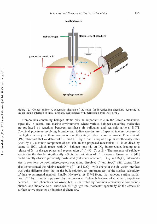
Compounds containing halogen atoms play an important role in the lower atmosphere,especially in coastal and marine environments where various halogen-containing moleculesare produced by reactions between gas-phase air pollutants and sea salt particles [197].Chemical processes involving bromine and iodine species are of special interest because ofthe high efficiency of these compounds in the catalytic destruction of ozone. Enami et al.[192] observed that oxidation of Br� and Cl� by ozone in liquid droplets is efficiently cata-lysed by I�, a minor component of sea salt. In the proposed mechanism, I� is oxidised byozone to HOI, which reacts with X� halogen ions via an IX�
2 intermediate, leading to arelease of X2 in the gas-phase and regeneration of I� (X =Cl or Br). The presence of sulphatespecies in the droplet significantly affects the oxidation of I� by ozone. Enami et al. [41]could directly observe previously postulated (but never observed) ISO�
3 and IS2O�3 intermedi-
ates in reactions between microdroplets containing dissolved I� and S2O2�3 with ozone. They
also demonstrated the relative reactivity of I� and S2O2�3 with ozone at the air–water interface
was quite different from that in the bulk solution, an important test of the surface selectivityof their experimental method. Finally, Hayase et al. [194] found that aqueous surface oxida-tion of I� by ozone is suppressed by the presence of phenols because of efficient competitionbetween I� and phenolates for ozone but is unaffected by common atmospheric compoundsbutanol and malonic acid. These results highlight the molecular specificity of the effects ofsurface-active organics on interfacial chemistry.
Figure 12. (Colour online) A schematic diagram of the setup for investigating chemistry occurring atthe air–liquid interface of small droplets. Reproduced with permission from Ref. [191].
International Reviews in Physical Chemistry 155
Dow
nloa
ded
by [
The
UC
Irv
ine
Lib
rari
es]
at 1
4:36
25
Febr
uary
201
3
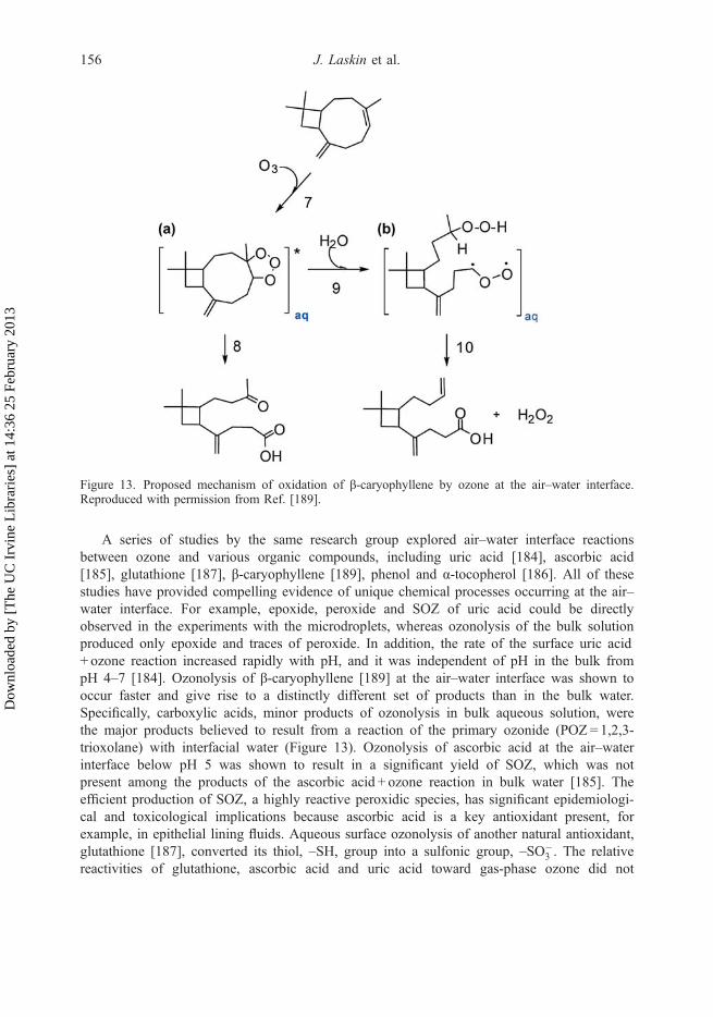
A series of studies by the same research group explored air–water interface reactionsbetween ozone and various organic compounds, including uric acid [184], ascorbic acid[185], glutathione [187], β-caryophyllene [189], phenol and α-tocopherol [186]. All of thesestudies have provided compelling evidence of unique chemical processes occurring at the air–water interface. For example, epoxide, peroxide and SOZ of uric acid could be directlyobserved in the experiments with the microdroplets, whereas ozonolysis of the bulk solutionproduced only epoxide and traces of peroxide. In addition, the rate of the surface uric acid+ ozone reaction increased rapidly with pH, and it was independent of pH in the bulk frompH 4–7 [184]. Ozonolysis of β-caryophyllene [189] at the air–water interface was shown tooccur faster and give rise to a distinctly different set of products than in the bulk water.Specifically, carboxylic acids, minor products of ozonolysis in bulk aqueous solution, werethe major products believed to result from a reaction of the primary ozonide (POZ = 1,2,3-trioxolane) with interfacial water (Figure 13). Ozonolysis of ascorbic acid at the air–waterinterface below pH 5 was shown to result in a significant yield of SOZ, which was notpresent among the products of the ascorbic acid + ozone reaction in bulk water [185]. Theefficient production of SOZ, a highly reactive peroxidic species, has significant epidemiologi-cal and toxicological implications because ascorbic acid is a key antioxidant present, forexample, in epithelial lining fluids. Aqueous surface ozonolysis of another natural antioxidant,glutathione [187], converted its thiol, –SH, group into a sulfonic group, –SO�
3 . The relativereactivities of glutathione, ascorbic acid and uric acid toward gas-phase ozone did not
Figure 13. Proposed mechanism of oxidation of β-caryophyllene by ozone at the air–water interface.Reproduced with permission from Ref. [189].
156 J. Laskin et al.
Dow
nloa
ded
by [
The
UC
Irv
ine
Lib
rari
es]
at 1
4:36
25
Febr
uary
201
3

match those toward aqueous ozone in the bulk solution. These studies make it clear that theair–water interface provides a unique reaction media for ozone chemistry, different from bothgas- and condensed-phase environments in terms of reaction rates and mechanisms. The air–water interface provides a similarly unique environment for reactions of other atmosphericoxidants, such as OH [198]. Therefore, extensions of these methods to other types of oxidantsare desirable.
Another series of papers by the same group examined the availability of protons at theair–water interface and the efficiency of proton transfer to and from water through such aninterface. Proton transfer equilibria are known to be dramatically sensitive to the solvationstate of the ions involved. For example, hydronium ions readily protonate most organiccompounds in the gas-phase, whereas only relatively strong bases can be protonated by asimilar reaction in bulk water because of the high free energy of hydration of the hydroniumion relative to that of the protonated base. Enami et al. [183] used reactions between gas-phase trimethylamine (TMA), a compound with high gas-phase basicity (227 kcal/mol), andmicrodroplets with different bulk pH to probe the apparent availability of protons at the air–water interface. The ‘titration curve’ representing the signal of TMA-H+ versus the bulksolution pH had an ‘equivalence point’ (i.e. the pH value below which the TMA-H+ signalincreased abruptly) at pHbulk = 3.8. This was quite different from the 9.8 pKa of TMA-H+, thevalue reproduced in the same experiments by spraying pre-mixed solutions of TMA chloridewith different initial pH. The authors concluded that hydronium ions are accessible to thegas-phase TMA species colliding with the surface of water microdroplets only below pHbulk
4. In the presence of certain surface-active organic compounds, such as fulvic acid, the pro-tonation could be achieved at a higher pHbulk, suggesting that these organic molecules mayact as proton donors instead of the hydronium ions [188]. A similar experiment also was con-ducted with n-hexanoic acid [191], the gas-phase basicity (187 kcal/mol) of which is closer tothat of water (158 kcal/mol). Although the protonated form of hexanoic acid is a strong acidwith a negative effective pKa <�3, the authors observed protonation of the –C(O)OH groupof hexanoic acid, resulting in –CðOHÞþ2 occurring at pHbulk as high as 3–4. Stated differently,at pHbulk < 4, the aqueous surface behaved as a ‘superacid’ with respect to the protonation ofthe molecules colliding with it. Because of the high kinetic isotope effect observed on H2Ovs. D2O microjets, they surmised that protonation involves proton tunneling through the out-ermost layers of water and is controlled by both the reaction energetics and the dynamics ofthe proton tunneling process. A new twist to the story has been provided by a recentexperiment, where aqueous microdroplets were exposed to HNO3 [196]. Although HNO3 isstrong acid, it did not produce NO�
3 upon collisions with water surfaces at neutral pHbulk
unless the solution already contained a small amount of other ions. Density functionalcalculations by the authors suggested that anions in the surface layers of the dropletelectrostatically assist in withdrawing the proton away from NO�
3 . It should be clear fromthese examples that the proton transfer processes at the interfaces may follow distinctlydifferent – and not always intuitive – predictable pathways.
Perhaps the most remarkable example of a unique proton transfer process at the air–waterinterface was the observation that isoprene, the second most important organic molecule emit-ted by the biosphere (after methane), is capable of protonation and cationic oligomerisationduring collisions with mildly acidic (pHbulk < 4) droplets [190]. It is known that highly acidicsurfaces such as >80wt.% sulphuric acid take up isoprene efficiency and convert it into
International Reviews in Physical Chemistry 157
Dow
nloa
ded
by [
The
UC
Irv
ine
Lib
rari
es]
at 1
4:36
25
Febr
uary
201
3

polymeric products [199]. It appears that a similar process may take place under more envi-ronmentally relevant conditions. This process, estimated to occur with an effective uptakecoefficient of �10�6 (one out of a million gas–aqueous surface collisions), has the potentialto contribute to nighttime removal of isoprene by thin water films on various environmentalsurfaces, including the forest canopy and aerosol particles. If such interface-specific processesturn out to be universally applicable to all biogenic terpenoids, we will have to reconsidertheir emission inventories, as well as the mechanisms of SOA formation, from suchcompounds.
4.3. Laser abblation mass spectrometry of levitated and supported droplets
Laser desorption/ionisation (LDI) of liquid droplets has many advantages for qualitativeanalysis of the mixture components compared to LDI on solids. In contrast to LA fromsolids, liquids represent a more homogenous sample, and the spot ablated by the laser ‘heals’quickly. Therefore, each subsequent laser pulse in effect probes a fresh sample. Jorabchi et al.[42] examined the enhancement of matrix-assisted laser desorption ionisation (MALDI) signalfrom immobilised liquid droplets in the presence of corona ions. In that study, a microliterdroplet was placed on a conducting stainless-steel or on a non-conducting Teflon supportbetween an entrance tube of a mass spectrometer and a corona needle. A pulsed nitrogenlaser was focused on the droplet from above. Using a clever way of timing the coronadischarge voltage with respect to the laser pulses, they were able to demonstrate that ionenhancement was not a result of postdesorption ionisation of neutral compounds by thecorona ions. Rather, it was a result of charging the electrically isolated droplet. The chargeddroplet was distorted in the applied field from the corona tip, which increased the ionformation efficiency at the droplet surface closest to the mass spectrometer entrance. Themain advantage of this mechanism, referred to as ‘CALDI’, by the authors, is that it requiresa significantly lower external field compared to the standard AP-MALDI or ESI methods.
Westphall et al. [200] took advantage of CALDI’s lower electric field requirements inwhat appears to be the first MS study of acoustically levitated microdroplets. Acoustic levita-tion is ideally suited for CALDI because this technique depends only on the density of thelevitated particle, not on its size or charge. Charging the droplet prior to the LDI pulse wasshown to be critical for the mass spectral detection of ions. In fact, no ions could be observedin these experiments if the droplet was not initially charged. Figure 14 illustrates the optimaltiming of the experiment, in which the droplet is first charged to the highest possible levelwith the corona discharge, then allowed to equilibrate in the acoustic trap, and finallysubjected to the LDI pulse. The entire timing sequence was repeated at the repetition rate ofthe laser (20Hz in these experiments). The resulting mass spectra of oligopeptides containedsingly charged protonated and sodiated ions with minimal fragmentation.
In the two previously described studies, the UV laser used for desorption/ionisationrequired significant amounts of chromophoric compounds and diethanolamine as part of theMALDI matrix, which suppressed partitioning of analytes to the surface. Jorabchi and Smith[201] described an alternative approach, in which a relatively low-power IR laser was usedfor preferential desorption of surface-active analytes from a droplet resting on a stainless-steelcapillary followed by postionisation with a methanol/formic acid electrospray. Theydemonstrated the surface bias of this method by an observation that more surface-activemolecules were depleted from the droplet faster as the droplet was subjected to repeated IR
158 J. Laskin et al.
Dow
nloa
ded
by [
The
UC
Irv
ine
Lib
rari
es]
at 1
4:36
25
Febr
uary
201
3

desorption pulses. These results were rationalised using a model that related the surfacepartitioning coefficients with the observed decay rates of the ion signals. The authors found acorrelation between the surface partitioning and the phase partitioning coefficients on a C18
HPLC column. Thus, the approach may become a viable separation tool for organic analytesin microdroplets.
4.4. Ion-mobility studies of nanodroplets
IMS [202] is a well-established technique based on separating ions by their mobility througha buffer gas. Mobility Z (cm2V�1 s�1) is defined as a proportionality constant between theterminal velocity of the ion, m (cm s�1), and the electrostatic field, E (V cm�1), as shown inEquation (7):
m ¼ Z � E ð7Þ
In the low-field limit, the mobility is inversely proportional to the product of the buffer gasdensity and the collisional cross section, which, in turn, depends on the ion’s size and shape. Atypical IMS instrument contains a pulsed ion source, linear drift region and suitable ion detec-tor. IMS instruments often are coupled to quadrupole, ion trap or TOF mass spectrometers toenable precise determination of the masses of ions exiting the drift region [203]. IMS-MSmethods traditionally have been applied to comparatively small molecular ions, cluster ionsand biomolecules, generally less than 1 nm in size [203,204]. Larger particles, with diametersexceeding several nanometers, are normally sized with mobility instruments of a very differentdesign [205], known as ‘differential mobility analysers’ (DMAs). The fundamental differencebetween IMS and DMA instruments is that the former separate packets of ions in time, whereasthe latter separate continuous streams of ions or charged particles in space. A standard DMAconsists of a continuous particle source, particle charger/neutraliser, cylindrical drift region anda particle detector, either a condensation particle counter or an electrometer. Commercial
Figure 14. (Colour online) Schematic diagram of the setup for taking CALDI mass spectra ofacoustically levitated particles and the timing sequence of the experiment involving charging the droplet,equilibration step and LDI step. Reproduced with permission from Ref. [200].
International Reviews in Physical Chemistry 159
Dow
nloa
ded
by [
The
UC
Irv
ine
Lib
rari
es]
at 1
4:36
25
Febr
uary
201
3
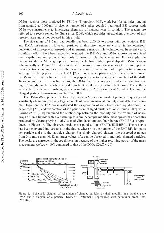
DMAs, such as those produced by TSI Inc. (Shoreview, MN), work best for particles rangingfrom about 5 to 1000 nm in size. A number of studies coupled traditional ESI sources withDMA-type instruments to investigate chemistry of nanoparticles in solution. The readers arereferred to a recent review by Guha et al. [206], which provides an excellent overview of thisresearch area and is not covered in this article.
The size range of 1–5 nm traditionally has been difficult to access with conventional IMSand DMA instruments. However, particles in this size range are critical in homogeneousnucleation of atmospheric aerosols and in emerging nanoparticle technologies. In recent years,significant efforts have been expended to morph the IMS-MS and DMA approaches to extendtheir capabilities and provide new tools for nanoparticle characterisation. For example, theFernandez de la Mora group incorporated a high-resolution parallel-plate DMA, shownschematically in Figure 15, into atmospheric pressure ionisation sources of various types ofmass spectrometers and described the design criteria for achieving both high ion transmissionand high resolving power of the DMA [207]. For smaller particle sizes, the resolving powerof DMAs is primarily limited by diffusion perpendicular to the intended direction of the drift.To overcome the diffusion limitations, the DMA had to be operated under the conditions ofhigh Reynolds numbers, where any design fault would result in turbulent flows. The authorswere able to achieve a resolving power in mobility (Z/ΔZ) in excess of 50 while keeping thecharged particle transmission greater than 50%.
The DMA-MS approach developed by the de la Mora group made it possible to quickly andsensitively obtain impressively large amounts of two-dimensional mobility-mass data. For exam-ple, Hogan and de la Mora investigated the evaporation of ions from ionic liquid-acetonitrilenanodrops [208] and evaporation of ion pairs from charged clusters of ionic liquids [209], whileLarriba et al. [210] examined the relationship between the mobility and the volume of nano-drops of ionic liquids with diameters up to 3 nm. A sample mobility-mass spectrum of particlesproduced by electrospraying 1-ethyl-3-methylimidazolium tetrafluoroborate (EMI-BF4) is repro-duced in Figure 16. The observed peaks correspond to ions (EMI+)z(EMI-BF4)n. The m/z-axishas been converted into n/z-axis in the figure, where n is the number of the EMI-BF4 ion pairsper particle and z is the particle’s charge. For singly charged clusters, the observed n rangesfrom 0 to more than 40. Even larger values of n can be observed in multiply charged particles.The peaks are narrower in the n/z dimension because of the higher resolving power of the massspectrometer (m/Δm � 104) compared to that of the DMA (Z/ΔZ � 50).
Figure 15. Schematic diagram of separation of charged particles by their mobility in a parallel plateDMA and a diagram of a practical DMA-MS instrument. Reproduced with permission from Refs.[207,208].
160 J. Laskin et al.
Dow
nloa
ded
by [
The
UC
Irv
ine
Lib
rari
es]
at 1
4:36
25
Febr
uary
201
3

Such a mobility-mass data-set is impressively rich with information. For example, theparticle densities can be directly calculated from the observed masses and mobilities using theStokes–Mulliken equation, which relates mobility to the volume-equivalent diameter andaccounts for the finite size of the gas-phase molecules colliding with the particle [211]. Theapparent particle densities approach bulk values for particles with diameters in excess of
Figure 16. (Colour online) A mobility-mass spectrum of particles generated by electrospraying asolution of ionic liquid EMI-BF4 in acetonitrile. Only singly charged ions EMI+(EMI-BF4)n are visiblein the top panel. The intensity scale has been adjusted in the bottom panel to better showcase themultiply charged ions (EMI+)z(EMI-BF4)n. Reproduced with permission from Ref. [210].
International Reviews in Physical Chemistry 161
Dow
nloa
ded
by [
The
UC
Irv
ine
Lib
rari
es]
at 1
4:36
25
Febr
uary
201
3
1.5 nm [210]. The apparent density is significantly reduced for smaller particles becauseparticles deviate from spherical shapes as they approach molecular dimensions. In addition,the importance of the ion-induced dipole interactions in particle–molecule collisions becomesgreater at smaller particle sizes. Both effects decrease the particle mobility relative to thatexpected for a spherical object drifting through a flow of non-polarisable molecules.
The dynamics of droplet evaporation have been examined using mobility-mass measure-ments. For each mobility value, several n/z peaks are observed for singly charged ions (z= 1)in Figure 16 instead of a single peak. This has been interpreted [209] as a result of ion-pairevaporation from the ionic liquid droplets occurring in the ion guide of the mass spectrometer(region Q0 in Figure 15) as a result of RF heating. Although ionic liquids generally areviewed as involatile, the droplet surface’s high degree of curvature enables evaporation of theEMI-BF4 ion pairs via the Kelvin effect (ion-pair evaporation from aerosolised ionic liquidsalso has been observed [212] using VUV photoionisation of the free ion pairs). Evaporationof ions from multiply charged droplets also was observed with the most common process cor-responding to the loss of (EMI+)(EMI-BF4). The highly efficient evaporation of the dropletsmakes it challenging to investigate more volatile nanodroplets made of water or commonorganic solvents using this technique. However, this limitation likely can be overcome in thefuture by using a buffer gas maintained at a predefined RH, similar to the approach used intandem DMA instruments designed to quantify hygroscopic growth of particles [213].
5. Summary and outlook
Recent developments of MS-based techniques highlighted in this review facilitate opportunitiesfor improved understanding of the mechanisms and kinetics of reactions affecting physical andchemical properties of ambient particles and droplets. Although the MS techniques reviewedhere are not universally applicable to all types of particles, and some are limited only to labora-tory studies by virtue of the instrumental design, the broad arsenal of the available MS methodsmakes it possible to pick one or several methods tailored to a specific application. For example,ambient surface ionisation techniques enable direct analysis of particles collected on substrateswithout special sample pre-treatment. When coupled with HR-MS, these techniques can beused for detailed characterisation of the chemical composition of the inherently complex SOAsamples. This capability is particularly important for understanding mechanisms of heteroge-neous reactions of SOA with common atmospheric gases responsible for chemical ageing ofSOA in the atmosphere. Furthermore, reactive analysis may be used for quantification ofreaction products in complex mixtures. Although a variety of ambient surface ionisationtechniques developed in the last decade have been used in forensics, biological materialsanalysis, counterfeit drug detection, chemical imaging and other applications, only a few ofthese methods have been applied for characterisation of aerosol samples. This presents a clearopportunity for future developments in the field of chemical analysis of aerosol samples.
Several vacuum-based techniques traditionally used for characterisation of mineralsurfaces and soil samples (e.g. NanoSIMS and LA-ICPMS) show great potential for depthprofiling and imaging of individual particles on substrates. Recent developments in SIMSinstrumentation [43] have resulted in improved sensitivity and better spatial resolution, whichenabled the first high-resolution elemental mapping of heterogeneous inorganic particles.Combined with depth profiling capabilities, this technique may be used in the future for 3Dchemical imaging of individual particles on substrates. Although images of individual
162 J. Laskin et al.
Dow
nloa
ded
by [
The
UC
Irv
ine
Lib
rari
es]
at 1
4:36
25
Febr
uary
201
3
particles deposited on the substrates may be affected by transformations of particle morpholo-gies upon sample collection, these effects can be minimised by gentle deposition of chargedparticles using ion soft landing [214–216] that can be further developed for particle deposi-tion. Alternatively, the advent of novel coherent light source facilities based on X-ray freeelectron laser technology has opened up an additional unique opportunity for on-the-flyimaging and mass spectrometric characterisation of individual particles [217].
Ambient liquid droplets are unique reactive vessels, in which both the rates and pathwaysof chemical reactions may be dramatically different from the bulk solution. For example, ratesof some organic reactions are enhanced by several orders of magnitude when the reactionoccurs in a submicron-sized droplet. MS approaches summarised in this review are ideallysuited for understanding chemistry on droplets with sizes spanning several orders ofmagnitude (from nanometers to millimeters in diameter). These techniques provide uniqueinsights on the surface chemistry of microdroplets. For example, MS has been used for exam-ining the availability of protons and proton transfer phenomena at the air–water interface.These studies demonstrated that at relatively mild acidic pH, the aqueous surface behaves asa superacid as reflected by the unexpectedly efficient protonation of gas-phase moleculesimpinging on the surface of the droplet. The observation of ozone + olefin reaction productsthat do not readily form in the bulk is another example of unusual chemistry occurring at theair–water interface that can now be investigated using emerging MS methods. These exam-ples highlight exciting future opportunities in the field of droplet MS. In addition, combiningexperimental data with molecular dynamics modelling may have a significant impact both onunderstanding chemistry in cloud and fog droplets and toward a fundamental understandingof physical chemistry underlying ambient ionisation techniques.
This review also makes the point that gathering comprehensive information on the chemicalcomposition, reactivity and transformations of environmental particles and surfaces is an espe-cially challenging task because no single method of analytical chemistry is capable of providingthe full range of necessary information. For example, methods of single-particle MS analysismay probe and sometimes even visualise particle morphology and internal structure at nanome-ter scale. However, they provide limited information on particle molecular content. HR-MSinterfaced with ambient surface ionisation approaches may provide detailed information onmolecular content of organic particles and surfaces, but these methods acquire integrated signalfrom an ensemble of particles and eliminate knowledge of particle individual composition.Therefore, the application of complementary analytical methods is needed to provide compre-hensive information, ranging from microscopy-level details of individual particles to advancedmolecular characterisation of complex organics comprising particulate matter.
AcknowledgementsThe authors acknowledge previous and ongoing support for their research projects described in thisreview: US Department of Energy’s (DOE) Office of Basic Energy Sciences, Division of ChemicalSciences, Geosciences and Biosciences (JL); DOE’s Office of Biological and Environmental Research(BER) Atmospheric System Research programme and the W.R. Wiley Environmental MolecularSciences Laboratory’s (EMSL) intramural research and development programme (AL); and the NationalScience Foundation grants AGS-1227579 and CHE-0909227 (SAN). Additionally, JL and ALacknowledge a multi-program Chemical Imaging Initiative funded through Pacific Northwest NationalLaboratory’s (PNNL) Laboratory Directed Research and Development programme that allowedcompilation of this review. EMSL is a national scientific user facility sponsored by DOE-BER and
International Reviews in Physical Chemistry 163
Dow
nloa
ded
by [
The
UC
Irv
ine
Lib
rari
es]
at 1
4:36
25
Febr
uary
201
3

located at PNNL. PNNL is operated by Battelle for the DOE under Contract No. DE-AC05-76RL01830.
References
[1] N.O.A. Kwamena, J. Buajarern, and J.P. Reid, J. Phys. Chem. A 114, 5787 (2010).[2] A.K. Bertram, S.T. Martin, S.J. Hanna, M.L. Smith, A. Bodsworth, Q. Chen, M. Kuwata, A.
Liu, Y. You, and S.R. Zorn, Atmos. Chem. Phys. 11, 10995 (2011).[3] B.J. Finlayson-Pitts, Phys. Chem. Chem. Phys. 11, 7760 (2009).[4] Y. Rudich, N.M. Donahue, and T.F. Mentel, Annu. Rev. Phys. Chem. 58, 321 (2007).[5] M.E. Monge, T. Rosenørn, O. Favez, M. Müller, G. Adler, A. Abo Riziq, Y. Rudich, H. Herr-
mann, C. George, and B. D’Anna, Proc. Natl. Acad. Sci. 109, 6840 (2012).[6] P. Kulkarni, P.A. Baron and K. Willeke, editors. Aerosol Measurement (John Wiley & Sons,
Hoboken, NJ, 2011).[7] R. Signorell and J.P. Reid, editors. Fundamentals and Applications in Aerosol Spectroscopy
(Taylor and Francis Books, Inc., CRC Press, Boca Raton, FL, 2011).[8] H.R. Pruppacher and J.D. Klett, Microphysics of Clouds and Precipitation. 2nd ed. (Kluwer
Academic, Boston, MA, 1997).[9] M.O. Andreae and D. Rosenfeld, Earth-Sci. Rev. 89, 13 (2008).[10] J.M. Waldman, J.W. Munger, D.J. Jacob, R.C. Flagan, J.J. Morgan, and M.R. Hoffmann, Science
218, 677 (1982).[11] J.P. Reid and R.M. Sayer, Chem. Soc. Rev. 32, 70 (2003).[12] D.J. Donaldson and K.T. Valsaraj, Environ. Sci. Technol. 44, 865 (2010).[13] B. Ervens, B.J. Turpin, and R.J. Weber, Atmos. Chem. Phys. 11, 11069 (2011).[14] D.J. Donaldson and V. Vaida, Chem. Rev. 106, 1445 (2006).[15] M.R. Canagaratna, J.T. Jayne, J.L. Jimenez, J.D. Allan, M.R. Alfarra, Q. Zhang, T.B.
Onasch, F. Drewnick, H. Coe, A. Middlebrook, A. Delia, L.R. Williams, A.M. Trimborn, M.J. Northway, P.F. DeCarlo, C.E. Kolb, P. Davidovits, and D.R. Worsnop, Mass Spectrom.Rev. 26, 185 (2007).
[16] K.A. Pratt and K.A. Prather, Mass Spectrom. Rev. 31, 1 (2012).[17] K.A. Pratt and K.A. Prather, Mass Spectrom. Rev. 31, 17 (2012).[18] S.A. Nizkorodov, J. Laskin, and A. Laskin, PCCP 13, 3612 (2011).[19] J. Zahardis, S. Geddes, and G.A. Petrucci, Anal. Chem. 83, 2409 (2011).[20] A. Zelenyuk and D. Imre, Int. Rev. Phys. Chem. 28, 309 (2009).[21] K. Hartonen, T. Laitinen, and M.L. Riekkola, TrAC, Trends Anal. Chem. 30, 1486 (2011).[22] T. Hoffmann, R.J. Huang, and M. Kalberer, Anal. Chem. 83, 4649 (2011).[23] Z.Y. Zhou, H.J. Guo, and F. Qi, TrAC, Trends Anal. Chem. 30, 1400 (2011).[24] B.R. Bzdek, M.R. Pennington, and M.V. Johnston, J. Aerosol Sci. (in press). <http://dx.doi.org/
10.1016/j.jaerosci.2012.1005.1001>.[25] A. Laskin, J. Laskin, and S.A. Nizkorodov, Environ. Chem. 9, 163 (2012).[26] C.E. Kolb and D.R. Worsnop, Annu. Rev. Phys. Chem. 63, 471 (2012).[27] T. Reemtsma, J. Chromatogr. A 1216, 3687 (2009).[28] R.G. Cooks, Z. Ouyang, Z. Takats, and J.M. Wiseman, Science 311, 1566 (2006).[29] G.A. Harris, L. Nyadong, and F.M. Fernandez, Analyst 133, 1297 (2008).[30] A. Venter, M. Nefliu, and R.G. Cooks, TrAC, Trends Anal. Chem. 27, 284 (2008).[31] H.W. Chen, G. Gamez, and R. Zenobi, J. Am. Soc. Mass. Spectrom. 20, 1947 (2009).[32] R.M. Alberici, R.C. Simas, G.B. Sanvido, W. Romao, P.M. Lalli, M. Benassi, I.B.S. Cunha,
and M.N. Eberlin, Anal. Bioanal. Chem. 398, 265 (2010).[33] G.A. Harris, A.S. Galhena, and F.M. Fernandez, Anal. Chem. 83, 4508 (2011).
164 J. Laskin et al.
Dow
nloa
ded
by [
The
UC
Irv
ine
Lib
rari
es]
at 1
4:36
25
Febr
uary
201
3

[34] J. Laskin, A. Laskin, P.J. Roach, G.W. Slysz, G.A. Anderson, S.A. Nizkorodov, D.L. Bones,and L.Q. Nguyen, Anal. Chem. 82, 2048 (2010).
[35] P.J. Roach, J. Laskin, and A. Laskin, Analyst. 135, 2233 (2010).[36] E.A. Bruns, V. Perraud, J. Greaves, and B.J. Finlayson-Pitts, Anal. Chem. 82, 5922 (2010).[37] L.A. Doezema, T. Longin, W. Cody, V. Perraud, M.L. Dawson, M.J. Ezell, J. Greaves, K.R.
Johnson, and B.J. Finlayson-Pitts, Rsc Adv. 2, 2930 (2012).[38] S.J. Fuller, Y. Zhao, S.S. Cliff, A.S. Wexler, and M. Kalberer, Anal. Chem. (2012).[39] R.L. Grimm and J.L. Beauchamp, J. Phys. Chem. B 107, 14161 (2003).[40] R.L. Grimm and J.L. Beauchamp, J. Phys. Chem. B 109, 8244 (2005).[41] S. Enami, C.D. Vecitis, J. Cheng, M.R. Hoffmann, and A.J. Colussi, J. Phys. Chem. A 111,
13032 (2007).[42] K. Jorabchi, M.S. Westphall, and L.M. Smith, J. Am. Soc. Mass. Spectrom. 19, 833 (2008).[43] J.S. Fletcher and J.C. Vickerman, Anal. Chem. (2012).[44] R. Hergenroder, O. Samek, and V. Hommes, Mass Spectrom. Rev. 25, 551 (2006).[45] R.E. Russo, X.L. Mao, H.C. Liu, J. Gonzalez, and S.S. Mao, Talanta 57, 425 (2002).[46] J. Pisonero, J. Koch, M. Wälle, W. Hartung, N.D. Spencer, and D. Günther, Anal. Chem. 79,
2325 (2007).[47] D. Weibel, S. Wong, N. Lockyer, P. Blenkinsopp, R. Hill, and J.C. Vickerman, Anal. Chem.
75, 1754 (2003).[48] G.J. Van Berkel, S.P. Pasilis, and O. Ovchinnikova, J. Mass Spectrom. 43, 1161 (2008).[49] D.R. Ifa, C.P. Wu, Z. Ouyang, and R.G. Cooks, Analyst. 135, 669 (2010).[50] D.J. Weston, Analyst 135, 661 (2010).[51] X.X. Ma, S.C. Zhang, and X.R. Zhang, TrAC, Trends Anal. Chem. 35, 50 (2012).[52] Z. Takats, J.M. Wiseman, B. Gologan, and R.G. Cooks, Science. 306, 471 (2004).[53] Z. Takáts, J.M. Wiseman, and R.G. Cooks, J. Mass Spectrom. 40, 1261 (2005).[54] A.B. Costa and R.G. Cooks, Chem. Commun. 3915 (2007). doi:3910.1039/B710511H.[55] A.B. Costa and R.G. Cooks, Chem. Phys. Lett. 464, 1 (2008).[56] V. Frankevich, R.J. Nieckarz, P.N. Sagulenko, K. Barylyuk, R. Zenobi, L.I. Levitsky, A.Y.
Agapov, T.Y. Perlova, M.V. Gorshkov, and I.A. Tarasova, Rapid Commun. Mass Spectrom. 26,1567 (2012).
[57] M.C. Wood, D.K. Busby, and P.B. Farnsworth, Anal. Chem. 81, 6407 (2009).[58] J. Laskin, B.S. Heath, P.J. Roach, L. Cazares, and O.J. Semmes, Anal. Chem. 84, 141 (2012).[59] J.B. Fenn, M. Mann, C.K. Meng, S.F. Wong, and C.M. Whitehouse, Science 246, 64 (1989).[60] F.M. Green, T.L. Salter, P. Stokes, I.S. Gilmore, and G. O'Connor, Surf. Interface Anal. 42,
347 (2010).[61] H. Chen, M. Li, Y.P. Zhang, X. Yang, J.J. Lian, and J.M. Chen, J. Am. Soc. Mass. Spectrom.
19, 450 (2008).[62] M. Li, H. Chen, B.F. Wang, X. Yang, J.J. Lian, and J.M. Chen, Int. J. Mass Spectrom. 281,
31 (2009).[63] M. Li, H. Chen, X. Yang, J.M. Chen, and C.L. Li, Atmos. Environ. 43, 2717 (2009).[64] T.B. Nguyen, J. Laskin, A. Laskin, and S.A. Nizkorodov, Environ. Sci. Technol. 45, 6908
(2011).[65] T.B. Nguyen, P.B. Lee, K.M. Updyke, D.L. Bones, J. Laskin, A. Laskin, and S.A. Nizkorodov, J.
Geophys. Res. Atm. 117, D01207 (2012). doi:01210.01029/02011jd016944.[66] T.B. Nguyen, P.J. Roach, J. Laskin, A. Laskin, and S.A. Nizkorodov, Atmos. Chem. Phys. 11,
6931 (2011).[67] R.E. O’Brien, A. Laskin, J. Laskin, S. Liu, R. Weber, L.M. Russell, and A.H. Goldstein, Atmos.
Environ. (submitted for publication).[68] R.E. O’Brien, T.B. Nguyen, A. Laskin, J. Laskin, P.L. Hayes, S. Liu, J.L. Jimenez, L.M. Russell,
S.A. Nizkorodov, and A.H. Goldstein, J. Geophys. Res. Atm. (submitted for publication).
International Reviews in Physical Chemistry 165
Dow
nloa
ded
by [
The
UC
Irv
ine
Lib
rari
es]
at 1
4:36
25
Febr
uary
201
3

[69] P.J. Roach, J. Laskin, and A. Laskin, Anal. Chem. 82, 7979 (2010).[70] J. Laskin, P.A. Eckert, P.J. Roach, B.S. Heath, S.A. Nizkorodov, and A. Laskin, Anal. Chem.
84, 7179 (2012).[71] D.L. Bones, D.K. Henricksen, S.A. Mang, M. Gonsior, A.P. Bateman, T.B. Nguyen, W.J. Cooper,
and S.A. Nizkorodov, J. Geophys. Res. Atm. 115, D05203 (2010). doi:05210.01029/02009jd012864.
[72] K.M Updyke, T.B. Nguyen, and S.A. Nizkorodov, Atmos. Environ. (accepted for publication).[73] T. Koop, J. Bookhold, M. Shiraiwa, and U. Poschl, PCCP 13, 19238 (2011).[74] T.D. Vaden, D. Imre, J. Beranek, M. Shrivastava, and A. Zelenyuk, Proc. Natl. Acad. Sci.
108, 2190 (2011).[75] V. Perraud, E.A. Bruns, M.J. Ezell, S.N. Johnson, Y. Yu, M.L. Alexander, A. Zelenyuk, D.
Imre, W.L. Chang, D. Dabdub, J.F. Pankow, and B.J. Finlayson-Pitts, Proc. Natl. Acad. Sci.109, 2836 (2012).
[76] P.A. Eckert, P.J. Roach, A. Laskin, and J. Laskin, Anal. Chem. 84, 1517 (2012).[77] H. Chen, I. Cotte-Rodriguez, and R.G. Cooks, Chem. Commun. 597 (2006).[78] C.P. Wu, D.R. Ifa, N.E. Manicke, and R.G. Cooks, Anal. Chem. 81, 7618 (2009).[79] A.M. Gaddis, R. Ellis, and G.T. Currie, Nature 191, 1391 (1961).[80] A.S. Stachissini and L. Doamaral, J. Org. Chem. 56, 1419 (1991).[81] M. Girod, E. Moyano, D.I. Campbell, and R.G. Cooks, Chem. Sci. 2, 501 (2011).[82] A.K. Badu-Tawiah, D.I. Campbell, and R.G. Cooks, J. Am. Soc. Mass. Spectrom. 23, 1077
(2012).[83] A.K. Badu-Tawiah, J. Cyriac, and R.G. Cooks, J. Am. Soc. Mass. Spectrom. 23, 842 (2012).[84] C.N. McEwen, R.G. McKay, and B.S. Larsen, Anal. Chem. 77, 7826 (2005).[85] M.J.P. Smith, N.R. Cameron, and J.A. Mosely, Analyst 137, 4524 (2012).[86] E.A. Bruns, J. Greaves, and B.J. Finlayson-Pitts, J. Phys. Chem. A 116, 5900 (2012).[87] C.D. Cappa, E.R. Lovejoy, and A.R. Ravishankara, J. Phys. Chem. A 111, 3099 (2007).[88] S. Trimpin, J. Mass Spectrom. 45, 471 (2010).[89] B.X. Wang, C.B. Lietz, E.D. Inutan, S.M. Leach, and S. Trimpin, Anal. Chem. 83, 4076
(2011).[90] H. Chen, A. Venter, and R.G. Cooks, Chem. Commun. 2042 (2006).[91] H. Zhang, J.D. Surratt, Y.H. Lin, J. Bapat, and R.M. Kamens, Atmos. Chem. Phys. 11, 6411
(2011).[92] M.M. Nudnova, L. Zhu, and R. Zenobi, Rapid Commun. Mass Spectrom. 26, 1447 (2012).[93] J. Ho, M.K. Tan, D.B. Go, L.Y. Yeo, J.R. Friend, and H.C. Chang, Anal. Chem. 83, 3260
(2011).[94] S. Guenther, K.C. Schafer, J. Balog, J. Denes, T. Majoros, K. Albrecht, M. Toth, B. Spen-
gler, and Z. Takats, J. Am. Soc. Mass. Spectrom. 22, 2082 (2011).[95] B. Hu, X.L. Zhang, M. Li, X.J. Peng, J. Han, S.P. Yang, Y.Z. Ouyang, and H.W. Chen,
Analyst 136, 4977 (2011).[96] A. Benninghoven, F.G. Rudenauer and H.W. Werner, Secondary Ion Mass Spectrometry: Basic
Concepts, Instrumental Aspects, Applications and Trends (Wiley, New York, NY, 1987).[97] R.J. Day, S.E. Unger, and R.G. Cooks, Anal. Chem. 52, A557 (1980).)[98] A. Benninghoven, Angew. Chem. Int. Ed. 33, 1023 (1994).[99] R.N.S. Sodhi, Analyst 129, 483 (2004).[100] H. Tervahattu, J. Juhanoja, and K. Kupiainen, J. Geophys. Res. Atm. 107, 4319 (2002).
doi:4310.1029/2001jd001403.[101] A. Laskin, D.J. Gaspar, W.H. Wang, S.W. Hunt, J.P. Cowin, S.D. Colson, and B.J. Finlay-
son-Pitts, Science 301, 340 (2003).[102] R.E. Peterson and B.J. Tyler, Appl. Surf. Sci. 203, 751 (2003).
166 J. Laskin et al.
Dow
nloa
ded
by [
The
UC
Irv
ine
Lib
rari
es]
at 1
4:36
25
Febr
uary
201
3

[103] H. Tervahattu, J. Juhanoja, V. Vaida, A.F. Tuck, J.V. Niemi, K. Kupiainen, M. Kulmala, and H.Vehkamaki, J. Geophys. Res. Atm. 110, D06207 (2005). doi:06210.01029/02004jd005400.
[104] R.J. Hopkins, Y. Desyaterik, A.V. Tivanski, R.A. Zaveri, C.M. Berkowitz, T. Tyliszczak, M.K.Gilles, and A. Laskin, J. Geophys. Res. Atm. 113, D04209 (2008). doi:04210.01029/02007jd008954.)
[105] Y. Liu, Z. Yang, Y. Desyaterik, P.L. Gassman, H. Wang, and A. Laskin, Anal. Chem. 80, 633(2008).
[106] Y. Liu, B. Minofar, Y. Desyaterik, E. Dames, Z. Zhu, J.P. Cain, R.J. Hopkins, M.K. Gilles,H. Wang, P. Jungwirth, and A. Laskin, PCCP 13, 11846 (2011).
[107] R. von Glasow and P.J. Crutzen, Atmos. Chem. Phys. 4, 589 (2004).[108] G.B. Ellison, A.F. Tuck, and V. Vaida, J. Geophys. Res. Atm. 104, 11633 (1999).[109] Y. Rudich, Chem. Rev. 103, 5097 (2003).[110] T. Sakamoto, M. Koizumi, J. Kawasaki, and J. Yamaguchi, Appl. Surf. Sci. 255, 1617 (2008).[111] J.S. Fletcher, S. Rabbani, A. Henderson, P. Blenkinsopp, S.P. Thompson, N.P. Lockyer, and J.
C. Vickerman, Anal. Chem. 80, 9058 (2008).[112] A. Carado, M.K. Passarelli, J. Kozole, J.E. Wingate, N. Winograd, and A.V. Loboda, Anal.
Chem. 80, 7921 (2008).[113] Y. Kalegowda and S.L. Harmer, Anal. Chem. 84, 2754 (2012).[114] J.L. Guerquin-Kern, T.D. Wu, C. Quintana, and A. Croisy, Biochim. Biophys. Acta-Gen. Subj.
1724, 228 (2005).[115] S.G. Boxer, M.L. Kraft, and P.K. Weber, Ann. Rev. Biophys. 38, 53 (2009).[116] K. Heister, C. Hoschen, G.J. Pronk, C.W. Mueller, and I. Kogel-Knabner, J. Soils Sediments
12, 35 (2012).[117] A.M. Herrmann, K. Ritz, N. Nunan, P.L. Clode, J. Pett-Ridge, M.R. Kilburn, D.V. Murphy,
A.G. O'Donnell, and E.A. Stockdale, Soil Biol. Biochem. 39, 1835 (2007).[118] L. Remusat, P.J. Hatton, P.S. Nico, B. Zeller, M. Kleber, and D. Derrien, Environ. Sci. Tech-
nol. 46, 3943 (2012).[119] T. Rennert, K.U. Totsche, K. Heister, M. Kersten, and J. Thieme, J. Soils Sediments 12, 3
(2012).[120] T.M. McIntire, O.S. Ryder, P.L. Gassman, Z. Zhu, S. Ghosal, and B.J. Finlayson-Pitts, Atmos.
Environ. 44, 939 (2010).[121] B. Winterholler, P. Hoppe, S. Foley, and M.O. Andreae, Int. J. Mass Spectrom. 272, 63 (2008).[122] E. Harris, B. Sinha, S. Foley, J.N. Crowley, S. Borrmann, and P. Hoppe, Atmos. Chem. Phys.
12, 4867 (2012).[123] E. Harris, B. Sinha, P. Hoppe, J.N. Crowley, S. Ono, and S. Foley, Atmos. Chem. Phys. 12,
407 (2012).[124] J. Langner and H. Rodhe, J. Atmos. Chem. 13, 225 (1991).[125] M.H. Thiemens, Annu. Rev. Earth Planetary Sci. 34, 217 (2006).[126] B.J. Tyler, S. Dambach, S. Galla, R.E. Peterson, and H.F. Arlinghaus, Anal. Chem. 84, 76
(2012).[127] R.E. Peterson, A. Nair, S. Dambach, H.F. Arlinghaus, and B.J. Tyler, Appl. Surf. Sci. 252,
7006 (2006).[128] S. Ebata, M. Ishihara, K. Uchino, S. Itose, M. Matsuya, M. Kudo, K. Bajo, and H. Yurimoto,
Surf. Interface Anal. 44, 635 (2012).[129] L.K. Takahashi, J. Zhou, K.R. Wilson, S.R. Leone, and M. Ahmed, J. Phys. Chem. A 113,
4035 (2009).[130] L.K. Takahashi, J. Zhou, O. Kostko, A. Golan, S.R. Leone, and M. Ahmed, J. Phys. Chem.
A 115, 3279 (2011).[131] S.R. Leone, M. Ahmed, and K.R. Wilson, PCCP 12, 6564 (2010).
International Reviews in Physical Chemistry 167
Dow
nloa
ded
by [
The
UC
Irv
ine
Lib
rari
es]
at 1
4:36
25
Febr
uary
201
3

[132] G. Pan, C.J. Hu, Z.Y. Wang, Y. Cheng, X.H. Zheng, X.J. Gu, W.X. Zhao, W.J. Zhang, J.Chen, F.Y. Liu, X.B. Shan, and L.S. Sheng, Rapid Commun. Mass Spectrom. 26, 189 (2012).
[133] W.Z. Fang, L. Gong, X.B. Shan, F.Y. Liu, Z.Y. Wang, and L.S. Sheng, Anal. Chem. 83, 9024(2011).
[134] W.Z. Fang, L. Gong, Q. Zhang, M.Q. Cao, Y.Q. Li, and L.S. Sheng, Environ. Sci. Technol.46, 3898 (2012).
[135] E. Gloaguen, E.R. Mysak, S.R. Leone, M. Ahmed, and K.R. Wilson, Int. J. Mass Spectrom.258, 74 (2006).
[136] S.J. Hanna, P. Campuzano-Jost, E.A. Simpson, D.B. Robb, I. Burak, M.W. Blades, J.W. Hep-burn, and A.K. Bertram, Int. J. Mass Spectrom. 279, 134 (2009).
[137] S.J. Hanna, P. Campuzano-Jost, E.A. Simpson, I. Burak, M.W. Blades, J.W. Hepburn, and A.K. Bertram, PCCP 11, 7963 (2009).
[138] E.A. Simpson, P. Campuzano-Jost, S.J. Hanna, K.M.M. Kanan, J.W. Hepburn, M.W. Blades,and A.K. Bertram, PCCP 12, 11565 (2010).
[139] S. Geddes, B. Nichols, K. Todd, J. Zahardis, and G.A. Petrucci, Atmos. Measure. Tech. 3,1175 (2010).
[140] R.E. Russo, X.L. Mao, C. Liu, and J. Gonzalez, J. Anal. Atm. Spectrom. 19, 1084 (2004).[141] F. Aubriet and V. Carre, Anal. Chim. Acta 659, 34 (2010).[142] P. Lorazo, L.J. Lewis, and M. Meunier, Phys. Rev. Lett. 91, 225502 (2003).[143] L.V. Zhigilei, E. Leveugle, B.J. Garrison, Y.G. Yingling, and M.I. Zeifman, Chem. Rev. 103,
321 (2003).[144] C.A. Heinrich, T. Pettke, W.E. Halter, M. Aigner-Torres, A. Audetat, D. Gunther, B. Hatten-
dorf, D. Bleiner, M. Guillong, and I. Horn, Geochim. Cosmochim. Acta 67, 3473 (2003).[145] G. Steinhoefel, J. Breuer, F. von Blanckenburg, I. Horn, D. Kaczorek, and M. Sommer,
Chem. Geol. 286, 280 (2011).[146] F. Pointurier, A.C. Pottin, and A. Hubert, Anal. Chem. 83, 7841 (2011).[147] Z. Abrego, A. Ugarte, N. Unceta, A. Fernandez-Isla, M.A. Goicolea, and R.J. Barrio, Anal.
Chem. 84, 2402 (2012).[148] B. Fernandez, F. Claverie, C. Pecheyran, and O.F.X. Donard, TrAC, Trends Anal. Chem. 26,
951 (2007).[149] J. Koch, S. Heiroth, T. Lippert, and D. Gunther, Spectroc. Acta Pt. B-Atom. Spectr. 65, 943
(2010).[150] L.A. Zhu, J. Stadler, T.A. Schmitz, F. Krumeich, and R. Zenobi, J. Phys. Chem. C 115, 1006
(2011).[151] J.A. Bradshaw, O.S. Ovchinnikova, K.A. Meyer, and D.E. Goeringer, Rapid Commun. Mass
Spectrom. 23, 3781 (2009).[152] K.A. Meyer, O. Ovchinnikova, K. Ng, and D.E. Goeringer, Rev. Sci. Instrum. 79 (2008).[153] T.A. Schmitz, G. Gamez, P.D. Setz, L. Zhu, and R. Zenobi, Anal. Chem. 80, 6537 (2008).[154] J. Cheng, A. Wucher, and N. Winograd, J. Phys. Chem. B 110, 8329 (2006).[155] P. Konarski, J. Haluszka, and M. Cwil, Appl. Surf. Sci. 252, 7010 (2006).[156] P. Konarski, K. Kaczorek, B. Balcerzak, J. Haluszka, M. Scibor, I. Iwanejko, and A. Zawada,
Surf. Interface Anal. 43, 470 (2011).[157] A. Delcorte and B.J. Garrison, Nucl. Instrum. Methods Phys. Res. Sec. B 180, 37 (2001).[158] A. Delcorte and B.J. Garrison, J. Phys. Chem. B 104, 6785 (2000).[159] A. Delcorte, X. Vanden Eynde, P. Bertrand, J.C. Vickerman, and B.J. Garrison, J. Phys. Chem.
B 104, 2673 (2000).[160] B.J. Garrison, A. Delcorte, and K.D. Krantzman, Accounts Chem. Res. 33, 69 (2000).[161] G. Friedbacher and H. Bubert, Surface and Thin Film Analysis (Wiley-VCH, Weinheim, 2011).[162] N. Winograd and B.J. Garrison, Annu. Rev. Phys. Chem. 61, 305 (2010).[163] C.M. Mahoney, Mass Spectrom. Rev. 29, 247 (2010).
168 J. Laskin et al.
Dow
nloa
ded
by [
The
UC
Irv
ine
Lib
rari
es]
at 1
4:36
25
Febr
uary
201
3

[164] Z. Postawa, B. Czerwinski, M. Szewczyk, E.J. Smiley, N. Winograd, and B.J. Garrison, Anal.Chem. 75, 4402 (2003).
[165] S. Rabbani, A.M. Barber, J.S. Fletcher, N.P. Lockyer, and J.C. Vickerman, Anal. Chem. 83,3793 (2011).
[166] T.T. Jarvi, J.A. Pakarinen, A. Kuronen, and K. Nordlund, Epl 82 (2008).[167] L. Yang, M.P. Seah, E.H. Anstis, I.S. Gilmore, and J.L.S. Lee, J. Phys. Chem. C 116, 9311
(2012).[168] J.T. Shelley, S.J. Ray, and G.M. Hieftje, Anal. Chem. 80, 8308 (2008).[169] R. Valledor, J. Pisonero, N. Bordel, J.I. Martin, C. Quiros, A. Tempez, and A. Sanz-Medel,
Anal. Bioanal. Chem. 396, 2881 (2010).[170] R. Valledor, J. Pisonero, T. Nelis, and N. Bordel, J. Anal. Atm. Spectrom. 26, 758 (2011).[171] Z. Xing, J.A. Wang, G.J. Han, B. Kuermaiti, S.C. Zhang, and X.R. Zhang, Anal. Chem. 82,
5872 (2010).[172] E. Woods, G.D. Smith, R.E. Miller, and T. Baer, Anal. Chem. 74, 1642 (2002).[173] T.D. Vaden, C. Song, R.A. Zaveri, D. Imre, and A. Zelenyuk, Proc. Natl. Acad. Sci. 107,
6658 (2010).[174] A. Zelenyuk, J. Yang, C. Song, R.A. Zaveri, and D. Imre, J. Phys. Chem. A 112, 669 (2008).[175] P. Kebarle and U.H. Verkerk, in Electrospray and MALDI Mass Spectrometry: Fundamentals,
Instrumentation, Practicalities, and Biological Applications, edited by R.B. Cole (John Wiley &Sons, Hoboken, NJ, 2010), pp. 3–48.
[176] G. Taylor, Proc Roy. Soc. Lond. Ser. A 280, 383 (1964).[177] K. Tang and R.D. Smith, J. Am. Soc. Mass. Spectrom. 12, 343 (2001).[178] A.C. MacMillan, J.B. Morrison, C.W. Harmon, and S.A. Nizkorodov, Aerosol Sci. Technol. 1239
(2012).[179] R.L. Grimm, R. Hodyss, and J.L. Beauchamp, Anal. Chem. 78, 3800 (2006).[180] H.I. Kim, H. Kim, Y.S. Shin, L.W. Beegle, W.A. Goddard, J.R. Heath, I. Kanik, and J.L.
Beauchamp, J. Phys. Chem. B 114, 9496 (2010).[181] J.Y. Ko, S.M. Choi, Y.M. Rhee, J.L. Beauchamp, and H.I. Kim, J. Am. Soc. Mass. Spectrom.
23, 141 (2012).[182] E.E. Dodd, J. Appl. Phys. 24, 73 (1953).[183] S. Enami, M.R. Hoffmann, and A.J. Colussi, J. Phys. Chem. Lett. 1, 1599 (2010).[184] S. Enami, M.R. Hoffmann, and A.J. Colussi, J. Phys. Chem. B 112, 4153 (2008).[185] S. Enami, M.R. Hoffmann, and A.J. Colussi, Proc. Natl. Acad. Sci. 105, 7365 (2008).[186] S.Enami, M.R. Hoffmann, and A.J. Colussi, J. Phys. Chem. A 113, 7002 (2009).[187] S. Enami, M.R. Hoffmann, and A.J. Colussi, Chem. Res. Toxicol. 22, 35 (2009).[188] S. Enami, M.R. Hoffmann, and A.J. Colussi, J. Phys. Chem. A 114, 5817 (2010).[189] S. Enami, M.R. Hoffmann, and A.J. Colussi, J. Phys. Chem. Lett. 1, 2374 (2010).[190] S. Enami, H. Mishra, M.R. Hoffmann, and A.J. Colussi, J. Phys. Chem. A 116, 6027 (2012).[191] S. Enami, L.A. Stewart, M.R. Hoffmann, and A.J. Colussi, J. Phys. Chem. Lett. 1, 3488
(2010).[192] S. Enami, C.D. Vecitis, J. Cheng, M.R. Hoffmann, and A.J. Colussi, J. Phys. Chem. A 111,
8749 (2007).[193] S. Enami, C.D. Vecitis, J. Cheng, M.R. Hoffmann, and A.J. Colussi, Chem. Phys. Lett. 455,
316 (2008).[194] S. Hayase, A. Yabushita, M. Kawasaki, S. Enami, M.R. Hoffmann, and A.J. Colussi, J. Phys.
Chem. A 114, 6016 (2010).[195] T. Kinugawa, S. Enami, A. Yabushita, M. Kawasaki, M.R. Hoffmann, and A.J. Colussi,
PCCP 13, 5144 (2011).[196] H. Mishra, S. Enami, R.J. Nielsen, M.R. Hoffmann, W.A. Goddard, III, and A.J. Colussi,
Proc. Natl. Acad. Sci. 109, 10228 (2012).
International Reviews in Physical Chemistry 169
Dow
nloa
ded
by [
The
UC
Irv
ine
Lib
rari
es]
at 1
4:36
25
Febr
uary
201
3

[197] B.J. Finlayson-Pitts, Chem. Rev. 103, 4801 (2003).[198] E.M. Knipping, M.J. Lakin, K.L. Foster, P. Jungwirth, D.J. Tobias, R.B. Gerber, D. Dabdub,
and B.J. Finlayson-Pitts, Science 288, 301 (2000).[199] B.M. Connelly and M.A. Tolbert, Environ. Sci. Technol. 44, 4603 (2010).[200] M.S. Westphall, K. Jorabchi, and L.M. Smith, Anal. Chem. 80, 5847 (2008).[201] K. Jorabchi and L.M. Smith, Anal. Chem. 81, 9682 (2009).[202] G.A. Eiceman and Z. Karpas, Ion Mobility Spectrometry. 2nd ed. (CRC Press, Taylor & Francis
Group, Boca Raton, FL, 2005).[203] A.B. Kanu, P. Dwivedi, M. Tam, L. Matz, and H.H. Hill, J. Mass Spectrom. 43, 1 (2008).[204] D.E. Clemmer and M.F. Jarrold, J. Mass Spectrom. 32, 577 (1997).[205] R.C. Flagan, Aerosol Sci. Technol. 28, 301 (1998).[206] S. Guha, M.D. Li, M.J. Tarlov, and M.R. Zechariah, Trends Biotechnol. 30, 291 (2012).[207] J. Rus, D. Moro, J.A. Sillero, J. Royuela, A. Casado, F. Estevez-Molinero, and J.F. de la
Mora, Int. J. Mass Spectrom. 298, 30 (2010).[208] J. Christopher, Jr. and J.F. de la Mora, PCCP 11, 8079 (2009).[209] C.J. Hogan and J.F. de la Mora, J. Am. Soc. Mass. Spectrom. 21, 1382 (2010).[210] C. Larriba, C.J. Hogan, Jr., M. Attoui, R. Borrajo, J.F. Garcia, and J.F. de la Mora, Aerosol
Sci. Technol. 45, 453 (2011).[211] B.K. Ku and J.F. de la Mora, Aerosol, Sci. Technol. 43, 241 (2009).[212] C.J. Koh, C.L. Liu, C.W. Harmon, D. Strasser, A. Golan, O. Kostko, S.D. Chambreau, G.L.
Vaghjiani, and S.R. Leone, J. Phys. Chem. A 115, 4630 (2011).[213] D.J. Rader and P.H. McMurry, J. Aerosol Sci. 17, 771 (1986).[214] B. Gologan, J.M. Wiseman, and R.G. Cooks, in Principles of Mass Spectrometry Applied to Bio-
molecules, edited by J. Laskin and C. Lifshitz (John Wiley & Sons, Hoboken, NJ, 2006).[215] G.E. Johnson, Q.C. Hu, and J. Laskin, Annu. Rev. Anal. Chem. 4, 83 (2011).[216] G. Verbeck, W. Hoffmann, and B. Walton, Analyst 137, 4393 (2012).[217] N.D. Loh, C.Y. Hampton, A.V. Martin, D. Starodub, R.G. Sierra, A. Barty, A. Aquila, J.
Schulz, L. Lomb, J. Steinbrener, R.L. Shoeman, S. Kassemeyer, C. Bostedt, J. Bozek, S.W.Epp, B. Erk, R. Hartmann, D. Rolles, A. Rudenko, B. Rudek, L. Foucar, N. Kimmel, G.Weidenspointner, G. Hauser, P. Holl, E. Pedersoli, M. Liang, M.M. Hunter, L. Gumprecht,N. Coppola, C. Wunderer, H. Graafsma, F.R.N.C. Maia, T. Ekeberg, M. Hantke, H. Flecken-stein, H. Hirsemann, K. Nass, T.A. White, H.J. Tobias, G.R. Farquar, W.H. Benner, S.P.Hau-Riege, C. Reich, A. Hartmann, H. Soltau, S. Marchesini, S. Bajt, M. Barthelmess, P.Bucksbaum, K.O. Hodgson, L. Struder, J. Ullrich, M. Frank, I. Schlichting, H.N. Chapman,and M.J. Bogan, Nature 486, 513 (2012).
170 J. Laskin et al.
Dow
nloa
ded
by [
The
UC
Irv
ine
Lib
rari
es]
at 1
4:36
25
Febr
uary
201
3