new preclinical antimalarial drugs potently inhibit...
TRANSCRIPT
New Preclinical Antimalarial Drugs Potently InhibitHepatitis C Virus Genotype 1b RNA ReplicationYouki Ueda1, Midori Takeda1, Kyoko Mori1, Hiromichi Dansako1, Takaji Wakita2, Hye-Sook Kim3,
Akira Sato3, Yusuke Wataya3, Masanori Ikeda1, Nobuyuki Kato1*
1Department of Tumor Virology, Okayama University Graduate School of Medicine, Dentistry, and Pharmaceutical Sciences, Shikata-cho, Okayama, Japan, 2Department
of Virology II, National Institute of Infectious Disease, Toyama, Shinjuku-ku, Tokyo, Japan, 3Department of Drug Informatics, Faculty of Pharmaceutical Sciences, Okayama
University, Tsushima-naka, Okayama, Japan
Abstract
Background: Persistent hepatitis C virus (HCV) infection causes chronic liver diseases and is a global health problem.Although new triple therapy (pegylated-interferon, ribavirin, and telaprevir/boceprevir) has recently been started and isexpected to achieve a sustained virologic response of more than 70% in HCV genotype 1 patients, there are severalproblems to be resolved, including skin rash/ageusia and advanced anemia. Thus a new type of anti-HCV drug is stillneeded.
Methodology/Principal Findings: Recently developed HCV drug assay systems using HCV-RNA-replicating cells (e.g., HuH-7-derived OR6 and Li23-derived ORL8) were used to evaluate the anti-HCV activity of drug candidates. During the course ofthe evaluation of anti-HCV candidates, we unexpectedly found that two preclinical antimalarial drugs (N-89 and itsderivative N-251) showed potent anti-HCV activities at tens of nanomolar concentrations irrespective of the cell lines andHCV strains of genotype 1b. We confirmed that replication of authentic HCV-RNA was inhibited by these drugs.Interestingly, however, this anti-HCV activity did not work for JFH-1 strain of genotype 2a. We demonstrated that HCV-RNA-replicating cells were cured by treatment with only N-89. A comparative time course assay using N-89 and interferon-ademonstrated that N-89-treated ORL8 cells had more rapid anti-HCV kinetics than did interferon-a-treated cells. This anti-HCV activity was largely canceled by vitamin E. In combination with interferon-a and/or ribavirin, N-89 or N-251 exhibited asynergistic inhibitory effect.
Conclusions/Significance: We found that the preclinical antimalarial drugs N-89 and N-251 exhibited very fast and potentanti-HCV activities using cell-based HCV-RNA-replication assay systems. N-89 and N-251 may be useful as a new type of anti-HCV reagents when used singly or in combination with interferon and/or ribavirin.
Citation: Ueda Y, Takeda M, Mori K, Dansako H, Wakita T, et al. (2013) New Preclinical Antimalarial Drugs Potently Inhibit Hepatitis C Virus Genotype 1b RNAReplication. PLoS ONE 8(8): e72519. doi:10.1371/journal.pone.0072519
Editor: Hak Hotta, Kobe University, Japan
Received April 11, 2013; Accepted July 5, 2013; Published August 30, 2013
Copyright: � 2013 Ueda et al. This is an open-access article distributed under the terms of the Creative Commons Attribution License, which permitsunrestricted use, distribution, and reproduction in any medium, provided the original author and source are credited.
Funding: This study was supported by a grant-in-aid for research on hepatitis from the Ministry of Health, Labor and Welfare of Japan. The funders had no role instudy design, data collection and analysis, decision to publish, or preparation of the manuscript.
Competing Interests: The authors have declared that no competing interests exist.
* E-mail: [email protected]
Introduction
Hepatitis C virus (HCV) infection causes chronic hepatitis,
which can lead to liver cirrhosis and hepatocellular carcinoma.
Approximately 170 million people are infected with HCV
worldwide, making HCV infection a serious global health problem
[1]. HCV is an enveloped virus with a positive single-stranded
RNA genome, and belongs to the Flaviviridae family. The HCV
genome encodes a large polyprotein precursor of approximately
3000 amino acids, which is cleaved into 10 proteins in the
following order: Core, envelope 1 (E1), E2, p7, non-structural 2
(NS2), NS3, NS4A, NS4B, NS5A, and NS5B [2,3].
Until last year, the combination of pegylated-interferon (PEG-
IFN) with ribavirin (RBV) was the standard therapy, resulting in a
sustained virologic response (SVR) in about half of the patients
receiving this treatment [4]. Two inhibitors of HCV NS3-4A
protease, telaprevir and boceprevir, were recently approved as the
first directly acting antiviral reagents for the treatment of HCV
genotype 1, and have been used in combination with PEG-IFN
and RBV [5]. The SVR rate in the treatment of HCV genotype 1
using the new triple therapy is expected to be more than 70%
[6,7]. However, several severe side effects have appeared, such as
skin rash by telaprevir, ageusia by boceprevir, and advanced
anemia by telaprevir/boceprevir [6,7]. Furthermore, the rapid
emergence of resistant viruses by treatment with telaprevir or
boceprevir is also a serious problem [8,9], since it is expected that
these resistant viruses will exhibit a resistant phenotype against
other NS3-4A inhibitors developed in the future [10]. Therefore, a
new type of anti-HCV reagent without severe side effects or
emergence of resistant virus is still needed [10], although several
anti-HCV candidates, such as NS5A and NS5B inhibitors, are
currently in phase II–III development [11].
To date, human hepatoma cell line HuH-7-derived cells are
used as the only the preferred culture system for robust HCV
replication, and most studies on anti-HCV reagents are currently
PLOS ONE | www.plosone.org 1 August 2013 | Volume 8 | Issue 8 | e72519

carried out using an HuH-7-derived cell culture system [12]. We
also developed an HuH-7-derived drug assay system (OR6), in
which genome-length HCV-RNA (O strain of genotype 1b
derived from an HCV-positive healthy carrier) encoding renilla
luciferase (RL) efficiently replicates [13]. Such reporter assay
systems could save time and facilitate the mass screening of anti-
HCV reagents, since the values of luciferase correlated well with
the level of HCV RNA after treatment with anti-HCV reagents
[13]. Furthermore, OR6 assay system became more useful as a
drug assay system [14] than the HCV subgenomic replicon-based
reporter assay systems developed to date [12,15], because the
older systems lack the core-NS2 regions containing structural
proteins likely to be involved in the events that take place in the
HCV-infected human liver. Indeed, by the screening of preexisting
drugs using the OR6 assay system, we have identified mizoribine
[16], statins [17], hydroxyurea [18], and teprenone [19] as new
anti-HCV drug candidates, indicating that the OR6 assay system
is useful for the discovery of anti-HCV reagents.
On the other hand, we recently found a new human hepatoma
cell line, Li23, that enables efficient HCV-RNA replication and
persistent HCV production, and we developed Li23-derived assay
systems (ORL8 and ORL11) [20] that are comparable to the OR6
assay system [13]. Since we indicated that the gene expression
profile of Li23 cells was distinct from that of HuH-7 cells [21], we
expected that anti-HCV targets in Li23-derived cells might be
distinct from those in HuH-7-derived cells. Indeed, we recently
found that 10 mM (a clinically achievable concentration) of RBV
efficiently inhibited HCV-RNA replication in the ORL8/ORL11
assays, but not in the OR6 assay [22]. This finding led us to clarify
the anti-HCV mechanism of RBV [22,23]. Furthermore, we
demonstrated that plural assay systems including OR6 and ORL8
were required for the objective evaluation of anti-HCV reagents
[24]. In that study, we observed that the antimalarial drug
artemisinin possessed weak anti-HCV activity, as reported
previously [25].
From these results, we considered that antimalarial drugs might
be good candidates for anti-HCV reagents, since the proliferation
of both HCV and malaria generally occurs in hepatocytes. We
therefore examined the anti-HCV activity of two preclinical
antimalarial drugs, N-89 and its derivative water soluble N-251,
which were previously discovered by our group as promising
antimalarial reagents [26–28]. Here we report that N-89 and N-
251 exhibit very fast and potent anti-HCV activities and have
promise as potential anti-HCV drugs.
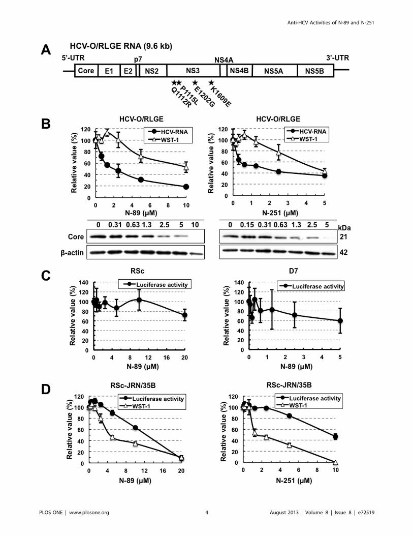
Figure 1. Anti-HCV activities of N-89 and N-251 detected in the OR6, ORL8, and ORL11 assays. (A) Structures of N-89 and N-251. (B)Effects of N-89 on genome-length HCV-RNA replication. OR6, ORL8, and ORL11 cells were treated with N-89 for 72 hrs, followed by RL assay (blackcircles in the upper panel) and WST-1 assay (open triangles in the upper panel). The relative value (%) calculated at each point, when the level in non-treated cells was assigned as 100%, is presented here. Data are expressed as the means6standard deviation of triplicate assays. Western blot analysisof the treated cells for the HCV Core was also performed (lower panel). b-actin was used as a control for the amount of protein loaded per lane. (C)Effects of N-251 on genome-length HCV-RNA replication. The RL assay, WST-1 assay, and Western blot analysis were performed as described in (B).doi:10.1371/journal.pone.0072519.g001
Anti-HCV Activities of N-89 and N-251
PLOS ONE | www.plosone.org 2 August 2013 | Volume 8 | Issue 8 | e72519
Materials and Methods
Cell CultureRSc and D7 cells were derived from the cell lines HuH-7 and
Li23, respectively, were cultured as described previously [20,29].
HuH-7-derived OR6 [13], AH1R [30], and 1B-4R [Ikeda et al.,
submitted] cells harboring genome-length HCV-RNA and HuH-
7-derived polyclonal sOR [31], and RSc-JRN/35B [Ikeda et al.,
submitted] cells harboring an HCV subgenomic replicon were
cultured with medium in the presence of G418 (0.3 mg/ml;
Geneticin, Invitrogen, Carlsbad, CA) as described previously [13].
Li23-derived ORL8 [20], ORL11 [20], 1B-4RL [Ikeda et al.,
submitted], and KAH5RL [Ikeda et al., submitted] cells harboring
genome-length HCV-RNA were maintained with medium in the
presence of G418 (0.3 mg/ml) as described previously [20]. Li23-
derived polyclonal sORL8 and sORL11 cells harboring an HCV
replicon, which were established by the transfection of ORN/3-
5B/QR,KE,SR RNA into the cured OL8 and OL11 cells,
respectively, were also cultured with medium in the presence of
G418 (0.3 mg/ml) as described previously [20]. Cured cells, from
which the HCV-RNA had been eliminated by IFN treatment,
were also maintained with medium in the absence of G418 as
described previously [13]. HCV-RNA-replicating cells possess the
G418-resistant phenotype because neomycin phosphotransferase
as a selective marker was produced by the efficient replication of
HCV-RNA. Therefore, when HCV-RNA is excluded from the
cells or when its level is decreased, the cells are killed in the
presence of G418.
ReagentsN-89 and N-251 were synthesized according to the methods
described previously [26–28]. RBV was kindly provided by
Yamasa (Chiba, Japan). Human IFN-a and vitamin E (VE) were
purchased from Sigma-Aldrich (St. Louis, MO). Cyclosporine A
(CsA) was purchased from Tokyo Chemical Industry (Tokyo,
Japan). Artemisinin was purchased from Alexis Biochemicals (San
Diego, CA).
RL AssayRL assay was performed as described previously [20,24].
Briefly, the cells were plated onto 24-well plates (26104 cells per
well) in triplicate and then treated with each reagent at several
concentrations for 72 hrs. After treatment, the cells were subjected
to luciferase assay using the RL assay system (Promega, Madison,
WI). The experiments were performed at least in triplicate. From
the assay results, the 50% effective concentration (EC50) of each
reagent was determined.
WST-1 Cell Proliferation AssayThe WST-1 cell proliferation assay was performed as described
previously [24]. Briefly, The cells were plated onto 96-well plates
(16103 cells per well) in triplicate and then treated with each
reagent at several concentrations for 72 hrs. After treatment, the
cells were subjected to the WST-1 cell proliferation assay (Takara
Bio, Otsu, Japan) according to the manufacturer’s protocol. This
assay is based on the enzymatic cleavage of the tetrazolium salt
WST-1 to formazan by cellular mitochondrial dehydrogenases
present in viable cells. Therefore, there are viable cells even if the
value of the WST-1 assay becomes zero. The experiments were
performed at least in triplicate. From the assay results, the 50%
cytotoxic concentration (CC50) of each reagent was determined.
Western Blot AnalysisThe preparation of cell lysates, sodium dodecyl sulfate-
polyacrylamide gel electrophoresis, and immunoblotting analysis
were performed as previously described [32]. The antibodies used
in this study were those against HCV Core (CP11; Institute of
Immunology, Tokyo, Japan), NS5B (a generous gift from Dr. M.
Kohara, Tokyo Metropolitan Institute of Medical Science), and b-actin (AC-15; Sigma-Aldrich) as the control for the amount of
protein loaded per lane.
Selective Index (SI)The SI value of each reagent was determined by dividing the
CC50 value by the EC50 value.
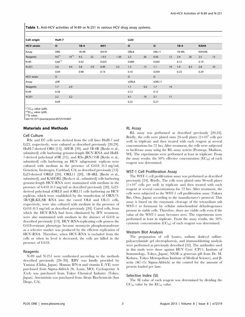
Table 1. Anti-HCV activities of N-89 or N-251 in various HCV drug assay systems.
Cell origin HuH-7 Li23
HCV strain O 1B-4 AH1 O O 1B-4 KAH5
Assay OR6 1B-4R AH1R ORL8 ORL11 1B-4RL KAH5RL
Reagents 9.0*1 14*3 9.3 22 .0.5 .20 2.3 26 0.56 12 2.4 20 2.5 13
N-89 0.66*2 0.42 0.025 0.089 0.045 0.12 0.19
N-251 3.0 4.4 3.8 3.9 0.49 3.5 1.3 13 1.1 19 1.9 8.3 2.8 10
0.69 0.98 0.14 0.10 0.059 0.23 0.29
HCV strain O O O
Assay sOR sORL8 sORL11
Reagents 1.7 2.9 1.1 9.2 1.7 14
N-89 0.58 0.12 0.12
N-251 2.2 3.2 4.1 19 3.1 11
0.69 0.22 0.27
*1CC50 value (mM),*2EC50 value (mM),*3SI value.doi:10.1371/journal.pone.0072519.t001
Anti-HCV Activities of N-89 and N-251
PLOS ONE | www.plosone.org 3 August 2013 | Volume 8 | Issue 8 | e72519
Anti-HCV Activities of N-89 and N-251
PLOS ONE | www.plosone.org 4 August 2013 | Volume 8 | Issue 8 | e72519

Quantitative RT-PCR AnalysisThe RNAs from HCV-RNA replicating cell lines were prepared
with an RNeasy extraction kit (Qiagen). The quantitative RT-
PCR analysis for HCV-RNA was performed using a real-time
LightCycler PCR (Roche Diagnostics, Basel, Switzerland) as
described previously [13,20].
HCV InfectionHCV infection was performed as described previously [29]. RSc
and D7 cells were inoculated with supernatant from RSc cells
replicating JR/C5B/BX-2 [29].
Statistical AnalysisDetermination of the significance of differences among groups
was assessed using the Student’s t-test. P,0.05 was considered
significant.
Results
Preclinical Antimalarial Drugs, N-89 and N-251, ShowedPotent Anti-HCV Activities in Both HuH-7- and Li23-derived Genome-length HCV-RNA-replicating CellsRecently we demonstrated that plural HCV assay systems
developed using both HuH-7 and Li23 cell lines or HCV strains
belonging to genotype 1b are required for the objective evaluation
of anti-HCV candidates [24]. In the present work, we used our
previously developed HCV assay systems to evaluate preclinical
antimalarial drugs (N-89 and N-251). N-89 (1,2,6,7-Tetraoxaspir-
o[7.11]nonadecane) is a chemically synthesized endoperoxide
compound (Fig. 1A) with potent antimalarial activity against
Plasmodium falciparum in vitro and Plasmodium berghei in vivo, and it
shows low levels of cytotoxicity in mice and rats (50% lethal dose:
.2000 mg/kg) [26,33,34]. N-251 (6-(1,2,6,7-tetraoxaspiro[7.11]-
nonadec-4-yl)hexan-1-ol), which bears a functional side chain
hydroxyl group that allows derivatization, is synthesized by
replacing the hydrogen at C-4 of N-89 with hexanol (Fig. 1A),
and it is as potent as N-89 against malaria parasites [27,28]. We
first evaluated the anti-HCV activities of N-89 and N-251 using
HuH-7-derived OR6 and Li23-derived ORL8 and ORL11 assay
systems. The results revealed that both N-89 and N-251 possessed
strong anti-HCV activities (Fig. 1B and C). The EC50 and SI
values of N-89 in each assay were calculated (EC50 0.66 mM, SI 14
in OR6 assay; EC50 0.089 mM, SI 26 in ORL8 assay; EC50
0.045 mM, SI 12 in ORL11 assay) (Table 1), and the anti-HCV
activity of N-251 was found to be as potent as that of N-89
(Table 1). The anti-HCV activities of N-89 and N-251 were
confirmed by Western blot analysis of HCV Core (Fig. 1B and C).
To further evaluate the activities of N-89 and N-251, as additional
assay systems, we used HuH-7-derived 1B-4R (1B-4 strain [31] of
Figure 2. Characterization of anti-HCV activities of N-89 and N-251. (A) Schematic gene organization of authentic HCV-RNA (HCV-O/RLGE).The positions of four adaptive mutations - Q1112R, P1115L, E1202G, and K1609E - are indicated by a black star. (B) N-89 and N-251 inhibited authenticHCV-RNA replication. The cells harboring HCV-O/RLGE RNA [19] were treated with N-89 (left panel) and N-251 (right panel) for 72 hrs, followed byreal-time LightCycler PCR (black circles in the upper panel) and WST-1 assay (open triangles in the upper panel). The relative value (%) calculated ateach point, when the level in non-treated cells was assigned as 100%, is presented here. Data are expressed as the means6standard deviation oftriplicate assays. Western blot analysis (lower panels) was performed as described in Fig. 1B. (C) N-89 did not inhibit the HCV-JFH-1 replication. RSc(left panel) and D7 (right panel) cells were inoculated with supernatant from RSc cells replicating JR/C5B/BX-2 [29]. The RL assay was performed asdescribed in Fig. 1B. (D) N-89 (left panel) and N-251 (right panel) did not inhibit the replication of HCV-JFH-1 subgenomic replicon. The RL and WST-1assays were performed as described in Fig. 1B.doi:10.1371/journal.pone.0072519.g002
Figure 3. OR6 and ORL8 cells were cured by treatment with only N-89. The treated cells were divided into two plates with or without G418,and then cultured for 2 weeks. The left panels show the cells stained with Coomassie brilliant blue. The right panels show the results of Western blotanalysis of the treated and non-treated cells for HCV proteins. Western blot analysis was performed as described in Fig. 1B.doi:10.1371/journal.pone.0072519.g003
Anti-HCV Activities of N-89 and N-251
PLOS ONE | www.plosone.org 5 August 2013 | Volume 8 | Issue 8 | e72519
genotype 1b derived from an HCV-positive healthy carrier) [Ikeda
et al., submitted] and AH1R (an AH1 strain [35] of genotype 1b
derived from a patient with acute hepatitis C) [30], and Li23-
derived 1B-4RL (1B-4 strain [31]) and KAH5RL (KAH5 strain
[31] of genotype 1b derived from a patient with acute hepatitis C)
[Ikeda et al., submitted]. These assays also showed that N-89 and
N-251 possessed potent anti-HCV activities (Fig. S1A–D and
Table 1). It was noteworthy that N-89 exhibited the strongest anti-
HCV activity (EC50 0.025 mM; SI .20) in the AH1R assay (Fig.
S1A and Table 1). These results suggest that the anti-HCV activity
of N-89 or N-251 is not influenced by the cell line or HCV strain.
We next examined the activities of N-89 and N-251 using
polyclonal cell-based assay systems (HuH-7-derived sOR [31],
Li23-derived sORL8 and sORL11 [22]) that facilitate the
monitoring replication of HCV subgenomic replicon RNA. These
assays also showed that N-89 and N-251 possessed anti-HCV
activity with EC50 values of less than 1 mM (Fig. S1E-G and
Table 1). Taken together, these results indicate that the anti-HCV
activities of N-89 and N-251 are not dependent on the specific
cloned cell line or HCV structural proteins.
N-89 and N-251 Inhibited Authentic HCV-RNA ReplicationThe genome-length HCV-RNA used in the assay systems
described above contains three non-natural elements: RL,
neomycin phosphotransferase, and an internal ribosomal entry
site of encephalomyocarditis virus. To exclude the possibility that
the anti-HCV activity of N-89 or N-251 was due to the inhibition
of these three exogenous elements, we examined the anti-HCV
activities of N-89 and N-251 using the authentic 9.6 kb HCV-
RNA-replicating HCV-O/RLGE cells [19], which were devel-
oped by the introduction of in vitro synthesized HCV-O/RLGE
RNA (Fig. 2A) into OR6c cured cells. We could demonstrate by
quantitative RT-PCR and Western blot analyses that N-89 and N-
251 at the expected concentrations efficiently prevented HCV-
RNA replication and HCV Core expression in HCV-O/RLGE
cells in a dose-dependent manner, respectively (Fig. 2B). The EC50
and SI values of N-89 and N-251 in this assay were calculated as
follows each: EC50 2.0 mM and SI .5.0 in N-89; EC50 1.6 mMand SI 2.8 in N-251. To further confirm that N-89 or N-251 does
not inhibit the RL activity, we examined the direct effect of each
reagent by adding it along with substrate to the cell lysate in the
RL assay. No suppressive effects by N-89 and N-251 were
observed in either the OR6 assay (Fig. S2A) or the ORL8 assay
(Fig. S2B). These results indicate that the anti-HCV activities of N-
89 and N-251 were due to the inhibition of HCV-RNA itself, but
not to exogenous elements contained in the genome-length HCV-
RNA.
N-89 and N-251 did not Inhibit RNA Replication of HCV-JFH-1 StrainWe next examined whether N-89 and N-251 worked in an
HCV production system using HCV-JFH-1 strain (genotype 2a).
Unexpectedly, the results using the JFH-1 reporter assay systems
[29], which were recently developed using HuH-7-derived RSc
and Li23-derived D7 cells, revealed that both N-89 and N-251 did
not show anti-HCV activity for the HCV-JFH-1 strain (Fig. 2C,
Fig. S3). To clarify whether anti-HCV activity depends on the
difference of genotype or assay model, we evaluated the activities
of N-89 and N-251 using RSc-JRN/35B [Ikeda et al., submitted]
cells harboring a subgenomic HCV-JFH-1 replicon as an
additional assay. The results revealed that N-89 and N-251 did
not show any anti-HCV activities in this assay system either
(Fig. 2D). Although the relative value of WST-1 almost became
zero when RSc-JRN/35B cells were treated with 10 mM of N-251,
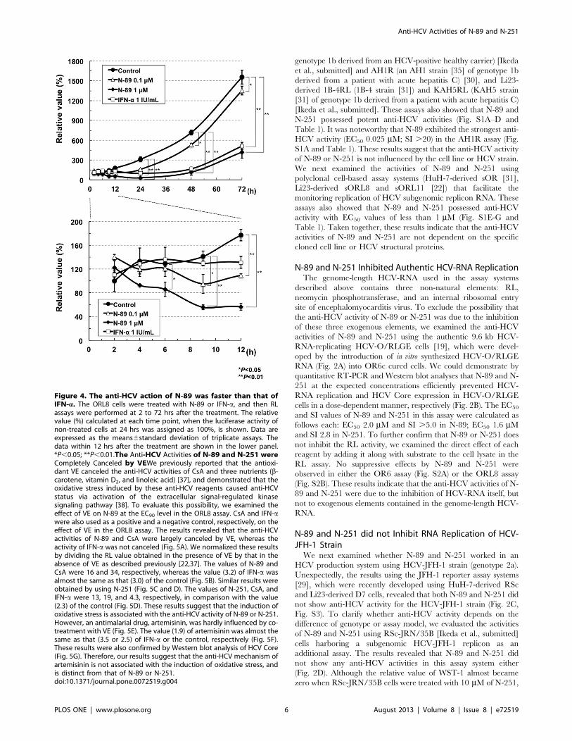
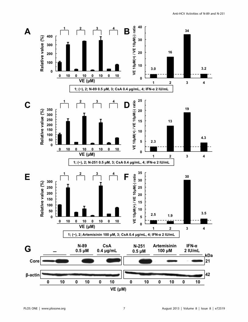
Figure 4. The anti-HCV action of N-89 was faster than that ofIFN-a. The ORL8 cells were treated with N-89 or IFN-a, and then RLassays were performed at 2 to 72 hrs after the treatment. The relativevalue (%) calculated at each time point, when the luciferase activity ofnon-treated cells at 24 hrs was assigned as 100%, is shown. Data areexpressed as the means6standard deviation of triplicate assays. Thedata within 12 hrs after the treatment are shown in the lower panel.*P,0.05; **P,0.01.The Anti-HCV Activities of N-89 and N-251 wereCompletely Canceled by VEWe previously reported that the antioxi-dant VE canceled the anti-HCV activities of CsA and three nutrients (b-carotene, vitamin D2, and linoleic acid) [37], and demonstrated that theoxidative stress induced by these anti-HCV reagents caused anti-HCVstatus via activation of the extracellular signal-regulated kinasesignaling pathway [38]. To evaluate this possibility, we examined theeffect of VE on N-89 at the EC90 level in the ORL8 assay. CsA and IFN-awere also used as a positive and a negative control, respectively, on theeffect of VE in the ORL8 assay. The results revealed that the anti-HCVactivities of N-89 and CsA were largely canceled by VE, whereas theactivity of IFN-a was not canceled (Fig. 5A). We normalized these resultsby dividing the RL value obtained in the presence of VE by that in theabsence of VE as described previously [22,37]. The values of N-89 andCsA were 16 and 34, respectively, whereas the value (3.2) of IFN-a wasalmost the same as that (3.0) of the control (Fig. 5B). Similar results wereobtained by using N-251 (Fig. 5C and D). The values of N-251, CsA, andIFN-a were 13, 19, and 4.3, respectively, in comparison with the value(2.3) of the control (Fig. 5D). These results suggest that the induction ofoxidative stress is associated with the anti-HCV activity of N-89 or N-251.However, an antimalarial drug, artemisinin, was hardly influenced by co-treatment with VE (Fig. 5E). The value (1.9) of artemisinin was almost thesame as that (3.5 or 2.5) of IFN-a or the control, respectively (Fig. 5F).These results were also confirmed by Western blot analysis of HCV Core(Fig. 5G). Therefore, our results suggest that the anti-HCV mechanism ofartemisinin is not associated with the induction of oxidative stress, andis distinct from that of N-89 or N-251.doi:10.1371/journal.pone.0072519.g004
Anti-HCV Activities of N-89 and N-251
PLOS ONE | www.plosone.org 6 August 2013 | Volume 8 | Issue 8 | e72519
Anti-HCV Activities of N-89 and N-251
PLOS ONE | www.plosone.org 7 August 2013 | Volume 8 | Issue 8 | e72519

cell counting after trypan blue dye treatment revealed that
approximately 30% of the cells were viable (data not shown).
These results suggest that the inhibitory effect of N-89 or N-251 on
HCV-RNA replication may depend on genotype 1b or not work
for only JFH-1 strain.
OR6 and ORL8 Cells were Cured by Treatment with onlyN-89To date, IFN-a alone or IFN-c alone has generally been used to
prepare cured cells from the cells harboring HCV-RNA [36].
Since we observed strong anti-HCV activity (.99% suppression)
at 8 mM of N-89 in OR6 cells or 1 mM of N-89 in ORL8 cells
without a decrease in cell viability (Fig. 1B), we expected that these
cells might be cured only by treatment with N-89. Accordingly,
OR6 and ORL8 cells were treated with 8 mM and 1 mM of N-89,
respectively, in the absence of G418. The treatment was continued
for 3 weeks with the addition of N-89 at 4-day intervals. All of the
treated cells were dead when cultured in the presence of G418 for
an additional two weeks, whereas the treated cells proliferated
efficiently in the absence of G418 (Fig. 3), suggesting that OR6
and ORL8 cells are cured by monotherapy with N-89. This
suggestion was confirmed by Western blot analysis (Fig. 3). These
results indicate that N-89 is a strong anti-HCV reagent, which can
be used to prepare cured cells by treatment at low concentration.
Comparative Time Course Assay of the Anti-HCVActivities of N-89 and IFN-aWe next performed a time course assay (2 to 72 hrs after
treatment) in the case of ORL8 cells treated with N-89 (0.1 mM or
1 mM) or IFN-a (1 IU/ml; corresponding to approximately EC80).
ORL8 cells treated with IFN-a (1 IU/ml) and N-89 (1 mM) had
almost the same anti-HCV kinetics over the first 24 hrs after
treatment (upper panel of Fig. 4); however, within the first 12 hrs
after treatment N-89-treated ORL8 cells had more rapid anti-
HCV kinetics than did the IFN-a-treated cells (lower panel of
Fig. 4). N-89 at concentrations of 0.1 mM and 1 mM led to
Figure 5. The anti-HCV activity of N-89 or N-251 was canceled by addition of VE. Effect of VE on the anti-HCV activity of N-89 (A), N-251 (C),Artemisinin (E), CsA, or IFN-a at the expected EC90. ORL8 cells were treated with control medium (2), N-89, CsA, or IFN-a in either the absence orpresence of VE for 72 hrs. After treatment, an RL assay of harvested ORL8 cell samples was performed. (B, D, and F) The ratio of RL activity in thepresence of VE to the RL activity in the absence of VE. The above ratio was calculated from the data of (A, C, and E). The horizontal line indicates thepromoting effect of VE alone on HCV-RNA replication as a baseline. (G) Western blot analysis was performed as described in Fig. 1B.doi:10.1371/journal.pone.0072519.g005
Figure 6. Synergistic anti-HCV effects of N-89 or N-251 in combination with IFN-a and/or RBV on HCV-RNA replication in ORL8 cells.Open symbols in the broken lines show the values expected as an additive anti-HCV effect and closed symbols in the solid lines show the valuesobtained by the ORL8 assay. ORL8 cells were treated with N-89 (upper panel) or N-251 (lower panel) in combination with IFN-a (A), RBV (B), or IFN-aand RBV (C) for 72 hrs and subjected to RL assay.doi:10.1371/journal.pone.0072519.g006
Anti-HCV Activities of N-89 and N-251
PLOS ONE | www.plosone.org 8 August 2013 | Volume 8 | Issue 8 | e72519

significantly decreased RL activity at 9 hrs and 6 hrs, respectively,
after treatment, whereas a decrease of RL activity in the cells
treated with 1 IU/ml of IFN-a began to be seen at 12 hrs after
treatment (lower panel of Fig. 4). These results suggest that the
action of N-89, and probably also that of N-251, is faster than that
of IFN-a, and the anti-HCV mechanism of N-89 is different from
that of IFN-a.
Synergistic Effect of Anti-HCV Activity by N-89 or N-251in Combination with IFN-a and/or RBVWe examined the anti-HCV activity of N-89 or N-251 in
combination with IFN-a using OR6 and ORL8 assay systems.
The results of the ORL8 assay revealed that the anti-HCV activity
of N-89 or N-251 in combination with IFN-a (more than 4 IU/ml)
was significantly stronger than that expected as an additive effect,
suggesting a synergistic effect of N-89 or N-251 and IFN-a(Fig. 6A). However, such an effect was not clear in the OR6 assay
(Fig. S4A). We recently demonstrated that 10 mM (a clinically
achievable concentration) of RBV efficiently inhibited HCV-RNA
replication in the ORL8 assay [22], and demonstrated that
adenosine kinase, which phosphorylates RBV to generate mono-
phosphorylated RBV possessing the inhibitory activity for inosine
monophosphate dehydrogenase, is an essential determinant of the
anti-HCV activity of RBV in cell culture [23]. Therefore, we next
examined the combination effect of RBV in the same way as IFN-
a using an ORL8 assay. We observed that the anti-HCV activity
of N-89 or N-251 in combination with RBV was significantly
stronger than that expected additively, suggesting that there was a
synergistic effect between N-89 or N-251 and RBV (Fig. 6B).
However, in the OR6 assay, we noticed that RBV showed an
additive anti-HCV effect in combination with N-89 or N-251 (Fig.
S4B). Since RBV has been shown to have little anti-HCV activity
in the OR6 assay system [22], some specific factor(s) in ORL8 cells
might contribute to the synergistic effect of N-89 or N-251 in
combination with RBV. Therefore, we further examined the effect
of N89 or N-251 in combination with both IFN-a and RBV using
an ORL8 assay. As expected, the anti-HCV activity of N-89 or N-
251 was synergistically enhanced in combination with both IFN-aand RBV in the ORL8 assay (Fig. 6C). On the other hand, in the
OR6 assay, a synergistic effect like that seen in the ORL8 assay
was not observed (Fig. S4C). We confirmed that any such
synergistic effect was not due to the cell toxic effect (Fig. S5).
Discussion
N-89 and its derivative N-251 are preclinical and promising
drugs possessing antimalarial activities in vitro and in vivo compa-
rable to those of artemisinin [26,27]. In the present study, using
cell-based HCV-RNA-replication assay systems, we found that N-
89 and N-251 possessed potent anti-HCV activities irrespective of
the cell lines and HCV strains of genotype 1b, and that they did
not work for JFH-1 strain of genotype 2a. Furthermore, We
demonstrated that the anti-HCV kinetics of N-89 was faster than
that of IFN-a, and that both N-89 and N-251 exhibited synergistic
effects in combination with IFN-a and/or RBV.
Along with the worldwide spread of HCV, high prevalence
areas of HCV infection have overlapped with endemic areas of
malaria infection [39,40]. It is also interesting that the liver is a
target organ for the replication of HCV and malaria. This fact
would again suggest that N-89 and N-251 target a common factor
that is required for the replication of HCV and malaria. At the
same time, N-89 and N-251 have become readily and cheaply
available due to their ease of synthesis [26,27]. Since we showed
that HCV-RNA-replicating cells were cured by monotherapy with
N-89, monotherapy with N-89 or N-251 would be simultaneously
effective for the diseases caused by malaria and HCV infection.
Furthermore, we recently showed that the blood concentration of
N-89 or N-251 reaches approximately 1 mM [Kim et al.,
unpublished data]. Since this concentration, which is equivalent
to the EC99 value of N-89 in the ORL8 assay, was used for the
preparation of cured cells, even monotherapy with N-89 would be
useful for patients with chronic hepatitis C.
In regard to the anti-HCV mechanism of N-89 and N-251, we
provided evidence that the anti-HCV activity of these reagents was
canceled by antioxidant VE, suggesting the induction of oxidative
stress. To identify the target factor(s) located downstream of ROS
production, we attempted microarray analysis using OR6 and
ORL8 cells treated with N-89. However, consequently, we failed
to obtain the candidate gene indicating the meaningful expression
level, although we identified several genes, which were commonly
upregulated or downregulated in the N-89-treated cells (Fig. S6).
On the other hand, it has been recently reported that Plasmodium
falciparum endoplasmic reticulum-resident calcium binding protein
is a possible target of N-89 and N-251 [41]. Therefore, this protein
may be involved in the anti-HCV activities of N-89 and N-251. To
clarify the factor(s), further analysis will be needed.
The synergistic anti-HCV effect of N-89 or N-251 in
combination with RBV rather than IFN-a is also interesting.
Using RBV-sensitive ORL8 cells, we recently clarified that the
anti-HCV mechanism of RBV was mediated by the inhibition of
IMPDH, which is required for HCV-RNA replication [22]. In
addition, since RBV is an important component of current IFN-
based therapies, including the recently developed triple therapy,
the use of N-89 or N-251 may further enhance the SVR rate
achieved with the current therapy. Furthermore, recent report
[42] that the lead-in four weeks of RBV treatment before starting a
standard course of PEG-IFN with RBV led a weak decrease of
viral replication (0.560.5 log10) is noteworthy. To evaluate this
possibility, we compared the SI values of N-89, N-251, RBV, and
CsA using the ORL8 assay system. The results revealed that the SI
values of N-89, N-251, RBV, and CsA were 26, 13, 10, and 15,
respectively, indicating that the anti-HCV activity of N-89 or N-
251 is equivalent to that of RBV or CsA. Since the treatment with
N-89/N-251 and RBV exhibits a synergistic effect, oral N-89 or
N-251 would be good compounds for inclusion in the current
triple therapy.
In conclusion, we found that two oral antimalarial drugs in the
preclinical stage of development (N-89 and N-251) exhibited
strong anti-HCV activities to genotype 1b. These compounds
would have potential as one component of a therapeutic regimen
based on combinations of HCV-specific inhibitors.
Supporting Information
Figure S1 Anti-HCV activities of N-89 and N-251detected in the several assay systems using genome-length HCV-RNA or HCV subgenomic replicon RNA. (A)Effects of N-89 and N-251 on genome-length HCV-RNA (AH1
strain of genotype 1b) replication in the AH1R assay. AH1R cells
were treated with N-89 or N-251 for 72 hrs, followed by RL assay
(black circles) and WST-1 assay (open triangles). The relative value
(%) calculated at each point, when the level in non-treated cells
was assigned as 100%, is presented here. Data are expressed as the
means 6 standard deviation of triplicate assays. (B) Effects of N-89
and N-251 on genome-length HCV-RNA (HCV 1B-4 strain of
genotype 1b) replication in the 1B-4R assay. The RL assay and
WST-1 assay were performed as described in (A). (C) Effects of N-
89 and N-251 on genome-length HCV-RNA (HCV 1B-4 strain of
Anti-HCV Activities of N-89 and N-251
PLOS ONE | www.plosone.org 9 August 2013 | Volume 8 | Issue 8 | e72519

genotype 1b) replication in the 1B-4RL assay. The RL assay and
WST-1 assay were performed as described in (A). (D) Effects of N-
89 and N-251 on genome-length HCV-RNA (HCV KAH5 strain
of genotype 1b) replication in the KAH5RL assay. The RL assay
and WST-1 assay were performed as described in (A). (E) Effects of
N-89 and N-251 on HCV subgenomic replicon RNA (HCV O
strain of genotype) replication in the sOR assay. The RL assay and
WST-1 assay were performed as described in (A). (F) Effects of N-
89 and N-251 on HCV subgenomic replicon RNA (HCV O strain
of genotype 1b) replication in the sORL8 assay. The RL assay and
WST-1 assay were performed as described in (A). (G) Effects of N-
89 and N-251 on HCV subgenomic replicon RNA (HCV O strain
of genotype 1b) replication in the sORL11 assay. The RL assay
and WST-1 assay were performed as described in (A).
(TIF)
Figure S2 No inhibition of RL activity by N-89 or N-251.(A) N-89 and N-251 did not inhibit the RL activity in the OR6 cell
lysate. N-89 or N-251 was added to the OR6 cell lysate, and then
an RL assay was performed. (B) N-89 and N-251 did not inhibit
the RL activity in the ORL8 cell lysate. N-89 or N-251 was added
to the ORL8 cell lysate, and then an RL assay was performed.
(TIF)
Figure S3 N-251 did not inhibit the HCV-JFH-1 replica-tion. RSc and D7 cells were inoculated with supernatant from
RSc cells replicating JR/C5B/BX-2 [2]. The RL assay was
performed as described in Fig. S1A.
(TIF)
Figure S4 Anti-HCV effects of N-89 or N-251 in combi-nation with IFN-a and/or RBV on HCV-RNA replicationin OR6 cells. Open symbols in the broken lines show the values
expected as an additive anti-HCV effect and closed symbols in the
solid lines show the values obtained by the OR6 assay. (A) Effect of
N-89 or N-251 in combination with IFN-a on OR6 assay. OR6
cells were treated with N-89 (upper panel) or N-251 (lower panel)
in combination with IFN-a for 72 hrs and subjected to RL assay.
(B) Effect of N-89 or N-251 in combination with RBV on OR6
assay. OR6 cells were treated with N-89 (upper panel) or N-251
(lower panel) in combination with RBV for 72 hrs and subjected to
RL assay. (C) Effect of N-89 or N-251 in combination with IFN-aand RBV on OR6 assay. OR6 cells were treated with N-89 (upper
panel) or N-251 (lower panel) in combination with IFN-a and
RBV for 72 hrs and subjected to RL assay.
(TIF)
Figure S5 Effects of N-89 or N-251 in combination withIFN-a and/or RBV on the growth of ORL8 or OR6 cells.ORL8 cells (A, B) or OR6 cells (C, D) were treated with N-89 (A,
C) or N-251 (B, D) in combination with IFN-a for 72 hrs and
subjected to the cell counting. The cell counting was carried out as
described in the Supporting Materials and methods.
(TIF)
Figure S6 Selection of genes whose expression levelswere commonly upregulated or downregulated in the N-89-treated OR6 and ORL8 cells. (A) Genes whose expression
levels were upregulated at ratios of more than 2 in the case of
OR6(2) versus OR6(N-89) or ORL8(2) versus ORL8(N-89) were
selected. 4 genes upregulated commonly in the N-89-treated cells
were listed. (B) Genes whose expression levels were downregulated
at ratios of less than 0.5 in the case of OR6(2) versus OR6(N-89)
or ORL8(2) versus ORL8(N-89) were selected. 5 genes down-
regulated commonly in the N-89-treated cells were listed.
(TIF)
Text S1.
(DOC)
Acknowledgments
We thank Yoshimi Kawae for her technical assistances. We also thank Dr.
Hiroyuki Doi (Okayama University, Japan) for his helpful suggestions.
Author Contributions
Conceived and designed the experiments: YU NK. Performed the
experiments: YU MT. Analyzed the data: YU NK. Contributed
reagents/materials/analysis tools: KM HD TW HSK AS YW MI. Wrote
the paper: YU NK.
References
1. Thomas DL (2000) Hepatitis C epidemiology. Curr Top Microbiol Immunol
242: 25–41.
2. Kato N (2001) Molecular virology of hepatitis C virus. Acta Med Okayama 55:
133–159.
3. Kato N, Hijikata M, Ootsuyama Y, Nakagawa M, Ohkoshi S, et al. (1990)
Molecular cloning of the human hepatitis C virus genome from Japanese
patients with non-A, non-B hepatitis. Proc Natl Acad Sci U S A 87: 9524–9528.
4. Chevaliez S, Pawlotsky JM (2007) Interferon-based therapy of hepatitis C. Adv
Drug Deliv Rev 59: 1222–1241.
5. Ghany MG, Nelson DR, Strader DB, Thomas DL, Seeff LB (2011) An update
on treatment of genotype 1 chronic hepatitis C virus infection: 2011 practice
guideline by the American Association for the Study of Liver Diseases.
Hepatology 54: 1433–1444.
6. Jacobson IM, McHutchison JG, Dusheiko G, Di Bisceglie AM, Reddy KR, et al.
(2011) Telaprevir for previously untreated chronic hepatitis C virus infection.
N Engl J Med 364: 2405–2416.
7. Poordad F, McCone J Jr, Bacon BR, Bruno S, Manns MP, et al. (2011)
Boceprevir for untreated chronic HCV genotype 1 infection. N Engl J Med 364:
1195–1206.
8. Reesink HW, Zeuzem S, Weegink CJ, Forestier N, van Vliet A, et al. (2006)
Rapid decline of viral RNA in hepatitis C patients treated with VX-950: a phase
Ib, placebo-controlled, randomized study. Gastroenterology 131: 997–1002.
9. Susser S, Welsch C, Wang Y, Zettler M, Domingues FS, et al. (2009)
Characterization of resistance to the protease inhibitor boceprevir in hepatitis C
virus-infected patients. Hepatology 50: 1709–1718.
10. Rosen HR (2011) Clinical practice. Chronic hepatitis C infection. N Engl J Med
364: 2429–2438.
11. Pawlotsky JM (2011) Treatment failure and resistance with direct-acting antiviral
drugs against hepatitis C virus. Hepatology 53: 1742–1751.
12. Bartenschlager R, Sparacio S (2007) Hepatitis C virus molecular clones and their
replication capacity in vivo and in cell culture. Virus Res 127: 195–207.
13. Ikeda M, Abe K, Dansako H, Nakamura T, Naka K, et al. (2005) Efficient
replication of a full-length hepatitis C virus genome, strain O, in cell culture, and
development of a luciferase reporter system. Biochem Biophys Res Commun
329: 1350–1359.
14. Ikeda M, Kato N (2007) Modulation of host metabolism as a target of new
antivirals. Adv Drug Deliv Rev 59: 1277–1289.
15. Moradpour D, Penin F, Rice CM (2007) Replication of hepatitis C virus. Nat
Rev Microbiol 5: 453–463.
16. Naka K, Ikeda M, Abe K, Dansako H, Kato N (2005) Mizoribine inhibits
hepatitis C virus RNA replication: effect of combination with interferon-alpha.
Biochem Biophys Res Commun 330: 871–879.
17. Ikeda M, Abe K, Yamada M, Dansako H, Naka K, et al. (2006) Different anti-
HCV profiles of statins and their potential for combination therapy with
interferon. Hepatology 44: 117–125.
18. Nozaki A, Morimoto M, Kondo M, Oshima T, Numata K, et al. (2010)
Hydroxyurea as an inhibitor of hepatitis C virus RNA replication. Arch Virol
155: 601–605.
19. Ikeda M, Kawai Y, Mori K, Yano M, Abe K, et al. (2011) Anti-ulcer agent
teprenone inhibits hepatitis C virus replication: potential treatment for hepatitis
C. Liver Int 31: 871–880.
20. Kato N, Mori K, Abe K, Dansako H, Kuroki M, et al. (2009) Efficient
replication systems for hepatitis C virus using a new human hepatoma cell line.
Virus Res 146: 41–50.
21. Mori K, Ikeda M, Ariumi Y, Kato N (2010) Gene expression profile of Li23, a
new human hepatoma cell line that enables robust hepatitis C virus replication:
Comparison with HuH-7 and other hepatic cell lines. Hepatol Res 40: 1248–
1253.
Anti-HCV Activities of N-89 and N-251
PLOS ONE | www.plosone.org 10 August 2013 | Volume 8 | Issue 8 | e72519

22. Mori K, Ikeda M, Ariumi Y, Dansako H, Wakita T, et al. (2011) Mechanism of
action of ribavirin in a novel hepatitis C virus replication cell system. Virus Res
157: 61–70.
23. Mori K, Hiraoka O, Ikeda M, Ariumi Y, Hiramoto A, et al. (2013) Adenosine
kinase is a key determinant for the anti-HCV activity of ribavirin. Hepatology in
press.
24. Ueda Y, Mori K, Ariumi Y, Ikeda M, Kato N (2011) Plural assay systems
derived from different cell lines and hepatitis C virus strains are required for the
objective evaluation of anti-hepatitis C virus reagents. Biochem Biophys Res
Commun 409: 663–668.
25. Paeshuyse J, Coelmont L, Vliegen I, Van Hemel J, Vandenkerckhove J, et al.
(2006) Hemin potentiates the anti-hepatitis C virus activity of the antimalarial
drug artemisinin. Biochem Biophys Res Commun 348: 139–144.
26. Kim HS, Nagai Y, Ono K, Begum K, Wataya Y, et al. (2001) Synthesis and
antimalarial activity of novel medium-sized 1,2,4,5-tetraoxacycloalkanes. J Med
Chem 44: 2357–2361.
27. Sato A, Hiramoto A, Morita M, Matsumoto M, Komich Y, et al. (2011)
Antimalarial activity of endoperoxide compound 6-(1,2,6,7-tetraoxaspiro[7.11]-
nonadec-4-yl)hexan-1-ol. Parasitol Int 60: 270–273.
28. Sato A, Kawai S, Hiramoto A, Morita M, Tanigawa N, et al. (2011)
Antimalarial activity of 6-(1,2,6,7-tetraoxaspiro[7.11]nonadec-4-yl)hexan-1-ol
(N-251) and its carboxylic acid derivatives. Parasitol Int 60: 488–492.
29. Takeda M, Ikeda M, Ariumi Y, Wakita T, Kato N (2012) Development of
hepatitis C virus production reporter-assay systems using two different hepatoma
cell lines. J Gen Virol 93: 1422–1431.
30. Mori K, Ueda Y, Ariumi Y, Dansako H, Ikeda M, et al. (2012) Development of
a drug assay system with hepatitis C virus genome derived from a patient with
acute hepatitis C. Virus Genes 44: 374–381.
31. Nishimura G, Ikeda M, Mori K, Nakazawa T, Ariumi Y, et al. (2009) Replicons
from genotype 1b HCV-positive sera exhibit diverse sensitivities to anti-HCV
reagents. Antiviral Res 82: 42–50.
32. Kato N, Sugiyama K, Namba K, Dansako H, Nakamura T, et al. (2003)
Establishment of a hepatitis C virus subgenomic replicon derived from humanhepatocytes infected in vitro. Biochem Biophys Res Commun 306: 756–766.
33. Aly NS, Hiramoto A, Sanai H, Hiraoka O, Hiramoto K, et al. (2007) Proteome
analysis of new antimalarial endoperoxide against Plasmodium falciparum.Parasitol Res 100: 1119–1124.
34. Kim HS, Begum K, Ogura N, Wataya Y, Nonami Y, et al. (2003) Antimalarialactivity of novel 1,2,5,6-tetraoxacycloalkanes and 1,2,5-trioxacycloalkanes. J Med
Chem 46: 1957–1961.
35. Mori K, Abe K, Dansako H, Ariumi Y, Ikeda M, et al. (2008) New efficientreplication system with hepatitis C virus genome derived from a patient with
acute hepatitis C. Biochem Biophys Res Commun 371: 104–109.36. Abe K, Ikeda M, Dansako H, Naka K, Kato N (2007) Cell culture-adaptive NS3
mutations required for the robust replication of genome-length hepatitis C virusRNA. Virus Res 125: 88–97.
37. Yano M, Ikeda M, Abe K, Dansako H, Ohkoshi S, et al. (2007) Comprehensive
analysis of the effects of ordinary nutrients on hepatitis C virus RNA replicationin cell culture. Antimicrob Agents Chemother 51: 2016–2027.
38. Yano M, Ikeda M, Abe K, Kawai Y, Kuroki M, et al. (2009) Oxidative stressinduces anti-hepatitis C virus status via the activation of extracellular signal-
regulated kinase. Hepatology 50: 678–688.
39. Feachem RG, Phillips AA, Hwang J, Cotter C, Wielgosz B, et al. (2010)Shrinking the malaria map: progress and prospects. Lancet 376: 1566–1578.
40. Shepard CW, Finelli L, Alter MJ (2005) Global epidemiology of hepatitis C virusinfection. Lancet Infect Dis 5: 558–567.
41. Morita M, Sanai H, Hiramoto A, Sato A, Hiraoka O, et al. (2012) Plasmodiumfalciparum endoplasmic reticulum-resident calcium binding protein is a possible
target of synthetic antimalarial endoperoxides, N-89 and N-251. J Proteome Res
11: 5704–5711.42. Rotman Y, Noureddin M, Feld JJ, Guedj J, Witthaus M, et al. (2013) Effect of
ribavirin on viral kinetics and liver gene expression in chronic hepatitis C. Gut inpress.
Anti-HCV Activities of N-89 and N-251
PLOS ONE | www.plosone.org 11 August 2013 | Volume 8 | Issue 8 | e72519