new roles for cysteine and transsulfuration enzymes ... roles for cysteine and...new roles for...
TRANSCRIPT

September 2004: 348–353
New Roles for Cysteine and Transsulfuration Enzymes:Production of H2S, A Neuromodulator and Smooth MuscleRelaxantJohn E. Dominy, B.S., and Martha H. Stipanuk, Ph.D.
The enzymes of the transsulfuration pathwayalso have the capacity to catalyze the desulf-hydration of cysteine. Recent studies demon-strate a role of the transsulfuration enzymes,cystathionine �-lyase and cystathionine �-syn-thase, in catalyzing the desulfhydration of cys-teine in brain and smooth muscle. The H2S pro-duced from cysteine functions as aneuromodulator and smooth muscle relaxant. Inglutamatergic neurons, the production of H2S bycystathionine �-synthase enhances N-methyl-D-aspartate (NMDA) receptor-mediated currents. Insmooth muscle cells, H2S produced by cystathi-onine �-lyase enhances the outward flux of po-tassium by opening potassium channels, leadingto hyperpolarization of membrane potential andsmooth muscle relaxation.Key words: brain, calmodulin, cysteine, cystathi-onine �-lyase, cystathionine �-synthase© 2004 International Life Sciences Institute
doi: 10.1301/nr.2004.sept.348–353
Introduction
The nutritional essentiality of the sulfur amino acids iswell established, and the current adult estimated av-erage requirement (EAR) and recommended daily al-lowance (RDA) for methionine plus cysteine are ap-proximately 15 mg kg�1 d�1 and 19 mg kg�1 d�1,respectively.1 The requirement for methionine is ab-solute, but cysteine can be synthesized via the unidi-rectional transsulfuration pathway that allows methi-onine to serve as the source of sulfur for cysteinesynthesis. The enzymes that catalyze the transsulfura-tion pathway are cystathionine �-synthase (CBS),
which catalyzes the condensation of homocysteinewith serine to form cystathionine, and cystathionine�-lyase (CGL), which catalyzes the �-cleavage ofcystathionine to yield cysteine, �-ketobutyrate, andammonia. While the roles of cysteine as a precursorfor the synthesis of protein, glutathione, coenzyme A,taurine, and sulfate and the pathways for their produc-tion are well established, the role of cysteine as aprecursor of reduced sulfur and the pathways forsynthesis of reduced sulfur are only now being eluci-dated.
Since the 1950s, scientists have known that mam-malian tissues contain enzymes capable of catalyzingthe desulfhydration of cysteine. These enzymes in-clude CBS, CGL, and possibly aminotransferases inconjunction with 3-mercaptopyruvate sulfurtrans-ferase. Stipanuk and Beck2 attempted to assess therelative roles of these enzymes in cysteine desulfhy-dration in rat tissues, performing experiments both invitro and in vivo, and concluded that both CBS andCGL catalyzed cysteine desulfhydration under physi-ologic conditions.
The enzymes involved in cysteine desulfhydration,CBS and CGL, are the same enzymes that catalyze thetwo steps in the methionine transsulfuration pathway.Both enzymes also accept cysteine as a substrate, but thereaction mechanisms for cysteine desulfhydration aresomewhat unclear. CBS can catalyze a wide range of�,�-elimination/replacement reactions, which includethe substitution of the thiol group of cysteine with avariety of thiol compounds to form H2S and the corre-sponding thioether.3–5 It is thought that CGL catalyzes an�,�-disulfide elimination reaction that results in the pro-duction of pyruvate, ammonia, and thiocysteine. Thio-cysteine may then react with cysteine or other thiols toform H2S and cystine, or the corresponding disulfide.Thiocysteine may also decompose to elemental sulfur,which may then be reduced to H2S, or more likely, thepersulfide sulfur may be transferred to an acceptor suchas sulfite.6–8 The affinity of these enzymes for cysteine isrelatively low, and H2S production increases with con-
Dominy and Stipanuk are with Division of Nutri-tional Sciences, Cornell University, Ithaca, NY, 14853,USA.
348 Nutrition Reviews�, Vol. 62, No. 9

centrations of up to �150 mM cysteine in assay sys-tems.2 Thus, the amount of H2S produced by theseenzymes would be expected to increase with any in-crease in tissue cysteine concentration.
Although reduced sulfur may be used in the syn-thesis of molecules requiring a source of reducedsulfur, it has generally been assumed that the H2Sproduced by these pathways would be readily oxidizedto thiosulfate (inner sulfur), sulfite, and finally sulfate.The reactions involved in sulfide oxidation were elu-cidated by Koj et al.9 and Szczepkowski et al.10 in the1960s.
2 HS��2 O2 3 �SSO3��2�H2O
�SSO3��2 � 2 GSH3 HS� � HSO3
� � GSSG
SO3�2 � 1⁄2 O23 SO4
�2
This series of steps for sulfide oxidation to sulfatehave been validated by subsequent studies demonstrat-ing the GSH dependence of sulfate production, as wellas thiosulfate accumulation by hepatocytes depleted ofGSH with bromoheptane and/or buthionine sulfoxi-mine and incubated with a high concentration ofcysteine.11 About half of the cysteine catabolism in ratenterocytes or renal cortical tubules occurred via cys-teinesulfinate-independent pathways, as evidenced bythiosulfate formation and inhibition by propargylgly-cine.12,13
Studies of cysteine desulfhydration in the 1970s and1980s were followed by recognition that cysteine desulf-hydration did not account for a quantitatively largeproportion of cysteine catabolism in animals fed ade-quate levels of protein or sulfur amino acids.13–16 Themajor pathway for cysteine catabolism involves hepaticcysteine dioxygenase, which oxidizes the thiol group ofcysteine to form cysteinesulfinate. Cysteinesulfinate isfurther oxidized, and cysteine sulfur is excreted as eithertaurine or sulfate. The Km of cysteine dioxygenase isapproximately 0.4 mM, which allows cysteine dioxyge-nase to respond to changes in cysteine concentrationswithin the physiologic range. Furthermore, cysteine di-oxygenase concentration is robustly upregulated in re-sponse to increases in dietary protein or sulfur aminoacid intake, effectively preventing accumulation of exesscysteine in the body but conserving it when intake islow.15,17 By ensuring that tissue cysteine concentrationsremain below 1 �mol/g (usually below 0.1 �mol/g),cysteine dioxygenase limits the extent of H2S productionvia cysteine desulfhydration.
Recently, another line of work has brought the focusback to cysteine desulfhydration pathways. Beginning inthe mid-1990s, several studies demonstrated that H2S has
important physiologic functions and that its production isregulated. Furthermore, the role of CBS and CGL inregulated production of H2S from cysteine in the nervoussystem and in smooth muscle tissues has been revealed.
Endogenous levels of H2S are relatively high inbrain, with 1.57 � 0.04 �g/g (�0.3 nmol/mg protein)being measured in rat brain18 and 0.49 � 0.07 nmol/mgprotein in human frontal cortex.19 Several lines of evi-dence indicate that CBS is the enzyme responsible forH2S production in brain. First, inhibitors or activators ofCBS affected the production of H2S from cysteine bywhole brain preparations.20 Second, CBS activity, pro-tein immunoreactivity, and mRNA levels were relativelyhigh in various brain regions (hippocampus, cerebellum,cerebral cortex, and brain stem), whereas CGL activityand mRNA levels were low in cerebellum comparedwith other brain regions.21–23 Additionally, very lowlevels of H2S in the brains of patients with Alzhei-mer’s disease, compared with the brains of age-matched normal individuals, were accompanied byreduced CBS activity, an elevated level of Hcy, and areduced level of S-adenosylmethionine (SAM), anactivator of CBS.19
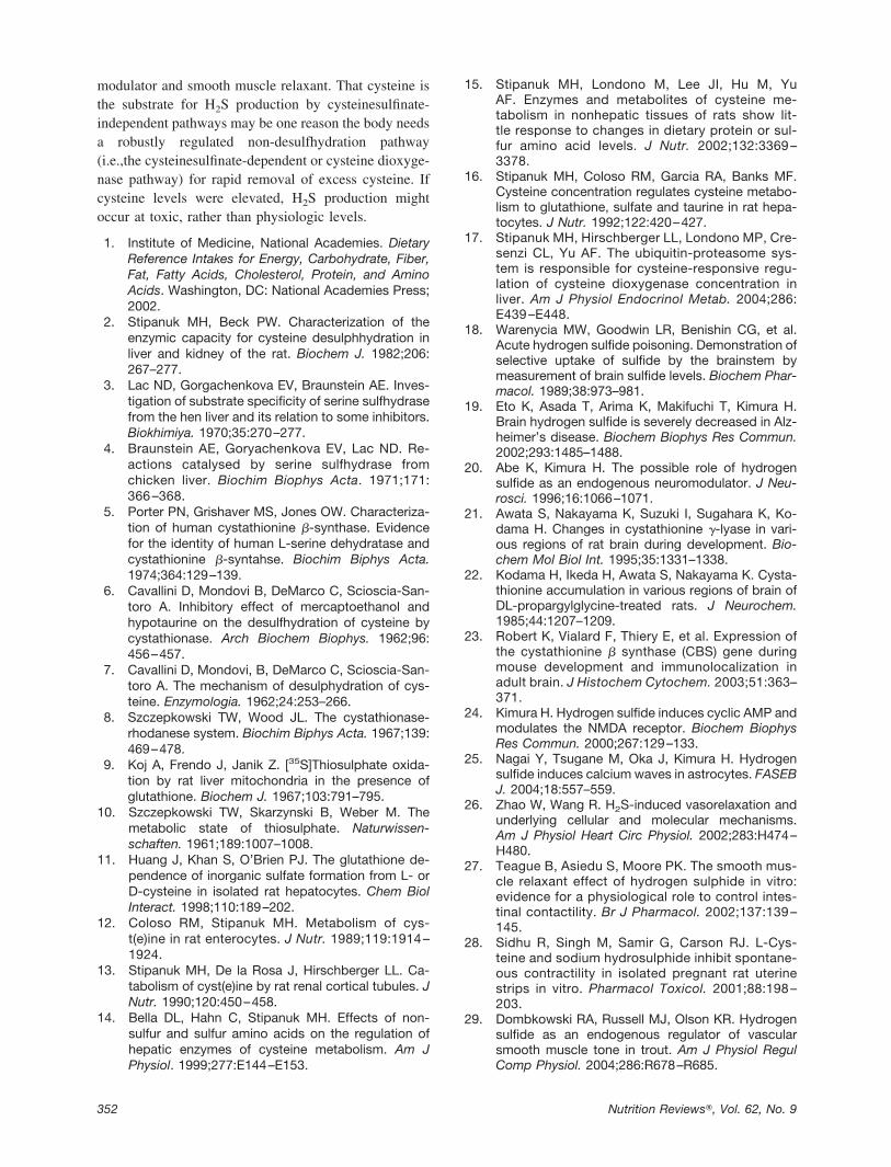
Physiologic concentrations of exogenously appliedH2S facilitate the induction of long-term potentiation(LTP) in rat hippocampal slices. Abe and Kimura20
showed that this enhancement of hippocampal LTP wasthe result of an enhancement of N-methyl-D-aspartate(NMDA) receptor-mediated currents in pyramidalcells of the CA1 layer. The mechanism underlying thisphenomenon is poorly understood. The prevailing the-ory is that enhancement of NMDA currents arises as asecondary result of H2S-induced alterations in intra-cellular cAMP metabolism (Figure 1). Indeed,Kimura24 demonstrated that sodium hydrosulfide(NaHS) increased cAMP concentrations in both pri-mary neuronal cultures and some transformed neuralcell lines.
An increase in cAMP levels in neurons could con-ceivably translate into alterations of NMDA channelconductance by means of protein kinase A (PKA) acti-vation and the subsequent phosphorylation of conservedPKA consensus sites found on NMDA receptor sub-units NMDAR1, NMDAR2A, and NMDAR2B. Em-ploying a Xenopus oocyte model that expressesNMDAR1 and NMDAR2A, Kimura24 showed thatNaHS (10 –30 �M) produced significant increases inintracellular cAMP and significant decreases in theonset of NMDA-induced membrane currents. The in-crease in cAMP and the reduction of activation laten-cies produced by NaHS could be prevented by anadenylyl cyclase-specific inhibitor.
Although these results suggest that changes incAMP concentrations could be involved in the ability of
349Nutrition Reviews�, Vol. 62, No. 9

H2S to modulate NMDA activity, further examination ofthe hypothesis using neuronal cultures and a combinationof adenylyl cyclase and PKA inhibitors needs to beconducted. Additional consideration should be given tothe participation of astrocytes in the neuromodulatoryproperties of H2S in situ. Accordingly, a recent study byNagai et al.25 showed that NaHS alone induced robust,propagatable increases in the intracellular Ca2� levels ofastrocytes from primary cultures and hippocampal slices.Co-cultured neurons, by contrast, showed no detectablechange in intracellular Ca2� following exposure toNaHS. The exact mechanism by which NaHS alteredintracellular Ca2� dynamics in astrocytes was not iden-tified. It is worth noting, however, that NaHS wasreported to increase the intracellular cAMP levels oftransformed astroglia.25 Whether Ca2� channel phos-phorylation by PKA is the cause of the dramatic
changes in the Ca2� levels of astrocytes exposed toNaHS is not known.
The effects of physiologic levels of exogenouslysupplied H2S on glutamatergic transmission throughNMDA receptors in vitro have led to the suggestion thatH2S is an endogenous gaseous neuromodulator. How-ever, definitive categorization of H2S as an endogenousneuromodulator requires proof that control of its synthe-sis is coordinated with neuronal activity. Although thereis no concrete evidence to indicate that this is the case,the localization of CBS expression within the soma andprocesses of many neurons hints at the possibility ofneuronal activity-mediated regulation.23 Future studiesremain to be conducted on the ability of glutamatergic-mediated changes in neuronal metabolic homeostasis,such as alterations in intracellular adenylate and calciumpools, to affect CBS activity.
Apart from its neuromodulatory properties, H2S mayalso function as an endogenous smooth muscle relaxantin a wide array of vertebrates. Zhao and Wang26 reportedthat H2S induced a concentration-dependent relaxation ofrat aortic tissues. Teague et al.27 similarly demonstrateda dose-related relaxation of isolated rabbit ileum and ratvas deferens. Additionally, Sidhu et al.28 demonstratedthat NaHS produced significant dose-dependent de-creases in the spontaneous contractility of uterine stripsfrom pregnant rats. Interestingly, the capacity for endog-enous synthesis of H2S in this tissue was hinted at by areduction in spontaneous contractility following preincu-bation with 1 mM cysteine. Even in species of fresh andsaltwater trout, H2S was shown to elicit dose-dependentrelaxation of isolated, pre-contracted branchial arteries.29
In vivo studies have supported the data gleaned fromwork done in vitro. Bolus intravenous injections of H2Sproduced transient, 12.5 to 29.8 mm Hg drops in meanarterial blood pressure in rats with no correspondingchange in heart rate—suggesting the exclusive involve-ment of arterial smooth muscle dilation.30 Given theblood levels of H2S reported for different strains of rat(10–50 �M), H2S may in fact be a tonic mediator ofvasoactivity.30,31
The in situ generation of H2S involved in peripheralsmooth muscle relaxation appears to be catalyzed pre-dominately by CGL. Inhibitors of CGL, but not of CBS,caused an increase in the contraction of guinea pig ileumin response to electrical stimulation.27 Hosoki et al.32
reported that CGL mRNA is expressed in the guinea pigileum, portal vein, and thoracic aorta, and inhibitorstudies suggested that the production of H2S in portalvein and thoracic aorta was catalyzed by CGL, whereasin ileum it was catalyzed by both CGL and CBS—whichwas also shown to be transcribed in this tissue. In intactrats, expression of CGL and its corresponding enzymeactivity in rat lung tissue were suppressed during hy-
Figure 1. A model combining the pathways responsible forH2S biosynthesis and its neuromodulatory effects within glu-tamatergic neurons. The membrane depolarization accompany-ing a glutamate-induced excitatory postsynaptic potential opensvoltage-gated calcium channels as well as NMDA channels (i).The associated influx of calcium (ii) raises intracellular calciumlevels and facilitates the activation of cystathionine �-synthase(CBS; iii). CBS catalyzes the production of H2S from cysteine(iv). H2S produced by CBS may remain in the neuron to exertautocrine effects, exit the cell down its concentration gradientand induce paracrine responses (v), or simply be inactivated bychemical and enzymatic means to sulfate (vi). The most notedbiologic effect of H2S in neurons, NMDA receptor modulation,may involve the activation of adenylyl cylase (vii). The conse-quent rise in intracellular cAMP (viii) activates protein kinaseA (PKA; ix), which in turn phosphorylates specific subunits ofNMDA receptors (x).
350 Nutrition Reviews�, Vol. 62, No. 9
poxic pulmonary hypertension, adding to evidence thatH2S production may be an important physiologic relaxorof smooth muscle.33
Within tissues that contain smooth muscle, it is notclear what cell types are contributing to the in situsynthesis of H2S. To date, it has been shown that CGL istranscribed in vascular smooth muscle cells but not theendothelia of rat arteries.30 Pharmacologic investigationswith CGL inhibitors and electrical field stimulation ofguinea pig ileum, on the other hand, indirectly suggestthat CGL may be expressed by intramural innervationwithin this tissue rather than smooth muscle cells.27
What bearing these differences in cell-specific expres-sion may have on the larger physiologic function ofH2S-induced relaxation remains unclear. In fact, there isno clear picture of the extent to which H2S contributes tosmooth muscle tone under physiologic conditions. Thisdeficit in understanding is due in part to how little isknown about the regulatory mechanisms that govern H2Sgeneration in peripheral tissues.
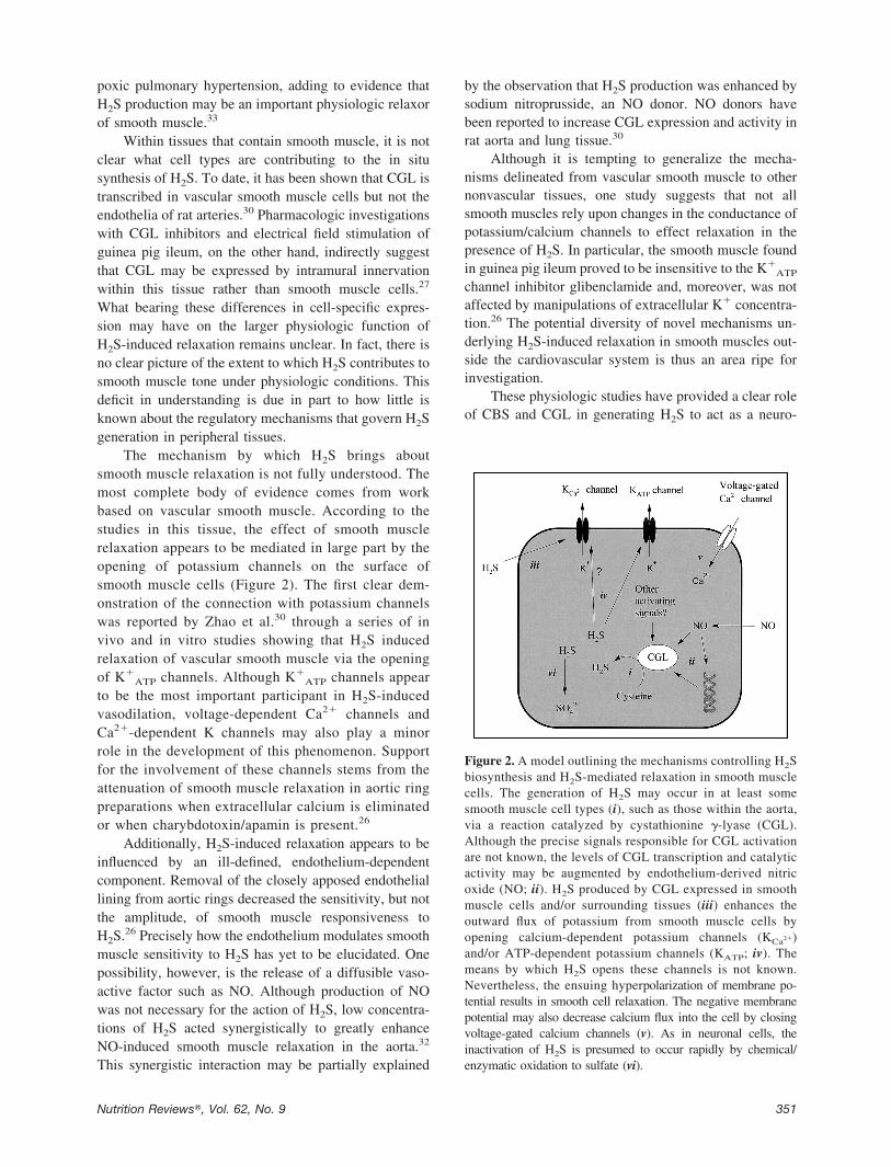
The mechanism by which H2S brings aboutsmooth muscle relaxation is not fully understood. Themost complete body of evidence comes from workbased on vascular smooth muscle. According to thestudies in this tissue, the effect of smooth musclerelaxation appears to be mediated in large part by theopening of potassium channels on the surface ofsmooth muscle cells (Figure 2). The first clear dem-onstration of the connection with potassium channelswas reported by Zhao et al.30 through a series of invivo and in vitro studies showing that H2S inducedrelaxation of vascular smooth muscle via the openingof K�
ATP channels. Although K�ATP channels appear
to be the most important participant in H2S-inducedvasodilation, voltage-dependent Ca2� channels andCa2�-dependent K channels may also play a minorrole in the development of this phenomenon. Supportfor the involvement of these channels stems from theattenuation of smooth muscle relaxation in aortic ringpreparations when extracellular calcium is eliminatedor when charybdotoxin/apamin is present.26
Additionally, H2S-induced relaxation appears to beinfluenced by an ill-defined, endothelium-dependentcomponent. Removal of the closely apposed endotheliallining from aortic rings decreased the sensitivity, but notthe amplitude, of smooth muscle responsiveness toH2S.26 Precisely how the endothelium modulates smoothmuscle sensitivity to H2S has yet to be elucidated. Onepossibility, however, is the release of a diffusible vaso-active factor such as NO. Although production of NOwas not necessary for the action of H2S, low concentra-tions of H2S acted synergistically to greatly enhanceNO-induced smooth muscle relaxation in the aorta.32
This synergistic interaction may be partially explained
by the observation that H2S production was enhanced bysodium nitroprusside, an NO donor. NO donors havebeen reported to increase CGL expression and activity inrat aorta and lung tissue.30
Although it is tempting to generalize the mecha-nisms delineated from vascular smooth muscle to othernonvascular tissues, one study suggests that not allsmooth muscles rely upon changes in the conductance ofpotassium/calcium channels to effect relaxation in thepresence of H2S. In particular, the smooth muscle foundin guinea pig ileum proved to be insensitive to the K�
ATP
channel inhibitor glibenclamide and, moreover, was notaffected by manipulations of extracellular K� concentra-tion.26 The potential diversity of novel mechanisms un-derlying H2S-induced relaxation in smooth muscles out-side the cardiovascular system is thus an area ripe forinvestigation.
These physiologic studies have provided a clear roleof CBS and CGL in generating H2S to act as a neuro-
Figure 2. A model outlining the mechanisms controlling H2Sbiosynthesis and H2S-mediated relaxation in smooth musclecells. The generation of H2S may occur in at least somesmooth muscle cell types (i), such as those within the aorta,via a reaction catalyzed by cystathionine �-lyase (CGL).Although the precise signals responsible for CGL activationare not known, the levels of CGL transcription and catalyticactivity may be augmented by endothelium-derived nitricoxide (NO; ii). H2S produced by CGL expressed in smoothmuscle cells and/or surrounding tissues (iii) enhances theoutward flux of potassium from smooth muscle cells byopening calcium-dependent potassium channels (KCa2�)and/or ATP-dependent potassium channels (KATP; iv). Themeans by which H2S opens these channels is not known.Nevertheless, the ensuing hyperpolarization of membrane po-tential results in smooth cell relaxation. The negative membranepotential may also decrease calcium flux into the cell by closingvoltage-gated calcium channels (v). As in neuronal cells, theinactivation of H2S is presumed to occur rapidly by chemical/enzymatic oxidation to sulfate (vi).
351Nutrition Reviews�, Vol. 62, No. 9
modulator and smooth muscle relaxant. That cysteine isthe substrate for H2S production by cysteinesulfinate-independent pathways may be one reason the body needsa robustly regulated non-desulfhydration pathway(i.e.,the cysteinesulfinate-dependent or cysteine dioxyge-nase pathway) for rapid removal of excess cysteine. Ifcysteine levels were elevated, H2S production mightoccur at toxic, rather than physiologic levels.
1. Institute of Medicine, National Academies. DietaryReference Intakes for Energy, Carbohydrate, Fiber,Fat, Fatty Acids, Cholesterol, Protein, and AminoAcids. Washington, DC: National Academies Press;2002.
2. Stipanuk MH, Beck PW. Characterization of theenzymic capacity for cysteine desulphhydration inliver and kidney of the rat. Biochem J. 1982;206:267–277.
3. Lac ND, Gorgachenkova EV, Braunstein AE. Inves-tigation of substrate specificity of serine sulfhydrasefrom the hen liver and its relation to some inhibitors.Biokhimiya. 1970;35:270–277.
4. Braunstein AE, Goryachenkova EV, Lac ND. Re-actions catalysed by serine sulfhydrase fromchicken liver. Biochim Biophys Acta. 1971;171:366 –368.
5. Porter PN, Grishaver MS, Jones OW. Characteriza-tion of human cystathionine �-synthase. Evidencefor the identity of human L-serine dehydratase andcystathionine �-syntahse. Biochim Biphys Acta.1974;364:129–139.
6. Cavallini D, Mondovi B, DeMarco C, Scioscia-San-toro A. Inhibitory effect of mercaptoethanol andhypotaurine on the desulfhydration of cysteine bycystathionase. Arch Biochem Biophys. 1962;96:456–457.
7. Cavallini D, Mondovi, B, DeMarco C, Scioscia-San-toro A. The mechanism of desulphydration of cys-teine. Enzymologia. 1962;24:253–266.
8. Szczepkowski TW, Wood JL. The cystathionase-rhodanese system. Biochim Biphys Acta. 1967;139:469–478.
9. Koj A, Frendo J, Janik Z. [35S]Thiosulphate oxida-tion by rat liver mitochondria in the presence ofglutathione. Biochem J. 1967;103:791–795.
10. Szczepkowski TW, Skarzynski B, Weber M. Themetabolic state of thiosulphate. Naturwissen-schaften. 1961;189:1007–1008.
11. Huang J, Khan S, O’Brien PJ. The glutathione de-pendence of inorganic sulfate formation from L- orD-cysteine in isolated rat hepatocytes. Chem BiolInteract. 1998;110:189–202.
12. Coloso RM, Stipanuk MH. Metabolism of cys-t(e)ine in rat enterocytes. J Nutr. 1989;119:1914 –1924.
13. Stipanuk MH, De la Rosa J, Hirschberger LL. Ca-tabolism of cyst(e)ine by rat renal cortical tubules. JNutr. 1990;120:450–458.
14. Bella DL, Hahn C, Stipanuk MH. Effects of non-sulfur and sulfur amino acids on the regulation ofhepatic enzymes of cysteine metabolism. Am JPhysiol. 1999;277:E144–E153.
15. Stipanuk MH, Londono M, Lee JI, Hu M, YuAF. Enzymes and metabolites of cysteine me-tabolism in nonhepatic tissues of rats show lit-tle response to changes in dietary protein or sul-fur amino acid levels. J Nutr. 2002;132:3369 –3378.
16. Stipanuk MH, Coloso RM, Garcia RA, Banks MF.Cysteine concentration regulates cysteine metabo-lism to glutathione, sulfate and taurine in rat hepa-tocytes. J Nutr. 1992;122:420–427.
17. Stipanuk MH, Hirschberger LL, Londono MP, Cre-senzi CL, Yu AF. The ubiquitin-proteasome sys-tem is responsible for cysteine-responsive regu-lation of cysteine dioxygenase concentration inliver. Am J Physiol Endocrinol Metab. 2004;286:E439 –E448.
18. Warenycia MW, Goodwin LR, Benishin CG, et al.Acute hydrogen sulfide poisoning. Demonstration ofselective uptake of sulfide by the brainstem bymeasurement of brain sulfide levels. Biochem Phar-macol. 1989;38:973–981.
19. Eto K, Asada T, Arima K, Makifuchi T, Kimura H.Brain hydrogen sulfide is severely decreased in Alz-heimer’s disease. Biochem Biophys Res Commun.2002;293:1485–1488.
20. Abe K, Kimura H. The possible role of hydrogensulfide as an endogenous neuromodulator. J Neu-rosci. 1996;16:1066–1071.
21. Awata S, Nakayama K, Suzuki I, Sugahara K, Ko-dama H. Changes in cystathionine �-lyase in vari-ous regions of rat brain during development. Bio-chem Mol Biol Int. 1995;35:1331–1338.
22. Kodama H, Ikeda H, Awata S, Nakayama K. Cysta-thionine accumulation in various regions of brain ofDL-propargylglycine-treated rats. J Neurochem.1985;44:1207–1209.
23. Robert K, Vialard F, Thiery E, et al. Expression ofthe cystathionine � synthase (CBS) gene duringmouse development and immunolocalization inadult brain. J Histochem Cytochem. 2003;51:363–371.
24. Kimura H. Hydrogen sulfide induces cyclic AMP andmodulates the NMDA receptor. Biochem BiophysRes Commun. 2000;267:129–133.
25. Nagai Y, Tsugane M, Oka J, Kimura H. Hydrogensulfide induces calcium waves in astrocytes. FASEBJ. 2004;18:557–559.
26. Zhao W, Wang R. H2S-induced vasorelaxation andunderlying cellular and molecular mechanisms.Am J Physiol Heart Circ Physiol. 2002;283:H474–H480.
27. Teague B, Asiedu S, Moore PK. The smooth mus-cle relaxant effect of hydrogen sulphide in vitro:evidence for a physiological role to control intes-tinal contactility. Br J Pharmacol. 2002;137:139 –145.
28. Sidhu R, Singh M, Samir G, Carson RJ. L-Cys-teine and sodium hydrosulphide inhibit spontane-ous contractility in isolated pregnant rat uterinestrips in vitro. Pharmacol Toxicol. 2001;88:198 –203.
29. Dombkowski RA, Russell MJ, Olson KR. Hydrogensulfide as an endogenous regulator of vascularsmooth muscle tone in trout. Am J Physiol RegulComp Physiol. 2004;286:R678–R685.
352 Nutrition Reviews�, Vol. 62, No. 9

30. Zhao W, Zhang J, Lu Y, Wang R. The vasorelaxanteffect of H2S as a novel endogenous gaseous KATP
channel opener. EMBO J. 2001;20:6008–6016.31. Mason J, Cardin CJ, Dennehy A. The role of sul-
phide and sulphide oxidation in the copper molyb-denum antagonism in rats and guinea pigs. Res VetSci. 1978;24:104–108.
32. Hosoki R, Matsuki N, Kimura H. The possible role of
hydrogen sulfide as an endogenous muscle relaxantin synergy with nitric oxide. Biochem Biophys ResCommun. 1997;237:528–531.
33. Chunyu Z, Janbao D, Dingfang B, Hui Y, Xiuying T,Chaoshu T. The regulatory effect of hydrogen sul-fide on hypoxic pulmonary hypertension in rats.Biochem Biophys Res Commun. 2003;302:810–816.
353Nutrition Reviews�, Vol. 62, No. 9
