new therapeutic approaches of mesenchymal stem cells
TRANSCRIPT

Janockova et al. J Biomed Sci (2021) 28:39 https://doi.org/10.1186/s12929-021-00736-4
REVIEW
New therapeutic approaches of mesenchymal stem cells-derived exosomesJana Janockova* , Lucia Slovinska, Denisa Harvanova, Timea Spakova and Jan Rosocha
Abstract
Mesenchymal stem cells (MSCs) have been demonstrated to have a great potential in the treatment of several diseases due to their differentiation and immunomodulatory capabilities and their ability to be easily cultured and manipulated. Recent investigations revealed that their therapeutic effect is largely mediated by the secretion of paracrine factors including exosomes. Exosomes reflect biophysical features of MSCs and are considered more effec-tive than MSCs themselves. Alternative approaches based on MSC-derived exosomes can offer appreciable promise in overcoming the limitations and practical challenges observed in cell-based therapy. Furthermore, MSC-derived exosomes may provide a potent therapeutic strategy for various diseases and are promising candidates for cell-based and cell-free regenerative medicine. This review briefly summarizes the development of MSCs as a treatment for human diseases as well as describes our current knowledge about exosomes: their biogenesis and molecular compo-sition, and how they exert their effects on target cells. Particularly, the therapeutic potential of MSC-derived exosomes in experimental models and recent clinical trials to evaluate their safety and efficacy are summarized in this study. Overall, this paper provides a current overview of exosomes as a new cell-free therapeutic agent.
Keywords: Mesenchymal stem cells, Exosomes, Cell-free therapy, Therapeutic potential
© The Author(s) 2021. Open Access This article is licensed under a Creative Commons Attribution 4.0 International License, which permits use, sharing, adaptation, distribution and reproduction in any medium or format, as long as you give appropriate credit to the original author(s) and the source, provide a link to the Creative Commons licence, and indicate if changes were made. The images or other third party material in this article are included in the article’s Creative Commons licence, unless indicated otherwise in a credit line to the material. If material is not included in the article’s Creative Commons licence and your intended use is not permitted by statutory regulation or exceeds the permitted use, you will need to obtain permission directly from the copyright holder. To view a copy of this licence, visit http:// creat iveco mmons. org/ licen ses/ by/4. 0/. The Creative Commons Public Domain Dedication waiver (http:// creat iveco mmons. org/ publi cdoma in/ zero/1. 0/) applies to the data made available in this article, unless otherwise stated in a credit line to the data.
BackgroundNowadays, multipotent mesenchymal stem cells (MSCs) have been extensively examined because of their usage in clinical trials. Their effective influence in cellular therapy and regenerative medicine is known for their strong immunosuppressive, immunomodulatory and regenerative activity [1, 2]. In addition, their considerable potential was demonstrated in the treatment of immune-mediated, inflammatory and degenerative diseases [3–9].
MSCs generally are multipotent, somatic progenitor/stem cells first isolated from adult bone marrow [10, 11] and successfully differentiated from marrow hematopoi-etic cells according to their adherent nature in in vitro cell lines and fibroblastic morphology. They are able to self-recover and retain variable differentiation potency
toward multi-lineages [12, 13]. The International Society for Cellular Therapy has officialy defined minimal criteria for MSCs, following as (a) being plastic-adherent cells, (b) having adipogenic, osteogenic and chondrogenic tri-lineage mesenchymal differentiation capacity and (c) being positive (> 95%) for surface antigens CD73, CD90 and CD105 and negative (< 2%) for hematopoietic mark-ers CD34, CD45, CD14 or CD11b, CD79α or CD19 and HLA-DR (typical markers of hematopoietic cells) [14]. Human MSCs were described in many tissues (Fig. 1), not only in those of mesodermal origin (bone marrow, bone, adipose, synovial membrane and muscle) but also in skin, heart, lungs, brain, kidneys, thymus, liver and pancreas [14, 15]. Another excellent sources of human MCSs are umbilical cord tissue and placenta [16–18]. However, it was revealed that MSCs obtained from various tissues have differences in gene expression, proliferation activity and differentiation potencial. In addition, some variations in surface antigens expression compared to requirements of minimal criteria were reported. Existing variances
Open Access
*Correspondence: [email protected] Tissue Bank, Faculty of Medicine, P. J. Safarik University in Kosice, Tr. SNP 1, 04011 Kosice, Slovakia

Page 2 of 26Janockova et al. J Biomed Sci (2021) 28:39
indicate specific features of MSCs from different tissues and organs or are related with isolation and cultivation protocols [19]. MSCs from different tissues can be cul-tured prior to clinical use. They can grow easily in the cul-ture dish which leads to an easy manipulation in terms of isolation and cultivation. Subsequently, prepared MSCs suspensions may be introduced intravenously or trough local injection to obtain the required therapeutic effects directly or indirectly [20]. Further characteristics include typical plasticity, intrinsic tropism towards injured or inflammed area (known as homing) and an extensive release of numerous useful growth factors, cytokines and another bioactive soluble factors as important indication of their potential clinical applications in tissue repair and regeneration [21].
There is an evidence of tissue alteration by MSCs through secretion of paracrine factors contained in extra-cellular vesicles (EVs). EVs are a group of cell-derived structures (composed of lipid bilayer membranes), which play an essential role in intercellular communication via transfer of bioactive proteins, lipids and RNAs and repre-sent a potential source for circulating biomarkers of dis-eases [22]. EVs are generally divided, depending on their biogenesis, into subgroups, like exosomes (40–150 nm in diameters), microvesicles (150–1000 nm in diameter) and apoptotic bodies (50–2000 nm in diameter) [23]. Recent studies suggest possible substitution of the bio-logical MSCs activity with MSC-derived exosomes [24–26]. Therefore, exosomes could represent a considerable alternative to cell therapy.
This review is focused on the characterization of MSCs-derived exosomes and their perspective using in
cell-free therapeutic applications, as well as on the sum-marization of important facts about general MSCs´ par-acrine secretion.
Paracrine secretion of MSCsMSCs perform their immunomodulatory activity not only through cell–cell interactions but also via strong paracrine impact. The MSCs´ paracrine effect was firstly described by Heynesworth et al. They notified secretion of a large spectrum of cytokines, chemokines and growth factors by MSCs with possible significant effects on cells in their periphery [27]. However, precise mechanism of action is still unknown and under examination. Numer-ous studies confirmed that factors secreted by MSCs could regenerate injured myocardium and improve car-diac function in porcine model [28], ameliorate acute renal failure and protect against limb tissue injury [29], promote in vitro and in vivo arteriogenesis [30] or sup-port neovascularization [31].
One of the main pattern representing MSCs secre-tion of biological factors is by EVs which are classified as membrane vesicles filled with plenty of different proteins, microRNAs or/and messenger RNAs and have been pro-gressively studied as the therapeutic agent in MSCs secre-tion [32]. The lipid bilayer of EVs encloses their bioactive capacity and protects them from enzymatic degradation. EVs are nowadays defined by their size, sedimentation rate, biogenesis pathway or protein delivery, but most of these parameters are neither terminal nor specific for any of EVs type. They have different structural and bio-chemical properties depending on their intracellular site of origin, which can affect their given functions [33]. Regardless of their origin, EVs are circular membrane particles possesing the characteristics of the origin cells, containing cytosol. In regard to their intracellular origin and the mechanisms of formation, EVs may be classified as exosomes, microvesicles and apoptotic bodies [23].
Apoptotic bodies are released as products of an appoptotic cell disassembly into subcellular fragments. There is an evidence that EVs generated during apop-tosis have an important immunoregulatory role in autoimmunity, infection and cancer [34]. Microvesi-cles, also called as ectosomes or shedding vesicles, rep-resent a heterogenous population formed by external budding and cleavage of the cell membrane. There is a large volume of phosphatidylserine on their surface and great number of proteins associated with lipid rafts (cholesterol-rich microdomains). Assembling of microvesicles is related to an increase of calcium ions which by calpain activation supports the cytoskeleton reorganization leading to the separation of plasma membrane protrusion from the cortical actin [35, 36]. Microvesicles may contain several plasma proteins
Fig. 1 The most common sources of MSC isolation. (Created with BioRender.com.)
Page 3 of 26Janockova et al. J Biomed Sci (2021) 28:39
depending on the type of the cell they originated and therefore specific markers are required for their identi-fication. The generic marker is Anexin V. CD45 is used to identify leukocyte-derived microvesicles, CD42b/CD31− and CD62P for plateled-derived microvesicles, and CD31+ /CD42−, CD62E and CD144 are used for characterization of endothelial-derived microvesicles [37]. In addition, microvesicles may contain selectins, integrins, metalloproteinases and CD40 ligand [38]. On the other hand, exosomes are smaller and homog-enous, have an endosomal origin and are formed by the internal budding of the multivesicular body membrane. The mechanism of their assembling and separation is still unknown [31]. Lipid bilayer of exosomes contains sphingomyelin, phosphatidylserine, phosphatidylcho-line, phosphatidylethanolamine, phosphatidylinosi-tol and monosialotetrahex-osylganglioside, which are similar to the cell plasma membrane composition [39]. Considered markers of exosomes are tetraspanins (CD9, CD63, CD81 and CD82), TSG101 (tumour sus-ceptibility gene 101), heat shock proteins HSP70 and HSP90 and ALIX [39].
In general, it was shown that EVs are able to effec-tively copy the therapeutic effect of MSCs, mainly in tis-sue repair and regeneration in some preclinical models, e.g. exosomes potentially applied in wound healing and cutaneous regeneration [40], human adult liver stem cells—derived microvesicles increased hepatocyte prolif-eration associated with an accelerated morphological and functional recovery in a rat model [41] or human bone marrow MSCs—derived microvesicles increased prolif-eration and reduced apoptosis of tubular cells in a mice model [42].
ExosomesPresently, the best characterized EVs are exosomes, which secretion into extracellular area by hematopoietic cells, more specifically by reticulocytes, was firstly described in late 1980s [43–45]. Initially, exosomes secreted from cells were considered as homeostasis secondary products or cellular waste from cell injury without any significant influence on cells nearby. Nowadays, exosomes are con-sidered as a special agent of intracellular communica-tion, playing a major role in cellular processes including immune response [46], antigen presentation [47] and signal transduction [48]. It was indicated that exosomes are produced and released by various types of healthy cells involving adipocytes, epithelial cells, fibrolasts, neurons, astrocytes and Schwann cells. In addition, they were found in numerous types of body fluids including cerebrospinal, synovial and amniotic fluid, urine, sperm, saliva, blood, ascites, vitreous and brest milk [49].
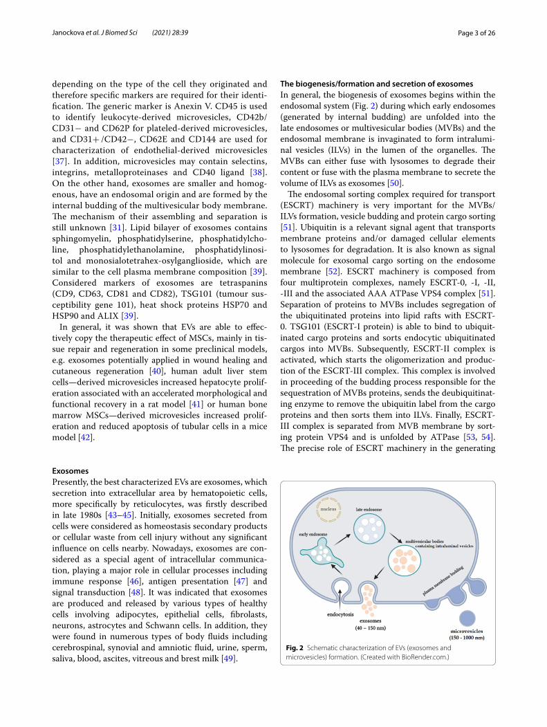
The biogenesis/formation and secretion of exosomesIn general, the biogenesis of exosomes begins within the endosomal system (Fig. 2) during which early endosomes (generated by internal budding) are unfolded into the late endosomes or multivesicular bodies (MVBs) and the endosomal membrane is invaginated to form intralumi-nal vesicles (ILVs) in the lumen of the organelles. The MVBs can either fuse with lysosomes to degrade their content or fuse with the plasma membrane to secrete the volume of ILVs as exosomes [50].
The endosomal sorting complex required for transport (ESCRT) machinery is very important for the MVBs/ILVs formation, vesicle budding and protein cargo sorting [51]. Ubiquitin is a relevant signal agent that transports membrane proteins and/or damaged cellular elements to lysosomes for degradation. It is also known as signal molecule for exosomal cargo sorting on the endosome membrane [52]. ESCRT machinery is composed from four multiprotein complexes, namely ESCRT-0, -I, -II, -III and the associated AAA ATPase VPS4 complex [51]. Separation of proteins to MVBs includes segregation of the ubiquitinated proteins into lipid rafts with ESCRT-0. TSG101 (ESCRT-I protein) is able to bind to ubiquit-inated cargo proteins and sorts endocytic ubiquitinated cargos into MVBs. Subsequently, ESCRT-II complex is activated, which starts the oligomerization and produc-tion of the ESCRT-III complex. This complex is involved in proceeding of the budding process responsible for the sequestration of MVBs proteins, sends the deubiquitinat-ing enzyme to remove the ubiquitin label from the cargo proteins and then sorts them into ILVs. Finally, ESCRT-III complex is separated from MVB membrane by sort-ing protein VPS4 and is unfolded by ATPase [53, 54]. The precise role of ESCRT machinery in the generating
Fig. 2 Schematic characterization of EVs (exosomes and microvesicles) formation. (Created with BioRender.com.)

Page 4 of 26Janockova et al. J Biomed Sci (2021) 28:39
of ILVs secreted later as exosomes is still unclear. In the screening study of RNA interference targeting ESCRT associated proteins in HeLa cells was shown that the depletion of Hrs, TSG101 and STAM1 proteins can reduce the exosomes secretion [55]. It was examined by nanoparticle tracking analysis that knockdown of Hrs reduced exosome secretion from head and neck squa-mous cell carcinoma cells [56]. Likewise, exosome secre-tion was increased by knockdown of the ESCRT-III and associated proteins ALIX, VTA1, VPS4B and CHMP4C [55]. Specifically, increase of exosomal level and typical exosomal markers (CD63, HSP70) was confirmed after syndecan – syntenin – ALIX depletion in MCF-7 cells [57].
Alternatively, sorting of exosomal cargo into MVBs and following ILVs formation can occur via ESCRT independ-ent mechanism. Proteolipid protein containing exosomes requires for their secretion ceramide which is able to ini-tiate the exosome budding into MVBs [58]. Expression of tetraspanins (transmembrane proteins rich in exosomes) CD9 and CD82 increased the exosomal release of β-catenin (involved in regulation and organization of cell–cell adhesion and gene transcription) from HEK293 cells [59]. The oligomerization of oligomers could play a significant role in exosome biogenesis based on CD43 exosomal sorting in Jurkat T-cells [60]. Observably, there are various possible mechanisms for separation of bioac-tive molecules into exosomes, either ESCRT dependent or independent, allow to work depending on the cell type and/or cellular homeostasis. In addition, it was shown that numerous diseases and other pathological condi-tions enhance exosome secretion. Increased quantity of exosomes were noticed in tumor cells, by progression of inflammation, angiogenesis and coagulation [61–63].
Molecular composition of exosomesThe molecular structure of exosomes is related not only to the cell type of origin but also to the microenviron-ment involving mechanical properties, biochemical impulses and topography, which could influence protein cargo regulation of the secreted exosomes [39]. Exosome secretion and their composition can also be modulated by other environmental factors such as oxygen level, type of disease, mechanical stress or media composition [64].
Exosomes are composed of various macromolecules involving unique lipid and protein structures and nucleic acids (Fig. 3). Exosomes are characterized by abundant amount of miRNAs with majority in the form of pre-miRNAs, which are inactive until their conversion to mature miRNAs [65]. Considering the endosomal origin of exosomes, they contain proteins participating in mem-brane transport and fusion (e.g. annexins, Rab, flotillin, GTPases), MVBs biogenesis (e.g. ALIX, TSG101) and
also proteins associated with lipid microdomains (inte-grins and tetraspanins). Besides that, another frequently determinated proteins are associated with cytoskel-eton (e.g. tubulin, myosin, actin) and metabolism (e.g. GADPH) [54], heat shock proteins (HSC70, HSC90), tis-sue specific proteins (e.g. MHC II located on the surface of exosomes secreted by dendritic cells or by B-lympho-cytes) or proteins specific for cancer cell lines (e.g. glioma EGFR, breast cancer HER2, ovarian cancer CD24) [66].
Specifically, numerous studies on the protein and RNA composition of MSC-derived exosomes have been reported. Lai et al. investigated the proteome of HPLC-purified human embryonic stem cells—derived exosomes using mass spectrometry and cytokine array. They iden-tified more than 850 proteins and detected total protein complement of a 20S proteasome with very high reli-ability [67]. Kang´s group realized proteomic analysis of the nanoscale size-based fractionation of exosomes from human neural stem cells and identified 103 proteins. Results from their study confirmed, that exosomes larger than ∼50 nm were morphologically different from those which were smaller than ∼50 nm [68]. MSC-derived exosomes were found to contain also all five enzymes involved in the ATP synthesis of glycolysis, namely
Fig. 3 Exosome´s composition briefing: MHC I, II—major histocompatibility complex I, II); MFGE8 -milk fat globule EGF factor 8 protein; ICAM-1—intercellular adhesion molecule 1; LAMP 1, 2—lysosomal-associated membrane protein 1, 2; proteins involved heat shock proteins (HSP60, HSP70, HSP90), MVB biogenesis proteins (Alix, TSG101, Ubiquitin, Clathrin), cytoskeleton proteins (profilin, cofilin, tubulin, actin, myosin, tropomyosin), signaling proteins (G protein, syntenin, MAPK, ERK ½, Rho); to enzymes belong pyruvate kinase, ATPase, PGK1, GADPH, aldolase, enolase; nucleic acids include mRNA, miRNA, siRNA, tRNA, DNA. (Created with BioRender.com.)

Page 5 of 26Janockova et al. J Biomed Sci (2021) 28:39
glyceraldehyde 3-phosphate dehydrogenase, phospho-glycerate kinase, phosphoglucomutase, enolase and pyruvate kinase m2 isoform [69]. Furthermore, Arslan´s group detected enzymatically active CD73 in MSC-derived exosomes responsible for the generating of extra-cellular adenosine from released adenine nucleotides [69]. Exosomes are able to activate adenosine receptors and thus generate adenosine-affected phosphorylation of ERK1/2 and Akt in H9C2 cardiomyocytes [70].
The genetic information in RNAs of exosomes which are endocytosed by acceptor cells is allow to influence the protein expression in those cells. Exosomes contain RNAs mostly in size range less than 700 nt. Chen et al. identified the presence of small RNAs (less than 30 nt) in human embryonic stem cells-derived MSCs´ condi-tioned medium, which were encapsulated in cholesterol rich phospholipid vesicles [65]. Plethora of miRNAs responsible for post-transcriptional maintainig of gene expression were detected in MSC-derived exosomes which are active in acceptor cells [71] and participate in physiological and pathological processes. Research group of Ratajczak et al. reported that embryonic stem cells – derived exosomes are highly enriched in mRNA (for numerous transcription factors, receptors and cytokines) [72]. Furthermore, Valadi et al. identified dif-ferent miRNAs including let-7, miR-1, miR-15, miR-16, miR-181 and miR-375 in exosomes isolated from mast-cell line (MC/9), primary bone marrow-derived mast cells (BMMC) and human mast-cell line (HMC-1) [73], which have been suggested to play an important role in exocytosis, tumorigenesis, angiogenesis and haemat-opoiesis [74]. Ono et al. reported that miR-23b promotes dormancy in breast cancer cells [75]. Exosomal miRNAs derived from umbilical cord MSCs, mainly represented by let-7f, miR-145, miR-199a and miR-221 supported the suppression of hepatitis C virus RNA replication [76]. Results of several sequencing studies also demonstrated, that exosomes isolated from human blood serum and urine contain marked amount of other RNA types, such as tRNA, rRNA, snRNA snoRNA, piRNA and scaRNA [77].
The current studies of the structure and composition of exosomes have relevant importance and are still under examination. Wang et al. compared paracrine functions in vivo and exosomal profiles of human endometrium-, bone marrow- and adipose-derived MSCs in a rat model of myocardial infarction. Analyses of exosomal micro-RNAs showed that miR-21 expression was improved in exosomes derived from endometrium [78], suggesting that innate differences of various MSC-derived exosomes have substantial influence on their clinical efficacy. The importance of exosomes has long been recognized also due to their capability to transfer important cellular
cargoes (proteins, DNA, mRNA, miRNAs) to target cells. Recent evidences suggest that exosomes are involved both in normal physiological functions and in pathologi-cal conditions. Deeper understanding of the exosomes content may influence the study of various diseases. Some research groups demonstrated that tetraspanin complexes significantly contributes to selective target binding of exosomes to target cells [79, 80]. Thakur et al. showed that the presence of dsDNA in exosomes repre-sented the whole genomic DNA and could be used for identification of mutations in parental tumor cells. They determined that tumor-derived exosomes carry dsDNA and may be use as a circulating biomarker in the early detection of cancer and metastasis [81]. Liang et al. used engineered exosomes for co-delivery of chemothera-peutic drug 5-fluorouracil and chemoresistance miR-21 inhibitor oligonucleotide to reduce the drug resistance in colorectal carcinoma and thus to improve the efficacy of cancer treatment [82]. Yang et al. demonstrated the capability of brain endothelial cell-derived exosomes to deliver siRNA across the brain-blood barrier in zebrafish and thus inhibit VEGF [83]. Results suggested potential application of natural exosome vesicles in the treatment of brain disease [83]. Raposo et al. showed that both human and murine B-lymphocytes secrete exosomes to induce antigen-specific MHC (major histocompatibility complex) II-restricted T cell responses, reffering to exo-some usefulness as biological instruments in immuno-therapy [84].
The therapeutic effect and biodistribution of exosomes is also greatly affected by the origin of exosome produc-ing cells. MSC-derived exosomes regarding to their inner properties and source of origin may play a relevant role in their clinical efficiency and represent an ideal delivery system for intermediate processes in specific target cells.
Therapeutic potential of MSC‑derived exosomesMSC-derived exosomes increasingly play an impor-tant role in intracellular communication mechanism and tissue repair and their clinical use may supply sub-stantial advantages in comparison with their live cells due to potential to reduce undesirable side effects after application as well as infusional toxicities, uncontrolled cell growth and possible tumor formation. Moreover exosomes transplantation seems to be less risky and may have several advantages in contrast to cell applications. Exosomes are neither able to mutate and duplicate, nor induce metastasis. They have been tested in various ani-mal models (Table 1) for human diseases (e.g. hypoxic pulmonary hypertension [85], acute kidney injury [86], liver fibrosis [87]) and it was detected that their functions are very similar to MSCs. First therapeutic potential of MSC-derived exosomes was described in a Langendorff

Page 6 of 26Janockova et al. J Biomed Sci (2021) 28:39
Tabl
e 1
List
of t
hera
peut
ic a
pplic
atio
n of
som
e M
SC-d
eriv
ed e
xoso
mes
from
diff
eren
t sou
rces
in a
nim
al m
odel
s of
hum
an d
isea
se
Tiss
ue ty
peM
SC o
rigi
nM
etho
d of
exo
som
e is
olat
ion
Exos
ome
char
acte
riza
tion
Dis
ease
focu
sA
nim
al ty
pe a
nd
mod
elEx
osom
e ad
min
istr
atio
nRe
late
d ex
osom
e ca
rgo/
path
way
Out
com
eRe
fs
Bone
mar
row
Hum
anCe
ntrif
ugat
ion,
ultr
a-ce
ntrif
ugat
ion
Det
erm
inat
ion
of to
tal p
rote
in
conc
entr
atio
n (B
CA
pr
otei
n as
say)
, TEM
, N
TA, w
este
rn b
lot-
ting
(CD
9, C
D63
, C
D81
, TSG
101,
and
A
lix m
arke
rs)
Live
r fibr
osis
CCl 4 i
nduc
ed li
ver
fibro
sis
in S
D ra
tsIn
ject
ion
thro
ugh
the
tail
vein
Wnt
/β-c
aten
in
path
way
MSC
-der
ived
ex
osom
es re
duce
d liv
er fi
bros
is in
viv
o th
roug
h th
e W
nt/β
-cat
enin
pa
thw
ayEx
osom
e tr
eatm
ent
redu
ces
the
expr
es-
sion
of P
PARγ
, W
nt3a
, Wnt
10b
and
β-ca
teni
n,
wha
t con
trib
uted
to
inhi
bitio
n of
do
wns
trea
m g
ene
expr
essi
on (W
ISP1
, Cy
clin
D1)
in b
oth
hepa
tic s
tella
te
cells
and
live
r fib
rosi
s tis
sue
[108
]
Hum
anPr
ecip
itatio
n (E
xo-
Qui
ck e
xoso
me
isol
atio
n)
Det
erm
inat
ion
of to
tal p
rote
in
conc
entr
atio
n (B
CA
pr
otei
n as
say)
, qN
ano
nano
pore
-ba
sed
dete
ctio
n,
SDS-
page
(CD
9,
CD
63, C
D81
), re
vers
ed-p
hase
ch
rom
atog
raph
y,
Q E
xact
ive
mas
s sp
ectr
omet
ry
Trau
mat
ic b
rain
in
jury
Cort
ical
impa
ct
Wis
tar r
ats
mod
el
of tr
aum
atic
bra
in
inju
ry
Intr
aven
ousl
y vi
a th
e ta
il ve
inN
.DEx
osom
es d
eriv
ed
from
hum
an B
M-
MSC
s in
2D
or 3
D
cultu
res
impr
oved
fu
nctio
nal r
ecov
-er
y, p
rom
oted
ne
urov
ascu
lar
rem
odel
ing
and
redu
ced
neur
o-in
flam
mat
ion
in
rats
aft
er tr
aum
atic
br
ain
inju
ry
[114
]
Hum
anG
radi
ent u
ltrac
entr
if-ug
atio
n, u
ltrafi
ltra-
tion
Det
erm
inat
ion
of to
tal p
rote
in
conc
entr
atio
n (B
CA
pr
otei
n as
say)
, ele
c-tr
on m
icro
scop
y,
flow
cyt
omet
ry
(CD
63)
Bone
def
ects
Calv
aria
l def
ects
in
SD ra
tsD
efec
ts tr
eate
d w
ith
hydg
ogel
+ E
Vsm
iR-1
96a,
miR
-27a
, m
iR-2
06In
vitr
o-EV
s po
sitiv
ely
regu
late
d ex
pres
-si
on o
f ost
eoge
nic
gene
s an
d os
teo-
blas
t diff
eren
tiatio
nIn
viv
o-EV
s st
imu-
late
d bo
ne fo
rma-
tion
in ra
ts w
ith
calv
aria
l def
ects
[119
]

Page 7 of 26Janockova et al. J Biomed Sci (2021) 28:39
Tabl
e 1
(con
tinue
d)
Tiss
ue ty
peM
SC o
rigi
nM
etho
d of
exo
som
e is
olat
ion
Exos
ome
char
acte
riza
tion
Dis
ease
focu
sA
nim
al ty
pe a
nd
mod
elEx
osom
e ad
min
istr
atio
nRe
late
d ex
osom
e ca
rgo/
path
way
Out
com
eRe
fs
Porc
ine
Ultr
afiltr
atio
nD
eter
min
atio
n of
to
tal p
rote
in c
on-
cent
ratio
n (B
rad-
ford
ass
ay),
NTA
, flo
w c
ytom
etry
(C
D44
and
CD
90)
Syno
vitis
Porc
ine
mod
el o
f an
tigen
-trig
gere
d sy
novi
tis
Intr
a-ar
ticul
ar in
jec-
tions
N.D
Exos
omes
dec
reas
ed
syno
vial
lym
-ph
ocyt
es, t
he
dow
nreg
ulat
ed
TNF-
α tr
ansc
ripts
an
d im
prov
ed
the
impu
lse
in
exos
ome-
trea
ted
join
ts
[116
]
Rat
Ultr
acen
trifu
gatio
nD
eter
min
atio
n of
tota
l pro
tein
co
ncen
trat
ion
(BC
A
prot
ein
assa
y), T
EM,
RT-P
CR
Acu
te k
idne
y in
jury
Acu
te k
idne
y in
jury
in
duce
d by
gen
-ta
mic
in in
Wis
tar
rats
Inje
ctio
n in
to c
auda
l ve
inRN
Aas
e, R
NA
car
ried
by th
e ex
osom
es/
mic
rove
sicl
es
BM c
ondi
tione
d m
edia
incr
ease
d th
e re
nal f
unct
ion
reco
very
. Pro
tec-
tive
effec
ts w
ere
med
iate
d by
the
exos
ome´
RN
A in
th
e co
nditi
oned
m
edia
[86]
Rat
Diff
eren
tial c
entr
ifu-
gatio
nD
eter
min
atio
n of
tota
l pro
tein
co
ncen
trat
ion
(BC
A
prot
ein
assa
y), D
LS,
conf
ocal
mic
ros-
copy
, SEM
, TEM
, EL
ISA
(CD
9), fl
ow
cyto
met
ry (C
D63
), w
este
rn b
lott
ing
(CD
81)
Acu
te li
ver i
njur
yIs
chem
ic/r
eper
fusi
on
liver
inju
ry a
nd C
Cl 4
indu
ced
acut
e liv
er
inju
ry in
rats
Inje
ctio
n th
roug
h he
patic
por
tal v
ein
Exos
ome-
rich
fract
iona
ted
secr
etom
e
In v
itro
– ex
osom
es
show
ed a
ntia
pop-
totic
and
pro
sur-
viva
l effe
ct, b
ette
r H
epG
2 ce
lls re
cov-
ery
and
redu
ced
cyto
toxi
city
In v
ivo
– ex
osom
es
impr
oved
live
r re
gene
ratio
n an
d re
cove
ry fr
om li
ver
inju
ry
[109
]
Page 8 of 26Janockova et al. J Biomed Sci (2021) 28:39
Tabl
e 1
(con
tinue
d)
Tiss
ue ty
peM
SC o
rigi
nM
etho
d of
exo
som
e is
olat
ion
Exos
ome
char
acte
riza
tion
Dis
ease
focu
sA
nim
al ty
pe a
nd
mod
elEx
osom
e ad
min
istr
atio
nRe
late
d ex
osom
e ca
rgo/
path
way
Out
com
eRe
fs
Rat
Prec
ipita
tion
(Exo
Qui
ck-T
C e
xo-
som
e is
olat
ion)
Det
erm
inat
ion
of to
tal p
rote
in
conc
entr
atio
n (B
CA
pr
otei
n as
say)
, flow
cy
tom
etry
(CD
63),
TEM
Myo
card
ial i
nfar
ctio
nA
cute
myo
card
ial
infa
rctio
n in
SD
rats
Intr
amyo
card
ial
inje
ctio
nN
.DEx
osom
es im
prov
ed
card
iac
func
tion
afte
r isc
hem
ic
inju
ryIn
vitr
o –
exos
omes
im
prov
ed th
e tu
be fo
rmat
ion
of
HU
VEC
cel
ls a
nd
impa
ired
T-ce
ll fu
nctio
n by
cel
l pr
olife
ratio
n in
hibi
-tio
nIn
viv
o –
exos
omes
re
duce
d in
farc
t si
ze a
nd re
tain
ed
card
iac
syst
olic
and
di
asto
lic p
erfo
r-m
ance
[111
]
Rat
Ultr
acen
trifu
gatio
nEl
ectr
on m
icro
scop
yM
yoca
rdia
l inf
arct
ion
SD ra
ts m
yoca
rdia
l is
chem
ia/r
eper
fu-
sion
mod
el
Intr
amyo
card
ial
inje
ctio
n in
to th
e le
ft v
entr
icul
ar w
all
Aut
opha
gy m
achi
n-er
yEx
osom
es in
hib-
ited
myo
card
ial
infa
rctio
n pa
tho-
gene
sis,
prob
-ab
ly b
y au
toph
agy
regu
latio
n.
Exos
omes
trea
t-m
ent s
uppr
esse
d th
e ex
pres
sion
of
Apa
f1 (a
pop-
totic
pro
teas
e ac
tivat
ing
fact
or
1) a
nd in
crea
sed
the
espr
essi
on o
f AT
G13
(aut
opha
gy-
rela
ted
prot
ein1
3)
[112
]
Page 9 of 26Janockova et al. J Biomed Sci (2021) 28:39
Tabl
e 1
(con
tinue
d)
Tiss
ue ty
peM
SC o
rigi
nM
etho
d of
exo
som
e is
olat
ion
Exos
ome
char
acte
riza
tion
Dis
ease
focu
sA
nim
al ty
pe a
nd
mod
elEx
osom
e ad
min
istr
atio
nRe
late
d ex
osom
e ca
rgo/
path
way
Out
com
eRe
fs
Rat
Mul
tiste
p ce
ntrif
uga-
tion
Det
erm
inat
ion
of
tota
l pro
tein
con
-ce
ntra
tion
(mic
ro
BCA
pro
tein
ass
ay)
Stro
keM
iddl
e ce
rebr
al
arte
ry o
cclu
sion
W
ista
r rat
s m
odel
Inje
ctio
n in
to th
e ta
il ve
inN
.DEx
osom
es im
prov
ed
neur
olog
ic o
ut-
com
e by
func
tiona
l re
cove
ry a
nd
enha
nced
neu
rite
rem
odel
ing,
neu
ro-
gene
sis
and
angi
o-ge
nesi
s. Ex
osom
es
syst
emic
trea
tmen
t im
prov
es n
euro
-lo
gic
outc
ome,
sig
-ni
fican
tly in
crea
sed
the
syna
ptop
hysi
n im
mun
orea
ctiv
e ar
ea in
isch
emic
bo
unda
ry z
one
[113
]
Mou
seFi
ltrat
ion,
diff
eren
tial
cent
rifug
atio
n,
ultr
acen
trifu
gatio
n
Det
erm
inat
ion
of to
tal p
rote
in
conc
entr
atio
n (B
CA
pr
otei
n as
say)
, DLS
, el
ectr
on m
icro
s-co
py
Card
iac
hype
rtro
phy
Tran
sver
se a
ortic
co
nstr
ictio
n m
ouse
m
odel
Intr
amyo
card
ial
inje
ctio
nN
.DIn
vitr
o—ex
osom
es
inhi
bite
d ce
ll hy
pert
roph
y st
imul
ated
with
an
giot
ensi
n II
in
cultu
red
myo
cyte
sIn
viv
o—ex
osom
es
sign
ifica
ntly
pr
otec
ted
myo
-ca
rdiu
m a
gain
st
card
iac
hype
r-tr
ophy
, inh
ibite
d m
yoca
rdia
l apo
p-to
sis
and
fibro
sis
and
reta
ined
hea
rt
func
tion
whe
n th
e pr
essu
re w
as
over
load
ed
[110
]

Page 10 of 26Janockova et al. J Biomed Sci (2021) 28:39
Tabl
e 1
(con
tinue
d)
Tiss
ue ty
peM
SC o
rigi
nM
etho
d of
exo
som
e is
olat
ion
Exos
ome
char
acte
riza
tion
Dis
ease
focu
sA
nim
al ty
pe a
nd
mod
elEx
osom
e ad
min
istr
atio
nRe
late
d ex
osom
e ca
rgo/
path
way
Out
com
eRe
fs
Mou
se
(isch
emic
pr
econ
di-
tione
d)
Prec
ipita
tion
(Exo
-Q
uick
exo
som
e is
olat
ion)
Det
erm
inat
ion
of to
tal p
rote
in
conc
entr
atio
n (B
CA
pr
otei
n as
say)
, w
este
rn b
lott
ing
(CD
9, C
D63
)
Alz
heim
er´s
dis
ease
Tran
sgen
ic A
PP/P
S1
mou
se m
odel
Inje
ctio
n th
roug
h la
tera
l cau
dal v
ein
miR
-21,
miR
-181
cEx
osom
es im
prov
ed
mem
ory
func
tions
an
d le
arni
ng
capa
bilit
ies
in m
ice.
H
ypox
ic M
SC-
deriv
ed e
xoso
mes
re
duce
d eff
ectiv
ely
Aβ
accu
mul
a-tio
n, in
crea
sed
the
expr
essi
on o
f sy
napt
ic p
rote
ins
and
enha
nced
the
leve
l of m
iR-2
1 in
th
e br
ains
of A
PP/
PS1
mic
e
[115
]
Um
bilic
al c
ord
Hum
an
(Wha
rton
’s je
lly) a
nd
mou
se B
M
Prec
ipita
tion,
col
umn
size
exc
lusi
on
chro
mat
ogra
phy,
ul
trac
entr
ifuga
tion
Elec
tron
mic
rosc
opic
an
alys
is, d
eter
-m
inat
ion
of to
tal
prot
ein
conc
en-
trat
ion
(Bra
dfor
d as
say)
wes
tern
bl
ottin
g (C
D63
, A
LIX,
TSG
101,
C
D81
, CD
9, h
sp90
, flo
tillin
-1, D
icer
), is
olat
ion
and
quan
tifica
tion
of
mic
roRN
As
Hyp
oxic
pul
mon
ary
hype
rten
sion
Hyp
oxia
indu
ced
pulm
onar
y hy
per-
tens
ion
in F
VB
stra
in m
ice
N.D
miR
-204
, miR
-17
MSC
-der
ived
ex
osom
es w
ere
able
to in
hibi
t pul
-m
onar
y hy
pert
en-
sion
by
inih
ition
of
hype
rpro
lifer
ativ
e pa
thw
ays,
iclu
d-in
g su
ppre
ssio
n of
the
hypo
xic
activ
atio
n of
STA
T3
sign
alin
g an
d th
e up
regu
latio
n of
the
miR
-17,
whe
reas
it
incr
ease
d lu
ng
leve
ls o
f miR
-204
[85]

Page 11 of 26Janockova et al. J Biomed Sci (2021) 28:39
Tabl
e 1
(con
tinue
d)
Tiss
ue ty
peM
SC o
rigi
nM
etho
d of
exo
som
e is
olat
ion
Exos
ome
char
acte
riza
tion
Dis
ease
focu
sA
nim
al ty
pe a
nd
mod
elEx
osom
e ad
min
istr
atio
nRe
late
d ex
osom
e ca
rgo/
path
way
Out
com
eRe
fs
Hum
anU
ltrac
entr
ifuga
tion
Det
erm
inat
ion
of to
tal p
rote
in
conc
entr
atio
n (B
CA
pr
otei
n as
say)
, TEM
, w
este
rn b
lott
ing
(CD
9, C
D81
)
liver
fibr
osis
CCl 4-
indu
ced
liver
fib
rosi
s in
mic
eIn
ject
ion
into
the
left
an
d rig
ht lo
bes
of
liver
s
(TG
F)-b
1/Sm
ad s
ign-
alin
g pa
thw
ayM
SC-d
eriv
ed
exos
omes
inhi
bite
d EM
T an
d im
prov
ed
CCl 4 i
nduc
ed li
ver
fibro
sis
In v
ivo—
exos
ome
tran
spla
ntat
ion
redu
ced
TGF-
β1
expr
essi
on,
inac
tivat
ed S
mad
2 ph
osph
oryl
atio
n an
d in
vert
ed li
ver
EMT
In v
itro
– Ex
o-so
me
trea
tmen
t of
HL7
702
cells
af
ter E
MT
caus
ed
reve
rsed
spi
ndle
-sh
aped
cel
ls a
nd
EMT
asso
ciat
ed
mar
ker e
xpre
ssio
n
[87]
Hum
anPr
ecip
itatio
n (E
xo-
Qui
ck U
LTRA
EV
isol
atio
n)
Elec
tron
mic
rosc
opy,
N
TA, w
este
rn
blot
ting
(TSG
101,
C
D63
, CD
81),
qPC
R an
alys
is, s
eque
nc-
ing
of m
iRN
As
Acu
te li
ver i
njur
yCC
l 4-in
duce
d ac
ute
liver
inju
ry a
nd
endo
toxe
mia
in
C57
BL/6
mic
e
Inje
ctio
nm
iR45
5-3p
Exos
omes
enr
iche
d in
miR
-455
-3p
wer
e ca
pabl
e to
inhi
b-ite
d th
e ov
erac
tiva-
tion
of m
onoc
yte/
mac
roph
ages
and
re
duce
d ac
ute
liver
in
jury
by
inhi
bitin
g IL
-6-r
elat
ed s
igna
l-in
g pa
thw
ays
[122
]
Hum
anU
ltrac
entr
ifuga
tion
TEM
, wes
tern
blo
t-tin
g (C
D9,
CD
63,
CD
81)
Acu
te k
idne
y in
jury
Cis
plat
in-in
duce
d ac
ute
kidn
ey in
jury
in
SD
rats
Rena
l cap
sule
inje
c-tio
np3
8MA
PK p
athw
ay,
ERK
1/2
path
way
Exos
omes
sup
-pr
esse
d ki
dney
in
jury
and
NRK
-52E
ce
ll in
jury
by
impr
ovem
ent o
f ox
idat
ive
stre
ss
and
cell
apop
tosi
s an
d pr
omot
ion
of
cell
prol
ifera
tion
thro
ugh
activ
atio
n of
ERK
1/2
in v
ivo
and
in v
itro
[123
]

Page 12 of 26Janockova et al. J Biomed Sci (2021) 28:39
Tabl
e 1
(con
tinue
d)
Tiss
ue ty
peM
SC o
rigi
nM
etho
d of
exo
som
e is
olat
ion
Exos
ome
char
acte
riza
tion
Dis
ease
focu
sA
nim
al ty
pe a
nd
mod
elEx
osom
e ad
min
istr
atio
nRe
late
d ex
osom
e ca
rgo/
path
way
Out
com
eRe
fs
Hum
anU
ltrac
entr
ifuga
tion
Det
erm
inat
ion
of
tota
l pro
tein
con
-ce
ntra
tion
(Bra
d-fo
rd a
ssay
), TE
M,
flow
cyt
omet
ry
Acu
te k
idne
y in
jury
Acu
te k
idne
y in
jury
m
odel
indu
ced
by
isch
emia
–rep
erfu
-si
on in
jury
in ra
ts
Intr
aven
ous
adm
inis
-tr
atio
nN
.DEx
osom
es re
duce
d ce
ll ap
opto
sis
and
impr
oved
pr
olife
ratio
n 24
h a
fter
kid
ney
inju
ry, p
rom
oted
an
giog
enes
is b
y in
duci
ng V
EGF
elev
atio
n th
roug
h H
IF-1
α in
depe
nd-
ent m
anne
r
[124
]
Hum
anD
iffer
entia
l cen
trifu
-ga
tion,
ultr
acen
-tr
ifuga
tion
Det
erm
inat
ion
of to
tal p
rote
in
conc
entr
atio
n (B
CA
pr
otei
n as
say)
, w
este
rn b
lott
ing
(CD
9, H
SP70
), TE
M,
NTA
Wou
nd h
ealin
g an
d an
giog
enes
isSk
in b
urn
wou
nd
mod
el in
rats
Subc
utan
eous
inje
c-tio
nW
nt4
In v
itro—
exos
omes
el
evat
ed e
ndot
he-
lial c
ell p
rolif
era-
tion,
mig
ratio
n an
d tu
be fo
rmat
ion
In v
ivo—
exos
omes
im
prov
ed
angi
ogen
esis
in th
e re
pair
of s
kin
burn
in
jury
by
deliv
erin
g W
nt4
to a
ctiv
ate
Wnt
/β-c
aten
in
sign
alin
g (t
issu
e re
pair
mec
hani
sm)
[125
]
Hum
anU
ltrac
entr
ifuga
tion
Det
erm
inat
ion
of to
tal p
rote
in
conc
entr
atio
n (B
CA
pr
otei
n as
say)
, TEM
, w
este
rn b
lott
ing
(CD
9, C
D63
, CD
81,
β-ca
teni
n, W
nt3a
, β-
actin
)
Frac
ture
hea
ling
Mod
el o
f fem
oral
e fra
ctur
e in
SD
rats
Inje
ctio
n of
the
mix
of
hyd
roge
l and
ex
osom
es in
to th
e fra
ctur
e
N.D
Exos
omes
par
tici-
pate
d in
the
repa
ir of
frac
ture
in ra
ts
thro
ugh
the
Wnt
si
gnal
ing
path
way
by
incr
easi
ng o
f β-
cate
nin
and
Wnt
3a p
rote
in
expr
essi
ons
[126
]

Page 13 of 26Janockova et al. J Biomed Sci (2021) 28:39
Tabl
e 1
(con
tinue
d)
Tiss
ue ty
peM
SC o
rigi
nM
etho
d of
exo
som
e is
olat
ion
Exos
ome
char
acte
riza
tion
Dis
ease
focu
sA
nim
al ty
pe a
nd
mod
elEx
osom
e ad
min
istr
atio
nRe
late
d ex
osom
e ca
rgo/
path
way
Out
com
eRe
fs
Hum
anU
ltrac
entr
ifuga
tion
Det
erm
inat
ion
of to
tal p
rote
in
conc
entr
atio
n (B
CA
pr
otei
n as
say)
, NTA
, w
este
rn b
lott
ing
(CD
81)
Wou
nd h
ealin
gSk
in-d
efec
t mod
el in
IC
R an
d BA
LB/c
-υ
mic
e
Inje
ctio
n of
the
mix
of
hyd
roge
l and
ex
osom
es a
roun
d th
e w
ound
miR
-21,
miR
-23a
, m
iR-1
25b,
miR
-145
Exos
omes
enr
iche
d in
spe
cific
mic
roR-
NA
s (m
iR-2
1, -2
3a,
-125
b, a
nd -1
45)
inhi
bite
d m
yofi-
brob
last
form
atio
n,
inhi
bite
d TG
F-β2
, TG
F-βR
2 an
d SM
AD
2 pa
thw
ay
and
acco
rdin
gly
supp
ress
ed th
e ex
pres
sion
of
α-SM
A g
ene
and
redu
ced
col-
lage
n I d
epos
ition
. Si
gnifi
cant
role
of
exo
som
es fo
r an
ti-sc
arrin
g ab
ility
an
d th
e m
yofib
ro-
blas
t-su
ppre
ssin
g w
as s
how
ed
both
in v
itro
and
in v
ivo
by b
lock
ing
miR
NA
s in
side
the
exos
omes
[127
]
Hum
anU
ltrac
entr
ifuga
tion
TEM
, det
erm
inat
ion
of to
tal p
rote
in
conc
entr
atio
n (B
CA
pr
otei
n as
say)
, NTA
, w
este
rn b
lott
ing
(CD
9, C
D63
, CD
81)
Infla
mm
ator
y bo
wel
di
seas
eD
SS-in
duce
d in
flam
-m
ator
y bo
wel
di
seas
e m
ouse
m
odel
Inje
ctio
n th
roug
h th
e ta
il ve
inN
.DEx
osom
es c
ould
im
prov
e in
flam
ma-
tory
bow
el d
isea
seIn
vitr
o –
cocu
lture
w
ith e
xoso
mes
su
ppre
ssed
the
expr
essi
on o
f iN
OS
and
IL-7
in m
ouse
en
tero
celia
mac
-ro
phag
esIn
viv
o –
exos
omes
re
duce
d th
e ex
pres
sion
of
pro-
infla
mm
ator
y cy
toki
nes T
NF-
α,
IL-1
β, IL
-6) a
nd
incr
ease
d th
e ex
pres
sion
of
anti-
infla
mm
ator
y cy
toki
ne (I
L10)
[128
]
Page 14 of 26Janockova et al. J Biomed Sci (2021) 28:39
Tabl
e 1
(con
tinue
d)
Tiss
ue ty
peM
SC o
rigi
nM
etho
d of
exo
som
e is
olat
ion
Exos
ome
char
acte
riza
tion
Dis
ease
focu
sA
nim
al ty
pe a
nd
mod
elEx
osom
e ad
min
istr
atio
nRe
late
d ex
osom
e ca
rgo/
path
way
Out
com
eRe
fs
Hum
anU
ltrac
entr
ifuga
tion
Det
erm
inat
ion
of
tota
l pro
tein
con
-ce
ntra
tion,
TEM
, Ze
tasi
zer,
wes
tern
bl
ottin
g (C
D9,
C
D63
, CD
81)
Colit
isCo
litis
indu
ctio
n in
C
57BL
/6 m
ice
Intr
aper
itone
al
inje
ctio
nN
.DIn
vitr
o –
exos
omes
de
crea
sed
pro-
infla
mm
ator
y cy
toki
nes
(IFN
-γ,
TNF-
α, IL
-1β)
co
ncen
trat
ion
and
enha
nced
th
e se
cret
ion
of
anti-
infla
mm
ator
y cy
toki
nes
(TG
F-β1
, IL
-10)
In v
ivo
– ex
osom
es
show
ed th
era-
peut
ic a
ctiv
ity in
ex
perim
enta
l col
itis
via
supp
ress
ing
infla
mm
atio
n m
achi
nery
, im
prov
ed c
linic
al
sym
ptom
s an
d hi
s-to
logi
cal s
ever
ity
[90]
Adi
pose
Hum
anU
ltrac
entr
ifuga
tion
Det
erm
inat
ion
of to
tal p
rote
in
conc
entr
atio
n (B
radf
ord
assa
y),
TEM
, NTA
, SIO
S,
Alz
heim
er’s
dise
ase
N.D
N.D
Nep
rilys
inEx
osom
es s
ecre
te
enzy
mat
ical
ly
activ
e ne
prily
-si
n. T
rans
fer o
f ex
osom
es to
N2a
ce
lls s
igni
fican
tly
decr
ease
d bo
th th
e in
trac
ellu
lar a
nd
extr
acel
lula
r Aβ4
0 an
d A
β42
leve
ls
[133
]
Pig
Ultr
acen
trifu
gatio
nN
TA, T
EM, w
este
rn
blot
ting
(CD
9,
CD
29, C
D63
), RT
PC
R (m
RNA
con
-te
nt o
f IL-
10)
Rena
l infl
amm
atio
nM
etab
olic
syn
drom
e an
d re
nal a
rter
y st
enos
is m
odel
in
dom
estic
pig
s
Intr
aren
al d
eliv
ery
IL-1
0Ex
osom
es re
duce
d re
nal i
nflam
ma-
tion,
enh
ance
d th
e re
para
tive
mac
-ro
phag
es n
umbe
r an
d in
crea
sed
expr
essi
on o
f IL-
10.
Exos
omes
wer
e ab
le to
redu
ce
rena
l fibr
osis
and
to
impr
ove
sten
otic
ki
dney
func
tion
[130
]

Page 15 of 26Janockova et al. J Biomed Sci (2021) 28:39
Tabl
e 1
(con
tinue
d)
Tiss
ue ty
peM
SC o
rigi
nM
etho
d of
exo
som
e is
olat
ion
Exos
ome
char
acte
riza
tion
Dis
ease
focu
sA
nim
al ty
pe a
nd
mod
elEx
osom
e ad
min
istr
atio
nRe
late
d ex
osom
e ca
rgo/
path
way
Out
com
eRe
fs
Rat
Ultr
afiltr
atio
n, u
ltra-
cent
rifug
atio
nD
eter
min
atio
n of
tota
l pro
tein
co
ncen
trat
ion
(BC
A
prot
ein
assa
y), T
EM,
wes
tern
blo
ttin
g (C
D9,
CD
63, H
SP70
, C
D81
)
Isch
emic
hea
rt
dise
ase
Myo
card
ial i
sche
mia
/re
perf
usio
n m
odel
in
SD
rats
Infu
sion
thro
ugh
the
tail
vein
N.D
Exos
omes
pro
-te
cted
isch
emic
m
yoca
rdiu
m fr
om
isch
emia
/rep
erfu
-si
on in
jury
thro
ugh
the
Wnt
/β-c
aten
in
sign
alin
g pa
thw
ay
activ
atio
nIn
vitr
o –
exos
omes
re
duce
d ce
ll ap
opto
sis
and
the
expr
essi
on o
f Ba
x, im
prov
ed c
ell
viab
ility
and
the
expr
essi
on o
f Bcl
-2
and
Cycl
in D
1 in
hy
poxi
a/re
oxy-
gena
tion-
indu
ced
H9c
2 ce
llsIn
viv
o –
exos
omes
si
gnifi
cant
ly
redu
ced
isch
emia
/re
perf
usio
n-in
duce
d m
yoca
r-di
al in
farc
tion,
it
was
sho
wed
de
crea
se in
ser
um
leve
ls o
f cre
atin
e ki
nase
-myo
card
ial
band
, lac
tate
deh
y-dr
ogen
ase,
and
ca
rdia
c tr
opon
in
I. A
fter
exo
som
e tr
eatm
ent w
as
obse
rved
att
enu-
atio
n of
isch
emia
/re
perf
usio
n-in
duce
d ap
opto
sis
acco
mpa
nied
by
the
incr
ease
of
Bcl-2
and
dec
reas
e of
Bax
, and
inhi
bi-
tion
of C
aspa
se
3 ac
tivity
in ra
t m
yoca
rdiu
m
[131
]
Page 16 of 26Janockova et al. J Biomed Sci (2021) 28:39
Tabl
e 1
(con
tinue
d)
Tiss
ue ty
peM
SC o
rigi
nM
etho
d of
exo
som
e is
olat
ion
Exos
ome
char
acte
riza
tion
Dis
ease
focu
sA
nim
al ty
pe a
nd
mod
elEx
osom
e ad
min
istr
atio
nRe
late
d ex
osom
e ca
rgo/
path
way
Out
com
eRe
fs
Mou
sePr
ecip
itatio
n an
d m
agne
tic b
eads
pu
rifica
tion
(Mag
-Ca
ptur
e Ex
osom
e Is
olat
ion)
Det
erm
inat
ion
of to
tal p
rote
in
conc
entr
atio
n (B
CA
pr
otei
n as
say)
, NTA
, TE
M, w
este
rn b
lot-
ting
(CD
29, C
D63
)
Acu
te m
yoca
rdia
l in
farc
tion
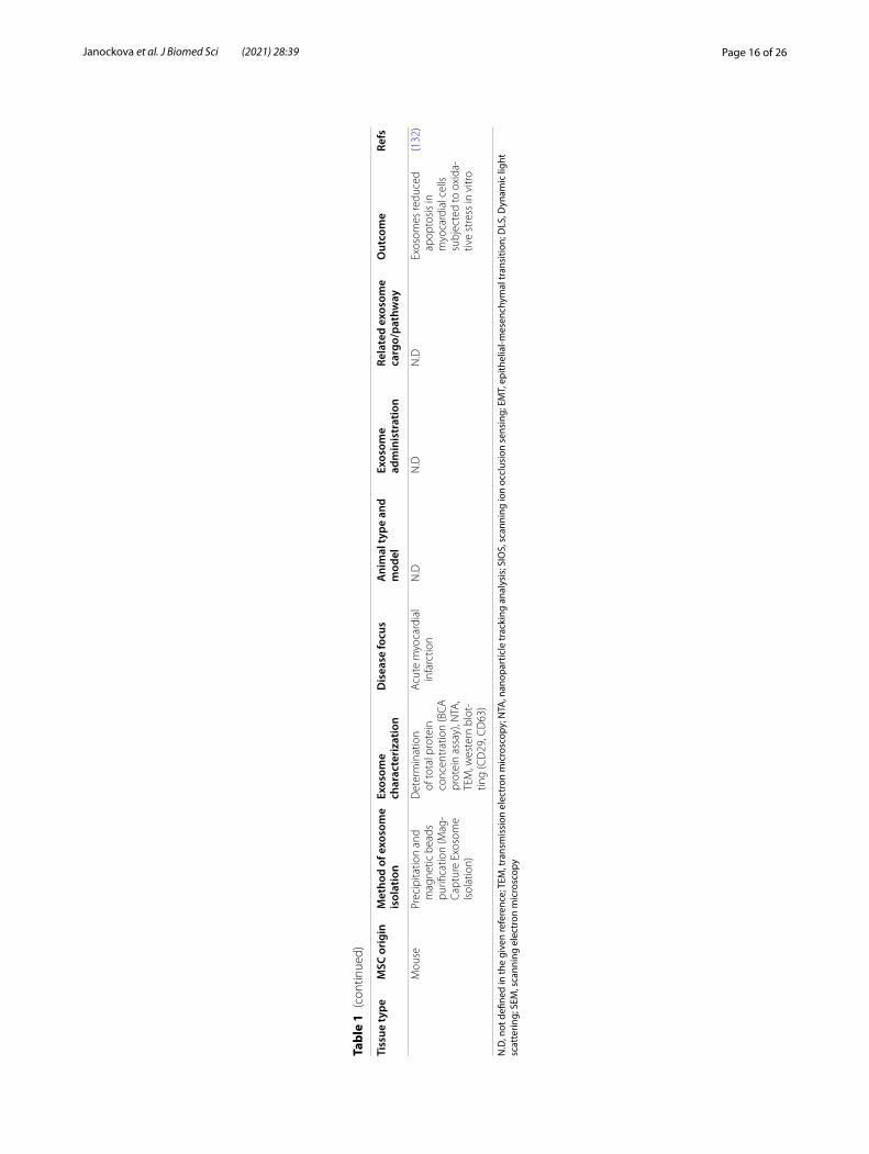
N.D
N.D
N.D
Exos
omes
redu
ced
apop
tosi
s in
m
yoca
rdia
l cel
ls
subj
ecte
d to
oxi
da-
tive
stre
ss in
vitr
o
(132
)
N.D
, not
defi
ned
in th
e gi
ven
refe
renc
e; T
EM, t
rans
mis
sion
ele
ctro
n m
icro
scop
y; N
TA, n
anop
artic
le tr
acki
ng a
naly
sis;
SIO
S, s
cann
ing
ion
occl
usio
n se
nsin
g; E
MT,
epi
thel
ial-m
esen
chym
al tr
ansi
tion;
DLS
, Dyn
amic
ligh
t sc
atte
ring;
SEM
, sca
nnin
g el
ectr
on m
icro
scop
y

Page 17 of 26Janockova et al. J Biomed Sci (2021) 28:39
heart model of acute myocardial ischemia/reperfusion injury in mice, where their cardioprotective effect was identified by myocardial infarct size reducing [88]. In this study, authors identified exosomes as cardioprotective elements in the MSCs´ paracrine secretion [88].
Several preclinical studies compared the beneficial effects of cell therapy based on MSCs and cell-free ther-apy based on MSC-derived EVs/exosomes and showed that they had similar therapeutic outcomes. Comparative analyses of MSCs and their EVs demonstrated different genetic cargo and protein content that play a significant role in biological processes, including angiogenesis, adi-pogenesis, apoptosis, regulation of inflammation, blood coagulation and extracellular matrix remodeling. Appli-cation of mice adipose MSCs in comparison with its conditioned medium had the same effect on sympotms of chronic colitis mouse model. Clinical symptoms and tissue damages were suppressed in treated mice [89]. Zhi et al. indicated that the application of umbilical cord MSC-derived exosomes (200 µg) resulted in ameliora-tion of clinical symptoms, reduction of colonic damage and decrease of the inflammatory state in mice colitis when compared with MSCs (1 × 10⁶ cells) administra-tion [90]. Shao et al. compared activity of rat bone mar-row MSCs and MSC-derived exosomes in a rat acute myocardial infarction model. It was showed a superior beneficial effects of MSC-derived exosomes in contrast to MSCs in cardiac repair. There were observed differ-ences in expression profiles of several miRNAs from that of MSCs detected through miRNA sequence analysis [91]. A recent cutaneous wound model study in rabbits reported that intradermal injection of EVs derived from adipose and bone marrow MSCs were superior to MSCs injection in vivo. Furthermore, adipose MSC-derived EVs enhanced wound healing better than EVs from bone mar-row [92]. In the study by Gatti et al. intravenous admin-istration of human bone marrow MSC-derived EVs had the same efficacy as MSCs on the treatment of acute kidney injury in rats by inhibiting apoptosis and stimu-lating tubular cell proliferation [93]. In an induced exper-imental autoimmune encephalomyelitis murine model of multiple sclerosis, both human placental MSCs and its MSC-derived EVs showed regenerative effects and pre-vented oligodendroglia degradation and demyelination [94]. Another preclinical study showed that MSC-derived exosomes could be a promising cell-free therapeutic strategy for the treatment of Alzheimer’s disease. It was demonstrated that 28 days after intervention of mice groups with 10 μg exosomes and 1 × 10⁶ MSCs separately had similar beneficial effects in improvement of neuro-genesis and cognitive functions [95].
From the preclinical studies of MSC-derived exosomes therapy to the clinical application, many critical
parameters should be resolved and determined, includ-ing clarification of important factors and conditions, defining optimal MSC culture conditions and protocols for precise monitoring of exosome formation, isolation, its characterization and storage. The biological effect of MCS-derived exosomes is mainly affected by the source of MSCs. The ideal source would be a high-exosome-yielding cell with a high expansion capacity [96, 97]. Fur-ther relevant requirement is the age of the donor tissue considering the exosome production might be indirectly connected with mentioned factor. Isolated exosomes are routinely identified by vesicle size and expression of typically tetraspanin markers CD63, CD9 and CD81. Production of exosomes could be enhanced by chang-ing of several cell cultivation conditions, like increasing of intracellular calcium, or serum starvation. The long lasting donor HEK293 cell cultivation and maintaining cells at acidic pH could results in considerably increased production of exosomes [98]. Pre-conditioning of MSCs with hypoxia [99, 100], cytokines [101, 102] and another biomoleculs or chemicals (e.g. LPS [103], thrombin [104], NO [105], H2O2 [106]) also evoked the increase of exosomes activity, directly or indirectly by increasing MSCs function. Further important requirements for exo-some preservation is an adequate storage. Sokolova et al. detected that the exosomes diameter decreased within 4 days at 4 °C and 2 days at 37 °C, indicating a struc-tural change or degradation of exosomes, but storage at − 20 °C did not affect their size [107]. Extensive ques-tions concerning of clinical grade exosomes production in sufficient quantity and of influence of different strate-gies on exosome potency are still under examination.
Bone marrow MSC‑derived exosomesImprovement of liver regeneration by BM MSC‑derived exosomesThe potential of bone marrow (BM) MSC-derived exosomes for the treatment of various disease pathologies seems to be obvious. Rong et al. demonstrated the abil-ity of human BM MSC-derived exosomes to reduce liver fibrosis in a carbon tetrachloride (CCl4)-induced liver fibrosis model of Sprague Dawley (SD) rats through the Wnt/β-catenin pathway. They also indicated the recovery of markers related to improved liver features, increasing hepatocyte regeneration and inhibition of inflammation process (significantly decreased inflammatory cytokines) [108]. Damania et al. studied the capability of rat BM MSC-derived exosomes present in fractionated MSC secretome to reduce liver injury in vitro in both 2D and 3D culture conditions of HepG2 cells and in in vivo rat models of acute liver injury caused by CCl4. Anti-apop-totic, anti-oxidative and prosurvival effects were shown in in vitro models of liver injury. In addition, the exosome

Page 18 of 26Janockova et al. J Biomed Sci (2021) 28:39
rich fraction of conditioned media improved liver regen-eration and recovery in vivo [109].
Cardioprotection by BM MSC‑derived exosomesThe multiple therapeutic effects of BM MSC-derived exosomes have also been detected in cardiovascular, ischemic and reperfusion diseases. Currently, Chen et al. established significant protection of myocardium against hypertrophy, inhibition of myocardial apopto-sis and reduction of cardiac fibrosis by using mice BM MSC-derived exosomes in the murine pressure overload induced cardiac hypertrophy model [110]. Teng et al. in their study hypothesized about a significant role of rat BM MSC-derived exosomes in the cardioprotection through angiogenesis and anti-inflammation in SD rats with acute myocardial infarction. They shown an efficacious action of exosomes in cardiac remodeling post-myocardial infarction in vivo. Accordingly, obtained results indicated that exosomes supported angiogesesis in vitro in human umbilical vein endothelial cell line (HUVEC). Further-more, the proliferation of CD3 stimulated T-cells was reduced after exosome treatment, which means decrease in proliferation of spleen lymphocytes [111]. The rat myoblast cell line H9c2 was used to study myocardial pathogenic processes as cellular hypoxia-reoxygenation model. Inhibition effect of cell proliferation, migration and also of suppresion of cardiomyocyte apoptosis during hypoxia-reoxygenation was revealed after rat BM MSC-derived exosome treatment [112]. In addition, quantity of both apoptosis- and autophagy-competent functional proteins and Apaf1 (apoptotic protease activating fac-tor 1) and ATG13 (autophagy-related protein 13) gene expression in these treated H9c2 cells exhibited modu-lations in accordance with SD rat myocardial ischemia/ reperfusion model. Apaf1 expression was consider-ably suppressed and ATG13 expression was significantly increased in vivo after exosome treatment. Authors con-cluded, that myocardial injury associated with myocar-dial infarction could be inhibited with BM MSC-derived exosomes, alternatively throught regulation of autophagy mechanism [112].
BM MSC‑derived exosomes and recovery after stroke and traumatic brain injuryIn a stroke model (middle cerebral artery occlusion model) in Wistar rats, Xin et al. indicated that systemic administration of rat BM MSC-derived exosomes signifi-cantly enhanced functional recovery and improved neu-rite remodeling, neurogenesis and angiogenesis [113]. Therefore, exosomes could be effectively used for stroke treatment. Zhang et al. used human BM MSC-derived exosomes for the treatment of experimental traumatic brain injury in controlled cortical impact model in Wistar
rats. Similarly, the improvement of functional recovery and promotion of neurovascular remodeling were dem-onstrated [114]. Administration of BM MSC-derived exosomes could regenerate cognition functions and memory impairment in neurological and neurodegen-erative diseases. Exosomes derived from MSCs precon-ditioned by hypoxia supressed amyloid β accumulation and enhanced the synaptic protein expression in the brains of transgenic APP/PS1 mice (Alzheimer´s disease mice). Furthermore, reduced activation of astrocytes and microglia and changes in levels of inflammatory fac-tors (increase of anti-inflammatoty cytokines IL-4, IL-10 and decrease of pro-inflammatory cytokines TNFα and IL-1β) were observed [115].
Anti‑inflammation mediated by BM MSC‑derived exosomesAnother promising therapeutic feature of porcine BM MSC-derived exosomes was evaluated by Casado et al. They showed the anti-inflammatory effect of exosomes in porcine model (large white pigs) of antigen-triggered synovitis. The local inflammation in animals caused by intra-articular injection of BSA leads to an elevated level of white blood cells in synovial fluid. Interestingly, there were found no differences of white blood cells in joints after exosome administration, but significant decrease in the lymphocytes accompanied by a noteworthy decline of only one (TNFα) from eight tested inflamma-tory cytokines in synovial fluid was revealed [116]. It is interesting, that TNFα antagonists (e.g. infliximab, goli-mumab, etanercept) are generally used for the treatment of rheumatoid arthritis [117].
Influence of BM MSC‑derived exosomes on bone regenerationOsteogenically differentiated human BM MSCs and sub-sequently derived EVs were used in the study of Martins et al.. They demonstrated, that human BM MSC-derived vesicles have osteoinductive potential characterized by early activation of alkaline phosphatase, early overexpres-sion of the activator bone morphogenetic protein 2, tran-sient increase in expression of Sp7 transcription factor (osterix) and secretion of phosphoprotein 1 (osteopon-tin) and integrin-binding sialoprotein (bone sialoprotein) [118]. Qin et al. tested the BM MSC-derived exosomes in the regulation of osteoblast activity in vitro and bone regeneration in vivo. Osteoblasts treated with miR-196a exhibited the best osteogenic activity in comparison with miR-27a and miR-206 treatment. Mentioned miR-NAs are typical osteogenic related RNAs and are highly enriched in BM MSC-derived exosomes [119]. They also generated calvarial bone defects in SD rats and then applied hydrogel with EVs, which resulted in accelerated

Page 19 of 26Janockova et al. J Biomed Sci (2021) 28:39
bone regeneration and indicated obvious improvement of the defect repair in comparison with hydrogel with-out EVs [119]. Narayanan et al. confirmed that exosomes from osteogenic human marrow MSCs are able to trigger lineage-specific differentiation of undifferentiated human BM MSCs [120]. Moreover, Shimbo et al. showed that the introduction of synthetic miR-143 into BM MSCs leads to an increase in not only the extracellular miR-143 but also increased secretion of exosomes. Such exosome-formed miR-143 was transferred to human osteosarcoma cell line 143B and caused suppression of their migration. It seems, that BM MSC-derived exosomes are also able to act as effective delivery system [121].
Umbilical cord MSC‑derived exosomesReduction of liver fibrosis and liver injury by UC MSC‑derived exosomesUmbilical cord (UC) MSCs and their exosomes have also extensive potential in regenerative medicine, but their fundamental mechanism of action is still unknown. Li et al. used UC MSC-derived exosomes to treat CCl4-induced mouse liver fibrosis on Kunmingbai strains mice. It was shown that transplantation of human UC MSC-derived exosomes caused successful decrease of the serum fibrotic marker hyaluronic acid, TGF-β1 and serum aspartate aminotransferase and reduced hepatic inflammation and collagen deposition. Entire improve-ment after liver injury was confirmed [87]. Likewise, Jiang et al. identified hepatoprotective activities of human UC MSC-derived exosomes throught antioxi-dant defenses in mouse models (BALB/c female mice) of acute and chronic liver injury and liver tumor induced by CCl4 injection. They detected suppression of the liver tumor development, inhibition of oxidative stress in liver tumor, reduction of oxidative stress, inhibition of apop-tosis in liver fibrosis and accordingly, reduction of oxida-tive stress and inhibition of apoptosis in acute liver injury after human UC MSC-derived exosome administra-tion [71]. Shao et al. described large production of miR-455-3p enriched exosomes by human UC MSCs and their ability to suppress macrofage activation and reduce acute liver injury in mice model by inhibiting IL-6 signaling pathway [122].
Influence of UC MSC‑derived exosomes on treatment of kidney injuryFurthermore, Zhou et al. studied the influence of human UC MSC-derived exosomes in SD rat model of kidney injury induced by cisplatin and in rat NRK-52E cells treated with or without cisplatin in vitro. There was indicated activation of the p38MAPK path-way followed by the increase of caspase 3 in NRK-52E cells after cisplatina treatment. Increase of apoptotic
cells and oxidative stress were also observed. By con-trast, these parameters were significantly reduced after human UC MSC-derived exosome administration. Accordingly, human UC MSC-derived exosomes mod-erated tubular oxidative damage, suppressed renal cell apoptosis and promoted renal cell proliferation in vivo in rats [123]. The major reason of acute kidney injury is ischemia/reperfusion injury in hospitalized patients. Therefore, Zou et al. in other study showed, that a sin-gle intravenous administration of human UC MSC-derived exosomes in rats with acute kidney injury induced by ischemia/reperfusion injury elevated renal capillary vessel density and alleviated renal fibrosis by increase of proangiogenic vascular endothelial growth factor (VEGF). In this process, reduction of HIF-1α (hypoxia inducible factor) was also observed. Exosomes were able to reduce cell apoptosis and improve prolif-eration after kidney injury [124].
Enhancement of fracture healing and wound healing by UC MSC‑derived exosomesZhang et al. demonstrated intensive support of cuta-neous wound healing and angiogenesis in vivo in a rat model of skin-deep second degree burn wound by human UC MSC-derived exosomes [125]. The Wnt signaling pathway plays an important role in angio-genesis mediated with the endothelial cell prolifera-tion modulation, migration, vascular sprouting and remodeling, and vascular system maturation. UC-MSC-derived exosomes promote the tube formation, proliferation and migration of endothelial cells in vitro. In addition to that, applied exosomes improved angio-genesis by delivering Wnt4 to activation of Wnt/β-catenin in endothelial cells which could be one of the possible mechanism for tissue repair [125]. Likewise, Zhou et al. investigated the role of human UC MSC-derived exosomes in the Wnt signaling and their influ-ence on femoral fracture healing in SD rats. Increase of β-catenin and Wnt3 expression indicating presumed participation of injected exosomes in repairing of the fracture was identified [126]. An important knowl-edge in this area is the study of Fang et al., in which they found that UC MSC-derived exosomes, especially exosomal microRNAs, decreased scar formation and myofibroblast accumulation in a skin-defect ICR mouse (Swiss-Hauschka mice) and nude mouse (BALB/c-υ) model. Myofibroblast formation can generally result in abnormal scarring. It was shown, that specific exo-somal microRNAs (miR-21, miR-23a, miR-125b, and miR-145) inhibited redundant α-smooth muscle actin (α-SMA) and collagen I deposition and also suppressed TGF-β/SMAD2 signaling pathway [127].

Page 20 of 26Janockova et al. J Biomed Sci (2021) 28:39
UC MSC‑derived exosomes relieve bowel diseasesUC MSC-derived exosomes have high potential in the treatment of inflammatory bowel diseases involv-ing chronic inflammation of the gastrointestinal tract, both Crohn´s disease and ulcerative colitis in the future. Mao et al. demonstrated decrease of pro-inflammatory cytokines IL-6, IL-1β, TNF-α expression and increase of anti-inflammatory cytokine IL-10 expression after UC MSC-derived exosomes treatment in inflammatory bowel disease in a mice model. Interestingly, significant inhibition of IL-7 expression was also observed in the colon mucosa tissues and spleens in a mice model [128]. The serum cytokine level of IL-7 is normally increased in inflammatory bowel disease patients [129]. Similarly, single intraperitoneal injection of UC MSC-derived exosomes resulted in a significant reduction of the clini-cal symptoms and colonic damages in the mouse model of dextran sodium sulfate-induced colitis through sup-pression of inflammation mechanism [90].
Adipose MSC‑derived exosomesAttenuation of kidney inflammation by AD MSC‑derived exosomesAdipose (AD) MSC-derived exosomes as well as exosomes derived from BM and UC MSCs present a multipotent and rich therapeutic role in the improve-ment of the injury repair of many tissues. AD MSC-derived exosomes are more abundant and have lower risk of side effects. A single intrarenal delivery of pig AD MSC-derived exosomes in a porcine model of metabolic syndrome and renal artery stenosis resulted in reduction of renal inflammation, enhancement of the reparative macrophages number and elevation of anti-inflamma-tory cytokine IL-10 expression. Furthermore, exosome administration lowered renal vein level of pro-inflamma-tory cytokines TNF-α, IL-1β and IL-6 [130]. Results in the study of Eirin et al., established attenuation of renal fibrosis and improvement of stenotic kidney function after AD MSC-derived exosome treatment [130].
Cardioprotection by AD MSC‑derived exosomesIt was observed, that AD MSC-derived exosomes are able to protect myocardium against acute ischemia/reperfu-sion induced necrosis and apoptosis in SD rat myocardial ischemia/reperfusion model. Ischemia/reperfusion injury in rats was accompanied with a remarkable decrease of Bcl-2 and an obvious increase in Bax expression. Both were eliminated after exosome administration. It was also observed, that AD MSC-derived exosomes attenuated hypoxia/reoxygenation induced apoptosis and promoted cell survival in H9c2 cell line [131]. In addition, Cui et al. hypothesized that AD MSC-derived exosome admin-istration could protect ischemic myocardium through
activation of Wnt/β-catenin signaling in vivo [131]. Liu et al. determined the protective influence of mouse AD MSC-derived exosomes on cardiomyocytes under oxida-tive stress in vitro [132].
Potential of AD MSC‑derived exosomes for Alzheimer´s disease treatmentInterestingly, Katsuda et al. demonstrated remarkable potential of AD MSC-derived exosomes for Alzheimer´s disease therapy [133]. They showed that AD MSC-derived exosomes exhibited neprilysin specific enzyme activity. Neprilysin is the most essential enzyme that degrade amyloid beta peptide in the brain. In addi-tion, transfer of mentioned exosomes to neuroblastoma N2a cells resulted in a decrease of both intracellular and extracellular amyloid beta peptide grades, suggesting a promising therapeutic approach for exosome-based Alzheimer´s disease treatment [133].
Role of AD MSC‑derived exosomes in tumor progressionRecently, the influence of MSC-derived exosomes on tumor progression in both inhibiting and supporting mode was intensively described. Reza et al. indicated that human AD MSC-derived exosomal miRNAs have sig-nificant inhibitory influence on the regulation of differ-ent ovarian cancer cells [134]. Exosomes collected from human AD MSC-derived conditioned medium inhib-ited the growth and proliferation of ovarian cancer cells A2780 and SKOV-3. Decreased cell viability and wound healing of cancer cells were also observed after exosome treatment. Furthermore, collected exosomes caused apoptosis by increasing of pro-apoptotic signalling mol-ecules Bax, caspase 3 and caspase 9 and by decreasing of anti-apoptotic bcl-2 protein [134].
Clinical perspectivesClinical applications using exosome technology as cell-free therapy has become an important field of research over the last years. Currently, 91 clinical trials involving exosomes are listed on www. clini caltr ials. gov. Exosomes used in these trials are mainly derived from several body fluids and are used as early diagnostic tools in prediction of various diseases.
The clinical use of human MSC-derived exosomes is limited due to rigorous resolution of critical parameters involved in the translation process of preclinical studies to the clinical ones. These paramaters include the optimal MSC culture conditions and protocols for exosome pro-duction, isolation, and storage with a considerable effect on the uniformity of optimal dose, exosome administra-tion and efficacy evaluation [2, 24]. Various approaches to optimize the therapeutic efficacy of exosomes are being developed. In general, the substantial requirement

Page 21 of 26Janockova et al. J Biomed Sci (2021) 28:39
Table 2 List of clinical trials of MSC-derived exosomes-based therapies (source: www. clini caltr ials. gov)
NCT number Study title Study start Condition Intervention Phase Status
NCT04602104 A Clinical Study of Mesenchymal Stem Cell Exosomes Nebulizer for the Treatment of ARDS
October 2020 Acute Respiratory Distress Syndrome
Allogenic humanMSC-Exos
Phase 1Phase 2
Not yet recruiting
NCT04602442 Safety and Efficiency of Method of Exo-some Inhalation in COVID-19 Associated Pneumonia (COVID-19EXO2)
October 1, 2020 Covid19;SARS-CoV-2 PNEU-
MONIA
MSC-Exos Phase 2 Enrolling by invitation
NCT04173650 MSC EVs in Dystrophic Epidermolysis Bul-losa
September 2020 Dystrophic Epider-molysis Bullosa
AGLE 102 Phase 1Phase 2
Not yet recruiting
NCT04356300 Exosome of Mesenchy-mal Stem Cells for Multiple Organ Dys-funtion Syndrome After Surgical Repaire of Acute Type A Aortic Dissection
September 1, 2020 Multiple Organ Failure UC MSC-Exos Not Applicable Not yet recruiting
NCT04491240 Evaluation of Safety and Efficiency of Method of Exosome Inhalation in SARS-CoV-2 Associated Pneumonia
July 20, 2020 Covid19;SARS-CoV-2 PNEU-
MONIA
MSC-Exos Phase 1Phase 2
Completedhas results
NCT04544215 A Clinical Study of Mesenchymal Progenitor Cell Exosomes Nebulizer for the Treatment of Pulmonary Infection
July 1, 2020 Drug-resistant Human ADMS progenitor cell-
Exos
Phase 1Phase 2
Recruiting
NCT04388982 the Safety and the Efficacy Evaluation of Allogenic Adipose MSC-Exos in Patients With Alzheimer’s Disease
July 1, 2020 Alzheimer´s Disease allogenicAD MSC-Exos
Phase 1Phase 2
Recruiting
NCT03608631 iExosomes in Treating Participants With Metastatic Pancreas Cancer With KrasG12D Mutation
March 2020 Pancreatic cancer with KrasG12D mutation;
Metastatic Pancreatic Adenocarcinoma;
Pancreatic Ductal Adenocarcinoma;
Stage IV Pancreatic Cancer AJCC v8
MSC-Exos with KRAS G12D siRNA
Phase 1 Not yet recruiting
NCT04313647 A Tolerance Clinical Study on Aerosol Inhalation of Mesen-chymal Stem Cells Exosomes In Healthy Volunteers
March 16, 2020 Healthy AllogenicAD MSC-Exos
Phase 1 Recruiting
NCT04213248 Effect of UMSCs Derived Exosomes on Dry Eye in Patients With cGVHD
February 18, 2020 Dry Eye UC MSC-Exos Phase 1Phase 2
Recruiting
NCT04276987 A Pilot Clinical Study on Inhalation of Mes-enchymal Stem Cells Exosomes Treating Severe Novel Coro-navirus Pneumonia
February 15, 2020 Coronavirus AllogenicAD MSC-Exos
Phase 1 Completed

Page 22 of 26Janockova et al. J Biomed Sci (2021) 28:39
is a standardization of the classification and extraction method of exosomes from various body fluids, includ-ing definition of using of lower biofluid volume, higher purity and yield. Also the identification and better char-acterization of specific EVs subgroups is needed because different EVs could involve different biological effects. Whereas actual extraction methods of exosomes are too diverse for confirmation of its purity, it is necessary to standardize the protocols and characterization meth-ods before application of exosomes in clinical trials. In addition, determination of the optimal dose, adequate time and appropriate method for exosome administra-tion with maximal targeted efficacy, biological safety and without adverse effects must be confirmed before their clinical use.
Up to date, there are 15 clinical trials related to MSC-derived exosomes, registered on Clinicaltrials.gov, which are summarized in Table 2. Some of these stud-ies have been completed or are recruiting/about to open to accrual. The trial NCT04491240 is focused on the evaluation of safe and effective method of MSC-derived exosomes aerosol inhalation in SARS-CoV-2 associated pneumonia and is only one trial which has been posted the results. Similar issue is performed in the completed pilot clinical trial NCT04276987 where the safety and efficiency of allogenic AD MSC-derived exosomes inha-lation in the treatment of patients hospitalized with new coronavirus pneumonia is investigated. In com-pleted trial NCT03562715 peripheral blood exosomes´ miRNA136, miRNA494 and miRNA495 genes expression in comparison to UC MSC conditioned media exosomes in patients with pre-eclampsia (pregnancy complica-tion) was indentified. Based on the received and pub-lished data, MSC-derived exosomes are going to be great
biological tools for diabetes, stroke, Alzheimer´s Disease and cancer therapy. Actually, it is hopeful to delve deeper into the potential of MSC-exosomes among SARS-CoV-2 pneumonia therapy and provide effective treatments with the highest safety.
ConclusionMSCs mainly exert their therapeutic effects through the secretion of paracrine factors to reduce inflammation, cellular injury and enhance cell and tissue repair. MSC-derived exosomes probably work in a similar manner and have the capacity to interact with multiple cell types, enabling the cells to recover, repair and regenerate within the tissue. Due to their ability to deliver genetic material, immunomodulatory proteins, enzymes, and growth fac-tors directly to the recipient cells, they also represent an ideal multifunctional delivery system. MSC-derived exo-some therapy may be an emerging and a promising tool for the treatment of various diseases, mainly of those with an inflammatory component. Whats more, encour-aging results of preclinical and clinical data predicted that MSC-derived exosome treatment could be supe-rior to cell-based therapy in the meaning of safety and versatility.
AbbreviationsMSCs: Mesenchymal stem cells; EVs: Extracellular vesicles; MVBs: Multivesicular bodies; ILVs: Intraluminal vesicles; BM: Bone marrow; UC: Umbilical cord; AD: Adipose; ESCRT : The endosomal sorting complex required for transport; SD rats: Sprague Dawley rats.
AcknowledgementsNot applicable.
Table 2 (continued)
NCT number Study title Study start Condition Intervention Phase Status
NCT03384433 Allogenic Mesen-chymal Stem Cell Derived Exosome in Patients With Acute Ischemic Stroke
April 17, 2019 Cerebrovascular Disorders
AllogenicMSC-Exos enriched by
miR-124
Phase 1Phase 2
Recruiting
NCT03437759 MSC-Exos Promote Healing of MHs
March 1, 2017 Macular Holes MSC-Exos Early Phase 1 Recruiting
NCT03562715 microRNAs Role in Pre-eclampsia Diagnosis
November 28, 2016 Pre-eclampsia Completed
NCT02138331 Effect of Microvesi-cles and Exosomes Therapy on β-cell Mass in Type I Diabe-tes Mellitus (T1DM)
April 2014 Diabetes Mellitus Type 1
UC blood MSC-Exos Phase 2Phase 3
Unknown
NCT number, ClinicalTrials.gov identifier; MSC-Exos, mesenchymal stem cells-derived exosomes

Page 23 of 26Janockova et al. J Biomed Sci (2021) 28:39
Authors’ contributionsJJ drafted, directed and finalized the manuscript. LS and DH participated in editing the manuscrift. TS gave suggestions during preparations, contributed to collecting information and editing manuscript. JR made critical revisions. All authors read and approved the final manuscript.
FundingThis publication is the result of the project implementation: “Open scientific community for modern interdisciplinary research in medicine (OPENMED)”, ITMS2014+ : 313011V455 supported by the Operational Programme Inte-grated Infrastructure, funded by the ERDF.
Availability of data and materialsNot applicable.
Declarations
Ethics approval and consent to participateNot applicable.
Consent for publicationNot applicable.
Competing interestsThe authors declare that they have no competing interests.
Received: 25 January 2021 Accepted: 14 May 2021
References 1. Galipeau J, Sensebe L. Mesenchymal stromal cells: clinical challenges
and therapeutic opportunities. Cell Stem Cell. 2018;22(6):824–33. 2. Squillaro T, Peluso G, Galderisi U. Clinical trials with mesenchymal stem
cells: an update. Cell Transplant. 2016;25(5):829–48. 3. Tanna T, Sachan V. Mesenchymal stem cells: potential in treatment of
neurodegenerative diseases. Curr Stem Cell Res Ther. 2014;9(6):513–21. 4. Chen XD, Wang SJ, Cao W. Mesenchymal stem cell-mediated immu-
nomodulation in cell therapy of neurodegenerative diseases. Cell Immunol. 2018;326:8–14.
5. Staff NP, Jones DT, Singer W. Mesenchymal stromal cell therapies for neurodegenerative diseases. Mayo Clin Proc. 2019;94(5):892–905.
6. Shi YF, Wang Y, Li Q, Liu KL, Hou JQ, Shao CS, et al. Immunoregulatory mechanisms of mesenchymal stem and stromal cells in inflammatory diseases. Nat Rev Nephrol. 2018;14(8):493–507.
7. Wang LT, Ting CH, Yen ML, Liu KJ, Sytwu HK, Wu KK, et al. Human mesenchymal stem cells (MSCs) for treatment towards immune- and inflammation-mediated diseases: review of current clinical trials. J Biomed Sci. 2016. https:// doi. org/ 10. 1186/ s12929- 016- 0289-5.
8. Katuchova J, Harvanova D, Spakova T, Kalanin R, Farkas D, Durny P, et al. Mesenchymal stem cells in the treatment of Type 1 diabetes mellitus. Endocr Pathol. 2015;26(2):95–103.
9. Katuchova J, Tothova T, Iannaccone SF, Toporcer T, Harvanova D, Hilde-brand T, et al. Impact of different pancreatic microenvironments on improvement in hyperglycemia and insulin deficiency in diabetic rats after transplantation of allogeneic mesenchymal stromal cells. J Surg Res. 2012;178(1):188–95.
10. Haynesworth SE, Goshima J, Goldberg VM, Caplan AI. Characteriza-tion of cells with osteogenic potential from human marrow. Bone. 1992;13(1):81–8.
11. Lazarus HM, Haynesworth SE, Gerson SL, Rosenthal NS, Caplan AI. Ex vivo expansion and subsequent infusion of human bone marrow-derived stromal progenitor cells (mesenchymal progenitor cells): impli-cations for therapeutic use. Bone Marrow Transplant. 1995;16(4):557–64.
12. Pittenger MF, Mackay AM, Beck SC, Jaiswal RK, Douglas R, Mosca JD, et al. Multilineage potential of adult human mesenchymal stem cells. Science. 1999;284(5411):143–7.
13. Caplan AI. Mesenchymal stem cells. J Orthopaedic Res. 1991;9(5):641–50.
14. Dominici M, Le Blanc K, Mueller I, Slaper-Cortenbach I, Marini FC, Krause DS, et al. Minimal criteria for defining multipotent mesenchymal stromal cells. The International Society for Cellular Therapy position statement. Cytotherapy. 2006;8(4):315–7.
15. Harvanová D, Tóthová T, Sarišský M, Amrichová J, Rosocha J. Isolation and characterization of synovial mesenchymal stem cells. Folia Biol. 2011;57(3):119–24.
16. Romanov YA, Svintsitskaya VA, Smirnov VN. Searching for alternative sources of postnatal human mesenchymal stem cells: candidate MSC-like cells from umbilical cord. Stem Cells. 2003;21(1):105–10.
17. He S, Gleason J, Fik-Rymarkiewicz E, Difiglia A, Bharathan M, Morschauser A, et al. Human placenta-derived mesenchymal stromal-like cells enhance angiogenesis via T cell-dependent reprogramming of macrophage differentiation. Stem Cells. 2017;35(6):1603–13.
18. Bacenkova D, Rosocha J, Tothova T, Rosocha L, Sarissky M. Isolation and basic characterization of human term amnion and chorion mesenchy-mal stromal cells. Cytotherapy. 2011;13(9):1047–56.
19. Kern S, Eichler H, Stoeve J, Kluter H, Bieback K. Comparative analysis of mesenchymal stem cells from bone marrow, umbilical cord blood, or adipose tissue. Stem Cells. 2006;24(5):1294–301.
20. Fitzsimmons REB, Mazurek MS, Soos A, Simmons CA. Mesenchymal Stromal/Stem Cells in Regenerative Medicine and Tissue Engineering. Stem Cells International. 2018;2018.
21. Saeedi P, Halabian R, Imani Fooladi AA. A revealing review of mesenchy-mal stem cells therapy, clinical perspectives and Modification strategies. Stem cell Investigation. 2019;6:34.
22. Shao H, Im H. New technologies for analysis of extracellular vesicles. Chem Rev. 2018;118(4):1917–50.
23. Heldring N, Mager I, Wood MJA, Le Blanc K, Andaloussi SEL. Therapeutic potential of multipotent mesenchymal stromal cells and their extracel-lular vesicles. Hum Gene Ther. 2015;26(8):506–17.
24. Mendt M, Rezvani K, Shpall E. Mesenchymal stem cell-derived exosomes for clinical use. Bone Marrow Transplant. 2019;54:789–92.
25. Lou GH, Chen Z, Zheng M, Liu YN. Mesenchymal stem cell-derived exosomes as a new therapeutic strategy for liver diseases. Exp Mol Med. 2017;49:e346.
26. Liew LC, Katsuda T, Gailhouste L, Nakagama H, Ochiya T. Mesenchymal stem cell-derived extracellular vesicles: a glimmer of hope in treating Alzheimer’s disease. Int Immunol. 2017;29(1):11–9.
27. Haynesworth SE, Baber MA, Caplan AI. Cytokine expression by human marrow-derived mesenchymal progenitor cells in vitro: effects of dexa-methasone and IL-1 alpha. J Cell Physiol. 1996;166(3):585–92.
28. Min JY, Sullivan MF, Yang Y, Zhang JP, Converso KL, Morgan JP, et al. Sig-nificant improvement of heart function by cotransplantation of human mesenchymal stem cells and fetal cardiomyocytes in postinfarcted pigs. Ann Thorac Surg. 2002;74(5):1568–75.
29. Patschan D, Plotkin M, Goligorsky MS. Therapeutic use of stem and endothelial progenitor cells in acute renal injury: ca ira. Curr Opin Pharmacol. 2006;6(2):176–83.
30. Kinnaird T, Stabile E, Burnett MS, Lee CW, Barr S, Fuchs S, et al. Marrow-derived stromal cells express genes encoding a broad spectrum of arteriogenic cytokines and promote in vitro and in vivo arteriogenesis through paracrine mechanisms. Circ Res. 2004;94(5):678–85.
31. Miyahara Y, Nagaya N, Kataoka M, Yanagawa B, Tanaka K, Hao H, et al. Monolayered mesenchymal stem cells repair scarred myocardium after myocardial infarction. Nat Med. 2006;12(4):459–65.
32. Akyurekli C, Le Y, Richardson RB, Fergusson D, Tay J, Allan DS. A sys-tematic review of preclinical studies on the therapeutic potential of mesenchymal stromal cell-derived microvesicles. Stem Cell Rev Rep. 2015;11(1):150–60.
33. Thery C, Ostrowski M, Segura E. Membrane vesicles as conveyors of immune responses. Nat Rev Immunol. 2009;9(8):581–93.
34. Caruso S, Poon IKH. Apoptotic cell-derived extracellular vesicles: more than just debris. Front Immunol. 2018;9:1486.
35. Pap E, Pallinger E, Pasztoi M, Falus A. Highlights of a new type of intercellular communication: microvesicle-based information transfer. Inflamm Res. 2009;58(1):1–8.

Page 24 of 26Janockova et al. J Biomed Sci (2021) 28:39
36. Camussi G, Deregibus MC, Bruno S, Grange C, Fonsato V, Tetta C. Exo-some/microvesicle-mediated epigenetic reprogramming of cells. Am J Cancer Res. 2011;1(1):98–110.
37. Jy W, Horstman LL, Jimenez JJ, Ahn YS. Measuring circulating cell-derived microparticles. J Thromb Haemost. 2004;2(10):1842–51.
38. Heijnen HFG, Schiel AE, Fijnheer R, Geuze HJ, Sixma JJ. Activated platelets release two types of membrane vesicles: microvesicles by surface shedding and exosomes derived from exocytosis of multive-sicular bodies and alpha-granules. Blood. 1999;94(11):3791–9.
39. Bjorge IM, Kim SY, Mano JF, Kalionis B, Chrzanowski W. Extracel-lular vesicles, exosomes and shedding vesicles in regenera-tive medicine—a new paradigm for tissue repair. Biomater Sci. 2017;6(1):60–78.
40. Hu P, Yang QX, Wang Q, Shi CS, Wang DL, Armato U, et al. Mesenchy-mal stromal cells-exosomes: a promising cell-free therapeutic tool for wound healing and cutaneous regeneration (vol 7, 38, 2019). Burns & Trauma. 2020;8.
41. Herrera MB, Fonsato V, Gatti S, Deregibus MC, Sordi A, Cantarella D, et al. Human liver stem cell-derived microvesicles acceler-ate hepatic regeneration in hepatectomized rats. J Cell Mol Med. 2010;14(6B):1605–18.
42. Bruno S, Grange C, Deregibus MC, Calogero RA, Saviozzi S, Collino F, et al. Mesenchymal stem cell-derived microvesicles protect against acute tubular injury. J Am Soc Nephrol. 2009;20(5):1053–67.
43. Johnstone RM, Adam M, Hammond JR, Orr L, Turbide C. Vesicle formation during reticulocyte maturation—association of plasma-membrane activities with released vesicles (exosomes). J Biol Chem. 1987;262(19):9412–20.
44. Harding C, Heuser J, Stahl P. Receptor-mediated endocytosis of transfer-rin and recycling of the transferrin receptor in rat reticulocytes. J Cell Biol. 1983;97(2):329–39.
45. Pan BT, Teng K, Wu C, Adam M, Johnstone RM. Electron-microscopic evidence for externalization of the transferrin receptor in vesicular form in sheep reticulocytes. J Cell Biol. 1985;101(3):942–8.
46. Greening DW, Gopal SK, Xu R, Simpson RJ, Chen WS. Exosomes and their roles in immune regulation and cancer. Semin Cell Dev Biol. 2015;40:72–81.
47. Mittelbrunn M, Gutierrez-Vazquez C, Villarroya-Beltri C, Gonzalez S, Sanchez-Cabo F, Gonzalez MA, et al. Unidirectional transfer of microRNA-loaded exosomes from T cells to antigen-presenting cells. Nat Commun. 2011. https:// doi. org/ 10. 1038/ ncomm s1285.
48. Gangoda L, Boukouris S, Liem M, Kalra H, Mathivanan S. Extracellular vesicles including exosomes are mediators of signal transduction: are they protective or pathogenic? Proteomics. 2015;15(2–3):260–71.
49. Isola AL, Chen SZ. Exosomes: the messengers of health and disease. Curr Neuropharmacol. 2017;15(1):157–65.
50. Kalluri R, LeBleu VS. The biology, function, and biomedical applications of exosomes. Science. 2020;367(6478):640.
51. Henne WM, Stenmark H, Emr SD. Molecular mechanisms of the membrane sculpting ESCRT pathway. Cold Spring Harbor Perspect Biol. 2013;5(9):a016766.
52. Piper RC, Luzio JP. Ubiquitin-dependent sorting of integral mem-brane proteins for degradation in lysosomes. Curr Opin Cell Biol. 2007;19(4):459–65.
53. Zhang Y, Liu YF, Liu HY, Tang WH. Exosomes: biogenesis, biologic func-tion and clinical potential. Cell Biosci. 2019. https:// doi. org/ 10. 1186/ s13578- 019- 0282-2.
54. Urbanelli L, Magini A, Buratta S, Brozzi A, Sagini K, Polchi A, et al. Signaling pathways in exosomes biogenesis. Secretion Fate Genes. 2013;4(2):152–70.
55. Colombo M, Moita C, van Niel G, Kowal J, Vigneron J, Benaroch P, et al. Analysis of ESCRT functions in exosome biogenesis, composition and secretion highlights the heterogeneity of extracellular vesicles. J Cell Sci. 2013;126(24):5553–65.
56. Hoshino D, Kirkbride KC, Costello K, Clark ES, Sinha S, Grega-Larson N, et al. Exosome secretion is enhanced by invadopodia and drives invasive behavior. Cell Rep. 2013;5(5):1159–68.
57. Baietti MF, Zhang Z, Mortier E, Melchior A, Degeest G, Geeraerts A, et al. Syndecan-syntenin-ALIX regulates the biogenesis of exosomes. Nat Cell Biol. 2012;14(7):677–85.
58. Trajkovic K, Hsu C, Chiantia S, Rajendran L, Wenzel D, Wieland F, et al. Ceramide triggers budding of exosome vesicles into multivesicular endosomes. Science. 2008;319(5867):1244–7.
59. Chairoungdua A, Smith DL, Pochard P, Hull M, Caplan MJ. Exosome release of beta-catenin: a novel mechanism that antagonizes Wnt signaling. J Cell Biol. 2010;190(6):1079–91.
60. Fang Y, Wu N, Gan X, Yan WH, Morrell JC, Gould SJ. Higher-order oligomerization targets plasma membrane proteins and HIV gag to exosomes. PLoS Biol. 2007;5(6):1267–83.
61. Janowska-Wieczorek A, Wysoczynski M, Kijowski J, Marquez-Curtis L, Machalinski B, Ratajczak J, et al. Microvesicles derived from activated platelets induce metastasis and angiogenesis in lung cancer. Int J Cancer. 2005;113(5):752–60.
62. Ailawadi S, Wang XH, Gu HT, Fan GC. Pathologic function and therapeu-tic potential of exosomes in cardiovascular disease. BBA Mol Basis Dis. 2015;1852(1):1–11.
63. Mitchell PJ, Welton J, Staffurth J, Court J, Mason MD, Tabi Z, et al. Can urinary exosomes act as treatment response markers in prostate can-cer? J Transl Med. 2009;7:4.
64. Atienzar-Aroca S, Flores-Bellver M, Serrano-Heras G, Martinez-Gil N, Barcia JM, Aparicio S, et al. Oxidative stress in retinal pigment epithe-lium cells increases exosome secretion and promotes angiogenesis in endothelial cells. J Cell Mol Med. 2016;20(8):1457–66.
65. Chen TS, Lai RC, Lee MM, Choo ABH, Lee CN, Lim SK. Mesenchymal stem cell secretes microparticles enriched in pre-microRNAs. Nucleic Acids Res. 2010;38(1):215–24.
66. Simpson RJ, Lim JWE, Moritz RL, Mathivanan S. Exosomes: prot-eomic insights and diagnostic potential. Expert Rev Proteomics. 2009;6(3):267–83.
67. Lai RC, Tan SS, Teh BJ, Sze SK, Arslan F, de Kleijn DP, et al. Proteolytic potential of the msc exosome proteome: implications for an exosome-mediated delivery of therapeutic proteasome. Int J Proteomics. 2012;2012:971907.
68. Kang DJ, Oh S, Ahn SM, Lee BH, Moon MH. Proteomic analysis of exosomes from human neural stem cells by flow field-flow fractiona-tion and nanoflow liquid chromatography-tandem mass spectrometry. J Proteome Res. 2008;7(8):3475–80.
69. Arslan F, Lai RC, Smeets MB, Akeroyd L, Choo A, Aguor EN, et al. Mes-enchymal stem cell-derived exosomes increase ATP levels, decrease oxidative stress and activate PI3K/Akt pathway to enhance myocardial viability and prevent adverse remodeling after myocardial ischemia/reperfusion injury. Stem Cell Res. 2013;10(3):301–12.
70. Li X, Arslan F, Ren Y, Adav SS, Poh KK, Sorokin V, et al. Metabolic adapta-tion to a disruption in oxygen supply during myocardial ischemia and reperfusion is underpinned by temporal and quantitative changes in the cardiac proteome. J Proteome Res. 2012;11(4):2331–46.
71. Deng H, Sun C, Sun YX, Li HH, Yang L, Wu DB, et al. Lipid, protein, and MicroRNA composition within mesenchymal stem cell-derived exosomes. Cell Reprogram. 2018;20(3):178–86.
72. Ratajczak J, Miekus K, Kucia M, Zhang J, Reca R, Dvorak P, et al. Embry-onic stem cell-derived microvesicles reprogram hematopoietic pro-genitors: evidence for horizontal transfer of mRNA and protein delivery. Leukemia. 2006;20(5):847–56.
73. Valadi H, Ekstrom K, Bossios A, Sjostrand M, Lee JJ, Lotvall JO. Exosome-mediated transfer of mRNAs and microRNAs is a novel mechanism of genetic exchange between cells. Nat Cell Biol. 2007;9(6):654-U72.
74. Alvarez-Garcia I, Miska EA. MicroRNA functions in animal development and human disease. Development. 2005;132(21):4653.
75. Ono M, Kosaka N, Tominaga N, Yoshioka Y, Takeshita F, Takahashi RU, et al. Exosomes from bone marrow mesenchymal stem cells contain a microRNA that promotes dormancy in metastatic breast cancer cells. Sci Signal. 2014;7(332):ra63.
76. Qian X, Xu C, Fang S, Zhao P, Wang Y, Liu H, et al. Exosomal MicroRNAs derived from umbilical mesenchymal stem cells inhibit Hepatitis C virus infection. Stem Cells Transl Med. 2016;5(9):1190–203.
77. Li M, Zeringer E, Barta T, Schageman J, Cheng AG, Vlassov AV. Analysis of the RNA content of the exosomes derived from blood serum and urine and its potential as biomarkers. Philos Trans R Soc B-Biol Sci. 2014;369(1652):20130502.
78. Wang K, Jiang Z, Webster KA, Chen JH, Hu HX, Zhou Y, et al. Enhanced cardioprotection by human endometrium mesenchymal stem

Page 25 of 26Janockova et al. J Biomed Sci (2021) 28:39
cells driven by exosomal MicroRNA-21. Stem Cells Transl Med. 2017;6(1):209–22.
79. Rana S, Yue SJ, Stadel D, Zoller M. Toward tailored exosomes: the exoso-mal tetraspanin web contributes to target cell selection. Int J Biochem Cell Biol. 2012;44(9):1574–84.
80. Nazarenko I, Rana S, Baumann A, McAlear J, Hellwig A, Trendelenburg M, et al. Cell surface tetraspanin Tspan8 contributes to molecular pathways of exosome-induced endothelial cell activation. Can Res. 2010;70(4):1668–78.
81. Williams C, Rodriguez-Barrueco R, Silva JM, Zhang WJ, Hearn S, Ele-mento O, et al. Double-stranded DNA in exosomes: a novel biomarker in cancer detection. Cell Res. 2014;24(6):766–9.
82. Liang GF, Zhu YL, Ali DJ, Tian T, Xu HT, Si K, et al. Engineered exosomes for targeted co-delivery of miR-21 inhibitor and chemotherapeutics to reverse drug resistance in colon cancer. J Nanobiotechnol. 2020. https:// doi. org/ 10. 1186/ s12951- 019- 0563-2.
83. Yang TZ, Fogarty B, LaForge B, Aziz S, Pham T, Lai LN, et al. Delivery of small interfering Rna to inhibit vascular endothelial growth factor in Zebrafish using natural brain endothelia cell-secreted exosome nan-ovesicles for the treatment of brain cancer. Aaps J. 2017;19(2):475–86.
84. Raposo G, Nijman HW, Stoorvogel W, Leijendekker R, Harding CV, Melief CJM, et al. B lymphocytes secrete antigen-presenting vesicles. J Exp Med. 1996;183(3):1161–72.
85. Lee C, Mitsialis SA, Aslam M, Vitali SH, Vergadi E, Konstantinou G, et al. Exosomes mediate the cytoprotective action of mesenchymal stromal cells on hypoxia-induced pulmonary hypertension. Circulation. 2012;126(22):2601.
86. Reis LA, Borges FT, Simões MJ, Borges AA, Sinigaglia-Coimbra R, Schor N. Bone marrow-derived mesenchymal stem cells repaired but did not prevent gentamicin-induced acute kidney injury through paracrine effects in rats. PLoS ONE. 2012;7(9):e44092.
87. Li TF, Yan YM, Wang BY, Qian H, Zhang X, Shen L, et al. Exosomes derived from human umbilical cord mesenchymal stem cells alleviate liver fibrosis. Stem Cells Dev. 2013;22(6):845–54.
88. Lai RC, Arslan F, Lee MM, Sze NSK, Choo A, Chen TS, et al. Exosome secreted by MSC reduces myocardial ischemia/reperfusion injury. Stem Cell Res. 2010;4(3):214–22.
89. Heidari M, Pouya S, Baghaei K, Aghdaei HA, Namaki S, Zali MR, et al. The immunomodulatory effects of adipose-derived mesenchymal stem cells and mesenchymal stem cells-conditioned medium in chronic colitis. J Cell Physiol. 2018;233(11):8754–66.
90. Jie Z, Wang YH, Li ZG, Wang Y, Li BY, Kang HY, et al. Immunosuppres-sive effect of exosomes from mesenchymal stromal cells in defined medium on experimental colitis. Int J Stem Cells. 2019;12(3):440–8.
91. Shao LB, Zhang Y, Lan BB, Wang JJ, Zhang ZW, Zhang LL, et al. MiRNA-sequence indicates that mesenchymal stem cells and exosomes have similar mechanism to enhance cardiac repair. Biomed Res Int. 2017. https:// doi. org/ 10. 1155/ 2017/ 41507 05.
92. Pelizzo G, Avanzini MA, Cornaglia AI, De Silvestri A, Mantelli M, Trava-glino P, et al. Extracellular vesicles derived from mesenchymal cells: perspective treatment for cutaneous wound healing in pediatrics. Regen Med. 2018;13(4):385–94.
93. Gatti S, Bruno S, Deregibus MC, Sordi A, Cantaluppi V, Tetta C, et al. Microvesicles derived from human adult mesenchymal stem cells pro-tect against ischaemia-reperfusion-induced acute and chronic kidney injury. Nephrol Dial Transplant. 2011;26(5):1474–83.
94. Clark K, Zhang S, Barthe S, Kumar P, Pivetti C, Kreutzberg N, et al. Pla-cental mesenchymal stem cell-derived extracellular vesicles promote myelin regeneration in an animal model of multiple sclerosis. Cells. 2019;8(12):1497.
95. Reza-Zaldivar EE, Hernandez-Sapiens MA, Gutierrez-Mercado YK, Sand-oval-Avila S, Gomez-Pinedo U, Marquez-Aguirre AL, et al. Mesenchymal stem cell-derived exosomes promote neurogenesis and cognitive func-tion recovery in a mouse model of Alzheimer’s disease. Neural Regen Res. 2019;14(9):1626–34.
96. Vakhshiteh F, Atyabi F, Ostad SN. Mesenchymal stem cell exosomes: a two-edged sword in cancer therapy. Int J Nanomed. 2019;14:2847–59.
97. Yeo RWY, Lai RC, Zhang B, Tan SS, Yin YJ, Teh BJ, et al. Mesenchymal stem cell: an efficient mass producer of exosomes for drug delivery. Adv Drug Deliv Rev. 2013;65(3):336–41.
98. Ban JJ, Lee M, Im W, Kim M. Low pH increases the yield of exosome isolation. Biochem Biophys Res Commun. 2015;461(1):76–9.
99. Zhang HC, Liu XB, Huang S, Bi XY, Wang HX, Xie LX, et al. Microvesicles derived from human umbilical cord mesenchymal stem cells stimu-lated by hypoxia promote angiogenesis both in vitro and in vivo. Stem Cells Dev. 2012;21(18):3289–97.
100. Lee SC, Jeong HJ, Lee SK, Kim SJ. Hypoxic conditioned medium from human adipose-derived stem cells promotes mouse liver regeneration through JAK/STAT3 signaling. Stem Cells Transl Med. 2016;5(6):816–25.
101. Domenis R, Cifu A, Quaglia S, Pistis C, Moretti M, Vicario A, et al. Pro inflammatory stimuli enhance the immunosuppressive functions of adipose mesenchymal stem cells derived exosomes. Sci Rep. 2018. https:// doi. org/ 10. 1038/ s41598- 018- 31707-9.
102. Zhang Q, Fu L, Liang Y, Guo Z, Wang L, Ma C, et al. Exosomes originating from MSCs stimulated with TGF-β and IFN-γ promote Treg differentia-tion. J Cell Physiol. 2018;233(9):6832–40.
103. Lee SC, Jeong HJ, Lee SK, Kim SJ. Lipopolysaccharide preconditioning of adipose-derived stem cells improves liver-regenerating activity of the secretome. Stem Cell Res Therapy. 2015. https:// doi. org/ 10. 1186/ s13287- 015- 0072-7.
104. Kim YE, Sung SI, Chang YS, Ahn SY, Sung DK, Park WS. Thrombin pre-conditioning enhances therapeutic efficacy of human wharton’s jelly-derived mesenchymal stem cells in severe neonatal hypoxic ischemic encephalopathy. Int J Mol Sci. 2019;20(10):2477.
105. Du W, Zhang KY, Zhang SQ, Wang R, Nie Y, Tao HY, et al. Enhanced proangiogenic potential of mesenchymal stem cell-derived exosomes stimulated by a nitric oxide releasing polymer. Biomaterials. 2017;133:70–81.
106. Bai Y, Han YD, Yan XL, Ren J, Zeng Q, Li XD, et al. Adipose mesenchy-mal stem cell-derived exosomes stimulated by hydrogen peroxide enhanced skin flap recovery in ischemia-reperfusion injury. Biochem Biophys Res Commun. 2018;500(2):310–7.
107. Sokolova V, Ludwig AK, Hornung S, Rotan O, Horn PA, Epple M, et al. Characterisation of exosomes derived from human cells by nanopar-ticle tracking analysis and scanning electron microscopy. Colloids and Surfaces B-Biointerfaces. 2011;87(1):146–50.
108. Rong X, Liu J, Yao X, Jiang T, Wang Y, Xie F. Human bone marrow mesen-chymal stem cells-derived exosomes alleviate liver fibrosis through the Wnt/β-catenin pathway. Stem Cell Res Therapy. 2019;10(1):98.
109. Damania A, Jaiman D, Teotia AK, Kumar A. Mesenchymal stromal cell-derived exosome-rich fractionated secretome confers a hepatoprotec-tive effect in liver injury. Stem Cell Res Therapy. 2018. https:// doi. org/ 10. 1186/ s13287- 017- 0752-6.
110. Chen F, Li XL, Zhao JX, Geng J, Xie J, Xu B. Bone marrow mesenchymal stem cell-derived exosomes attenuate cardiac hypertrophy and fibrosis in pressure overload induced remodeling. Vitro Cellular Dev Biol Ani-mal. 2020;56(7):567–76.
111. Teng XM, Chen L, Chen WQ, Yang JJ, Yang ZY, Shen ZY. Mesenchy-mal stem cell-derived exosomes improve the microenvironment of infarcted myocardium contributing to angiogenesis and anti-inflamma-tion. Cell Physiol Biochem. 2015;37(6):2415–24.
112. Zou LY, Ma XK, Lin S, Wu BY, Chen Y, Peng CQ. Bone marrow mesenchy-mal stem cell-derived exosomes protect against myocardial infarction by promoting autophagy. Exp Ther Med. 2019;18(4):2574–82.
113. Xin HQ, Li Y, Cui YS, Yang JJ, Zhang ZG, Chopp M. Systemic administra-tion of exosomes released from mesenchymal stromal cells promote functional recovery and neurovascular plasticity after stroke in rats. J Cereb Blood Flow Metab. 2013;33(11):1711–5.
114. Zhang YL, Chopp M, Zhang ZG, Katakowski M, Xin HQ, Qu CS, et al. Systemic administration of cell-free exosomes generated by human bone marrow derived mesenchymal stem cells cultured under 2D and 3D conditions improves functional recovery in rats after traumatic brain injury. Neurochem Int. 2017;111:69–81.
115. Cui GH, Wu J, Mou FF, Xie WH, Wang FB, Wang QL, et al. Exosomes derived from hypoxia-preconditioned mesenchymal stromal cells ameliorate cognitive decline by rescuing synaptic dysfunction and regulating inflammatory responses in APP/PS1 mice. FASEB J. 2018;32(2):654–68.
116. Casado JG, Blazquez R, Vela FJ, Alvarez V, Tarazona R, Sanchez-Margallo FM. Mesenchymal stem cell-derived exosomes: immunomodulatory

Page 26 of 26Janockova et al. J Biomed Sci (2021) 28:39
• fast, convenient online submission
•
thorough peer review by experienced researchers in your field
• rapid publication on acceptance
• support for research data, including large and complex data types
•
gold Open Access which fosters wider collaboration and increased citations
maximum visibility for your research: over 100M website views per year •
At BMC, research is always in progress.
Learn more biomedcentral.com/submissions
Ready to submit your researchReady to submit your research ? Choose BMC and benefit from: ? Choose BMC and benefit from:
evaluation in an antigen-induced synovitis porcine model. Front Veteri-nary Sci. 2017;4:39.
117. Ma XX, Xu SQ. TNF inhibitor therapy for rheumatoid arthritis (Review). Biomedical Rep. 2013;1(2):177–84.
118. Martins M, Ribeiro D, Martins A, Reis RL, Neves NM. Extracellular vesicles derived from osteogenically induced human bone marrow mesen-chymal stem cells can modulate lineage commitment. Stem Cell Rep. 2016;6(3):284–91.
119. Qin Y, Wang L, Gao Z, Chen G, Zhang C. Bone marrow stromal/stem cell-derived extracellular vesicles regulate osteoblast activity and dif-ferentiation in vitro and promote bone regeneration in vivo. Sci Rep. 2016;6(1):21961.
120. Narayanan R, Huang C-C, Ravindran S. Hijacking the cellular mail: exo-some mediated differentiation of mesenchymal stem cells. Stem Cells Int. 2016;2016:3808674.
121. Shimbo K, Miyaki S, Ishitobi H, Kato Y, Kubo T, Shimose S, et al. Exosome-formed synthetic microRNA-143 is transferred to osteosarcoma cells and inhibits their migration. Biochem Biophys Res Commun. 2014;445(2):381–7.
122. Shao M, Xu Q, Wu Z, Chen Y, Shu Y, Cao X, et al. Exosomes derived from human umbilical cord mesenchymal stem cells ameliorate IL-6-induced acute liver injury through miR-455-3p. Stem Cell Res Ther. 2020;11(1):37.
123. Zhou Y, Xu HT, Xu WR, Wang BY, Wu HY, Tao Y, et al. Exosomes released by human umbilical cord mesenchymal stem cells protect against cisplatin-induced renal oxidative stress and apoptosis in vivo and in vitro. Stem Cell Res Therapy. 2013;4:34.
124. Zou XY, Gu D, Xing XY, Cheng ZL, Gong DL, Zhang GY, et al. Human mesenchymal stromal cell-derived extracellular vesicles alleviate renal ischemic reperfusion injury and enhance angiogenesis in rats. Am J Transl Res. 2016;8(10):4289–99.
125. Zhang B, Wu X, Zhang X, Sun Y, Yan Y, Shi H, et al. Human umbilical cord mesenchymal stem cell exosomes enhance angiogenesis through the Wnt4/β-catenin pathway. Stem Cells Transl Med. 2015;4(5):513–22.
126. Zhou J, Liu HX, Li SH, Gong YS, Zhou MW, Zhang JH, et al. Effects of human umbilical cord mesenchymal stem cells-derived exosomes on fracture healing in rats through the Wnt signaling pathway. Eur Rev Med Pharmacol Sci. 2019;23(11):4954–60.
127. Fang S, Xu C, Zhang Y, Xue C, Yang C, Bi H, et al. Umbilical cord-derived mesenchymal stem cell-derived exosomal micrornas suppress
myofibroblast differentiation by inhibiting the transforming growth Factor-β/SMAD2 pathway during wound healing. Stem Cells Transl Med. 2016;5(10):1425–39.
128. Mao F, Wu YB, Tang XD, Kang JJ, Zhang B, Yan YM, et al. Exosomes derived from human umbilical cord mesenchymal stem cells relieve inflammatory bowel disease in mice. Biomed Res Int. 2017;2017:1.
129. Korolkova OY, Myers JN, Pellom ST, Wang L, M’Koma AE. Characteriza-tion of serum cytokine profile in predominantly colonic inflammatory bowel disease to delineate ulcerative and crohn’s colitides. Clin Med Insights Gastroenterol. 2015;8:29–44.
130. Eirin A, Zhu XY, Puranik AS, Tang H, McGurren KA, van Wijnen AJ, et al. Mesenchymal stem cell-derived extracellular vesicles attenuate kidney inflammation. Kidney Int. 2017;92(1):114–24.
131. Cui X, He Z, Liang Z, Chen Z, Wang H, Zhang J. Exosomes from adipose-derived mesenchymal stem cells protect the myocardium against ischemia/reperfusion injury through Wnt/β-catenin signaling pathway. J Cardiovasc Pharmacol. 2017;70(4):225–31.
132. Liu Z, Xu YD, Wan YG, Gao J, Chu YY, Li J. Exosomes from adipose-derived mesenchymal stem cells prevent cardiomyocyte apoptosis induced by oxidative stress. Cell Death Discov. 2019. https:// doi. org/ 10. 1038/ s41420- 019- 0159-5.
133. Katsuda T, Tsuchiya R, Kosaka N, Yoshioka Y, Takagaki K, Oki K, et al. Human adipose tissue-derived mesenchymal stem cells secrete func-tional neprilysin-bound exosomes. Sci Rep. 2013. https:// doi. org/ 10. 1038/ srep0 1197.
134. Reza A, Choi YJ, Yasuda H, Kim JH. Human adipose mesenchymal stem cell-derived exosomal-miRNAs are critical factors for inducing anti-proliferation signalling to A2780 and SKOV-3 ovarian cancer cells. Sci Rep. 2016. https:// doi. org/ 10. 1038/ srep3 8498.
Publisher’s NoteSpringer Nature remains neutral with regard to jurisdictional claims in pub-lished maps and institutional affiliations.