newborn screening - unisi.it · newborn screening also for umbria 8500 newborns/year. 3 prelievo...
TRANSCRIPT
1
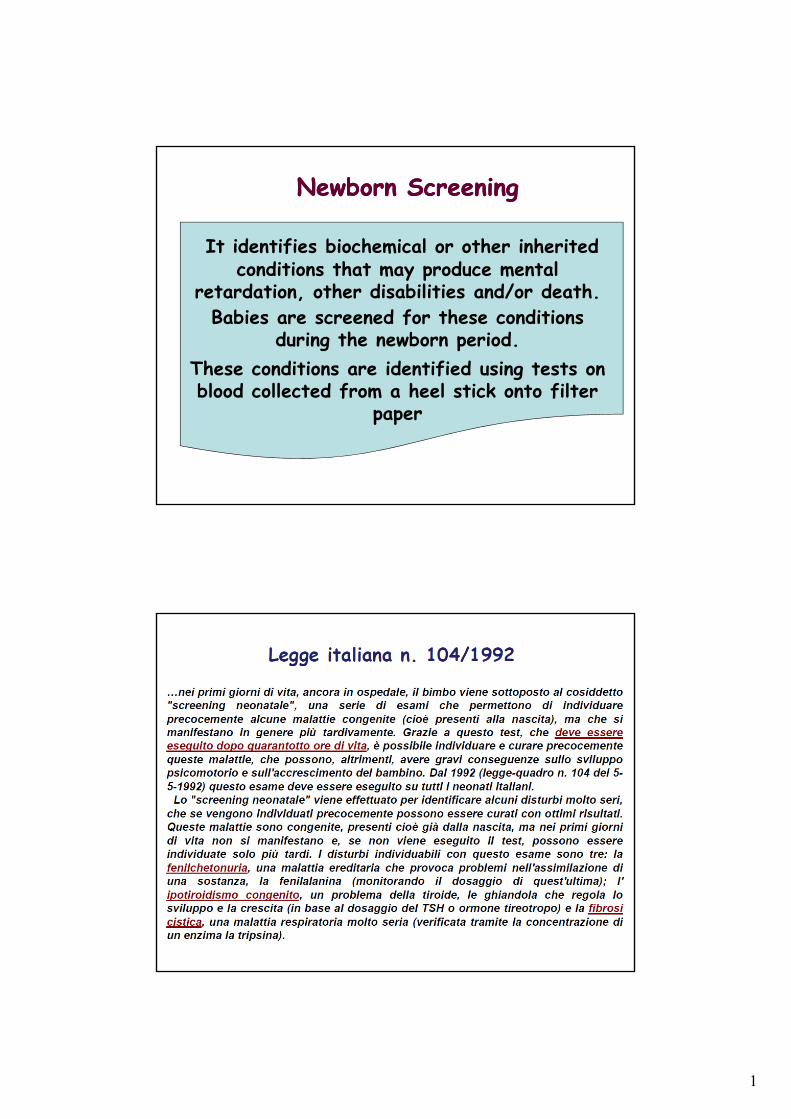
Newborn ScreeningNewborn Screening
It identifies biochemical or other inherited conditions that may produce mental
retardation, other disabilities and/or death. Babies are screened for these conditions
during the newborn period. These conditions are identified using tests on blood collected from a heel stick onto filter
paper
2
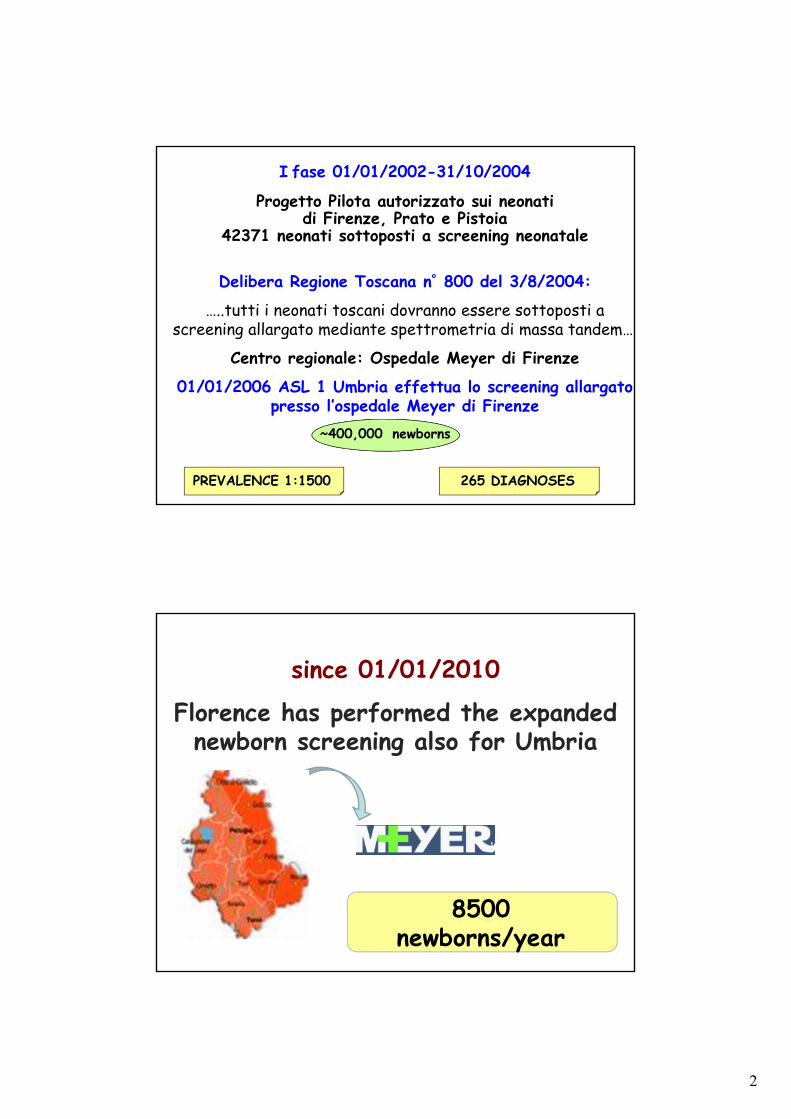
I fase 01/01/2002-31/10/2004
Progetto Pilota autorizzato sui neonati di Firenze, Prato e Pistoia
42371 neonati sottoposti a screening neonatale
Delibera Regione Toscana n˚ 800 del 3/8/2004:
…..tutti i neonati toscani dovranno essere sottoposti a screening allargato mediante spettrometria di massa tandem….
Centro regionale: Ospedale Meyer di Firenze
01/01/2006 ASL 1 Umbria effettua lo screening allargato presso l’ospedale Meyer di Firenze
PREVALENCE 1:1500 265 DIAGNOSES
~400,000 newborns
since 01/01/2010
Florence has performed the expanded newborn screening also for Umbria
8500 newborns/year
3
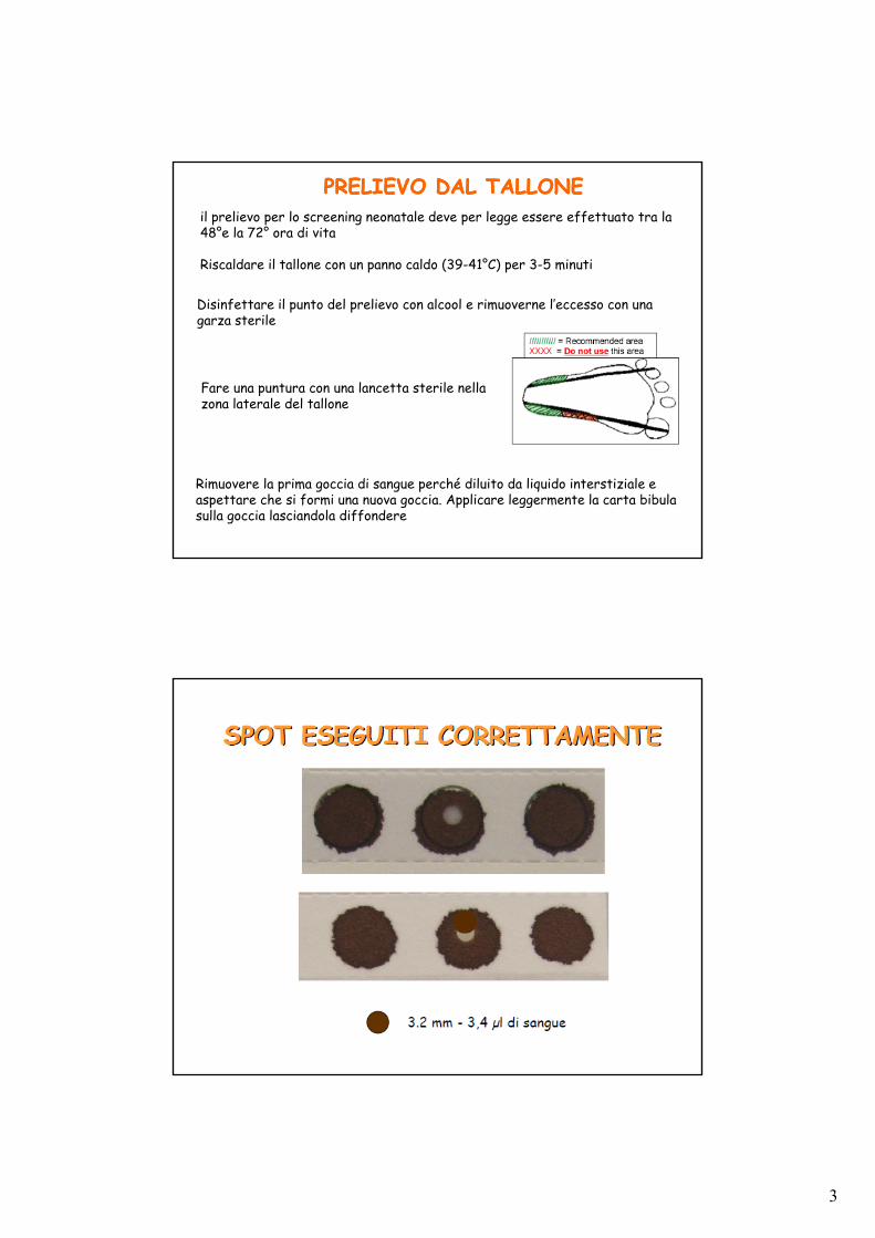
PRELIEVO DAL TALLONEPRELIEVO DAL TALLONEil prelievo per lo screening neonatale deve per legge essere effettuato tra la 48°e la 72° ora di vita
Riscaldare il tallone con un panno caldo (39-41°C) per 3-5 minuti
Disinfettare il punto del prelievo con alcool e rimuoverne l’eccesso con una garza sterile
Fare una puntura con una lancetta sterile nella zona laterale del tallone
Rimuovere la prima goccia di sangue perché diluito da liquido interstiziale e aspettare che si formi una nuova goccia. Applicare leggermente la carta bibula sulla goccia lasciandola diffondere
4
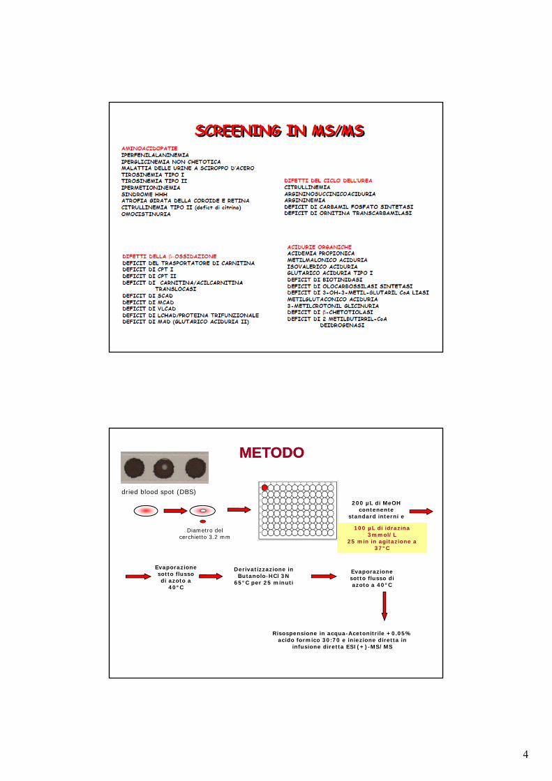
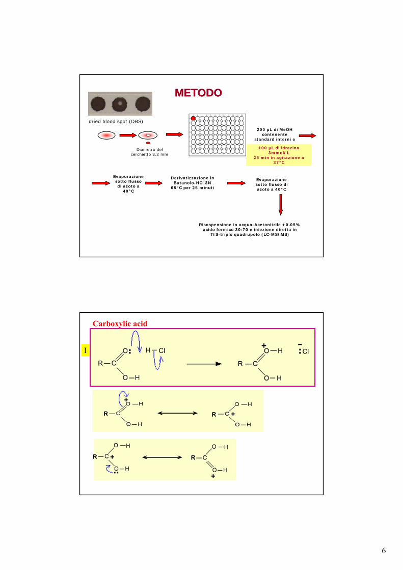
,
dried blood spot (DBS)
Evaporazione sotto flusso di azoto a
40°C
Derivatizzazione in Butanolo-HCl 3N
65°C per 25 minuti
200 µL di MeOH contenente
standard interni e
Risospensione in acqua-Acetonitrile +0.05% acido formico 30:70 e iniezione diretta in
infusione diretta ESI(+)-MS/MS
Diametro del cerchietto 3.2 mm
Evaporazione sotto flusso di azoto a 40°C
100 µL di idrazina 3mmol/L
25 min in agitazione a 37°C
100 µL di idrazina 3mmol/L
25 min in agitazione a 37°C
METODO METODO
5
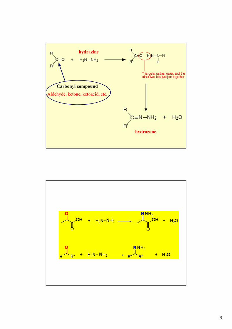
Carbonyl compound
Aldehyde, ketone, ketoacid, etc.
hydrazone
hydrazine
6
,
dried blood spot (DBS)
Evaporazione sotto flusso di azoto a
40°C
Derivatizzazione in Butanolo-HCl 3N
65°C per 25 minuti
200 µL di MeOH contenente
standard interni e
Risospensione in acqua-Acetonitrile +0.05% acido formico 30:70 e iniezione diretta in
TIS-triplo quadrupolo (LC-MS/MS)
Diametro del cerchietto 3.2 mm
Evaporazione sotto flusso di azoto a 40°C
100 µL di idrazina 3mmol/L
25 min in agitazione a 37°C
100 µL di idrazina 3mmol/L
25 min in agitazione a 37°C
METODO METODO
R
Cl Cl
R
R R
R R
I
Carboxylic acid
7
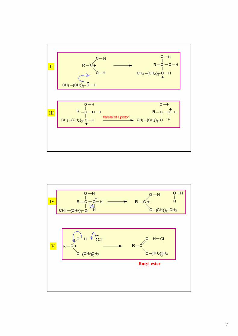
( )3
( )3
R R
( )3 ( )3
R R
II
III
IV
( )3
R
( )3
R
V
( )3
R
( )3
R
Butyl ester
Cl Cl
8
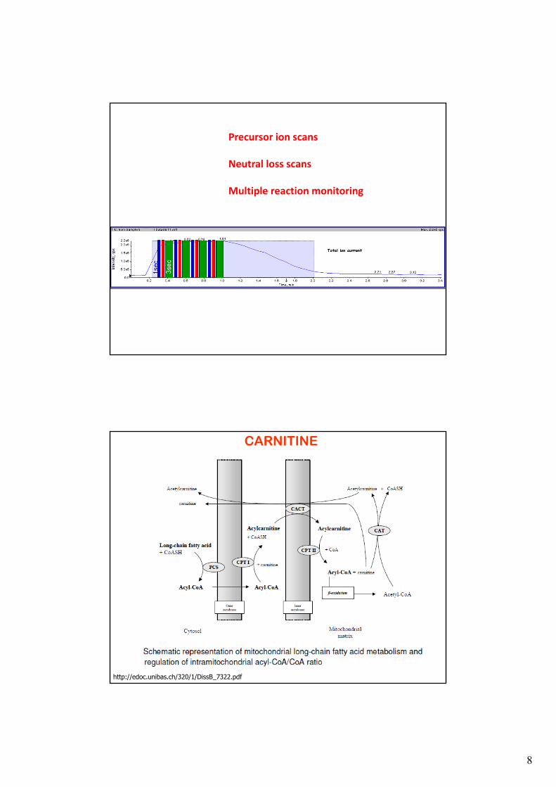
Precursor ion scans
Neutral loss scans
Multiple reaction monitoring
http://edoc.unibas.ch/320/1/DissB_7322.pdf
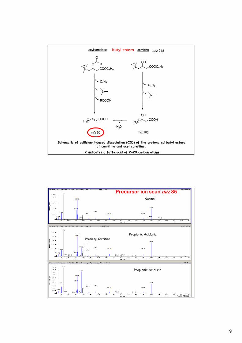
CARNITINE
9
m/z 218
Schematic of collision-induced dissociation (CID) of the protonated butyl esters of carnitine and acyl carnitine.
R indicates a fatty acid of 2-20 carbon atoms
butyl esters
Propionyl CarnitinePropionic Aciduria
Normal
Propionic Aciduria
Precursor ion scan m/z 85
G. la Marca
10
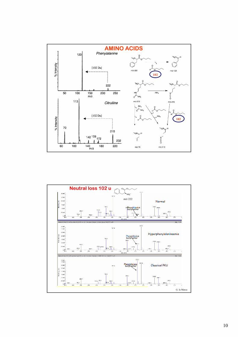
102
102
AMINO ACIDS
Neutral loss 102 u
butyl ester
NH3+
O O
m/z 222
G. la Marca
e
e
e
butyl ester
butyl ester
11
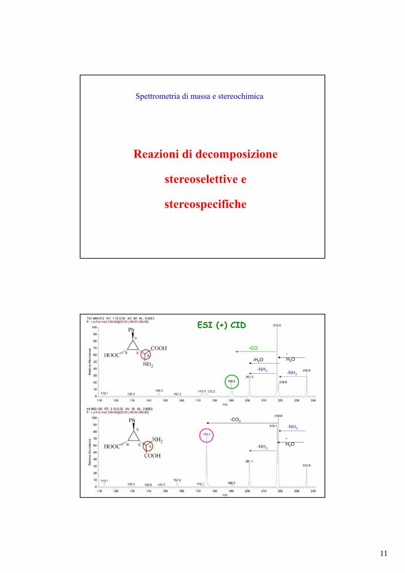
Reazioni di decomposizione
stereoselettive e
stereospecifiche
Spettrometria di massa e stereochimica
-H2O
-NH3
-H2O
-NH3
-CO
-CO2
-NH3
-NH3
-H2O
ESI (+) CID
12
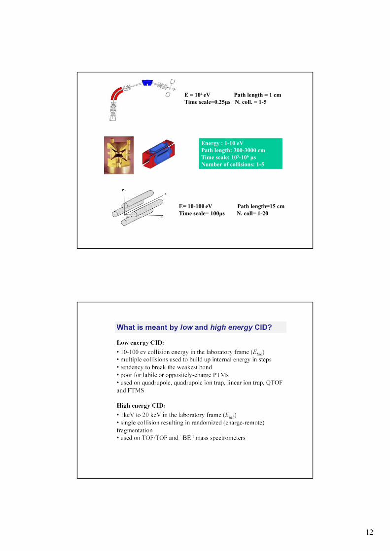
E = 104 eV Path length = 1 cmTime scale=0.25μs N. coll. = 1-5
Energy : 1-10 eVPath length: 300-3000 cm Time scale: 105-106 μsNumber of collisions: 1-5
E= 10-100 eV Path length=15 cmTime scale= 100μs N. coll= 1-20
BE
13
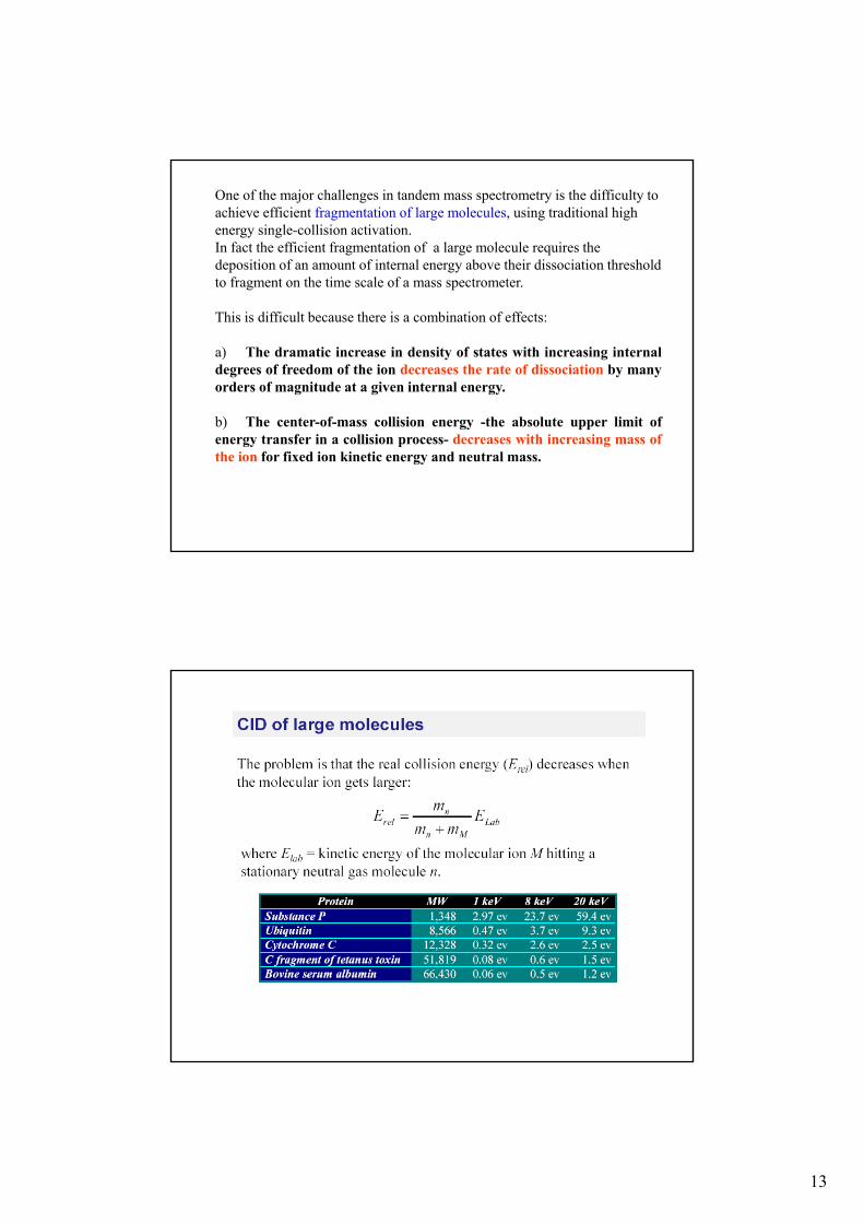
One of the major challenges in tandem mass spectrometry is the difficulty to achieve efficient fragmentation of large molecules, using traditional high energy single-collision activation.In fact the efficient fragmentation of a large molecule requires the deposition of an amount of internal energy above their dissociation threshold to fragment on the time scale of a mass spectrometer.
This is difficult because there is a combination of effects:
a) The dramatic increase in density of states with increasing internaldegrees of freedom of the ion decreases the rate of dissociation by manyorders of magnitude at a given internal energy.
b) The center-of-mass collision energy -the absolute upper limit ofenergy transfer in a collision process- decreases with increasing mass ofthe ion for fixed ion kinetic energy and neutral mass.
14
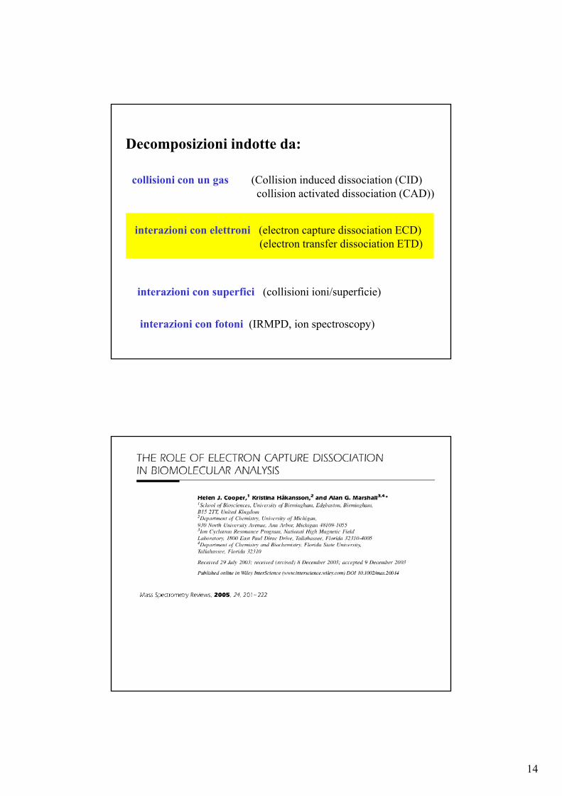
Decomposizioni indotte da:
collisioni con un gas (Collision induced dissociation (CID)collision activated dissociation (CAD))
interazioni con elettroni (electron capture dissociation ECD)(electron transfer dissociation ETD)
interazioni con superfici (collisioni ioni/superficie)
interazioni con fotoni (IRMPD, ion spectroscopy)
15
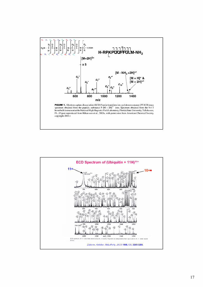
16
17
x3
a1 b1
y3
c1
z3 x2
a2 b2
y2
c2
z2
a3
x1
b3
y1
c3
z1
18
ETD
Electron Transfer Dissociation
interazioni con elettroni
“Ok. Now I want you to figure outhow to do ETD on our ion trapsso we can sequence more phosphopeptides.”
Donald F. HuntK P Q A R K G pS M A D V P K
Ion Trap CID Spectra of Phosphopeptides Are Dominated by Losses of Phosphoric Acid

19
ETD Reaction Scheme
odd-electronprotonated
peptide
Multiply charged analyte(n≥ 2)
Electron-transfer
Cleavage ofN-Cα bondn+ + (n-1)+-
Prerequisite: multiply charged precursor ions, n ≥ 2 !ETD is not applicable to 1+ or negatively charged ions
Reagent radical anion
multiply charged fragment ions n =11, 10, 9, 8, ...
+ n+ n+n+ n+Electron
Transfer12+ ‐
20
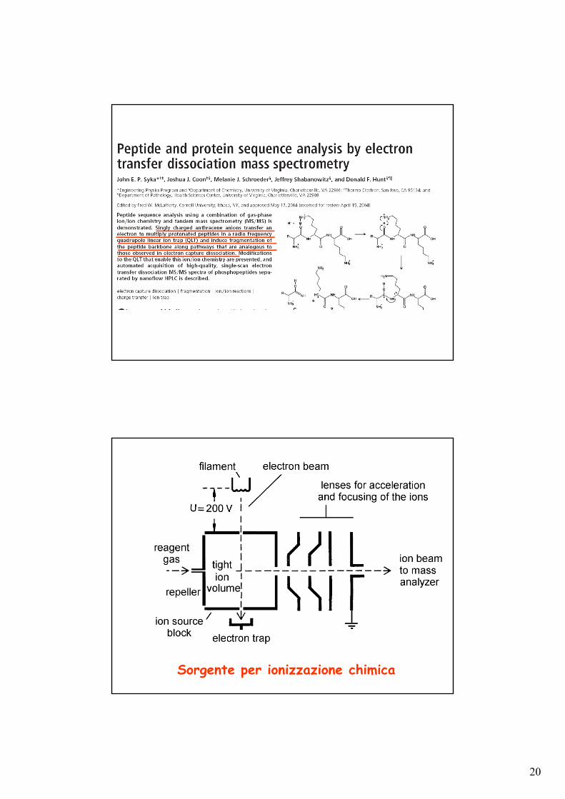
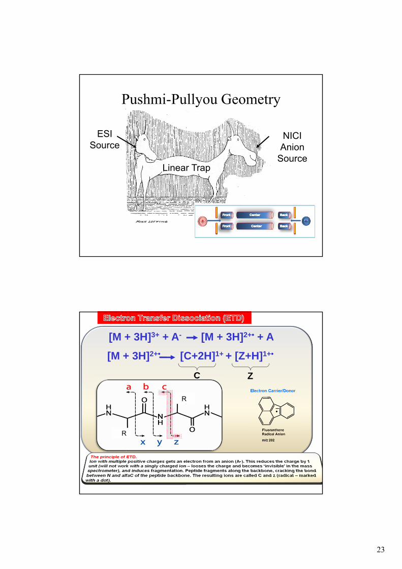
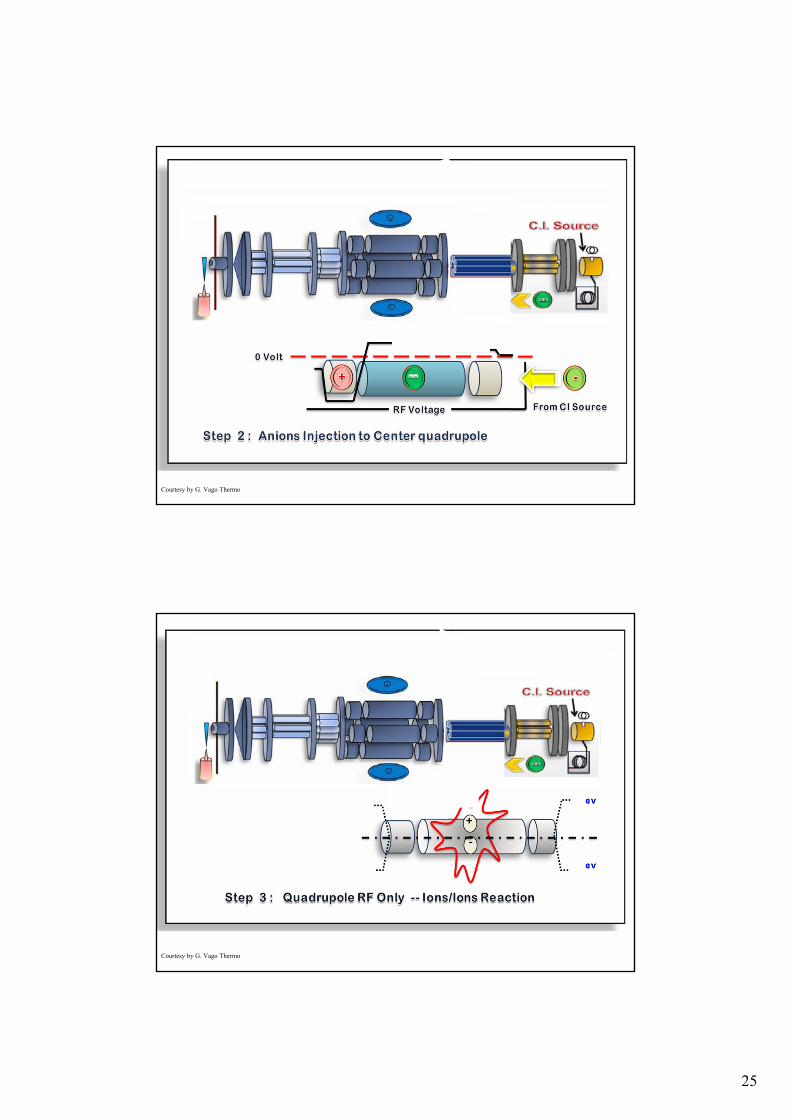
Sorgente per ionizzazione chimica
21
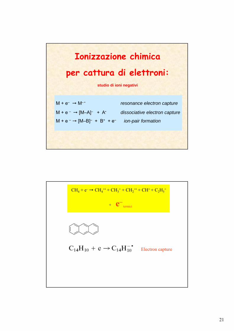
Ionizzazione chimica
per cattura di elettroni:studio di ioni negativi
M + e– M– • resonance electron capture
M + e – [M–A]– + A• dissociative electron capture
M + e – [M–B]– + B+ + e– ion-pair formation
Electron capture
CH4 + e– CH4+• + CH3
+ + CH2+• + CH+ + C2H5
+
+ e–termici
22
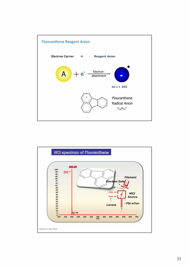
Fluoranthene Reagent Anion
Electron Carrier Reagent Anion
A e-+ Electron-attachment
FlourantheneRadical Anion
C16H10•-
m/z = 202
-
-•
100 150 200 250 300 350 400 450 500 550 600 650 700m/z
05
101520253035404550556065707580859095
100
217.09
NICISource
Filament
e-
~700 mTorrLenses
Electron Gate
[M]–
Courtesy by G. Vago Thermo
23
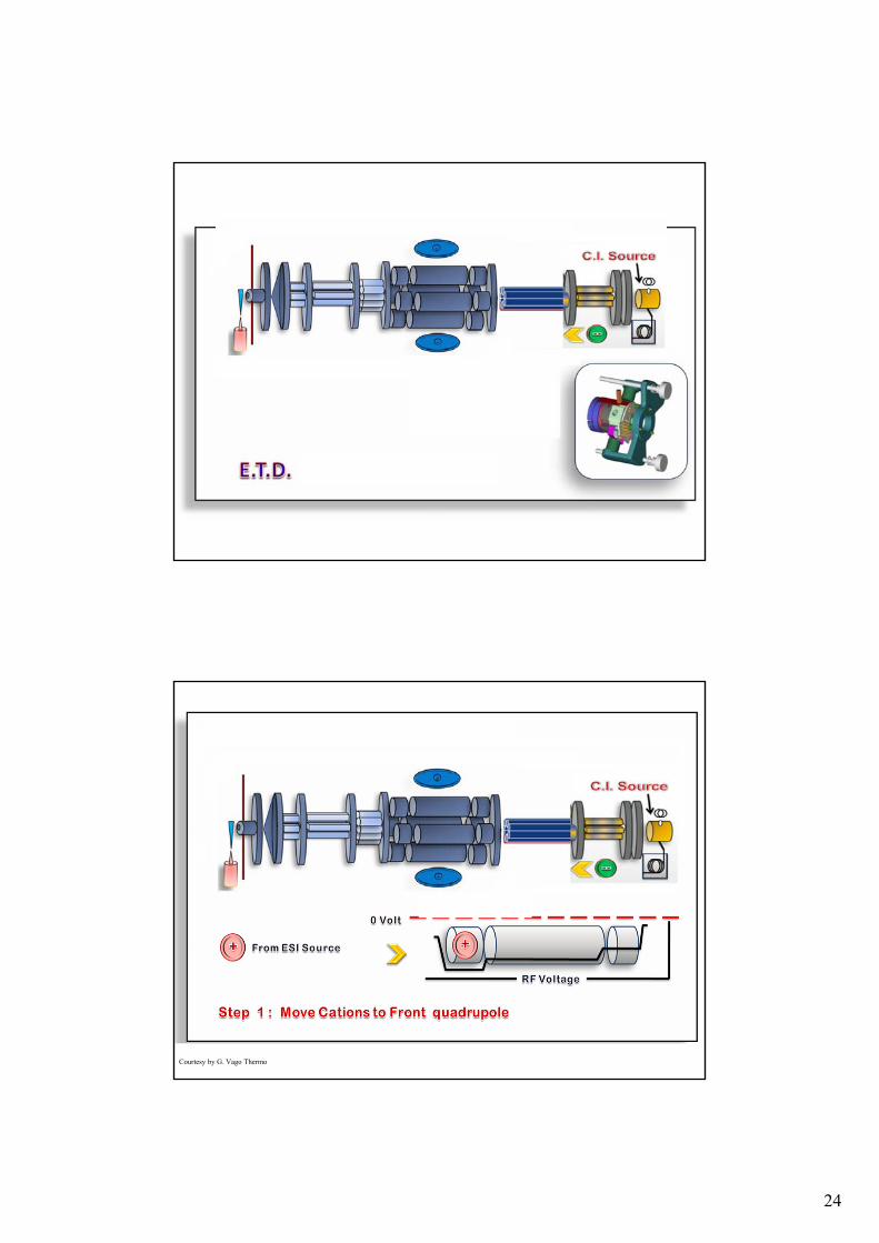
Pushmi-Pullyou Geometry
ESISource
NICIAnion
SourceLinear Trap
[M + 3H]3+ + A- [M + 3H]2+• + A
[M + 3H]2+• [C+2H]1+ + [Z+H]1+•
C Z
R
R
24
s
Courtesy by G. Vago Thermo
25
s
Courtesy by G. Vago Thermo
s
Courtesy by G. Vago Thermo
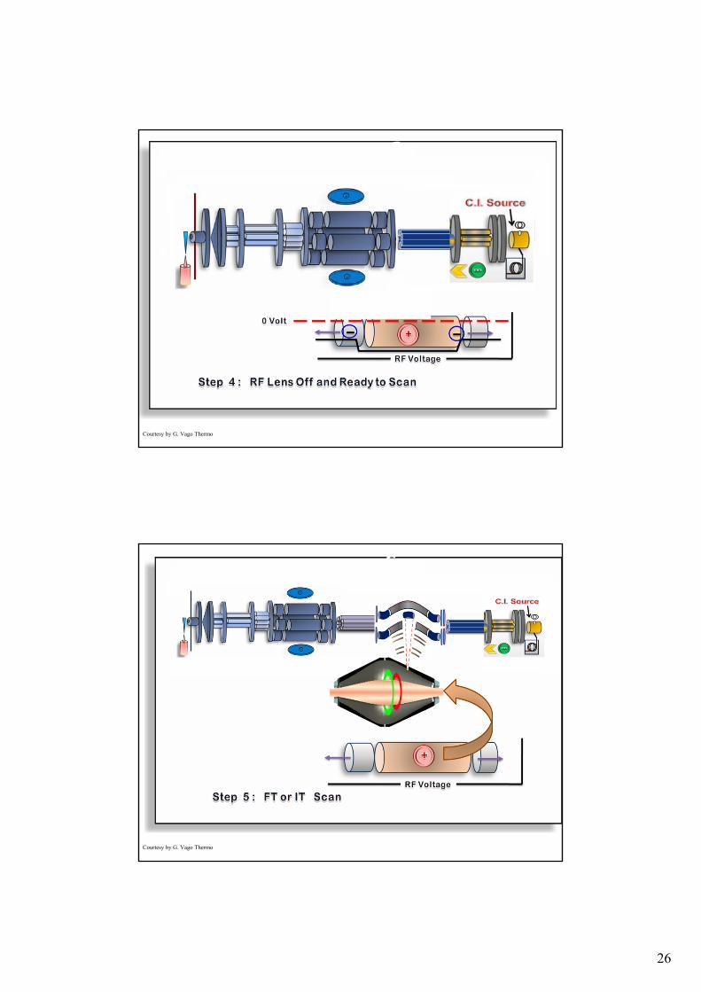
26
s
Courtesy by G. Vago Thermo
s
Courtesy by G. Vago Thermo
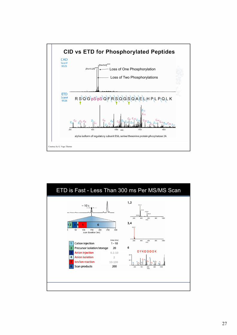
27
Loss of One Phosphorylation
Loss of Two Phosphorylations
Courtesy by G. Vago Thermo
54
ETD is Fast - Less Than 300 ms Per MS/MS Scan
2
10-100
0.1-10
28
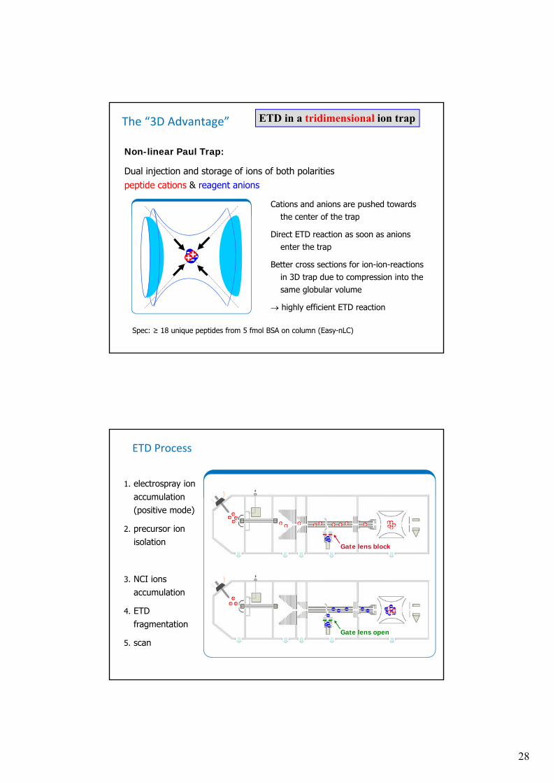
The “3D Advantage”
Cations and anions are pushed towards the center of the trap
Direct ETD reaction as soon as anions enter the trap
Better cross sections for ion-ion-reactions in 3D trap due to compression into the same globular volume
highly efficient ETD reaction
Non-linear Paul Trap:
Dual injection and storage of ions of both polarities peptide cations & reagent anions
Spec: ≥ 18 unique peptides from 5 fmol BSA on column (Easy-nLC)
ETD in a tridimensional ion trap
ETD Process
1. electrospray ion accumulation(positive mode)
2. precursor ion isolation
3. NCI ions accumulation
4. ETD fragmentation
5. scan
Gate lens block
Gate lens open
29
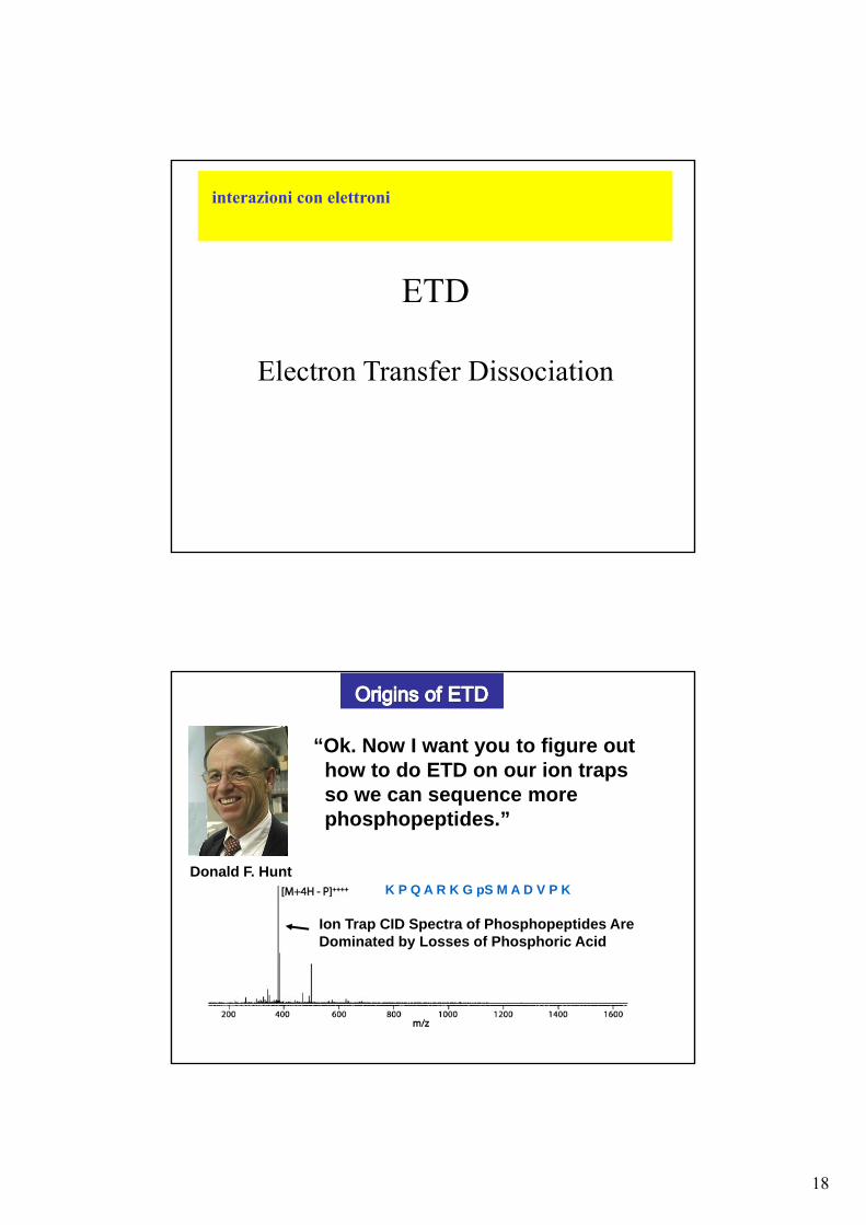
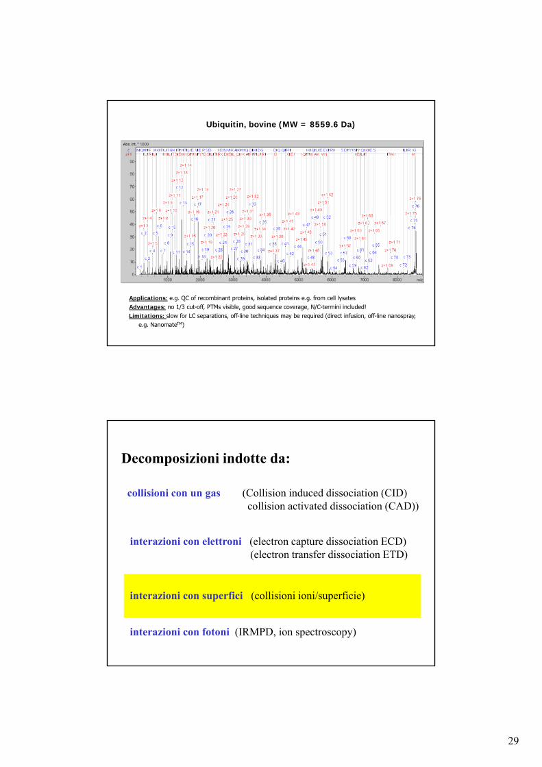
Ubiquitin, bovine (MW = 8559.6 Da)
Applications: e.g. QC of recombinant proteins, isolated proteins e.g. from cell lysatesAdvantages: no 1/3 cut-off, PTMs visible, good sequence coverage, N/C-termini included!Limitations: slow for LC separations, off-line techniques may be required (direct infusion, off-line nanospray,
e.g. NanomateTM)
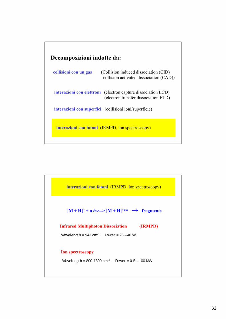
Decomposizioni indotte da:
collisioni con un gas (Collision induced dissociation (CID)collision activated dissociation (CAD))
interazioni con elettroni (electron capture dissociation ECD)(electron transfer dissociation ETD)
interazioni con superfici (collisioni ioni/superficie)
interazioni con fotoni (IRMPD, ion spectroscopy)
30
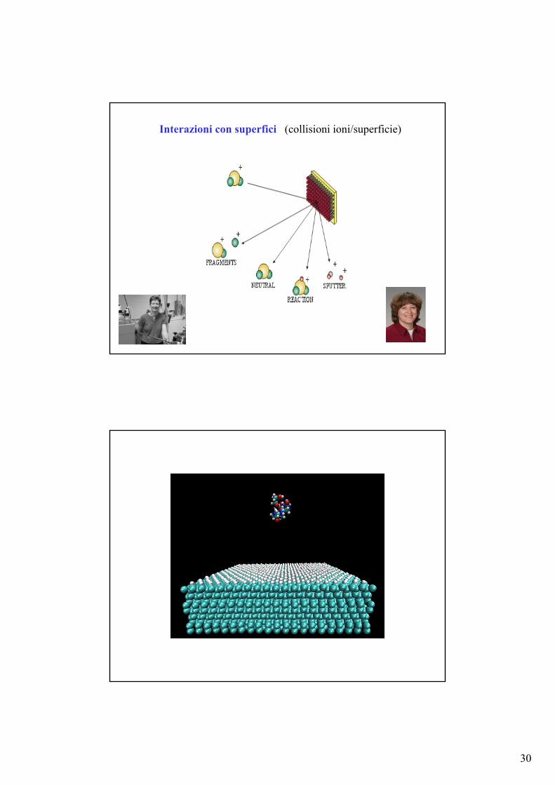
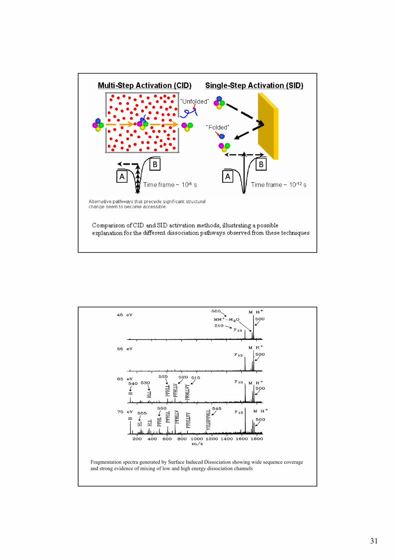
Interazioni con superfici (collisioni ioni/superficie)
31
Fragmentation spectra generated by Surface Induced Dissociation showing wide sequence coverage and strong evidence of mixing of low and high energy dissociation channels
32
Decomposizioni indotte da:
collisioni con un gas (Collision induced dissociation (CID)collision activated dissociation (CAD))
interazioni con elettroni (electron capture dissociation ECD)(electron transfer dissociation ETD)
interazioni con superfici (collisioni ioni/superficie)
interazioni con fotoni (IRMPD, ion spectroscopy)
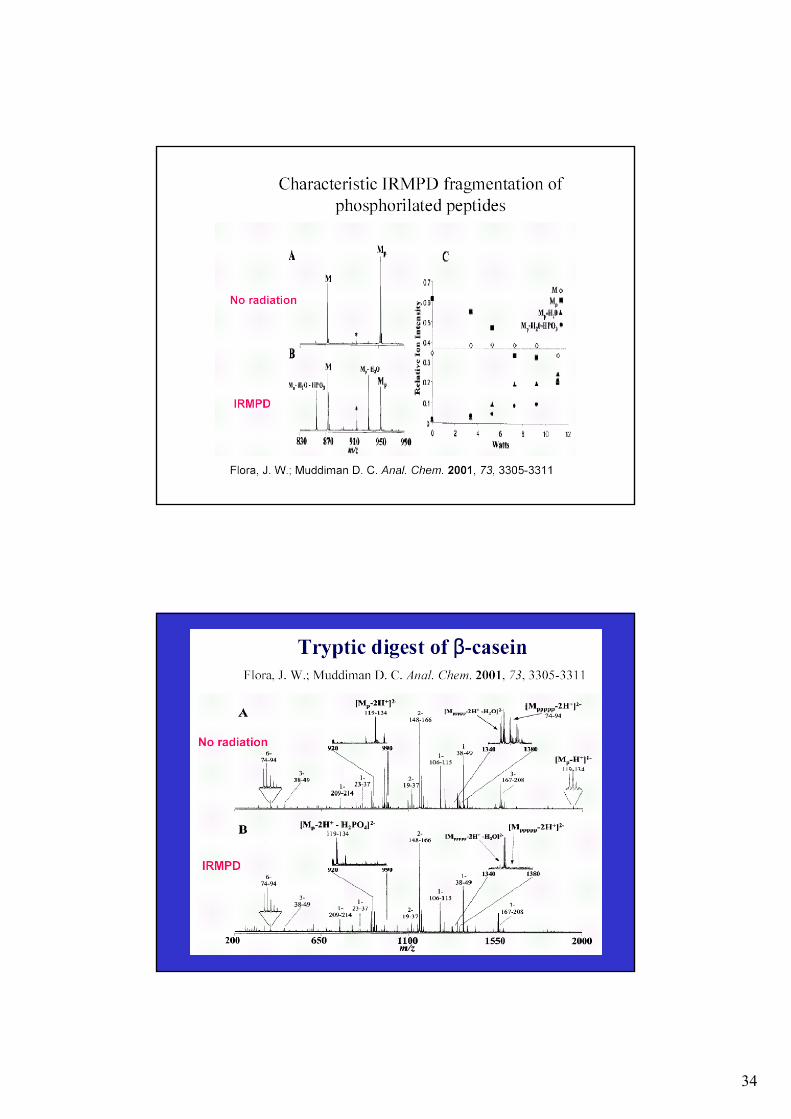
Infrared Multiphoton Dissociation (IRMPD)
interazioni con fotoni (IRMPD, ion spectroscopy)
Wavelength = 943 cm-1 Power = 25 – 40 W
[M + H]+ + n h --> [M + H]+** → fragments
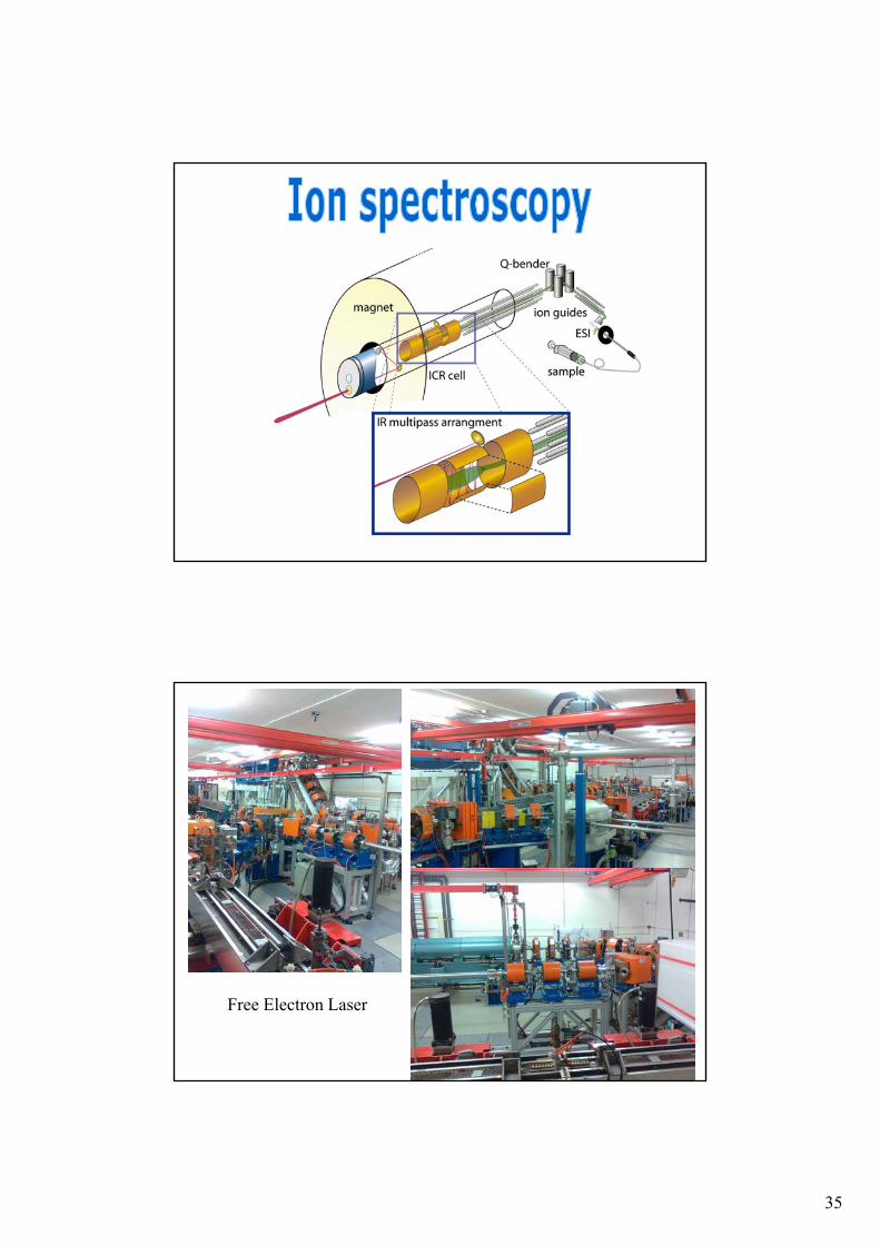
Ion spectroscopy
Wavelength = 800-1800 cm-1 Power = 0.5 – 100 MW
33
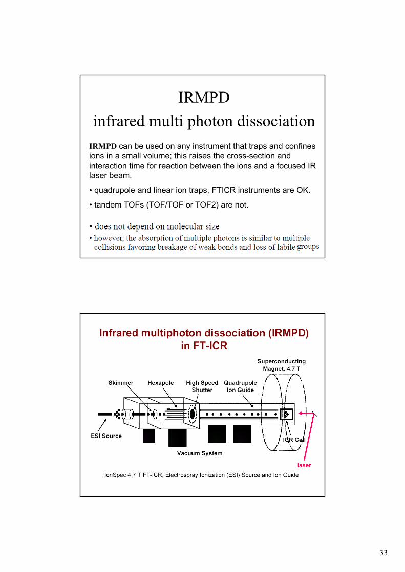
IRMPD
infrared multi photon dissociation
IRMPD can be used on any instrument that traps and confines ions in a small volume; this raises the cross-section and interaction time for reaction between the ions and a focused IR laser beam.
• quadrupole and linear ion traps, FTICR instruments are OK.
• tandem TOFs (TOF/TOF or TOF2) are not.
34
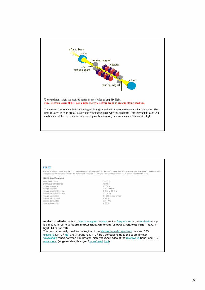
35
Free Electron Laser
36
'Conventional' lasers use excited atoms or molecules to amplify light.Free-electron lasers (FEL) use a high-energy electron beam as an amplifying medium.
The electron beam emits light as it wiggles through a periodic magnetic structure called ondulator. The light is stored in in an optical cavity, and can interact back with the electrons. This interaction leads to a modulation of the electronic density, and a growth in intensity and coherence of the emitted light.
terahertz radiation refers to electromagnetic waves sent at frequencies in the terahertz range.It is also referred to as submillimeter radiation, terahertz waves, terahertz light, T-rays, T-light, T-lux and THz.The term is normally used for the region of the electromagnetic spectrum between 300 gigahertz (3x10¹¹ Hz) and 3 terahertz (3x10¹² Hz), corresponding to the submillimeter wavelength range between 1 millimeter (high-frequency edge of the microwave band) and 100 micrometer (long-wavelength edge of far-infrared light).
37
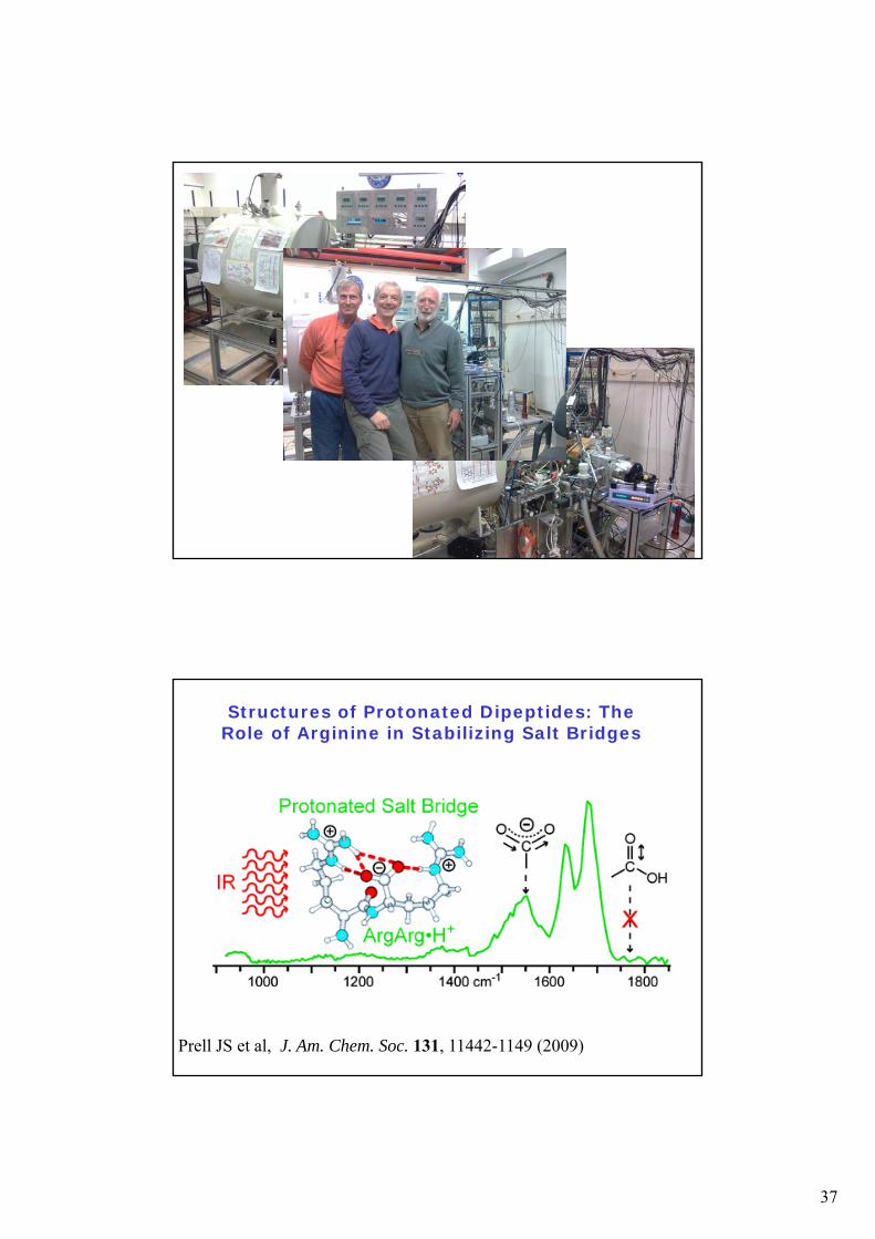
Structures of Protonated Dipeptides: The Role of Arginine in Stabilizing Salt Bridges
Prell JS et al, J. Am. Chem. Soc. 131, 11442-1149 (2009)
38
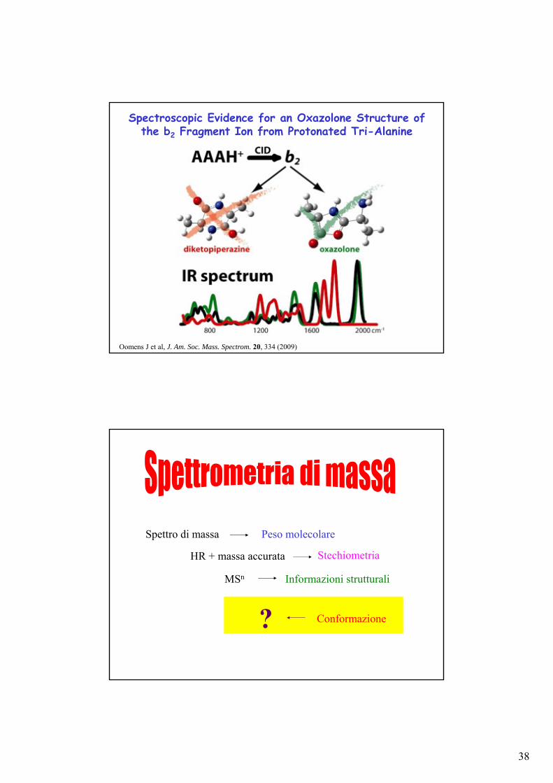
Spectroscopic Evidence for an Oxazolone Structure of the b2 Fragment Ion from Protonated Tri-Alanine
Oomens J et al, J. Am. Soc. Mass. Spectrom. 20, 334 (2009)
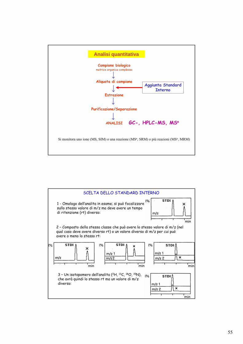
Conformazione
Spettro di massa Peso molecolare
HR + massa accurata Stechiometria
MSn Informazioni strutturali
?
39
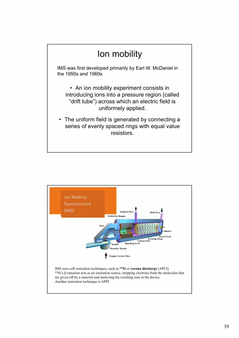
Ion mobility
• An ion mobility experiment consists in introducing ions into a pressure region (called
“drift tube”) across which an electric field is uniformely applied.
• The uniform field is generated by connecting a series of evenly spaced rings with equal value
resistors.
IMS was first developed primarily by Earl W. McDaniel in the 1950s and 1960s
IMS uses soft ionization techniques, such as 63Ni or corona discharge (APCI).63Ni’s β emission acts as an ionization source, stripping electrons from the molecules that are given off by a material and analyzing the resulting ions in the device.Another ionization technique is APPI.
40
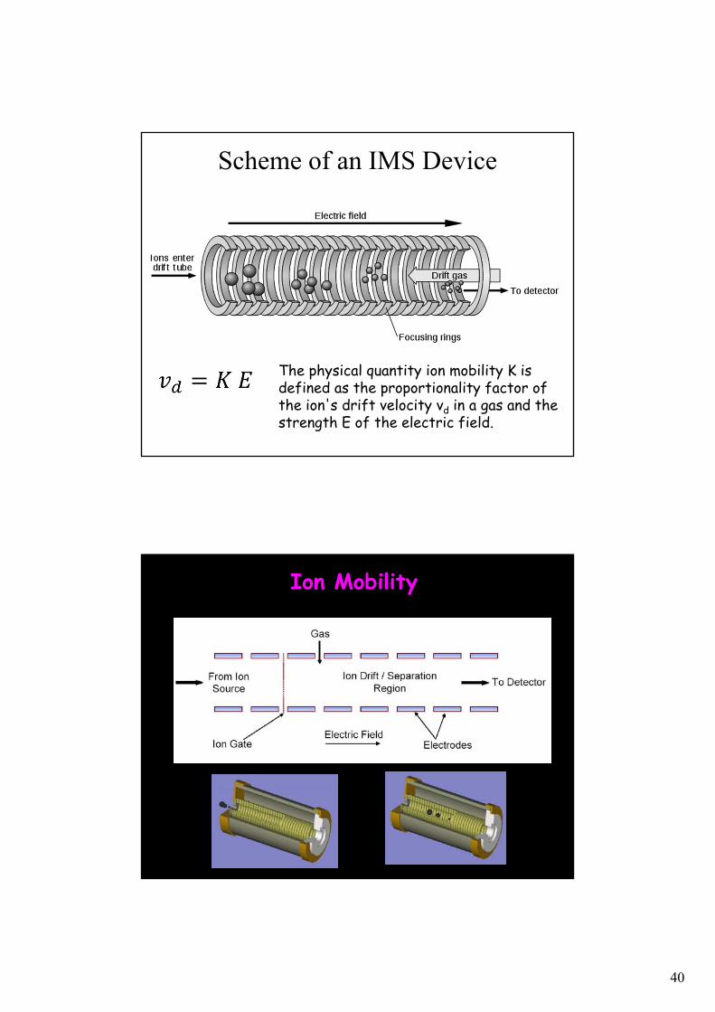
Scheme of an IMS Device
The physical quantity ion mobility K is defined as the proportionality factor of the ion's drift velocity vd in a gas and the strength E of the electric field.
Ion Mobility
41
42
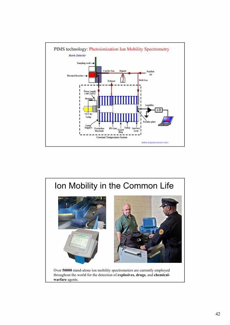
PIMS technology: Photoionization Ion Mobility Spectrometry
Ion Mobility in the Common Life
Over 50000 stand-alone ion mobility spectrometers are currently employed throughout the world for the detection of explosives, drugs, and chemical-warfare agents.
43
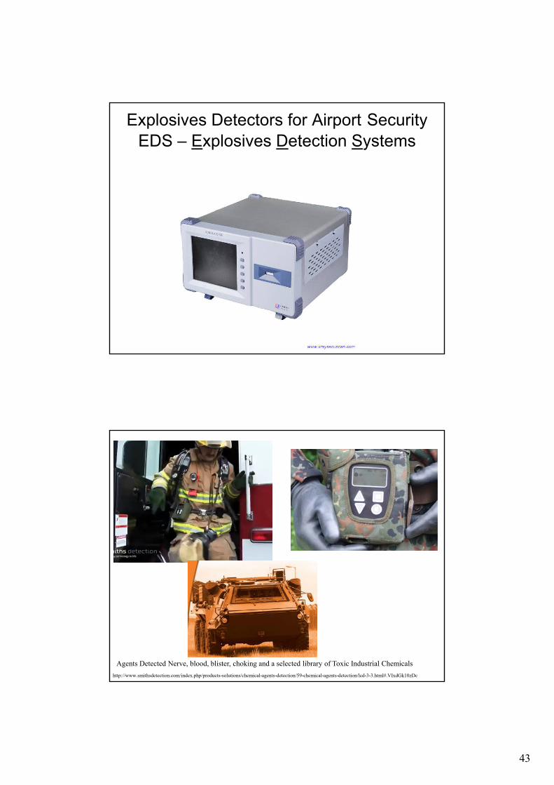
Explosives Detectors for Airport SecurityEDS – Explosives Detection Systems
http://www.smithsdetection.com/index.php/products-solutions/chemical-agents-detection/59-chemical-agents-detection/lcd-3-3.html#.VIxdGk10zDc
Agents Detected Nerve, blood, blister, choking and a selected library of Toxic Industrial Chemicals
44
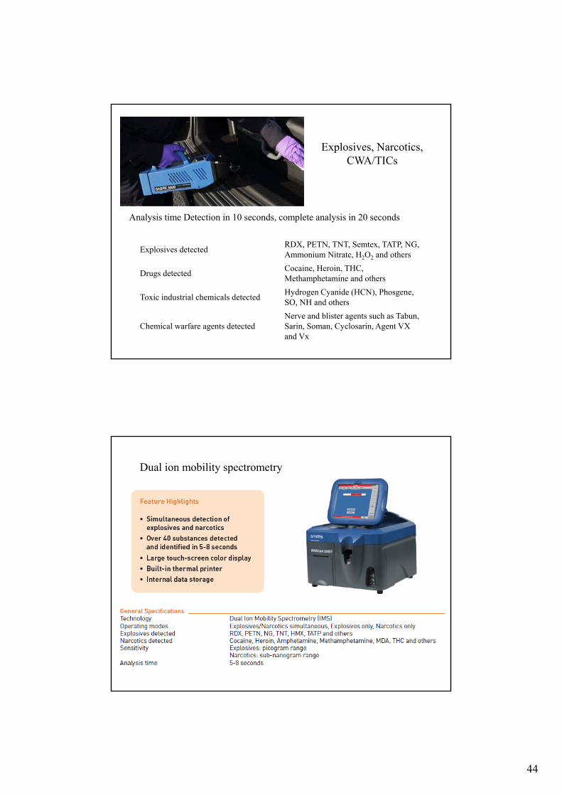
Explosives, Narcotics, CWA/TICs
Explosives detectedRDX, PETN, TNT, Semtex, TATP, NG, Ammonium Nitrate, H2O2 and others
Drugs detectedCocaine, Heroin, THC, Methamphetamine and others
Toxic industrial chemicals detectedHydrogen Cyanide (HCN), Phosgene, SO, NH and others
Chemical warfare agents detectedNerve and blister agents such as Tabun, Sarin, Soman, Cyclosarin, Agent VX and Vx
Analysis time Detection in 10 seconds, complete analysis in 20 seconds
Dual ion mobility spectrometry
45
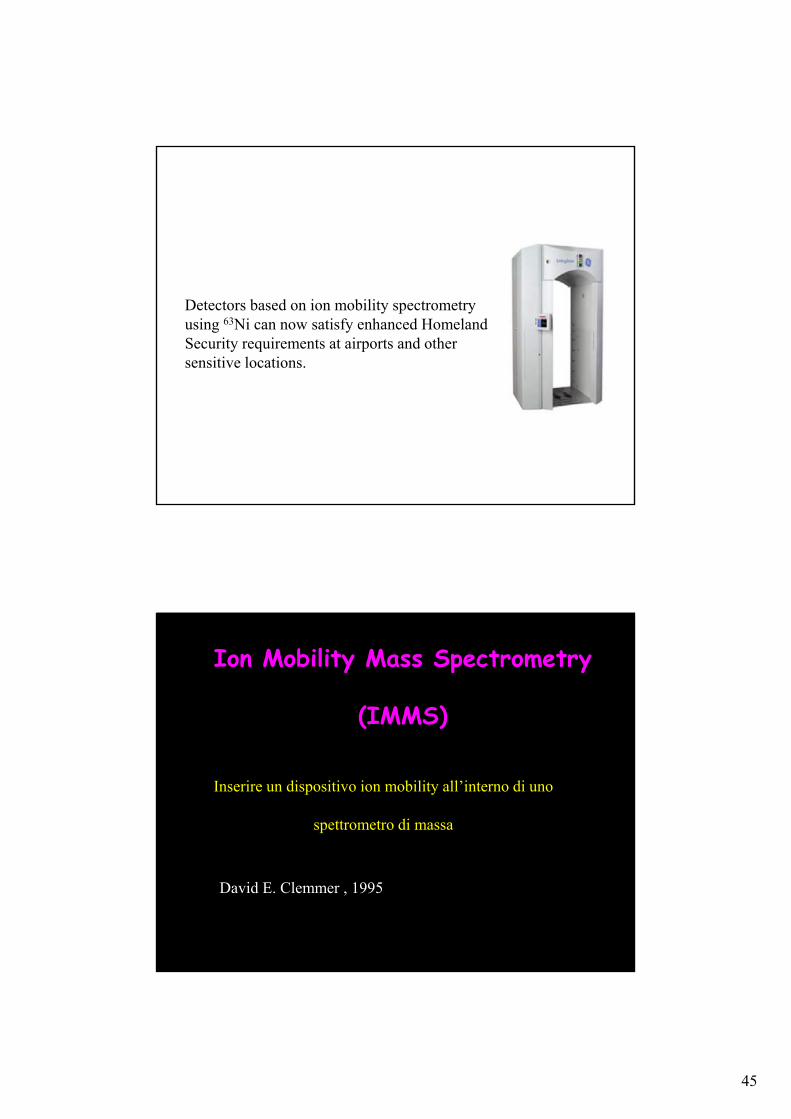
Detectors based on ion mobility spectrometry using 63Ni can now satisfy enhanced Homeland Security requirements at airports and other sensitive locations.
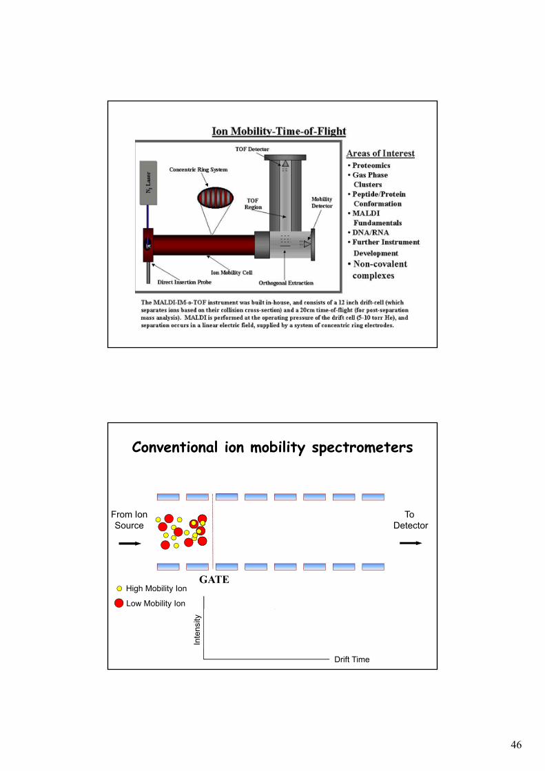
Ion Mobility Mass Spectrometry
(IMMS)
Inserire un dispositivo ion mobility all’interno di uno
spettrometro di massa
David E. Clemmer , 1995
46
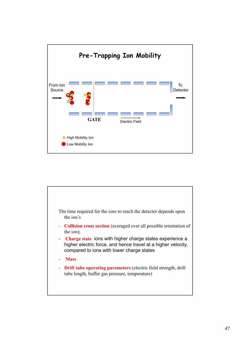
GATE
Low Mobility Ion
High Mobility Ion
Inte
nsity
Drift Time
From IonSource
To Detector
Conventional ion mobility spectrometers
47
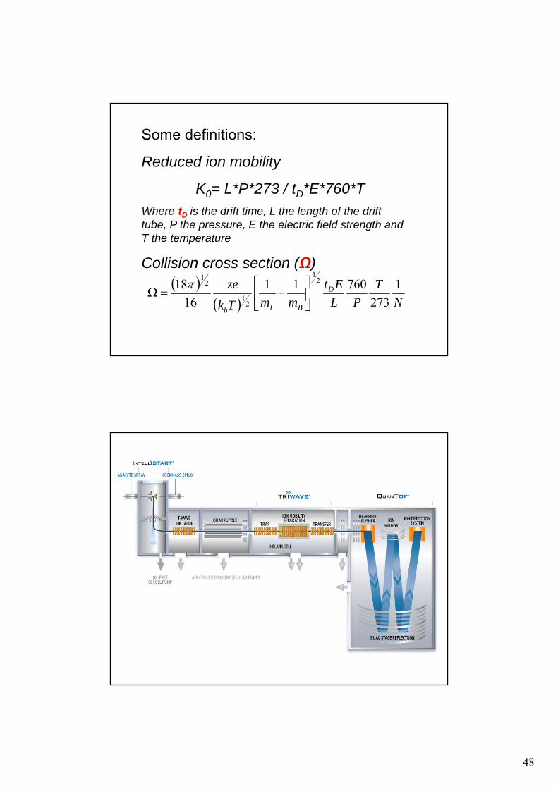
GATE
To Detector
From IonSource
Electric Field
Low Mobility Ion
High Mobility Ion
Pre-Trapping Ion Mobility
The time required for the ions to reach the detector depends upon the ion’s
- Collision cross section (averaged over all possible orientation of the ion);
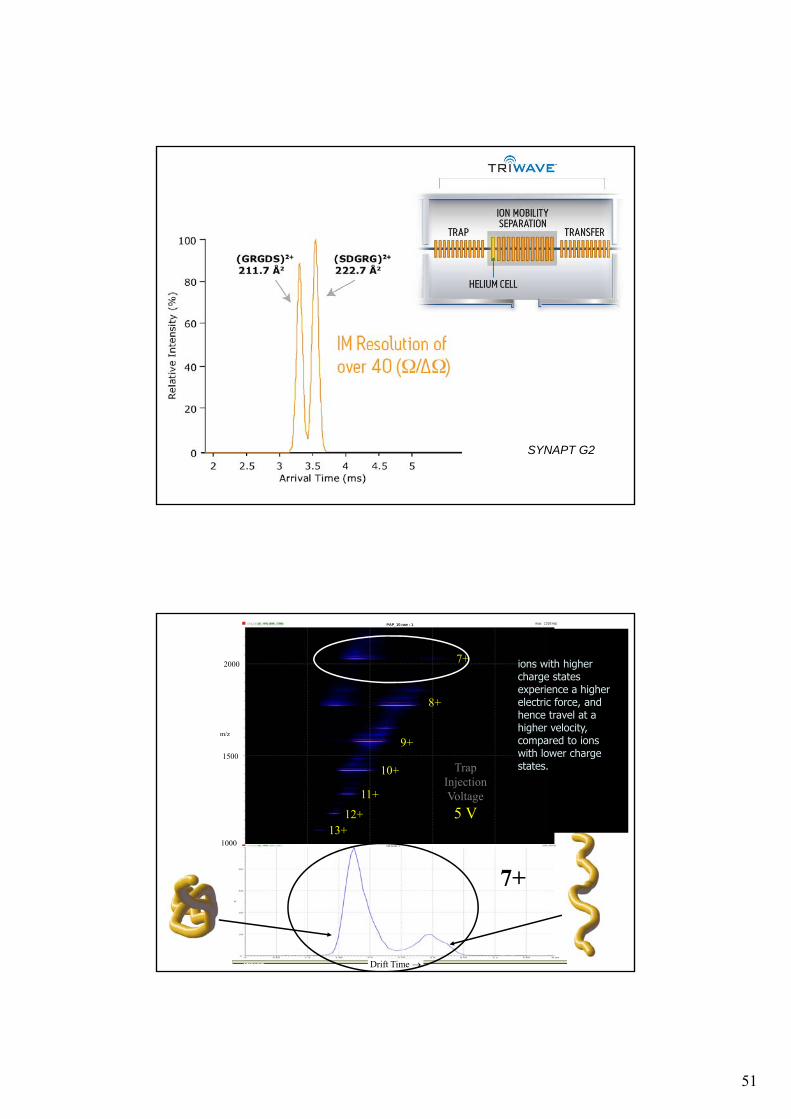
- Charge state ions with higher charge states experience a higher electric force, and hence travel at a higher velocity, compared to ions with lower charge states
- Mass
- Drift tube operating parameters (electric field strength, drift tube length, buffer gas pressure, temperature)
48
Some definitions:
Reduced ion mobility
K0= L*P*273 / tD*E*760*T
Where tD is the drift time, L the length of the drift tube, P the pressure, E the electric field strength and T the temperature
Collision cross section (Ω)
N
T
PL
Et
mmTk
ze D
BIb
1
273
76011
16
18 21
21
21
49
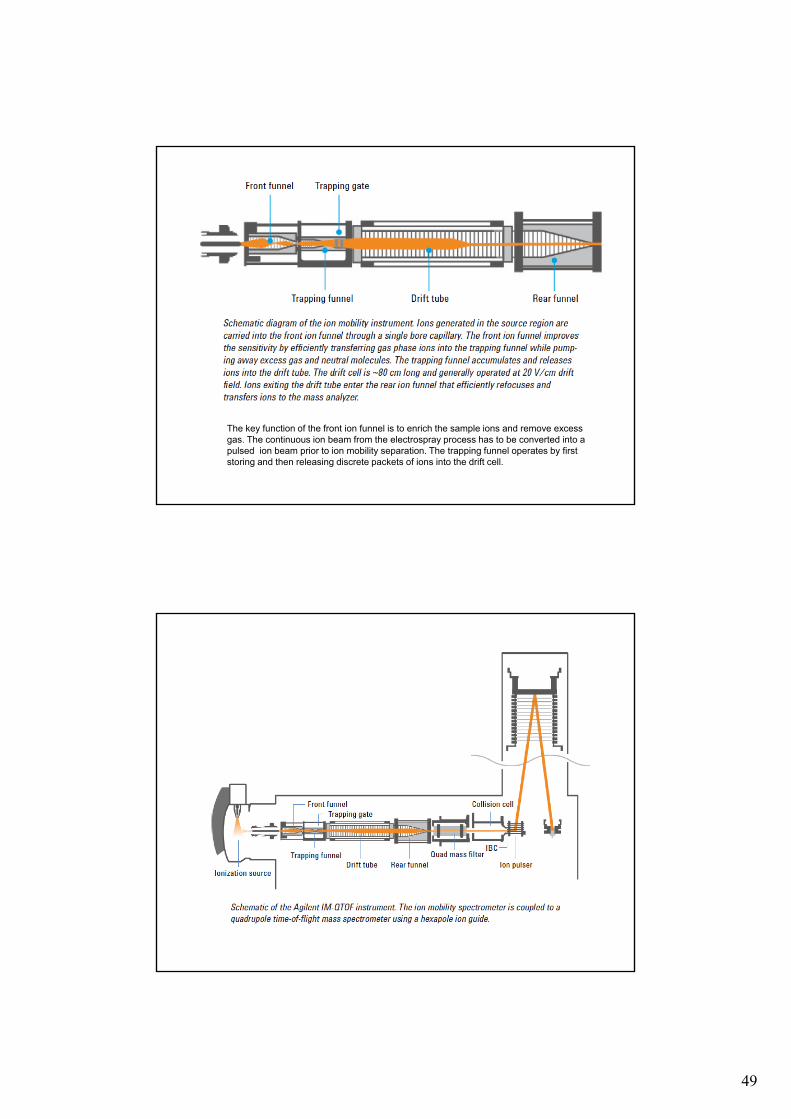
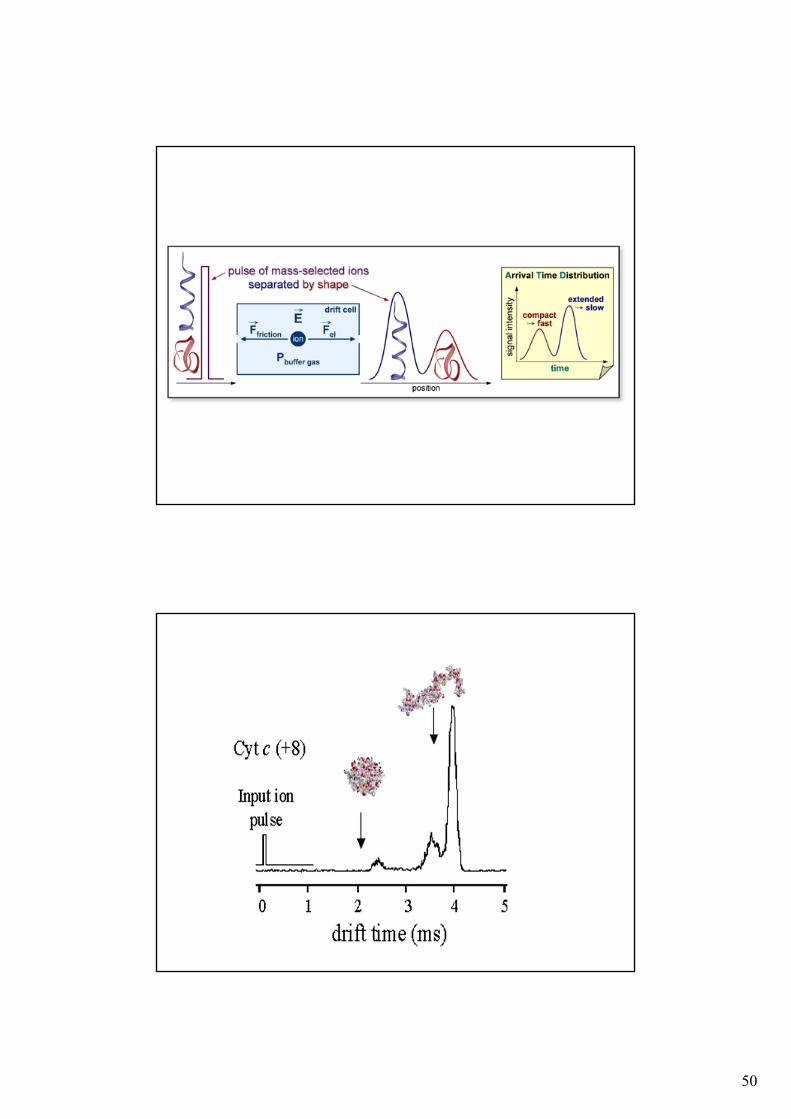
The key function of the front ion funnel is to enrich the sample ions and remove excess gas. The continuous ion beam from the electrospray process has to be converted into a pulsed ion beam prior to ion mobility separation. The trapping funnel operates by first storing and then releasing discrete packets of ions into the drift cell.
50
51
SYNAPT G2
PAP_10.raw:1
PAP_10.raw : 1
PAP_10.raw:1
PAP_10.raw : 1
7+
8+
9+
10+
11+
12+13+
7+
Trap Injection Voltage
5 V
m/z
1000
1500
2000
Drift Time
ions with higher charge states experience a higher electric force, and hence travel at ahigher velocity, compared to ions with lower charge states.
52
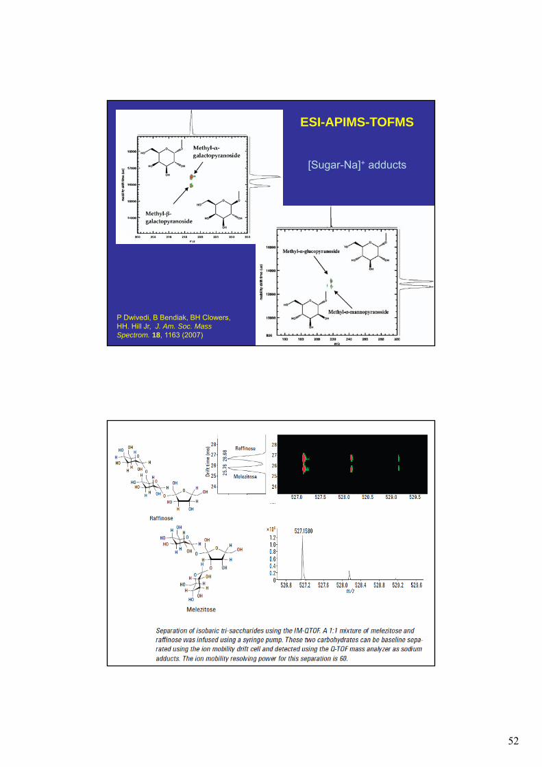
[Sugar-Na]+ adducts
P Dwivedi, B Bendiak, BH Clowers, HH. Hill Jr, J. Am. Soc. Mass Spectrom. 18, 1163 (2007)
ESI-APIMS-TOFMS
53
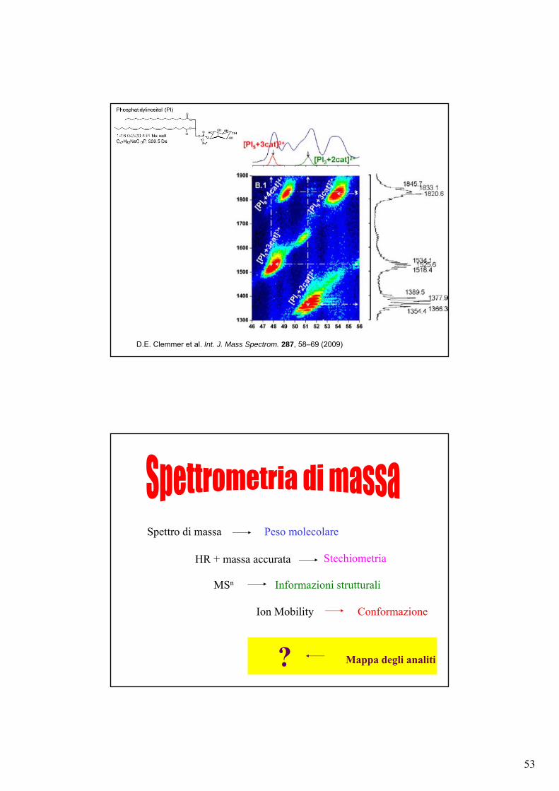
D.E. Clemmer et al. Int. J. Mass Spectrom. 287, 58–69 (2009)
Mappa degli analiti
Spettro di massa Peso molecolare
HR + massa accurata Stechiometria
MSn Informazioni strutturali
?
ConformazioneIon Mobility
54
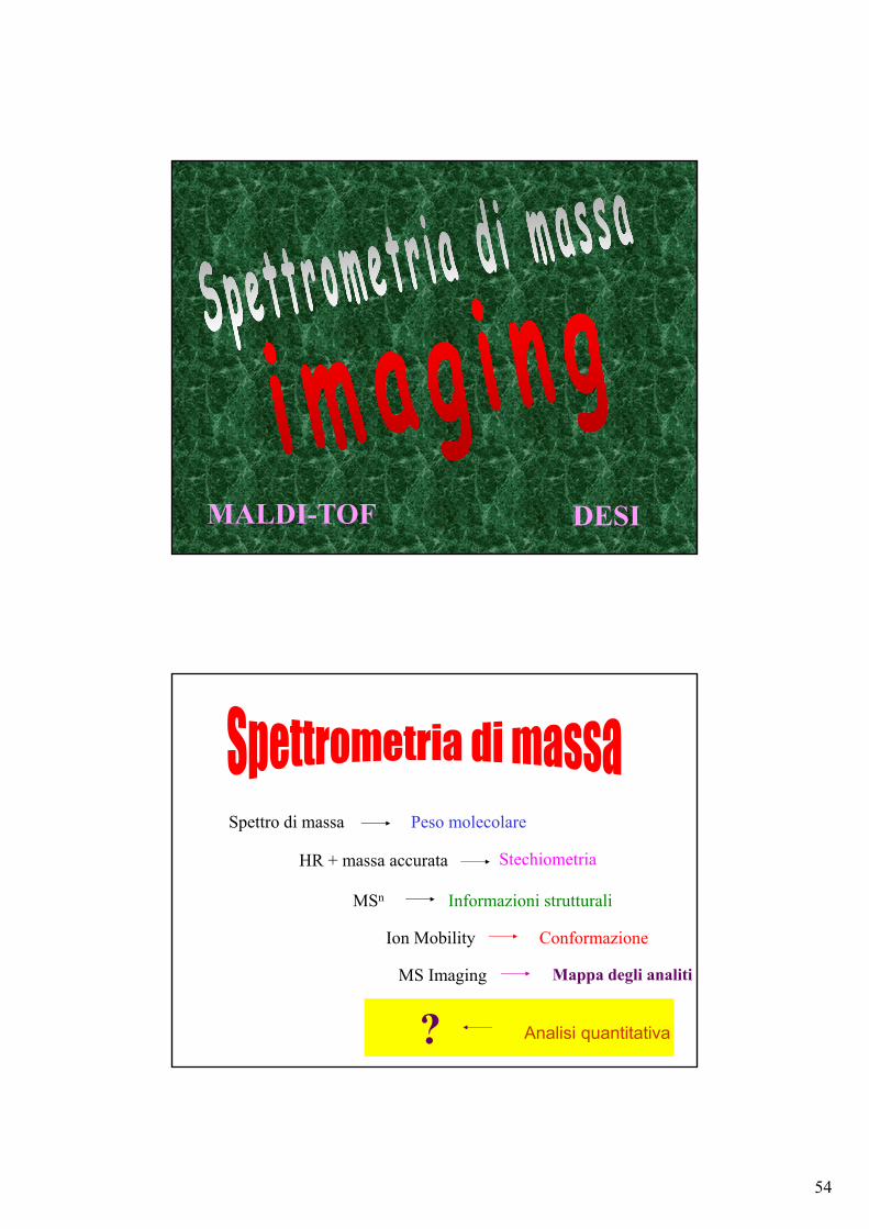
MALDI-TOF DESI
Analisi quantitativa
Spettro di massa Peso molecolare
HR + massa accurata Stechiometria
MSn Informazioni strutturali
?
Mappa degli analiti
ConformazioneIon Mobility
MS Imaging
55
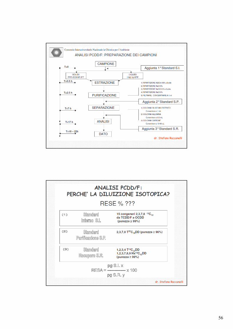
Campione biologicomatrice organica complessa
Aliquota di campione
Estrazione
Purificazione/Separazione
ANALISI
Aggiunta Standard Interno
Analisi quantitativa
GC-, HPLC-MS, MSn
Si monitora uno ione (MS, SIM) o una reazione (MSn, SRM) o più reazioni (MSn, MRM)
SCELTA DELLO STANDARD INTERNO
1 - Omologo dell’analita in esame; si può focalizzare sullo stesso valore di m/z ma deve avere un tempo di ritenzione (rt) diverso:
2 – Composto della stessa classe che può avere lo stesso valore di m/z (nel qual caso deve avere diverso rt) o un valore diverso di m/z per cui può avere o meno lo stesso rt:
3 – Un isotopomero dell’analita (2H, 13C, 18O, 15N), che avrà quindi lo stesso rt ma un valore di m/z diverso:
56
dr. Stefano Raccanelli
dr. Stefano Raccanelli
57
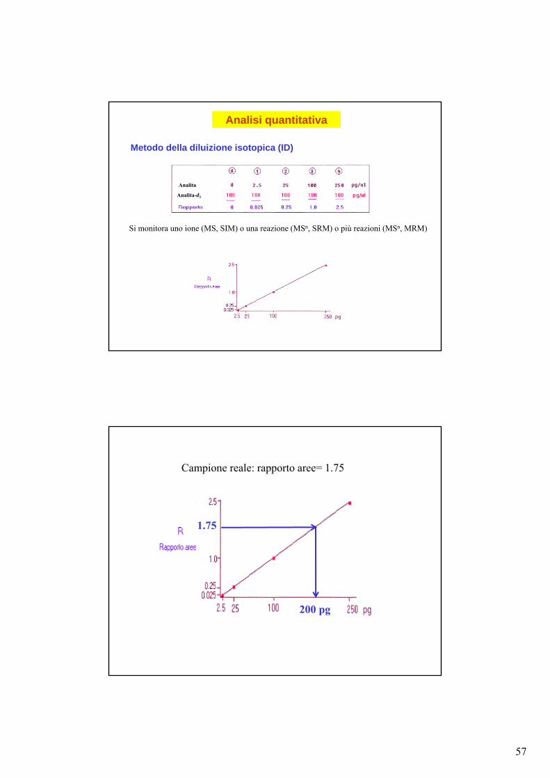
Analisi quantitativa
Metodo della diluizione isotopica (ID)
Analita
Analita-d3
Si monitora uno ione (MS, SIM) o una reazione (MSn, SRM) o più reazioni (MSn, MRM)
Campione reale: rapporto aree= 1.75
200 pg
1.75
58
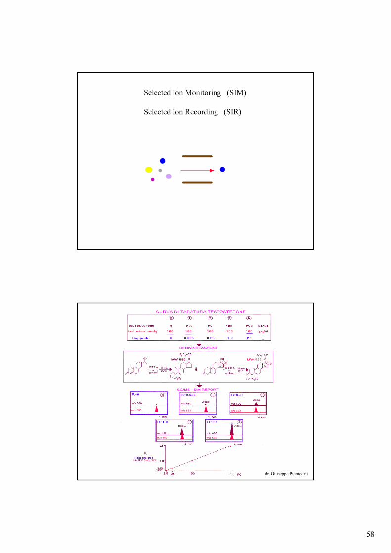
Selected Ion Monitoring (SIM)
Selected Ion Recording (SIR)
dr. Giuseppe Pieraccini
59
dr. Giuseppe Pieraccini
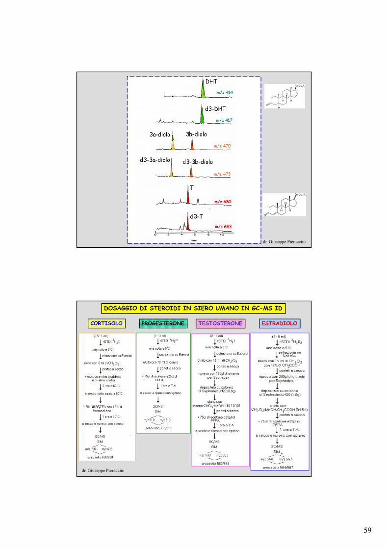
DOSAGGIO DI STEROIDI IN SIERO UMANO IN GC-MS ID
CORTISOLO PROGESTERONE TESTOSTERONE ESTRADIOLO
dr. Giuseppe Pieraccini

60
Ion Trap Tandem Mass Spectrometric Determinationof F2-Isoprostanes
Cinzia Signorini, Mario Comporti, Gianluca Giorgi *, J. Mass Spectrom. 38, 1067-1074 (2003)
GC-NICI-ITMS/MS
•F2-isoprostanes, are formed in vivo by non-enzymatic free radical-induced oxidation of arachidonic acid. •8-epi-PGF2, the most abundant isomer formed, can exist as two main distereoisomers differing in the stereochemistry at C(15), namely 15(R) and 15(S).•F2-Isoprostanes have many characteristics to be considered as a reliable index of oxidative stress in vivo.
OH
HOCH3
COOH
OH
8-epi-15(R)-PGF2
OH
HOCH3
OH
COOH
8-epi-15(S)-PGF2
m/z 573
m/z 569
OSi(CH3)3
(CH3)3SiOCH3
OSi(CH3)3
COO CH2
F F
F
FF
569
181
OSi(CH3)3
(CH3)3SiOCH3
OSi(CH3)3
D D
D DCOO CH2
F F
F
FF181
573
GC-NICI-ITMS/MSof isoprostanes
- TMSOH
- 2 TMSOH
- 3 TMSOH
- CO2
61
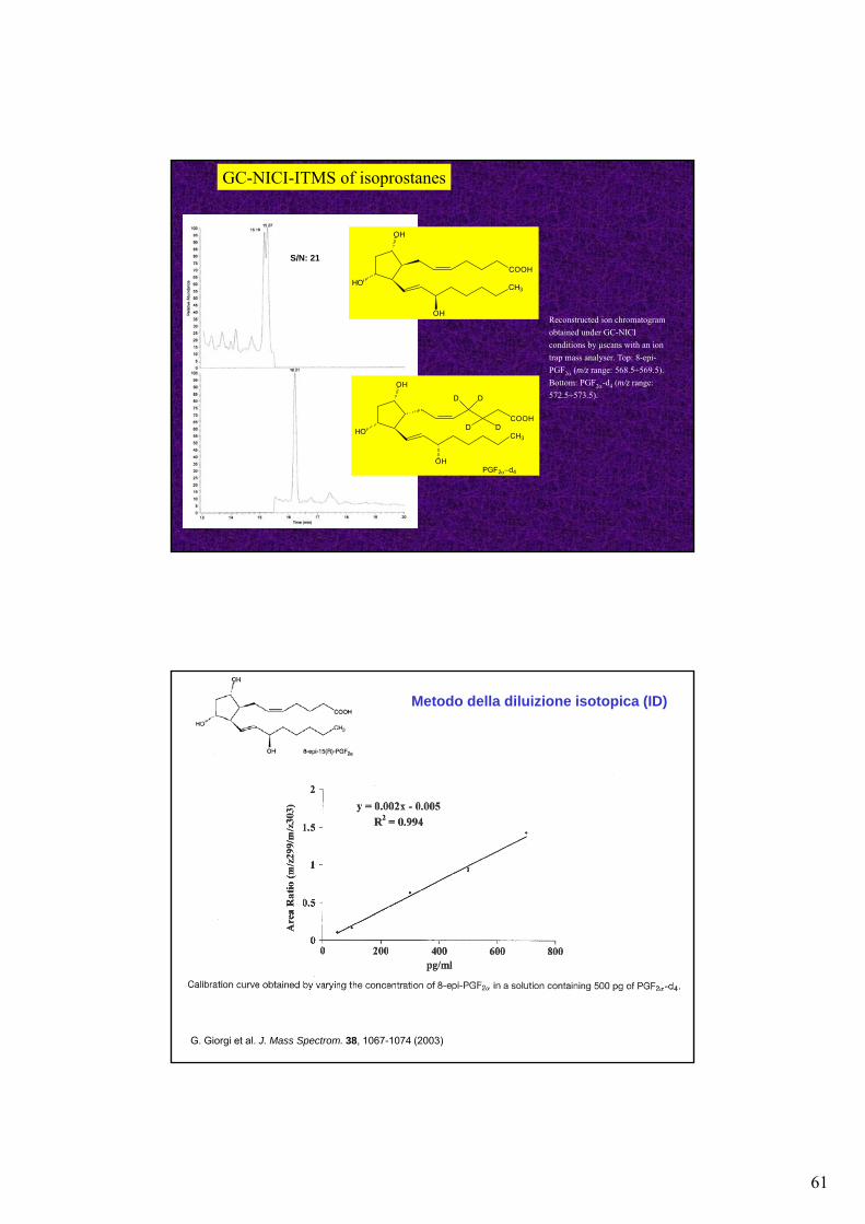
GC-NICI-ITMS of isoprostanes
Reconstructed ion chromatogram
obtained under GC-NICI
conditions by μscans with an ion
trap mass analyser. Top: 8-epi-
PGF2 (m/z range: 568.5÷569.5).
Bottom: PGF2-d4 (m/z range:
572.5÷573.5).
OH
HOCH3
COOH
OH
PGF2d4
OH
HO
COOH
CH3
OH
D D
D D
S/N: 21
G. Giorgi et al. J. Mass Spectrom. 38, 1067-1074 (2003)
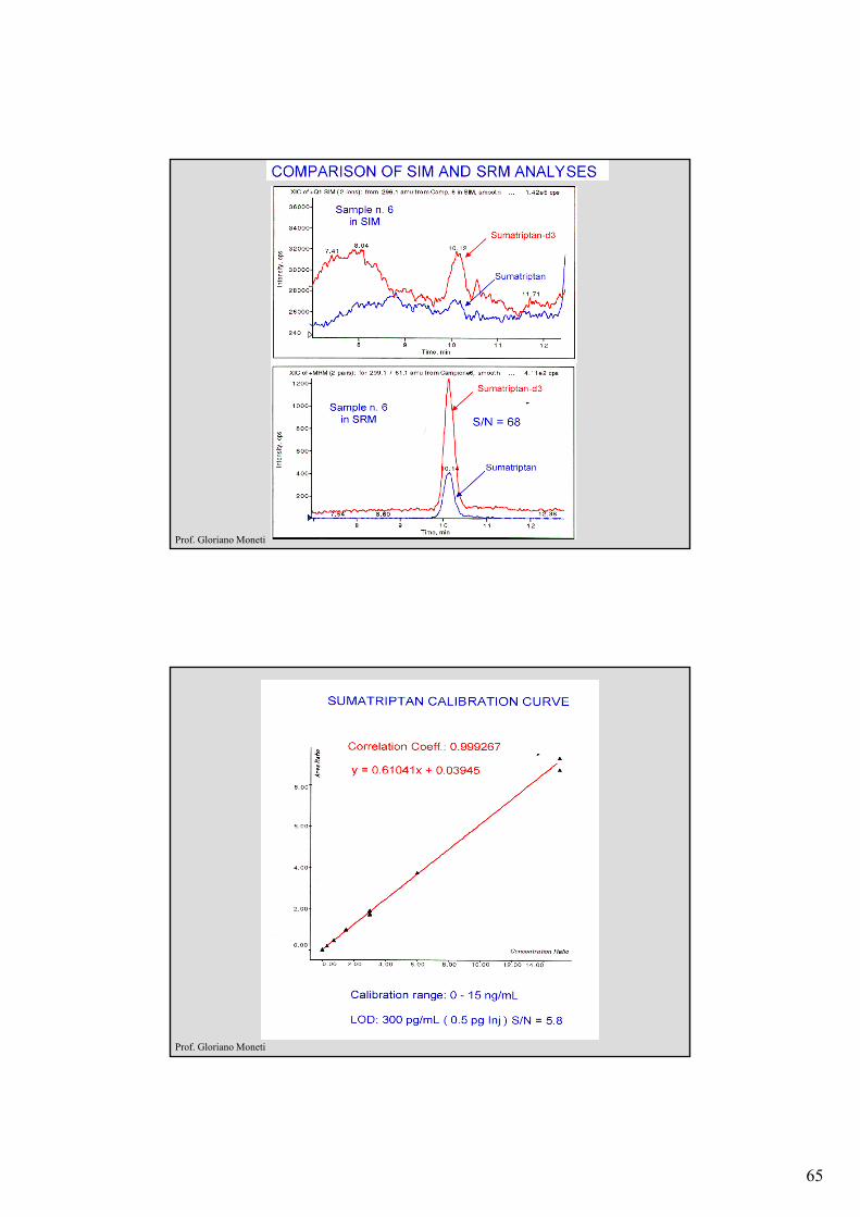
Metodo della diluizione isotopica (ID)
62
GC-NICI/SIM
GC-NICI/MS/MS
GC-NICI-ITMS/MSof isoprostanes
healthy adult human plasma
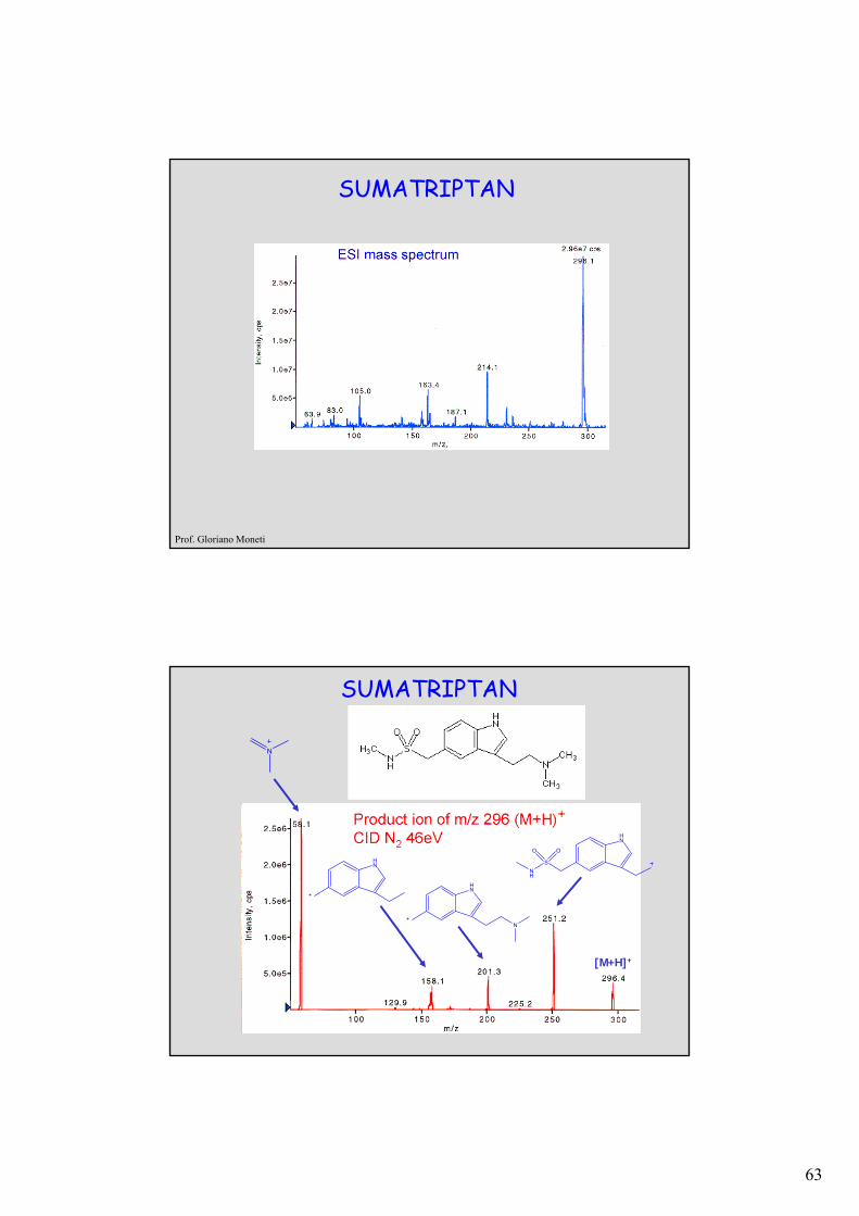
CAS Registry number: [103628-46-2]
CA name(s): 3-[2-(Dimethylamino)ethyl]-N-methyl-1H-indole-5-methanesulfonamide;
Drug code(s): GR-43175. Derivative: Succinate
CAS Registry number: [103628-48-4] Drug code(s): GR-43175C
Trade name(s): Imigran (Glaxo), Imitrex (Glaxo). THERAP. CAT.: Antimigraine.
Molecular formula: C14H21N3O2S . C4H6O4
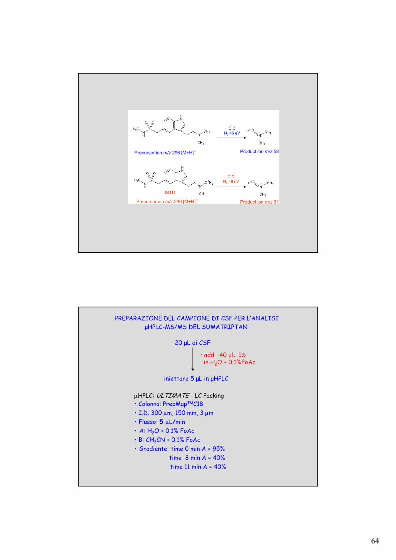
SUMATRIPTAN
63
SUMATRIPTAN
Prof. Gloriano Moneti
SUMATRIPTAN
HN
HN
N
HN
S
NH
O O
N
[M+H]+
64
CID N2 46 eV
CID N2 46 eV
Precursor ion m/z 296 [M+H]+ Product ion m/z 58
Precursor ion m/z 299 [M+H]+ Product ion m/z 61
ISTD
PREPARAZIONE DEL CAMPIONE DI CSF PER L’ANALISIμHPLC-MS/MS DEL SUMATRIPTAN
20 μL di CSF
• add. 40 μL ISin H2O + 0.1%FoAc
iniettare 5 μL in μHPLC
HPLC: ULTIMATE - LC Packing• Colonna: PrepMapTMC18 • I.D. 300 m, 150 mm, 3 m• Flusso: 5 L/min• A: H2O + 0.1% FoAc• B: CH3CN + 0.1% FoAc• Gradiente: time 0 min A = 95%
time 8 min A = 40%time 11 min A = 40%
65
Prof. Gloriano Moneti
Prof. Gloriano Moneti