newly formed cementum induced by the osteogenic proteins
TRANSCRIPT

I
Newly formed cementum induced by the osteogenic proteins of the TGF-ß
supergene family
A comparative histomorphometric study of newly formed cementum as induced by different
applications of osteogenic proteins
Safiyya Hassam Essa
A research report submitted to the Faculty of Health Sciences, University of the Witwatersrand,
Johannesburg, in partial fulfilment of the requirements for the degree
Master of Dentistry (Periodontics and Oral Medicine)
March 2015

II
Declaration I hereby declare that this research report is my own unaided work, except where due
acknowledgment for assistance received has been made. It is being submitted for the degree of
Master of Dentistry (Periodontology and Oral medicine) in the University of Witwatersrand,
Johannesburg. It has not been presented before for any degree or examination at this or any
other University.
……………………………………….
Safiyya Hassam Essa
Signed this ……………………………day of…………………………………..2015
The work reported in this research report was performed at the Bone Research Unit, School of Oral
Health Sciences, Faculty of Health Sciences University of Witwatersrand, Johannesburg, South Africa

III
Abstract
Background and Objective: Osteogenic proteins/ Bone morphogenetic proteins are soluble
signals that have potent and pleiotropic functions which have enticed researchers to explore
their role in periodontal regeneration. The potential of these proteins to induce cementum
formation in a periodontal healing context has been previously demonstrated. However, is the
ability to induce cementum uniformly applicable to different BMPs? Do some BMPs have more
of a cementogenic effect than others? This study was designed to measure newly formed
cementum induced by different osteogenic proteins of the transforming growth factor-ß
supergene family.
Material and Methods: Histological sections prepared from previous periodontal regeneration
studies of surgically created furcation defects of 26 mandibular molar teeth of Papio ursinus,
exposed to different applications of BMPs were assessed and compared. The BMPs used in
these periodontal studies included recombinant human osteogenic protein-1 (hOP-1),
recombinant human bone morphogenetic protein-2 (hBMP-2) , binary application of hOP-1 and
hBMP-2, recombinant human transforming growth factor β₃ (hTGF-β₃), synergistic application
of hTGF- β₃ and hOP-1 as well as naturally-derived BMPs. Histomorphometric measurements of
the extent of cementum formation along the mesial and distal root surfaces were made. The
thickness of newly formed cementum was measured in three regions along the root surface in
apical, middle and coronal regions.
Results: The BMPs which yielded the most favourable cementogenic outcome included hOP-1,
hTGF-β₃ and the synergistic application of hOP-1 and hTGF-β₃. Naturally-derived highly purified
BMPs and hOP-1 applications showed greater cementogenic effects with regards to the extent
of cementum formed. Applications of hOP-1, hTGF-β₃ and the synergistic application of hOP-1
and hTGF-β₃ showed positive cementogenic effects with regards to the thickness of cementum
formed along root surfaces. The hBMP-2 applications were found to be the least inductive
category of BMPs in forming new cementum along the root surfaces, both in extent and
thickness measurements.

IV
Conclusion: BMPs show variable inductive potential for cementogenesis along exposed root
surfaces of P. ursinus possibly reflecting a structure-activity profile of each tested protein.

V
Acknowledgements I would like to express my sincere gratitude and appreciation to the following persons whose
contributions were invaluable to the completion of this work:
Professor Ugo Ripamonti, MD, MDent (Periodontics and Oral Medicine), Ph.D., Director,
Bone Research Laboratory.
Professor Sindisiwe Shangase, MDent (Periodontics and Oral Medicine)
Mrs Petra Gaylard at Data Management and Statistical Analysis, for her assistance in the
analysis of the data and statistical insights.
My husband, daughter and family for supporting me through the process of preparing this
manuscript

VI
Table of contents Declaration .................................................................................................................................................................... II
Abstract ........................................................................................................................................................................ III
Acknowledgements ...................................................................................................................................................... V
Table of contents ......................................................................................................................................................... VI
List of figures .............................................................................................................................................................. VII
List of tables ............................................................................................................................................................... VIII
Abbreviations ................................................................................................................................................................ IX
1. Introduction .......................................................................................................................................................... 1
1.1 Aim ............................................................................................................................................................... 2
1.2 Objectives ..................................................................................................................................................... 2
2. Review of the literature ........................................................................................................................................ 3
2.1 Bone morphogenetic proteins ..................................................................................................................... 5
2.2 Tooth morphogenesis .................................................................................................................................. 6
2.3 Structure-activity profile .............................................................................................................................. 8
2.4 Mechanistic insights ..................................................................................................................................... 9
3. Materials and Methods ....................................................................................................................................... 10
3.1 Study sample .............................................................................................................................................. 12
3.2 Study Design ............................................................................................................................................... 12
3.3 Data Collection and Statistical analysis ...................................................................................................... 14
4. Results ................................................................................................................................................................. 16
5. Discussion ........................................................................................................................................................... 26
6. Conclusion ........................................................................................................................................................... 29
7. References .......................................................................................................................................................... 30
Appendices .................................................................................................................................................................. 36

VII
List of figures
Figure Description Page
1. Schematic diagram of measurements 14
2. Graph depicting extent of cementum along the mesial
root (mean values)
17
3. Graph depicting extent of cementum along the distal
root (mean values)
18
4. Graph depicting thickness of cementum in the apical
region of root surfaces (mean values)
19
5. Graph depicting thickness of cementum in the middle
region of root surfaces (mean values)
19
6. Graph depicting thickness of cementum in the coronal
region of root surfaces (mean values)
20
7. Histological images showing extent of cementum
formation by different BMP applications
22-23
8. Histological images showing extent of cementum
formation by different BMP applications
24-25

VIII
List of tables
Table Descriptions Page
A. Treatment Effect Sizes 15
B. Distribution of the number of teeth per treatment
group
16
C. Source table for extent of cementum formation 17
D. Source table for thickness of cementum 18

IX
Abbreviations
BMPs Bone morphogenetic proteins
BSP Bone sialoprotein
CAM Cell adhesion molecules
ECM Extracellular matrix
FGF Fibroblast growth factor
Gdn HCL Guanidinium hydrochlroide
hOP-1 Recombinant human osteogenic protein-1
hBMP-2 Recombinant human bone morphogenetic protein-2
hTGF-β₃ Recombinant human transforming growth factor-β₃
μm Micrometer
OCN Osteocalcin
P. ursinus Papio ursinus (Chacma baboon)
Runx-2 Runt-related transcription factor-2
Shh Sonic hedgehog proteins
Wnt Wingless intergrated transcription factor

1
1. Introduction
The periodontium is defined as ‘the tissues that invest and support the teeth and include the
gingiva, alveolar mucosa, cementum, periodontal ligament and alveolar bone’ (Glossary of
periodontal terms, 2001). It is a collective term describing the tooth supporting structures and
tissues that is, the root cementum, periodontal ligament, alveolar bone and gingiva (Page &
Schroeder, 1982). Periodontal diseases lead to the loss or damage of the periodontal tissues
(Page & Schroeder, 1982). Periodontal therapy involves two primary components. First, the
elimination of the periodontal infection and second, the regeneration of damaged or lost
components of the periodontal tissues (Garrett, 1996). Tissue regeneration in postnatal life
recapitulates events which occur in the normal course of embryonic development (Ripamonti,
2007). Development of the periodontium is initiated with the process of root formation, as the
apical proliferating mesenchyme forms a bi-layered epithelial root sheath. These epithelial root
sheath cells are thought to produce basement membrane containing chemotactic proteins,
which facilitate the migration and differentiation of pre-cementoblast cells, hence contributing
to cementogenesis (Zeichner-David, 2006). Additionally, the cells of HERS undergo epithelial-
mesenchymal transformation to become functional cementoblasts contributing to production
of acellular and cellular cementum (Zeichner-David, 2006).
Several genes and their secreted products have been identified as crucial for regulating this
process of tooth morphogenesis (Levander, 1945; Thesleff & Nieminen, 1996; Äberg, Wozney,
& Thesleff, 1997; Thesleff & Sharpe, 1997). The pleiotropic activity of the soluble molecular
signals of the transforming growth factor-ß (TGF-ß) supergene family including the bone
morphogenetic proteins (BMPs), initiate cementogenesis as well as the assembly of the
periodontal attachment apparatus (Ripamonti & Petit, 2009). Originally BMPs have been
isolated and identified by the osteoinductive capacity of demineralised bone matrix (DBM)
when implanted in heterotopic extraskeletal sites (Urist, 1965; Reddi and Huggins 1972).The
BMPs are pleiotropic proteins with several different functions and activities in the context of
different organs and tissues modulated by various extracellular matrix components (Ripamonti,

2
2007). BMPs are soluble molecular signals that are deployed during tooth development and
morphogenesis (Hogan , 1996; Thesleff & Sharpe, 1997; Ripamonti, 2007). This is at the crux of
therapeutic tissue induction and morphogenesis by recombinant human BMPs, which may
additionally be deployed recapitulating events which occur in the normal course of embryonic
development to induce periodontal tissue regeneration, specifically cementogenesis along
exposed root surfaces.
1.1 Aim
To compare the effect of different osteogenic proteins, namely hOP-1, hBMP-2,
hTGF-β₃, the binary application of hOP-1 and hBMP-2, the synergistic application
of hOP-1 and hTGF-β₃, and naturally-derived bone morphogenetic proteins, on
the induction of newly formed cementum on root surfaces of molar teeth in
surgically created furcation defects of P.ursinus.
1.2 Objectives
1. Evaluate whether all the osteogenic proteins were capable of inducing
cementum formation
2. To quantify the newly formed cementum induced by various osteogenic
proteins by:
a. Measuring the extent of newly formed cementum along mesial and distal
roots of molar teeth
b. Measuring the thickness of newly formed cementum along different
regions of the roots of molar teeth to establish if any significant
differences in cementum induction were present
3
2. Review of the literature
Untreated periodontal diseases may lead to tooth loss due to destruction of the tooth
supporting structures. Regeneration of these lost structures is a strategy which is central to
successful periodontal therapy, but yet remains an elusive outcome in clinical periodontal
practice (AAP Position paper, 2005). Historical treatment approaches, such as flap debridement,
have served as a reliable method for access to root surfaces to facilitate removal of infective
aetiology. This treatment approach has however yielded limited, if any, potential for
regeneration of lost components.
Regenerative therapies aimed at reconstitution of tooth supporting structures including
alveolar bone, cementum and periodontal ligament have yielded more positive outcomes
(Reynolds et al., 2003). These therapies such as osseous grafting and guided tissue regeneration
(GTR) have been used in the management of infrabony periodontal defects and furcation
defects, predominantly yielding bone fill, and not complete recapitulation of the periodontium
i.e. cementum and PDL formation (Reynolds et al., 2003). It is on this premise, that
developments in biology and material sciences could provide new regenerative materials and
delivery systems for predictable regeneration of all components of the periodontium (Position
paper AAP, 2005).
The induction of newly formed cementum is an essential ingredient to engineer periodontal
tissue regeneration (Ripamonti, 2006). In the past several years, tissue induction and
regeneration has witnessed a rapid growth due to several discoveries in regenerative medicine
but particularly in bone tissue induction. The essence of tissue engineering and regeneration is
to use morphogenetic signals or morphogens first described by Turing (1952) as ‘forms
generating substances’ (Turing, 1952). Morphogens, initiate the multi-step cascade of gene
expression, protein synthesis, and secretions resulting in the induction of tissue formation or
morphogenesis (Reddi, 1988; Reddi, 1994; Reddi, 1997; Ripamonti & Duneas, 1998; Reddi,
2000.)
4
The discovery of these regulatory morphogens with novel biological activities and therapeutic
potential has been the subject of exciting research. The recognition of the extracellular matrix
of bone as a multi-factorial repository of locally active pleiotropic morphogenetic proteins that
initiate and modulate bone formation by induction, has been at the crux of continuously
developing research (Ripamonti & Reddi, 1992; Reddi, 2000; Ripamonti, 2006). Progression in
the isolation and characterization of regulatory morphogens within the bone matrix has been
hampered by the fact that the extracellular matrix of bone is in the solid state (Reddi, 1997).
The bulk of the extracellular matrix proteins are tightly bound to the collagenous bone matrix,
further cemented by the mineralised component of the bone matrix. Reddi and co-workers
were the first to unlock the problem of the bone matrix in the solid state (Reddi, 1997).
Sampath and Reddi used chaotropic agents such as urea or guanidinium hydrochloride (Gdn
HCL) to extract and solubilize the putative osteogenic proteins contained within the
demineralised bone marix (Sampath & Reddi, 1981). Solubilized putative osteogenic proteins
were then reconstituted with the insoluble collagenous matrix or residue obtained after the
dissociative extraction of the bone matrix (Sampath & Reddi, 1981). This operational
reconstitution restored the biological activity of the intact demineralised bone matrix (Sampath
& Reddi, 1981; Reddi, 1997).
A futher contribution by Sampath and Reddi was an experiment which addressed the species
specificity of the bone matrix, showing homology among the osteoinductive proteins from
diverse species of mammals. The species specificity of matrix-induced bone formation is due to
species-specific alloantigens or inhibitors (or both) present in the Gdn HCl-extracted allogenic
insoluble collagenous bone matrix and the Gdn HCI-solubilized extracellular bone matrix
components of >50,000 daltons (Sampath & Reddi, 1983).
The above study highlighted that the osteogenic proteins extracted from the bone matrix are
homologous amongst mammalian species. On the contrary, the insoluble collagenous bone
matrix carries the antigenic load across mammalian species, and the restoration of the

5
biological activity in vivo is only achieved when using allogenic collagenous matrix components
(Sampath & Reddi, 1983). These key experiments, showing the combination of an insoluble
signal or substratum, with solubilised osteogenic soluble signals, propelled the bone induction
principle into the pre-clinical and clinical arena culminating in the isolation and purification of
an entirely new family of protein initiators, the BMPs which were found to be members of the
TGF-β supergene family (Sampath & Reddi, 1981; Wozney et al., 1988; Celeste et al., 1990;
Özkaynak et al., 1990; Ripamonti et al., 1996).
2.1 Bone morphogenetic proteins
The BMPs are a family of highly conserved secreted proteins that have potent and pleiotropic
functions and activities in the context of different organs and tissues, further modulated by the
biomimetism of the extracellular matrix (Thesleff, 1995; Hogan, 1996; Thomadakis et al., 1999,
Reddi, 2000, Ripamonti, 2006). BMPs show sequence homologies with members of the
transforming growth factor β (TGF-β) family (Wozney et al., 1988; Celeste et al., 1990; Özkaynak
et al.1990).
A striking and discriminatory prerogative of highly purified naturally derived BMPs is the
induction of de novo bone formation. Classic experiments by Urist , Reddi and Huggins have
demontrated the ‘bone induction principle’(Huggins, 1931; Urist, 1965; Urist et al., 1967; Reddi
& Huggins, 1972; Reddi , 1997).
Increasing purification schemes of large quantities of naturally derived bovine BMPs yielded
protein extracts and final purification bands including electroendosmotic elution that provided
amino acid sequencing information (Wang et al., 1988; Sampath, et al., 1992). This was
followed by expression cloning of the recombinant human proteins (Wozney et al., 1988;
Celeste et al., 1990; Özkaynak et al., 1990). More than 40 related proteins with BMP-like
sequences and activities have been sequenced and cloned (Ripamonti, 2006).

6
In addition to bone induction in postnatal life, the BMPs are involved in inductive events that
control pattern formation during morphogenesis and organogenesis in such disparate tissues
and organs as the kidney, eye, nervous system, lung, teeth, skin and heart (Hogan, 1996; Äberg
et al., 1997; Chinsembu, 2012). The impressive evolutionary conservation of the BMPs genes
indicate that the secreted proteins are critical in development and are invovled in several
unrelated events that control pattern formation during both embryonic development and
postnatal tissue morphogenesis and regeneration (Hogan, 1996; Äberg et al., 1997; Chinsembu,
2012).
2.2 Tooth morphogenesis
Tooth morphogenesis is a classic example of epithelial-mesenchymal interaction involving
reciprocal signalling events mediated by several proteins (Thesleff et al., 1995). Signalling
between the epithelial and mesenchymal tissues regulates morphogenesis and cell
determination from the very beginning of odontogenesis.
Soluble signals such as the fibroblast growth factors (FGFs) and the BMPs, and transcription
factors such as the wingless intergrated (Wnt) and sonic hedgehog (Shh) proteins play a crucial
role in tooth initiation, morphogenesis and differentiation (Äberg et al., 1997; Thesleff, 2006;
Chinsembu, 2012). In the developing tooth BMPs secreted by the epithelium (BMP-2 and/or
BMP-4) are suggested to be early signals that stimulate expression of the homeobox-containing
genes Msx-1 and Msx-2, which have central regulatory roles in tooth morphogenesis as
demonstrated in knockout mice experiments (Gao et al., 1998; Thesleff, 2006). A weak BMP
signal, ensuing from a loss of BMP receptors or overexpression of BMP inhibitors, results in
various defects in different cusps and teeth (Chinsembu, 2012). During the bud-to-cap stage
transition, BMP-2 signalling from the condensing mesenchyme plays a critical role in the
induction of the enamel knot (Chinsembu, 2012). Synchronously, BMP-4 within the oral

7
epithelium leads to an up-regulation of enamel knot markers such as p21 (Caton & Tucker,
2009). Loss of BMP-4 signalling by knockout of the receptor Bmpr1a gene leads to an arrest of
tooth development at the bud stage (Chinsembu, 2012).
The expression of BMPs is not confined to early tooth development since its expression is
sustained during root morphogenesis (Thomadakis et al., 1999). As mantle dentine is secreted,
the epithelial root sheath disintegrates, followed by migration of mesenchymal cells of the
dental follicle towards the root surface and differentiation into cementoblasts, secreting
cementoid which later mineralises. The outer cells of the dental follicle differentiate into
osteoblasts lining the alveolar bone, whilst the more centrally located cells differentiate into
fibroblasts that produce collagen that become embedded in both bone and cementum as
Sharpey’s fibres (Zeichner-David, 2006). In this context, the localisation of BMP-3 and OP-1
during morphogenesis of the murine root, suggests that the secreted proteins play a role during
cementogenesis and the assembly of the periodontal ligament fibres (Thomadakis et al., 1999).
The localisation of BMP-2 in alveolar bone alone and BMP-3 and OP-1 in all three components
of the periodontium suggests that the morphogenesis of periodontal tissues may involve a
composite pattern of co-ordinated expression of different signalling BMPs, each endowed with
a specific pleiotropic biological activity (Thomadakis et al., 1999).
Experimental studies in non-human primates have also shown that BMPs in addition to the
induction of bone formation also induce cementogenesis (Ripamonti et al., 1994). Naturally-
derived highly purified BMPs implanted in furcation defects of the non-human primate Chacma
baboon Papio ursinus showed tissue induction of the three essential components of the
periodontium, that is cementum, periodontal ligament and alveolar bone (Ripamonti et al.,
1994). Short term studies in Papio ursinus showed that furcation defects implanted with human
recombinant osteogenic protein-1 (hOP-1) primarily induces cementogenesis, with minimal, if
any, bone formation (Ripamonti et al 1996). Importantly, within the context of the periodontal
wound and in contact with the dentine extracellular matrix, hOP-1 is primarily cementogenic,

8
whilst hBMP-2, when implanted in identical periodontal defects of P. ursinus is primarily
osteogenic (Ripamonti et al., 2001; Ripamonti, 2007).
2.3 Structure-activity profile
Morphological differences in periodontal tissue induction, as demonstrated by experimental
studies, highlight the pleiotropic functions of members of the BMPs family (Ripamonti, 2006;
Ripamonti, 2007). Ripamonti (2007) has stated “The mosaicism of BMPs’ expression, synthesis
and localization during periodontal tissue development in embryogenesis indicates the
presense of several BMP isoforms synchronously expressed” (Ripamonti, 2007). The expression
of multiple forms of BMPs may reflect different functions in vivo which could have therapeutic
significance. In vivo studies, highlighted in primate studies, have indicated that amino acid
sequence variations in the active carboxy-terminal domain of a morphogenetic protein confer
specialised pleiotropic properties to each BMP isoform. The amino acid sequence variations in
the carboxy-terminal domain form the molecular basis that determine the structure-activity
profile of each morphogenetic protein (Ripamonti, 2006; Ripamonti, 2007).
In vivo studies in P. ursinus, have demonstrated this novel concept, that there is a structure-
activity profile of each BMP that results in the induction of different tissue morphologies when
evaluated in periodontal regenerative procedures (Ripamonti et al., 2001; Ripamonti et al.,
2009). These experiments focused on the efficacy of highly purified naturally derived and
different recombinant human BMPs for periodontal tissue regeneration after implantation in
mandibular furcation defects (Ripamonti, 2007). Experimental application of recombinant hOP-
1 in contact with dentine extracellular matrix, shows a specific preferential cementogenic
function, with newly induced cementum and with inserted Sharpey’s fibres visible on
morphologic examination of treated sites (Ripamonti et al., 1996; Ripamonti et al., 2001). The
predominant cementogenic effect of hOP-1 is further highlighted in a study which compares
the induction of periodontal regeneration by doses of recombinant hOP-1 and hBMP-2 applied
singly or in combination (Ripamonti et al., 2001). Histomorphometric analysis showed hOP-1 to

9
be strongly cementogenic in its single application. On the other hand hBMP-2 showed limited
cementum formation. However, hBMP-2 induced greater amounts of bone formation when
applied alone or when combined morphogens were applied (Ripamonti et al., 2001). The data
supports the notion of an existing structure-activity profile, as tissue morphogenesis induced by
hOP-1 and hBMP-2 is qualitatively different when the morphogens are applied singly to
furcation defects (Ripamonti et al., 2001). Choi et al (2002) using a canine experimental model
investigated the effects of hBMP-2 in 3-wall intrabony periodontal defects. The study showed
positive healing outcomes for hBMP-2 with regards to alveolar bone, where accelerated and
enhanced bone formation was noted. However, cementum regeneration was minimal, with the
authors concluding that hBMP-2 does not appear to have a significant effect on cementum
regeneration (Choi et al., 2002).
2.4 Mechanistic insights
In vitro studies have attempted to provide mechanistic insights which support the concept of a
structure-activity profile of BMPs. Hakki et al (2010) studied the effects of hOP-1 on murine
cementoblast cells in vitro. The study focused on the effect of hOP-1 on regulating mineralised
tissue-associated genes in cementoblasts and the expression profile of cementoblasts,
extracellular matrix (ECM) components and cell adhesion molecules (CAM) (Hakki et al., 2010).
The findings of the study, demonstrated that hOP-1 at a concentration of 50ng/ml upregulated
the expression of mineralised tissue associated genes such as osteocalcin (OCN), bone
sialoprotein (BSP) and runt-related transcription factor-2 (Runx-2) mRNA expression on the
cementoblasts. These effects of hOP-1 on gene regulation possibly modulate the response of
cementoblasts during tissue induction, mineralisation and tissue turnover, highlighting the role
of hOP-1 during cementogenesis (Hakki et al., 2010). The effect of hBMP-2 on periodontal
regeneration using in vitro experimental models has also been explored (Zhao et al., 2003). The
study by Zhao et al. (2003) examining the effects of hBMP-2 on murine cementoblasts in vitro
showed that hBMP-2 inhibited cementoblast-mediated mineral nodule formation at

10
concentrations of 10 ng/mL, with a downregulation of gene and protein expression. hBMP-2
does however play a promotive role in the process of osteoblastic differentiation as
demonstrated by osteoblastic differentiation of preogenitor cells upon exposure to hBMP-2
(Gorri et al., 1999; Zhao et al., 2003).
3. Materials and Methods
This was a retrospective study which used different tissue sections from six previous studies of
periodontal regeneration in furcation defects of P. ursinus (Ripamonti et al., 1994; Ripamonti et
al., 1996; Ripamonti et al., 2001; Ripamonti et al., 2009b; Teare et al., 2012). In these studies,
furcation defects of mandibular molars of P. ursinus were exposed to different applications and
concentrations of morphogenetic proteins, the effect of which was examined
histomorphometrically on undecalcified serial sections. The surgical methodology of the
furcation defect model of tissue morphogenesis by bone derived and recombinant BMPs in the
adult baboon has been described in detail in Addendum A (Ripamonti et al., 1994; Ripamonti et
al., 1996; Ripamonti, 2001). The surgical methodology was the same for all six studies with the
exception of the application used in each study. The same operator cut all the furcation defects
within the reported studies providing a highly reproducible furcation defect model for
sequential morphological and morphometric analyses (Ripamonti et al., 1994; Ripamonti et al.,
1996). All animals were killed 60 days after soluble morphogen application. Preparation of
serial histological sections from these experiments for histomorphometric analysis have also
been described in detail in Addendum A (Ripamonti et al., 2001).
In summary, the applications used in the previous studies were:
1. hOP-1
100 µg recombinant human osteogenic protein-1 combined with a bovine insoluble
collagenous bone matrix as a carrier.
2. hBMP-2

11
100 µg recombinant human bone morphogenetic protein-2 combined with a bovine
insoluble collagenous bone matrix as a carrier.
3. Binary application of hOP-1 and hBMP-2
100 µg recombinant human osteogenic protein-1 and 100 µg recombinant human bone
morphogenetic protein 2 combined with a bovine insoluble collagenous bone matrix as a
carrier.
4. hTGF-β₃
75 µg recombinant human transforming growth factor-β₃ combined with Matrigel® matrix
as a carrier.
75 µg recombinant human transforming growth factor-β₃ combined with Matrigel® matrix
and morcellated fragments of rectus abdominis muscle.
5. Synergistic binary application of hOP-1 and hTGF-β₃
25µg recombinant human osteogenic protein-1 and 1.25 µg recombinant human
transforming growth factor-β₃ combined with Matrigel® matrix as a carrier
25µg recombinant human osteogenic protein-1 and 1.25 µg recombinant human
transforming growth factor-β₃ combined with Matrigel® matrix and morcellated fragments
of rectus abdominis muscle
6. Naturally-derived highly purified bone morphogenetic protein
250 µg naturally derived highly purified bone morphogenetic protein from bovine bone
matrix combined with allogenic insoluble collagenous bone matrix as a carrier.
12
3.1 Study sample
Prepared histological sections of periodontal tissues of treated mandibular teeth from
the six studies were accessed from the archives at the Bone Research Laboratory of the
University of Witwatersrand. For inclusion into the study the slides had to display the
following:
Good staining properties to allow for discrimination between the different
components of the periodontium
Complete view of both roots for measurement purposes
Identifiable apical notches on each root for measurement reference point
Identifiable furcation region between the two roots of selected teeth
All histological sections with poor staining preservation and resultant unclear distinction
between the cementum and other hard tissue were excluded from the study.
Histological sections displaying anatomical distortion of roots, apical notch and furcation
regions were also not considered for evaluation in this study.
The total number of teeth analysed in this study were dictated by these inclusion and
exclusion criteria. Hence for each study the number of teeth analysed did differ as
reflected in Table B.
3.2 Study Design
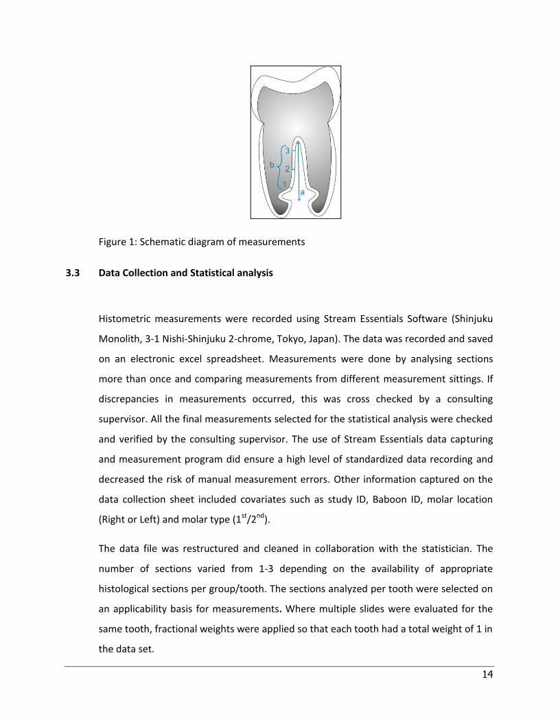
Histometric measurements of the extent and thickness of cementum formed along
individual mesial and distal roots of each tooth were taken (Figure 1). Selected sections
were analysed at objective 10x (cementum extent measurements) and objective 4x
(cementum thickness measurements) with a Provis AX70 research microscope (Olympus

13
Optical Tokyo, Japan). Histometric measurements (in μm) were made and recorded
using Stream Essentials software (Shinjuku Monolith, 3-1 Nishi-Shinjuku 2-chrome,
Tokyo, Japan).
a. Apico-coronal extent of cementum formation from the apical border of the
radicular notch to the most coronal extent of cementum. Measurements were
recorded for mesial and distal roots independently. The measurements were
recorded as a proportion of the total root length.
b. Measurement of the thickness of cementum formed along the root surfaces.
Three points along each root (mesial and distal root), were selected for
measurement. Mesial and distal root measurements for each region were
combined and a mean value was calculated. The reference points along the root
surface for each measurement were equidistant and described as follows:
1. Apical reference point: Thickness of newly formed cementum was measured
coronal to the apical notch
2. Middle reference point: Thickness of newly formed cementum was measured
from a measured halfway reference point between the apical and coronal
aspect of the root surface
3. Coronal reference point: Thickness of newly formed cementum was
measured corresponding to the most coronal extension of cementum along
the root surface
14
Figure 1: Schematic diagram of measurements
3.3 Data Collection and Statistical analysis
Histometric measurements were recorded using Stream Essentials Software (Shinjuku
Monolith, 3-1 Nishi-Shinjuku 2-chrome, Tokyo, Japan). The data was recorded and saved
on an electronic excel spreadsheet. Measurements were done by analysing sections
more than once and comparing measurements from different measurement sittings. If
discrepancies in measurements occurred, this was cross checked by a consulting
supervisor. All the final measurements selected for the statistical analysis were checked
and verified by the consulting supervisor. The use of Stream Essentials data capturing
and measurement program did ensure a high level of standardized data recording and
decreased the risk of manual measurement errors. Other information captured on the
data collection sheet included covariates such as study ID, Baboon ID, molar location
(Right or Left) and molar type (1st/2nd).
The data file was restructured and cleaned in collaboration with the statistician. The
number of sections varied from 1-3 depending on the availability of appropriate
histological sections per group/tooth. The sections analyzed per tooth were selected on
an applicability basis for measurements. Where multiple slides were evaluated for the
same tooth, fractional weights were applied so that each tooth had a total weight of 1 in
the data set.

15
Differences between treatments were assessed using a General Linear Model (GLM)
procedure for each dependent variable (DV), with treatment and the covariates as
independent variables. Post-hoc tests were conducted using the Tukey-Kramer test.
Effect sizes were calculated using Cohen’s d, which were interpreted according to Table
A. The 5% significance level was used throughout, unless specified otherwise. In other
words, p-values <0.05 indicate significant results.
*Cohen’s d Effect sizes
0.80 and above large effect
0.50 to 0.79 moderate effect
0.20 to 0.39 small effect
below 0.20 near zero effect
Table A: *Cohen’s d (effect size used to indicate standardised difference between two means)

16
4. Results
There were 27 teeth in the study, spread over six treatments, with 1-9 teeth per treatment. It
must be noted at the outset that the sample size per treatment is small, and this affects the
power of the study that is the ability to detect a significant difference.
Treatment Frequency Percent
1. hBMP-2 4 14.81
2. hBMP-2 and hOP-1 1 3.70
3. Naturally-derived BMPs 4 14.81
4. hOP-1 9 33.33
5. Synergistic hOP-1 and hTGF-β₃ 4 14.81
6. hTGF-β₃ 5 18.52
Table B: Distribution of the number of teeth per treatment group
The data were taken from different studies (Ripamonti et al., 1994; Ripamonti et al., 1996;
Ripamonti et al., 2001; Ripamonti et al., 2009; Teare et al., 2012), with treatment 2 only
contributing one tooth. The data were obtained from 14 baboons, each contributing between 1
and 4 (the maximum) teeth. The study teeth were reasonably well-balanced between the 1st
and 2nd molars (59% and 41% respectively). The teeth were predominantly from the right
mandible (67%).
Between-group analysis for the dependent variables
A one-way analysis of variance (ANOVA) was conducted for each dependent variable with
Treatment as factor. Baboon ID (nested in Treatment), molar type (1st/2nd) and molar location
(L/R) were used as blocking variables. Study ID (nested in Treatment) could not be used as an

17
additional blocking variable due to the confounding between Treatment, Study ID and Baboon
ID. A natural log transformation of the dependent variable was used in order to meet the
assumptions of the ANOVA technique.
Extent of cementum Source DF Type III SS Mean Square F Value Pr > F
Mesial root Treatment 5 0.93 0.19 3.97 0.013
Distal root Treatment 5 0.19 0.04 2.31 0.087
Table C: Source table for extent of cementum formation
Figure 2: Extent of cementum along the mesial root (mean values)
The effect of treatment was significant (p=0.013) for the mesial root between treatment
groups. Post-hoc tests showed that the mean extent of cementum on the mesial root was
significantly higher for Naturally-Derived, hOP-1 and synergistic binary application of hTGF-β₃
and hOP-1, compared to hBMP-2 (Figure 7 A-E; Figure 8 E). The effect sizes were large (Cohen’s
d=1.9, 1.0, and 2.4 respectively). The Least-Squares (LS) Means are illustrated below (the error
bars denote the 95% confidence intervals for the means).
18
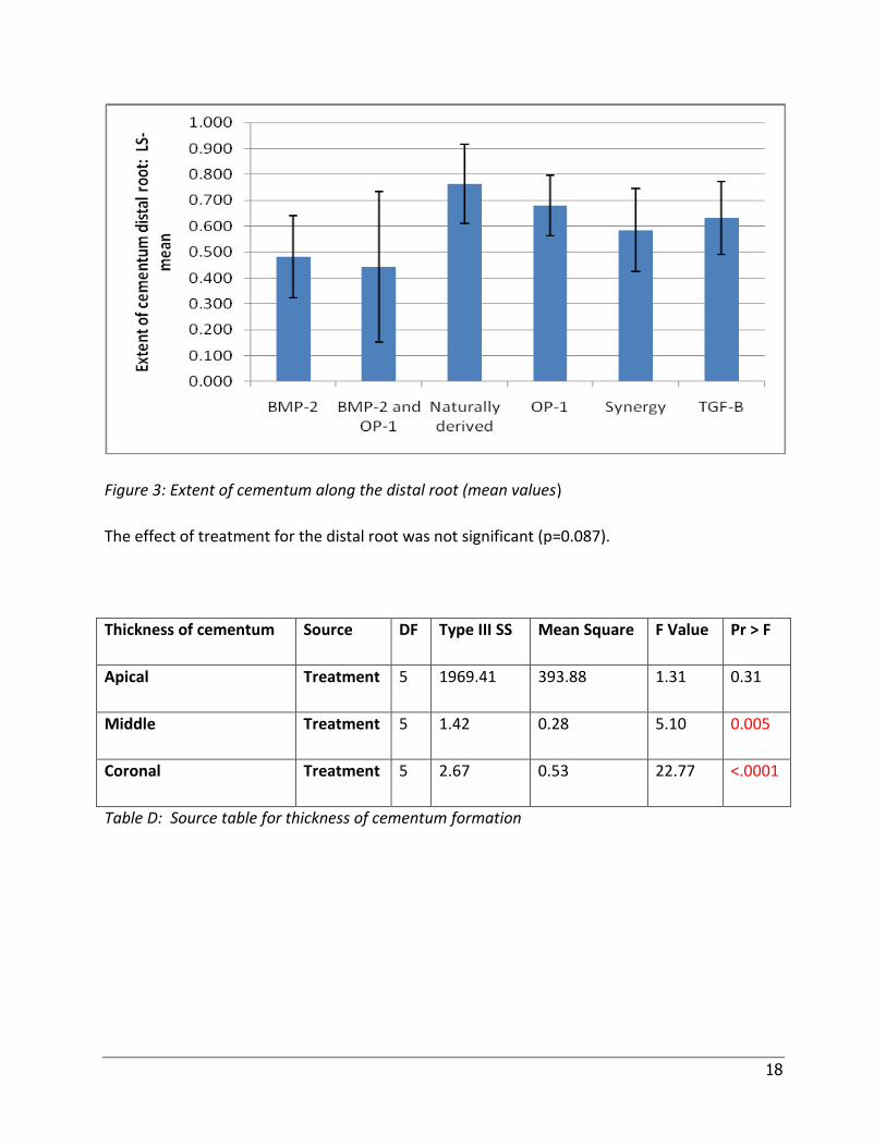
Figure 3: Extent of cementum along the distal root (mean values)
The effect of treatment for the distal root was not significant (p=0.087).
Thickness of cementum Source DF Type III SS Mean Square F Value Pr > F
Apical Treatment 5 1969.41 393.88 1.31 0.31
Middle Treatment 5 1.42 0.28 5.10 0.005
Coronal Treatment 5 2.67 0.53 22.77 <.0001
Table D: Source table for thickness of cementum formation

19
Figure 4: Thickness of cementum in the apical region (mean values calculated for combined
mesial and distal root measurements)
Three outliers were removed (this did not change the conclusions). The effect of treatment was
not significant (p=0.31).
.

20
Figure 5: Thickness of cementum in the middle region (mean values calculated for combined
mesial and distal root measurements)
The effect of treatment was significant (p=0.005). Post-hoc tests showed that the thickness of
cementum in the middle region of the root was significantly higher for the synergistic
application of TGF-β₃ and hOP-1, compared to hBMP-2 and hBMP-2 with hOP-1 (Figure 8 A-E).
The effect sizes were large (Cohen’s d=2.7 and 1.7 respectively). The Least-Squares (LS) Means
are illustrated in Figure 5 above.
Figure 6: Thickness of cementum in the coronal region (mean values calculated for combined
mesial and distal root measurements)
Three outliers were removed. The effect of treatment was significant (p<0.0001). Post-hoc
tests showed that the thickness of cementum in the coronal region of the root was significantly
higher for hOP-1, compared to hBMP-2 and hBMP-2 with hOP-1, and also higher for Naturally
Derived, synergistic application of TGF-β₃ and hOP-1 and hTGF-β₃ compared to hBMP-2 (Figure

21
8). The effect sizes were large (Cohen’s d=3.3, 1.9, 5.4, 1.9, 2.5 respectively). The Least-
Squares (LS) Means are illustrated in Figure 6 above.

22
B
)
A
)
C
)
D
)
E
)
F
)
23
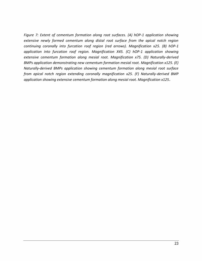
Figure 7: Extent of cementum formation along root surfaces. (A) hOP-1 application showing
extensive newly formed cementum along distal root surface from the apical notch region
continuing coronally into furcation roof region (red arrows). Magnification x25. (B) hOP-1
application into furcation roof region. Magnification X45. (C) hOP-1 application showing
extensive cementum formation along mesial root. Magnification x75. (D) Naturally-derived
BMPs application demonstrating new cementum formation mesial root. Magnification x125. (E)
Naturally-derived BMPs application showing cementum formation along mesial root surface
from apical notch region extending coronally magnification x25. (F) Naturally-derived BMP
application showing extensive cementum formation along mesial root. Magnification x125.

24
A
)
B
)
C
)
D
)
E
)

25
Figure 8: Thickness of cementum along different regions of root surfaces. (A) hTGF-β₃
application showing thickness of newly formed cementum along mesial root surface in the
middle region with an appreciable “bulge” observed (red arrows). Magnification x150. (B)
hTGF-β₃ application showing thickness of newly formed cementum along distal root surface in
the coronal region. Magnification x125. (C) High-power detail of synergistic binary application
of hOP-1 and hTGF-β₃ showing thickened areas of newly formed cementum along distal root
surface in the middle region (red arrows). Magnification x300. (D) Synergistic binary application
of hOP-1 and hTGF-β₃ showing irregular thickened areas of newly formed cementum along
mesial root surface in the middle region. Magnification x150. (E) hBMP-2 application with
limited new cementum formation along distal root surface confined to apical region of root
surface not extending coronally (red arrows). Extent and thickness of new cementum are
minimal. Magnification x75.
26
5. Discussion
Experiments in the non human primate P.ursinus have given insight into the morphogenetic
potential of BMPs in periodontal tissue regeneration (Ripamonti et al, 1994). The ability of
BMPs to induce cementogenesis has been highlighted by a number of studies (Ripamonti et al.,
1994; Ripamonti et al., 1996; Ripamonti et al., 2001; Ripamonti et al., 2009; Teare et al., 2012).
The presence of multiple forms of BMPs and their varying potential to induce cementogenesis
has therapeutic significance (Ripamonti et al., 2006). The molecular basis for the specialised and
pleiotropic activities of BMPs is the result of the amino acid sequence variations in the carboxy
terminal domain. This study, in assessing the magnitude of cementum formation (extent and
thickness) in periodontal defects reinforces the concept of a structure-activity profile amongst
BMPs (Ripamonti et al.,2006).
In this study, newly formed cementum can be observed on all the histological sections
assessed, but variations in the magnitude of newly formed cementum can be appreciated
between the different BMPs applications. The BMPs that show the most cementogenic effect in
terms of extent of cementum formed along the root surfaces are hOP-1, the synergistic
application of hOP-1 and hTGF-β₃ and naturally-derived highly purified BMPs. A comparison of
the quantity of newly formed cementum measured amongst the different BMPs applications
does demonstrate these specific groups to be favourable in terms of cementum deposition
along the connective tissue root interface, albeit in relation to a small sample size.
The positive cementogenic effect of hOP-1 confirmed in this study, could be associated with its
ability to directly influence the expression levels of other morphogens, thus enhancing its
regenerative potential. A previous study in the primate P. ursinus has provided evidence that
hOP-1 directly influences the expression levels of OP-1, type IV collagen, BMP-3 and TGF-β1
mRNAs, demonstrating a transcriptional cascade during regeneration (Ripamonti, 2005). In vitro
studies suggesting that hOP-1 can modulate the expression profile of cementoblasts in relation
to ECM components and related genes, provide mechanistic insights into the cementogenic

27
potential of hOP-1 (Hakki et al., 2010). The upregulation of adhesion molecules and
proliferating genes as a result of hOP-1 applications suggest a positive contributory role to
cementum regeneration (Hakki et al., 2010). A previous experimental study has also alluded to
the role of naturally-derived highly purified BMPs contributing positively to periodontal
regeneration, specifically cementogenesis (Ripamonti et al., 1994) . The findings of this study
are consistent with the proposed notion about naturally-derived BMPs being highly
cementogenic in relation to other BMPs.
In this study, evaluation and measurement of the thickness of newly formed cementum along
the root surfaces, showed that the apical regions do not show a high level of variation between
BMPs groups. This observation could be as a result of the defect configuration of the apical
notch area which would favour formation of cementum by virtue of the process of
cementogenesis being initiated in the apical region, regardless of the BMPs applied.
Differences in cementum thickness were however appreciated, as cementogenesis continued
coronally along the root surfaces. The middle regions and coronal regions demonstrated
differences in cementum thickness between BMP groups with the synergistic binary application
of hOP-1 and TGF-β₃ yielding the most promising potential to induce cementogenesis. The
binary application of these morphogens as well as the addition of morcellated autogenous
rectus abdominis muscle fragments containing myoblastic stem cells could have accounted for
this positive effect. However, a study by Teare et al. (2012) failed to show greater periodontal
regeneration with the addition of morcellated muscle fragments highlighting that their precise
role in periodontal regeneration is still unclear (Teare et al., 2012).
Thickness measurements in the coronal extent of the root surfaces did not differ significantly
between the synergistic binary application of hOP-1 and hTGF-β₃ group and the hTGF-β₃ applied
singly, although an appreciable difference relative to hBMP-2 group is observed. hBMP-2
applications yielded the least amount of cementum in the coronal extent suggestive of its poor
inductive capacity for cementogenesis, although due to a small sample size, concrete
conclusions cannot be made.

28
Previous studies highlighted minimal cementum formation when applications without BMPs
and TGF-βs were used (Ripamonti et al., 1994). For this reason, measurements of cementum
formation in the control sections where insoluble bone matrix without BMPs and TGF-βs were
applied were not done and thus, were not accounted for in the results of this study, .This
however, in retrospect, is a significant limitation within this study.
The comparative applications of different BMPs highlight the possibililty that different
morphogens would initiate a variable periodontal regeneration outcome when in initial contact
with the tooth root surface. The suggestion that multiple forms of BMPs have biologically
different effects, modulated and selectively potentiated by different extracellular matrix
components, underscores the critical regulatory role of the extracellular matrix substrata
(Ripamonti et al., 1994).
The results of this study would categorise hOP-1 and hTGF-β₃, used singly or in synergistic
binary application as being favourable for cementum formation whilst hBMP-2 showed the
least capacity to induce cementogenesis. However, the observation should be interpreted in
the context of a small sample size.

29
6. Conclusion
This study comparing the effects of different BMPs on cementum formation, gives insights into
favourable therapeutic BMP applications which would facilitate periodontal regeneration.
Variation in the inductive capacity of different BMP applications in forming new cementum can
be appreciated and understood in context of an existing structure-activity profile of each BMP
and the influences of the extracellular matrix on their functions. Future research should focus
on the complex molecular and cellular cascades of regenerating periodontal tissues induced by
BMPs to achieve predictable periodontal regenerative outcomes.

30
7. References
AAP Position paper (2005). Periodontal regeneration. Journal of Periodontology , 76, 1601-
1622.
Äberg, T., Wozney, J., & Thesleff, I. (1997). Expression patterns of bone morphogenetic proteins
(BMPs) in the developing mouse model suggest roles in morphogenesis and cell differentiation.
Development Dynamics , 210, 383-96.
Bowers, G., Felton, F., & Middleton, C. (1991). Histological comparison of regeneration in
human intrabony defects when ostegenin is combined with demineralised freeze-dried bone
allograft and with purified bovine collagen. Journal of Periodontology , 62, 690-702.
Caton, J., & Tucker, A. (2009). Current knowledge of tooth development: patterning and
mineralization of the murine dentition. Journal of Anatomy , 214, 502-515.
Celeste, A., Ianazzi, J., Taylor, R., Hewick, R., Rosen, V., Wang, E., et al. (1990). Identification of
transforming growth factor beta family members present in bone inductive proteins purified
from bovine bone. Proceedings of the Natural Academy Sciences USA, 87, 9843-9847.
Chinsembu, K. (2012). Teeth and bones: Signature genes and molecules that underwrite
odontogenesis. Journal of Medical Genetics and Genomics , 4, 13-24.
Choi, S.-H., Kim, C.-K., Cho, K.-S., Huh, J.-S., Sorensen, R., Wozney, J., et al. (2002). Effect of
recombinant human bone morphogenetic protein 2/Absorbable collagen sponge in 3-wall
intrabony defects in dogs. Journal of Periodontology , 73, 63-72.
Gao, J., Symons, A., & Bartold, P. (1998). Expression of transforming growth factor-beta 1 (TGF-
β₁) in the developing periodontium of rats. Journal of Dental Research , 77, 1708-1716.
Garrett, S. (1996). Periodontal regeneration around natural teeth. Annals of Periodontology , 1,
621-666.

31
Glossary of Periodontal Terms 4th edition. (2001).
Gorri, F., Thomas, T., Hicok, K., Spelsberg, T., & Riggs, B. (1999). Differentiation of human
marrow stromal precursor cells: bone morphogenetic protein-2 increases OSF2/CBFA1,
enhances osteoblast commitment, and inhibits late adipocyte maturation. Journal Bone Mineral
Research , 14, 1522-1535.
Hakki, S., Foster, B., Nagatomo, K., Bozkurt, S., Hakki, E., Somerman, M., et al. (2010). Bone
morphogenetic protein-7 enhances cementoblast function in vitro. Journal of Periodontology ,
81, 1663-1674.
Hogan, B. (1996). Bone morphogenetic proteins: Multifunctional regulators of vertebrate
development. Genes Development , 10, 1580-94.
Huggins, C. (1931). The formation of bone under the influence of epithelium of the urinary
tract. Archives of Surgery , 22, 377-408.
Levander, G. (1945). Tissue induction. Nature , 155, 148-149.
Özkaynak, E., Rueger, D., & Drier, E. (1990). OP-1 cDNA encodes an osteogenic protein in the
TGF-β family. European Molecular Biology Organisation Journal , 9, 2085-2093.
Page, R., & Schroeder, H. (1982). Periodontitis in man and other animals: A Comparative
Review. Basel: Kruger.
Reddi, A., & Huggins, C. (1972). Biochemical sequences in the transformation of normal
fibroblasts in adolescent rats. Proceedings of the Natural Academy of Sciences USA, 69, 1601-
1605.
Reddi, A. (1988). Role of morphogenetic proteins in skeletal tissue engineering and
regeneration. Nature Biotechnology , 16, 247-50.
Reddi, A. (1994). Symbiosis of biotechnology and biomaterials: Applications in tissue
engineering of bone and cartilage. Journal of Cell and Biochemistry , 56, 192-95.

32
Reddi, A. (1997). Bone morphogenesis and modeling: soluble signals sculpt osteosomes in the
solid state. Cell , 89, 159-161.
Reddi, A. (2000). Morphogenesis and tissue engineering of bone and cartilage: Inductive signals,
stem cells, and biomimetic biomaterials. Tissue Engineering , 6, 351-359.
Reynolds, M., Aichelmann-Reidy, M., Branch-Mays, G., & Gunsolley, J. (2003). The efficacy of
bone replacement grafts in the treatment of periodontal osseous defects. A systematic review.
Annals of Periodontology , 8, 227-265.
Ripamonti, U., & Reddi, A. (1992). Growth and morphogenetic factors in bone induction: Role of
osteogenin and related bone morphogenetic proteins in craniofacial and periodontal bone
repair. Critical Review Oral Biology Medicine , 3, 1-14.
Ripamonti, U., Heliotis, M., van den Heever, B., & Reddi, A. (1994). Bone morphogenetic
proteins induce periodontal regeneration in the baboon (Papio ursinus). Journal of Periodontal
Research , 29, 439-445.
Ripamonti, U., Heliotis, M., Rueger, D., & Sampath, T. (1996). Induction of cementogenesis by
recombinant human osteogenic protein-1 (hOP-1/BMP-7) in the baboon (Papio ursinus).
Archives of Oral Biology , 41, 121-126.
Ripamonti, U., & Duneas, N. (1998). Tissue morphogenesis and regeneration by bone
morphogenetic proteins. Plastic and Reconstructive Surgery , 101, 227-31.
Ripamonti, U., Crooks, J., Petit, J.-C., & Rueger, D. (2001). Periodontal tissue regeneration by
combined applications of recombinant human osteogenic protein-1 and bone morphogenetic
protein-2. A pilot study in Chacma baboons. European Journal of Oral Science , 109, 241-248.
Ripamonti, U. (2005). Bone induction by recombinant human osteogenic protein-1 (hOP-
1/BMP-7) in the primate Papio ursinus with expression of mRNA of gene products of the TGF-β
superfamily. Journal of Cellular and Molecular Medicine , 9, 911-928.

33
Ripamonti, U. (2006a). Soluble osteogenic molecular signals and the induction of bone
formation. Biomaterials , 27, 807-822.
Ripamonti, U., Teare, J., & Petit, J.-C. (2006b). Pleiotropism of bone morphogenetic proteins:
From bone induction to cementogenesis and periodontal ligament regeneration. Journal of the
International Academy of Periodontology , 8, 23-32.
Ripamonti, U. (2007). Recapitulating Development: A template for periodontal tissue
engineering. Tissue Engineering , 13, 51-71.
Ripamonti, U. (2008). Induction of cementogenesis and periodontal ligament regeneration by
the bone morphogenetic proteins. In S. Vukicevic, & K. Sampath, Bone morphogenetic proteins:
From Local to Systemic Therapeutics (pp. 233-256). Basel: Birkauser.
Ripamonti, U., & Petit, J.-C. (2009a). Bone morphogenetic proteins, cementogenesis, myoblastic
stem cells and the induction of periodontal tissue regeneration. Cytokine and Growth Factor
Reviews , 20, 489-499.
Ripamonti, U., Parak, R., & Petit, J.-C. (2009b). Induction of cementogenesis and periodontal
ligament regeneration by recombinant human transforming growth factor-β₃ in Matrigel with
rectus abdominis responding cells. Journal of Periodontal Research , 44, 81-87.
Ripamonti, U., Petit, J.-C., & Teare, J. (2009c). Cementogenesis and the induction of periodontal
tissue regeneration by the osteogenic proteins of the transforming growth factor-β superfamily.
Journal of Periodontal Research , 44, 141-152.
Sampath, T., & Reddi, A. (1981). Dissociative extraction and reconstitution of extracellular
matrix components involved in local bone differentiation. Proceedings of the Natural Academy
of Sciences USA, 78, 7599-7603.
Sampath, T., & Reddi, A. (1983). Homology of bone-inductive proteins from human, monkey,
bovine and rat extracellular matrix. Proceedings of the Natural Academy of Sciences USA. , 80,
6591-6595.

34
Sampath, T., Maliakal, J., Hauschka, P., Jones, W., Sasak, H., Tucker, R., et al. (1992).
Recombinant human osteogenic protein-1 (hOP-1) induces new bone formation in vivo with a
specific activity comparable with natural bovine osteogenic protein and stimulates osteoblast
proliferation and differentiation in vitro. Journal of Biology Chemistry , 267, 20352-20362.
Teare, J., Petit, J.-C., & Ripamonti, U. (2012). Synergistic induction of periodontal tissue
regeneration by binary application of human osteogenic protein-1 and human transforming
growth factor-β₃ in class II furcation defects of Papio ursinus. Journal of Periodontal Research ,
47, 336-344.
Thesleff, I. (1995). Homeobox genes and growth factors in regulation of craniofacial and tooth
morphogenesis. Acta Odontologica Scandinavica , 53, 129-134.
Thesleff, I., Vaahtokari, A., Kettunen, P., & Äsberg, T. (1995b). Epithelial-mesenchymal
signalling during tooth development. Connective Tissue Research , 32, 9-15.
Thesleff, I., & Nieminen, P. (1996). Tooth morphogenesis and cell differentiation. Current
Opinion Cell Biology , 8, 844-50.
Thesleff, I., & Sharpe, P. (1997). Signalling networks regulating dental development.
Mechanisms of Development , 67, 111-23
Thesleff, I. (2006). The genetic basis of tooth development and dental defects. American Journal
of Medical Genetics Part A , 140A, 2530-2535.
Thomadakis, G., Ramoshebi, L., Crooks, J., Rueger, D., & Ripamonti, U. (1999).
Immunolocalization of bone morphogenetic protein-2 and -3 and osteogenic protein-1 during
murine tooth root morphogenesis and in other craniofacial structures. European Journal of Oral
Science , 107, 368-377.
Turing, A. (1952). The chemical basis of morphogenesis. Philosophical Transaction of Royal
Society of London , 237, 37.
Urist, M. (1965). Bone: formation by autoinduction. Science , 150, 893-899.

35
Urist, M., Silverman, B., Buring, K., Dubuc, F., & Rosenberg, J. (1967). The bone induction
principle. Clinical Orthopedics and Related Research , 53, 243-283.
Wang, E., Rosen, V., Cordes, P., Hewick, R., Kriz, M., Luxenberg, D., et al. (1988). Purification and
characterization of other distinct bone-inducing factors. Proceedings of the Natural Academy of
Sciences USA, 85, 9484-9488.
Wozney, J., Rosen, V., Celeste, A., Mitsock, L., Whitters, M., Kriz, R., et al. (1988). Novel
regulators of bone formation: Molecular clones and activities. Science , 242, 1528-1534.
Zeichner-David, M. (2006). Regeneration of periodontal tissues: cementogenesis revisited.
Periodontology 2000 , 41, 196-217.
Zhao, M., Berry, J., & Somerman, M. (2003). Bone morphogenetic protein-2 inhibits
differentiation and mineralisation of cementoblasts in vitro. Journal of Dental Research , 82, 23-
27.

36
Appendices
Addendum A
Surgical methodology of furcation defect model as described previously by Ripamonti et al,
1994; Ripamonti et al, 2001.
Healthy adult Chacma baboons (Papio ursinus) with intact dentitions were selected for the
experiments. Class II furcation defects were surgically prepared bilaterally in the first and
second mandibular molars. Exposed roots were curetted to remove periodontal ligament fibers
and cementum, and notched with small burs at the level of the residual bone housing. The
depth of each furcation defect extended for at least 10 to 12 mm buccolingually as measured
from the buccal entrance of the exposed furcations of the 1st and 2nd molars measured
respectively. Applications of BMPs were selected and applied as per study objective. BMPs
either naturally derived or in recombinant form were recombined with insoluble collagenous
bone matrix as a carrier, prepared after dissociative extraction of demineralised bone matrix
and lypholyzed, to form a pellet suitable for implantation into the furcation defect. Insoluble
bone matrix without BMPs and TGF-βs were used as controls during these experiments. The
surgically raised flaps were closely readapted and sutured. Sutures were removed 10 days after
surgical intervention with a plaque control regimen instituted weekly until 60 days after the
operation when animals were killed with an intravenous overdose of sodium pentobarbitone.
Histological slide preparation model as described previously by Ripamonti et al, 1994;
Ripamonti et al, 2001.
Specimen blocks including 1st and 2nd molars with surrounding bone and soft tissues were
embedded, undecalcified in methylmethacrylate and trimmed along the buccal aspects until
apical notches along the root surfaces could be detected. Serial sections, including dentine and
associated periodontal tissues, were then cut at 3 to 7 μm in the mesiodistal plane throughout
the entire buccolingual extension of each furcation defect using tungsten-carbide knives on a
Reichert-Jung Polycut-S microtome (Reichert, Heidelberg, Germany). Every 14th section,

37
approximately 100 μm apart, was stained using the free-floating method with Goldner’s
trichome stain for undecalcified bone. Three-step serial sections, each 600 μm apart from each
other, representing the buccal, internal and central regions of the buccal half of the defects
were selected for histomophometry and histometry. The buccal half of the defect was
evaluated by cutting the specimen blocks until the mesio-distal fossa was reached. At this level,
serial sections were representative of the central region of the defects.

38